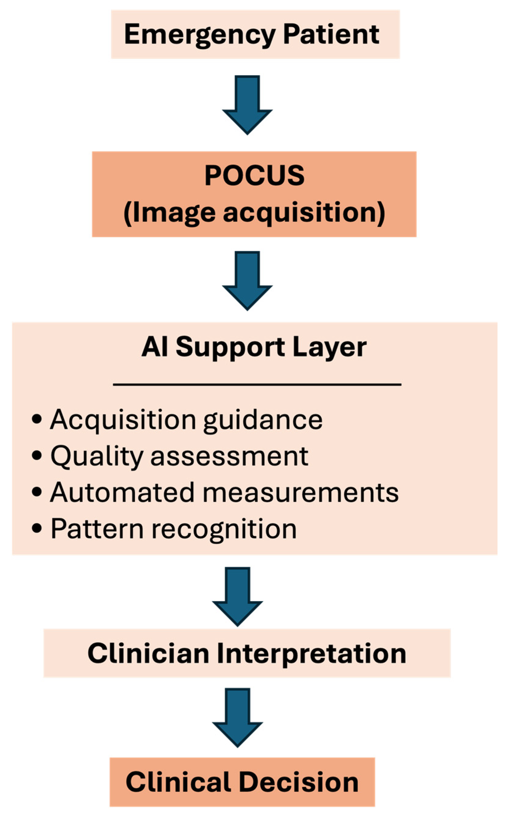
AI-Enhanced POCUS in Emergency Care

 ,
,  , ,
, ,  , ,
, ,  and
and
Abstract
1. Introduction
2. Materials and Methods
- (i)
- Studies explicitly addressing the application of artificial intelligence in point-of-care ultrasound;
- (ii)
- Relevance to emergency medicine or acute care settings;
- (iii)
- Original research articles, technical development studies, clinical validation studies, or narrative/scoping reviews.
- (i)
- Case reports or small case series;
- (ii)
- Publications in languages other than English;
- (iii)
- Studies not specifically involving AI-based applications in POCUS.
3. Trauma and AI-Enhanced POCUS
4. Non-Traumatic Emergencies and AI-Enhanced POCUS
4.1. Cardiovascular Assessment
4.2. Lung Assessment
4.3. Abdominal Assessment
4.4. Integrated Applications and Resource-Limited Settings
4.5. Education and Training—AI-Enhanced POCUS Learning
5. Limitations
6. Conclusions
Author Contributions
Funding
Institutional Review Board Statement
Informed Consent Statement
Data Availability Statement
Conflicts of Interest
References
- Osterwalder, J.; Polyzogopoulou, E.; Hoffmann, B. Point-of-Care Ultrasound—History, Current and Evolving Clinical Concepts in Emergency Medicine. Medicina 2023, 59, 2179. [Google Scholar] [CrossRef] [Scilit] [PubMed]
- Kim, J.; Maranna, S.; Watson, C.; Parange, N. A scoping review on the integration of artificial intelligence in point-of-care ultrasound: Current clinical applications. Am. J. Emerg. Med. 2025, 92, 172–181. [Google Scholar] [CrossRef] [Scilit]
- East, S.A.; Wang, Y.; Yanamala, N.; Maganti, K.; Sengupta, P.P. Artificial Intelligence-Enabled Point-of-Care Echocardiography: Bringing Precision Imaging to the Bedside. Curr. Atheroscler. Rep. 2025, 27, 70. [Google Scholar] [CrossRef] [Scilit]
- Yordanova, M.Z. The Applications of Artificial Intelligence in Radiology: Opportunities and Challenges. Eur. J. Med. Health Sci. 2024, 6, 11–14. [Google Scholar] [CrossRef] [Scilit]
- Yan, L.; Li, Q.; Fu, K.; Zhou, X.; Zhang, K. Progress in the Application of Artificial Intelligence in Ultrasound-Assisted Medical Diagnosis. Bioengineering 2025, 12, 288. [Google Scholar] [CrossRef] [Scilit]
- He, L.; Luan, L.; Hu, D. Deep learning-based image classification for AI-assisted integration of pathology and radiology in medical imaging. Front. Med. 2025, 12, 1574514. [Google Scholar] [CrossRef] [Scilit] [PubMed]
- Zhang, X.M.; Gao, T.H.; Cai, Q.Y.; Xia, J.B.; Sun, Y.N.; Yang, J.; Li, W.-H.; Zhang, S.-X.; Lou, H.-R.; Yu, X.-T.; et al. Artificial intelligence in digital pathology diagnosis and analysis: Technologies, challenges, and future prospects. Mil. Med. Res. 2026, 12, 93. [Google Scholar] [CrossRef] [Scilit] [PubMed]
- Myhre, P.L.; Grenne, B.; Asch, F.M.; Delgado, V.; Khera, R.; Lafitte, S.; Lang, R.M.; Pellikka, P.A.; Sengupta, P.P.; Vemulapalli, S.; et al. Artificial intelligence-enhanced echocardiography in cardiovascular disease management. Nat. Rev. Cardiol. 2025, 14, 1669. [Google Scholar] [CrossRef] [Scilit]
- Kim, S.; Fischetti, C.; Guy, M.; Hsu, E.; Fox, J.; Young, S.D. Artificial Intelligence (AI) Applications for Point of Care Ultrasound (POCUS) in Low-Resource Settings: A Scoping Review. Diagnostics 2024, 14, 1669. [Google Scholar] [CrossRef] [Scilit]
- Wong, A.; Roslan, N.L.; McDonald, R.; Noor, J.; Hutchings, S.; D’Costa, P.; Via, G.; Corradi, F. Clinical obstacles to machine-learning POCUS adoption and system-wide AI implementation (The COMPASS-AI survey). Ultrasound J. 2025, 17, 32. [Google Scholar] [CrossRef] [Scilit]
- Koçak, B.; Ponsiglione, A.; Stanzione, A.; Bluethgen, C.; Santinha, J.; Ugga, L.; Huisman, M.; Klontzas, M.E.; Cannella, R.; Cuocolo, R. Bias in artificial intelligence for medical imaging: Fundamentals, detection, avoidance, mitigation, challenges, ethics, and prospects. Diagn. Interv. Radiol. 2024, 25, 949–955. [Google Scholar] [CrossRef] [Scilit]
- Osifowokan, A.S.; Agbadamasi, T.O.; Adukpo, T.K.; Mensah, N. Regulatory and legal challenges of Artificial Intelligence in the U.S. Healthcare System: Liability, Compliance, and Patient Safety. World J. Adv. Res. Rev. 2025, 25, 949–955. [Google Scholar] [CrossRef] [Scilit]
- Hernandez Torres, S.I.; Holland, L.; Winter, T.; Ortiz, R.; Amezcua, K.L.; Ruiz, A.; Thorpe, C.R.; Snider, E.J. Real-Time Deployment of Ultrasound Image Interpretation AI Models for Emergency Medicine Triage Using a Swine Model. Technologies 2025, 13, 29. [Google Scholar] [CrossRef] [Scilit]
- Kim, K.; Macruz, F.; Wu, D.; Bridge, C.; McKinney, S.; Al Saud, A.A.; Sharaf, E.; Sesic, I.; Pely, A.; Danset, P.; et al. Point-of-care AI-assisted stepwise ultrasound pneumothorax diagnosis. Phys. Med. Biol. 2023, 68, 205013. [Google Scholar] [CrossRef] [Scilit]
- Montgomery, S.; Li, F.; Funk, C.; Peethumangsin, E.; Morris, M.; Anderson, J.T.; Hersh, A.M.; Aylward, S. Detection of pneumothorax on ultrasound using artificial intelligence. J. Trauma. Acute Care Surg. 2023, 94, 379–384. [Google Scholar] [CrossRef] [Scilit] [PubMed]
- Ruiz, A.J.; Hernández Torres, S.I.; Snider, E.J. Development of Deep Learning Models for Real-Time Thoracic Ultrasound Image Interpretation. J. Imaging 2025, 11, 222. [Google Scholar] [CrossRef] [Scilit]
- Yıldız Potter, İ.; Leo, M.M.; Vaziri, A.; Feldman, J.A. Automated detection and localization of pericardial effusion from point-of-care cardiac ultrasound examination. Med. Biol. Eng. Comput. 2023, 61, 1947–1959. [Google Scholar] [CrossRef] [Scilit] [PubMed]
- Leo, M.M.; Potter, I.Y.; Zahiri, M.; Vaziri, A.; Jung, C.F.; Feldman, J.A. Using Deep Learning to Detect the Presence and Location of Hemoperitoneum on the Focused Assessment with Sonography in Trauma (FAST) Examination in Adults. J. Digit. Imaging 2023, 36, 2035–2050. [Google Scholar] [CrossRef] [Scilit] [PubMed]
- Gohar, E.; Herling, A.; Mazuz, M.; Tsaban, G.; Gat, T.; Kobal, S.; Fuchs, L. Artificial Intelligence (AI) versus POCUS Expert: A Validation Study of Three Automatic AI-Based, Real-Time, Hemodynamic Echocardiographic Assessment Tools. J. Clin. Med. 2023, 12, 1352. [Google Scholar] [CrossRef] [Scilit]
- Mika, S.; Gola, W.; Gil-Mika, M.; Wilk, M.; Misiołek, H. Overview of artificial intelligence in point-of-care ultrasound. New horizons for respiratory system diagnoses. Anaesthesiol. Intensive Ther. 2024, 56, 1–8. [Google Scholar] [CrossRef] [Scilit]
- Oikonomou, E.K.; Vaid, A.; Holste, G.; Coppi, A.; McNamara, R.L.; Baloescu, C.; Krumholz, H.M.; Wang, Z.; Apakama, D.J.; Nadkarni, G.N.; et al. Artificial intelligence-guided detection of under-recognised cardiomyopathies on point-of-care cardiac ultrasonography: A multicentre study. Lancet Digit. Health 2025, 7, e113–e123. [Google Scholar] [CrossRef] [Scilit]
- Singh, Y. Echocardiography in the neonatal unit: Current status and future prospects. Expert Rev. Med. Devices 2024, 21, 307–316. [Google Scholar] [CrossRef] [Scilit]
- Crockett, D.; Kelly, C.; Brundage, J.; Jones, J.; Ockerse, P. A Stress Test of Artificial Intelligence: Can Deep Learning Models Trained From Formal Echocardiography Accurately Interpret Point-of-Care Ultrasound? J. Ultrasound Med. 2022, 41, 3003–3012. [Google Scholar] [CrossRef] [Scilit] [PubMed]
- Blaivas, M.; Blaivas, L.N.; Campbell, K.; Thomas, J.; Shah, S.; Yadav, K.; Liu, Y.T. Making Artificial Intelligence Lemonade Out of Data Lemons: Adaptation of a Public Apical Echo Database for Creation of a Subxiphoid Visual Estimation Automatic Ejection Fraction Machine Learning Algorithm. J. Ultrasound Med. 2022, 41, 2059–2069. [Google Scholar] [CrossRef] [Scilit] [PubMed]
- Vega, R.; Dehghan, M.; Nagdev, A.; Buchanan, B.; Kapur, J.; Jaremko, J.L.; Zonoobi, D. Overcoming barriers in the use of artificial intelligence in point of care ultrasound. npj Digit. Med. 2025, 8, 213. [Google Scholar] [CrossRef] [Scilit]
- Gottlieb, M.; Schraft, E.; O’Brien, J.; Patel, D. Diagnostic accuracy of artificial intelligence for identifying systolic and diastolic cardiac dysfunction in the emergency department. Am. J. Emerg. Med. 2024, 86, 115–119. [Google Scholar] [CrossRef] [Scilit]
- Vega, R.; Nagdev, A.; Dehghan, M.; Seyed Bolouri, S.E.; Buchanan, B.; Kapur, J.; Jaremko, J.L.; Zonoobi, D. A wall tracking method to estimate ejection fraction from the parasternal long axis view in point of care ultrasound. WFUMB Ultrasound Open 2025, 3, 100097. [Google Scholar] [CrossRef] [Scilit]
- Blaivas, M.; Blaivas, L.N.; Tsung, J.W. Deep learning algorithm performance compared to experts in visual evaluation of interior vena cava collapse on ultrasound to determine intravenous fluid need in dehydration management. Signa Vitae 2021, 17, 34–41. [Google Scholar] [CrossRef] [Scilit]
- Efrimescu, C.I.; Moorthy, A.; Griffin, M. Rescue Transesophageal Echocardiography: A Narrative Review of Current Knowledge and Practice. J. Cardiothorac. Vasc. Anesth. 2023, 37, 584–600. [Google Scholar] [CrossRef] [Scilit] [PubMed]
- Ienghong, K.; Cheung, L.W.; Gaysonsiri, D.; Apiratwarakul, K. The diagnostic performance of automatic B-lines detection for evaluating pulmonary edema in the emergency department among novice point-of-care ultrasound practitioners. Emerg. Radiol. 2025, 32, 241–246. [Google Scholar] [CrossRef] [Scilit]
- Duggan, N.M.; Jin, M.; Duran Mendicuti, M.A.; Hallisey, S.; Bernier, D.; Selame, L.A.; Asgari-Targhi, A.; Fischetti, C.E.; Lucassen, R.; Samir, A.E.; et al. Gamified Crowdsourcing as a Novel Approach to Lung Ultrasound Data Set Labeling: Prospective Analysis. J. Med. Internet Res. 2024, 26, e51397. [Google Scholar] [CrossRef] [Scilit] [PubMed]
- Nti, B.; Lehmann, A.S.; Haddad, A.; Kennedy, S.K.; Russell, F.M. Artificial Intelligence-Augmented Pediatric Lung POCUS: A Pilot Study of Novice Learners. J. Ultrasound Med. 2022, 41, 2965–2972. [Google Scholar] [CrossRef] [Scilit]
- Moore, C.L.; Wang, J.; Battisti, A.J.; Chen, A.; Fincke, J.; Wang, A.; Wagner, M.; Raju, B.; Baloescu, C. Interobserver Agreement and Correlation of an Automated Algorithm for B-Line Identification and Quantification with Expert Sonologist Review in a Handheld Ultrasound Device. J. Ultrasound Med. 2022, 41, 2487–2495. [Google Scholar] [CrossRef] [Scilit]
- Jeffers, K.; Keim, S.M.; Long, B.; Gottlieb, M.; Adhikari, S.R. What is the Utility of Point-of-Care Ultrasound for Diagnosing Pulmonary Edema? J. Emerg. Med. 2025, 77, 21–29. [Google Scholar] [CrossRef] [Scilit]
- Song, F.; Liu, H.; Ma, H.; Chen, X.; Wang, S.; Qin, T.; Liang, H.; Huang, D. AI Model Based on Diaphragm Ultrasound to Improve the Predictive Performance of Invasive Mechanical Ventilation Weaning: Prospective Cohort Study. JMIR Form Res 2025, 9, e72482. [Google Scholar] [CrossRef] [Scilit]
- Labaf, A.; Åhman-Persson, L.; Husu, L.S.; Smith, J.G.; Ingvarsson, A.; Evaldsson, A.W. Performance of a point-of-care ultrasound platform for artificial intelligence-enabled assessment of pulmonary B-lines. Cardiovasc. Ultrasound 2025, 23, 3. [Google Scholar] [CrossRef] [Scilit]
- Sultan, L.R.; Haertter, A.; Al-Hasani, M.; Demiris, G.; Cary, T.W.; Tung-Chen, Y.; Sehgal, C.M. Can Artificial Intelligence Aid Diagnosis by Teleguided Point-of-Care Ultrasound? A Pilot Study for Evaluating a Novel Computer Algorithm for COVID-19 Diagnosis Using Lung Ultrasound. AI 2023, 4, 875–887. [Google Scholar] [CrossRef] [Scilit]
- Schneider, E.; Maimon, N.; Hasidim, A.; Shnaider, A.; Migliozzi, G.; Haviv, Y.S.; Halpern, D.; Abu Ganem, B.; Fuchs, L. Can Dialysis Patients Identify and Diagnose Pulmonary Congestion Using Self-Lung Ultrasound? J. Clin. Med. 2023, 12, 3829. [Google Scholar] [CrossRef] [Scilit] [PubMed]
- Kuroda, Y.; Kaneko, T.; Yoshikawa, H.; Uchiyama, S.; Nagata, Y.; Matsushita, Y.; Hiki, M.; Minamino, T.; Takahashi, K.; Daida, H.; et al. Artificial intelligence-based point-of-care lung ultrasound for screening COVID-19 pneumoniae: Comparison with CT scans. Valera-Calero JA, editor. PLoS ONE 2023, 18, e0281127. [Google Scholar]
- Song, J.; Ebadi, A.; Florea, A.; Xi, P.; Tremblay, S.; Wong, A. COVID-Net USPro: An Explainable Few-Shot Deep Prototypical Network for COVID-19 Screening Using Point-of-Care Ultrasound. Sensors 2023, 23, 2621. [Google Scholar] [CrossRef] [Scilit] [PubMed]
- Baloescu, C.; Toporek, G.; Kim, S.; McNamara, K.; Liu, R.; Shaw, M.M.; McNamara, R.L.; Raju, B.I.; Moore, C.L. Automated Lung Ultrasound B-Line Assessment Using a Deep Learning Algorithm. IEEE Trans. Ultrason. Ferroelectr. Freq. Control 2020, 67, 2312–2320. [Google Scholar] [CrossRef] [Scilit]
- Mathyk, B.; Pandya, S.; Wright Beatty, H.; Anderson, M.L.; Kohut, A. Handheld Point-of-Care Ultrasonography for Gynecology: Insights Into Space Travel Through Parabolic Flight. Obstet. Gynecol. 2025, 146, 751–757. [Google Scholar] [CrossRef] [Scilit]
- Zgool, T.; Antico, M.; Edwards, C.; Fontanarosa, D. Point-of-Care Ultrasound Imaging for Automated Detection of Abdominal Haemorrhage: A Systematic Review. Ultrasound Med. Biol. 2025, 51, 1888–1900. [Google Scholar] [CrossRef] [Scilit]
- Lee, K.; Kim, M.; Lim, C.; Song, T.K. Reverse Scan Conversion and Efficient Deep Learning Network Architecture for Ultrasound Imaging on a Mobile Device. Sensors 2021, 21, 2629. [Google Scholar] [CrossRef] [Scilit]
- Yu, C.J.; Yeh, H.J.; Chang, C.C.; Tang, J.H.; Kao, W.Y.; Chen, W.C.; Huang, Y.-J.; Li, C.-H.; Chang, W.-H.; Lin, Y.-T.; et al. Lightweight deep neural networks for cholelithiasis and cholecystitis detection by point-of-care ultrasound. Comput. Methods Programs Biomed. 2021, 211, 106382. [Google Scholar] [CrossRef] [Scilit] [PubMed]
- Zahid, M.A.; Nasir, H.; Abid, S.; Shuja Ul Islam, M.H.S.; Sawwa, A.; Jamil, S. Why is it necessary for every medical specialty to learn point-of-care ultrasound (POCUS)? A comprehensive review. Anaesth. Pain. Intensive Care 2025, 29, 392–399. [Google Scholar] [CrossRef] [Scilit]
- Kaffas, A.E.; Vo-Phamhi, J.M.; Griffin, J.F.; Hoyt, K. Critical Advances for Democratizing Ultrasound Diagnostics in Human and Veterinary Medicine. Annu. Rev. Biomed. Eng. 2024, 26, 49–65. [Google Scholar] [CrossRef]
- Venkatayogi, N.; Gupta, M.; Gupta, A.; Nallaparaju, S.; Cheemalamarri, N.; Gilari, K.; Pathak, S.; Vishwanath, K.; Soney, C.; Bhattacharya, T.; et al. From Seeing to Knowing with Artificial Intelligence: A Scoping Review of Point-of-Care Ultrasound in Low-Resource Settings. Appl. Sci. 2023, 13, 8427. [Google Scholar] [CrossRef] [Scilit]
- Alfoti, B.O.O.; Alfoti, F.O.O.; Alothman, S.T.H.; Al-Dhafiri, T.M.A.; Al-Harbi, N.H.M.; Al-Khalidi, A.M.; Alzahrany, N.D.B.; Abutalib, F.M.; Almojam, S.A.; Ali, M.A.-H.S.; et al. Utilization of Point-of-Care Ultrasound (POCUS) in Emergency and Critical Care: Role of Nursing for Enhancing Diagnostic Accuracy and Efficiency-Systematic Review. Egypt. J. Chem. 2024, 67, 705–716. [Google Scholar] [CrossRef] [Scilit]
- Zeng, E.Z.; Ebadi, A.; Florea, A.; Wong, A. COVID-Net L2C-ULTRA: An Explainable Linear-Convex Ultrasound Augmentation Learning Framework to Improve COVID-19 Assessment and Monitoring. Sensors 2024, 24, 1664. [Google Scholar] [CrossRef] [Scilit]
- Yazici, M.M.; Yavaşi, Ö. The development of point-of-care ultrasound (POCUS): Worldwide contributions and publication trends. J. Clin. Ultrasound 2025, 53, 129–138. [Google Scholar] [CrossRef] [Scilit]
- Park, Y.; Han, J.; Leikin, S.; Díaz-Gómez, J.L. Essential Point-of-Care Ultrasound Insights for 2024. Semin. Ultrasound CT MRI 2024, 45, 22–28. [Google Scholar] [CrossRef] [Scilit]
- Blaivas, M.; Arntfield, R.; White, M. Creation and Testing of a Deep Learning Algorithm to Automatically Identify and Label Vessels, Nerves, Tendons, and Bones on Cross-sectional Point-of-Care Ultrasound Scans for Peripheral Intravenous Catheter Placement by Novices. J. Ultrasound Med. 2020, 39, 1721–1727. [Google Scholar] [CrossRef] [Scilit] [PubMed]
- Rowe, M.; Ferrada, P. Ultrasound to guide critical decisions: All that you need to know. J. Trauma Acute Care Surg. 2025, 17, 32. [Google Scholar] [CrossRef] [Scilit] [PubMed]
- Blaivas, M.; Arntfield, R.; White, M. DIY AI, deep learning network development for automated image classification in a point-of-care ultrasound quality assurance program. J. Am. Coll. Emerg. Physicians Open 2020, 1, 124–131. [Google Scholar] [CrossRef] [Scilit] [PubMed]
- Chou, H.H.; Chang, Y.C.; Lien, W.C.; Lin, L.C.; Lin, X.Z.; Hsu, T.E.; Liu, Y.-P.; Liu, L.; Chan, Y.-T.; Kuan, F.-S. Efficient Deep Learning Models Revolutionize Doctor’s Training for Point-of-Care Ultrasound. IEEE Access 2025, 13, 76038–76046. [Google Scholar] [CrossRef] [Scilit]
- Karni, O.; Shitrit, I.B.; Perlin, A.; Jedwab, R.; Wacht, O.; Fuchs, L. AI-enhanced guidance demonstrated improvement in novices’ Apical-4-chamber and Apical-5-chamber views. BMC Med. Educ. 2025, 25, 558. [Google Scholar] [CrossRef] [Scilit]
- Aronovitz, N.; Hazan, I.; Jedwab, R.; Ben Shitrit, I.; Quinn, A.; Wacht, O.; Fuchs, L. The effect of real-time EF automatic tool on cardiac ultrasound performance among medical students. AbdelMassih AF, editor. PLoS ONE 2024, 19, e0299461. [Google Scholar] [CrossRef] [Scilit]
- Dadon, Z.; Orlev, A.; Butnaru, A.; Rosenmann, D.; Glikson, M.; Gottlieb, S.; Alpert, E.A. Empowering Medical Students: Harnessing Artificial Intelligence for Precision Point-of-Care Echocardiography Assessment of Left Ventricular Ejection Fraction. Int. J. Clin. Pract. 2023, 2023, 5225872. [Google Scholar] [CrossRef] [Scilit] [PubMed]
- Zhai, S.; Wang, H.; Sun, L.; Zhang, B.; Huo, F.; Qiu, S.; Wu, X.; Ma, J.; Wu, Y.; Duan, J. Artificial intelligence (AI) versus expert: A comparison of left ventricular outflow tract velocity time integral (LVOT-VTI) assessment between ICU doctors and an AI tool. J. Appl. Clin. Med. Phys. 2022, 23, e13724. [Google Scholar] [CrossRef] [Scilit]
- Lin-Martore, M.; Kornblith, A.; Firnberg, M.; Haque, A.; O’Brien, B. Trust of Artificial Intelligence-Augmented Point-of-Care Ultrasound Among Pediatric Emergency Physicians. J. Am. Coll. Emerg. Physicians Open 2025, 6, 100173. [Google Scholar] [CrossRef] [Scilit]
- Shokoohi, H.; Liteplo, A.S.; Montoya, K.; Patnode, C.; Hutchinson, A.B.; Zalis, M.E.; Gottlieb, M.; Raja, A.S.; Slutzman, J.E. Climate-Smart Diagnostic Medical Imaging and Point-of-Care Ultrasound: An Evidence-Based Perspective. J. Emerg. Med. 2025, 75, 150–157. [Google Scholar] [CrossRef] [Scilit]
- Kayarian, F.; Patel, D.; O’Brien, J.R.; Schraft, E.K.; Gottlieb, M. Artificial intelligence and point-of-care ultrasound: Benefits, limitations, and implications for the future. Am. J. Emerg. Med. 2024, 80, 119–122. [Google Scholar] [CrossRef] [Scilit]
- Blaivas, M.; Blaivas, L.N.; Tsung, J.W. Deep Learning Pitfall: Impact of Novel Ultrasound Equipment Introduction on Algorithm Performance and the Realities of Domain Adaptation. J. Ultrasound Med. 2022, 41, 855–863. [Google Scholar] [CrossRef] [Scilit] [PubMed]
- Suttels, V.; Du Toit, J.D.; Fiogbé, A.A.; Wachinou, A.P.; Guendehou, B.; Alovokpinhou, F.; Toukoui, P.; Hada, A.R.; Sefou, F.; Vinasse, P.; et al. Point-of-care ultrasound for tuberculosis management in Sub-Saharan Africa—A balanced SWOT analysis. Int. J. Infect. Dis. 2022, 123, 46–51. [Google Scholar] [CrossRef] [Scilit] [PubMed]
- Blaivas, L.; Blaivas, M. Are Convolutional Neural Networks Trained on ImageNet Images Wearing Rose-Colored Glasses?: A Quantitative Comparison of IMAGENET, Computed Tomographic, Magnetic Resonance, Chest X-Ray, and Point-of-Care Ultrasound Images for Quality. J. Ultrasound Med. 2021, 40, 377–383. [Google Scholar] [CrossRef] [Scilit] [PubMed]
- Kameda, T.; Ishii, H.; Oya, S.; Katabami, K.; Kodama, T.; Sera, M.; Takei, H.; Taniguchi, H.; Nakao, S.; Funakoshi, H.; et al. Guidance for clinical practice using emergency and point-of-care ultrasonography. Acute Med. Surg. 2024, 11, e974. [Google Scholar] [CrossRef] [Scilit]


| Setting/Focus | AI Approach | Key Findings | Sample Size (Approx.) | Validation Type | Dataset Origin | References |
|---|---|---|---|---|---|---|
| Emergency eFAST | Real-time anatomical landmark detection + classifier | Combined image guidance and interpretation demonstrated feasibility in emergency triage. | 200–400 exams | Internal | Single-centre | [13] |
| Lung ultrasound pneumothorax | Stepwise DL for QA and sliding classification | AUC ~0.89 for full pipeline; high reliability for diagnostic support. | 800–1200 video clips | External | Multi-centre | [14] |
| Pneumothorax detection | Neural network on POCUS | Sensitivity ~86% for PTX detection, illustrating potential clinical performance. | 500–1000 images | Internal | Multi-centre | [15] |
| Thoracic trauma (swine model) | CNN classification (MobileNetV3) | Real-time M-mode PTX/HTX detection with ~85% accuracy; reduces required expertise threshold. | 300–500 M-mode clips | Internal | Single-centre | [16] |
| Lung trauma (pneumothorax) | CNN-based deep learning | Automated pneumothorax detection with high diagnostic accuracy | 500–1000 lung US frames/clips | Internal | Single-centre | [17] |
| Lung trauma (pneumothorax) | Deep learning (CNN) | Automated pneumothorax detection on lung ultrasound with strong diagnostic performance | 400–800 lung US images/clips | Internal | Single-centre | [18] |
| Setting/Population | AI Task | Key Findings | Sample Size (Approx.) | Validation Type | Dataset Origin | References |
|---|---|---|---|---|---|---|
| ED/ICU patients; cardiac POCUS clips | On-device AI (auto-EF, auto-VTI, auto-IVC) | Moderate–good agreement with expert POCUS for high-quality views (κ ≈ 0.50–0.66) | 200–400 clips | Internal | Single-centre | [19] |
| Emergency department adults ≥45 years | Vendor AI for systolic/diastolic dysfunction | Sensitivity 85–92%; specificity 94–95% vs. expert reviewers | ~200 patients | External | Single-centre | [26] |
| Unstable ED/ICU patients; PLAX POCUS | CNN-based wall-tracking | Accurate EF classification (85–87%) from parasternal long-axis view | 500–700 studies | Internal | Multi-centre | [27] |
| ED and community cardiac POCUS | CNN screening for cardiomyopathies | AUROC~0.90–0.97; early detection of HCM and ATTR-CM | >40,000 videos | External | Multi-centre | [21] |
| Emergency department cardiac POCUS | Deep learning (EchoNet-Dynamic) | Reduced performance on POCUS vs. formal echo (Dice~0.72; κ~0.16) | 300–400 videos | External | Single-centre | [23] |
| Subxiphoid cardiac POCUS views | Machine learning with data augmentation | Feasible EF estimation; higher error at mid-range EF values | 500–700 clips | Internal | Single-centre | [24] |
| Neonatal non-traumatic emergencies | ML/DL-assisted targeted echocardiography | Early-stage and conceptual applications for bedside hemodynamic assessment | Not specified | Narrative/Conceptual | Multi-centre | [22] |
| Cardiovascular POCUS platforms | Integrated AI quantification tools (AutoEF, SmartVTI) | Demonstrated technical maturity and clinical feasibility in acute care | Not specified | Narrative/Technology overview | Multi-centre | [25] |
| Cardiac ultrasound (LV function) | Machine learning–based EF estimation | Feasible automated estimation of left ventricular function from ultrasound images | 100–300 studies | Internal | Single-centre | [28] |
| Perioperative and critical care cardiac POCUS | AI-assisted cardiac function assessment | AI-supported quantification of cardiac function feasible and clinically relevant in acute care settings | 150–300 examinations | Internal | Single-centre | [29] |
| Clinical Context | Lung POCUS Application | AI Approach | Main Findings | Sample Size (Approx.) | Validation Type | Dataset Origin | References |
|---|---|---|---|---|---|---|---|
| Emergency department; acute dyspnea | Detection of B-lines and pleural abnormalities | CNN-based image classification | Good discrimination between normal and abnormal lung patterns (preliminary) | Not specified | Not specified | Not specified | [34] |
| Suspected pneumothorax | Pleural sliding analysis; pneumothorax detection | Deep learning on annotated ultrasound video loops | High diagnostic performance (AUC > 0.90) vs. expert interpretation | Several hundred-1000 clips (reported as large dataset) | Internal (development/validation) | Single-centre/not clearly stated | [30] |
| Acute dyspnea/heart failure | Automated B-line quantification | Machine learning–based feature extraction and classification | Strong correlation with expert annotations and congestion biomarkers | Not specified | Not specified | Not specified | [31] |
| Pleural disease evaluation | Differentiation of pleural effusion types | Pattern recognition algorithms | Improved accuracy in pleural fluid characterization | Not specified | Internal | Single-centre | [32] |
| Emergency lung ultrasound workflows | Image acquisition support and standardization | Prototype AI-guided acquisition system | Feasibility of AI feedback for improving scan quality | Not specified | Prototype/feasibility | Not specified | [33] |
| Lung ultrasound data development | Lung POCUS image labeling for AI training | Crowdsourcing-assisted annotation with ML support | Demonstrated feasibility of scalable, high-quality annotation for lung ultrasound datasets | Large annotated dataset (exact size reported in study) | Not applicable (data development) | Multi-centre/crowdsourced | [35] |
| Acute care/emergency lung ultrasound | Automated lung ultrasound pattern recognition | Deep learning (CNN-based) | Demonstrated feasibility of AI-assisted lung pattern classification on POCUS images | Several hundred images/clips | Internal | Single-centre | [36] |
| Lung ultrasound (acute and emergency care) | Automated lung ultrasound image analysis | Deep learning (CNN-based) | Demonstrated feasibility of AI-based lung ultrasound pattern recognition | Tens to low hundreds of images/clips | Internal | Single-centre | [37] |
| Emergency and acute care lung ultrasound | Automated lung ultrasound interpretation | Deep learning (CNN-based) | Demonstrated feasibility of AI-assisted lung ultrasound analysis with clinically relevant performance | Several hundred lung US images/clips | Internal | Single-centre | [38] |
| Acute and emergency lung ultrasound | Automated lung ultrasound pattern classification | Deep learning (CNN-based) | AI model achieved reliable lung pattern classification on POCUS images | Several hundred lung US images/clips | Internal | Single-centre | [39] |
| Lung ultrasound image analysis | Automated lung ultrasound feature and pattern detection | Deep learning (CNN-based) | Demonstrated feasibility of automated lung ultrasound image analysis with promising classification performance | Several hundred images | Internal | Single-centre | [40] |
| Lung ultrasound image analysis | Automated lung ultrasound pattern classification | Deep learning (CNN-based) | Demonstrated accurate automated classification of lung ultrasound patterns under controlled conditions | Retrospective analysis; not specific to emergency workflows | Several hundred images/clips | Internal | [41] |
| Clinical Context | Abdominal Application | AI Approach | Main Findings | Sample Size (Approx.) | Validation Type | Dataset Origin | References |
|---|---|---|---|---|---|---|---|
| Obstetric and gynecologic emergencies | Early pregnancy assessment; risk stratification | Machine learning–based image interpretation | AI-assisted POCUS supported rapid differentiation of intrauterine vs. ectopic pregnancy | Not specified | Internal | Single-centre | [42] |
| Acute abdominal evaluation | Detection of free intraperitoneal fluid | CNN-based image and video classification | Demonstrated feasibility of automated free fluid detection | Not specified | Not specified | Not specified | [43] |
| General abdominal ultrasound | Automated image classification | Deep learning models | Performance comparable to human readers for selected tasks | Several hundred images | Internal | Single-centre | [44] |
| Abdominal ultrasound image analysis | Automated abdominal organ and pathology classification | Deep learning (CNN-based) | Demonstrated accurate automated classification of abdominal ultrasound images | Several hundred images | Internal | Single-centre/curated dataset | [45] |
| Setting/Focus | AI Application | AI Approach | Key Contribution | Sample Size | Validation Type | Dataset Origin | References |
|---|---|---|---|---|---|---|---|
| Low- and middle-income countries (LMICs) | AI-assisted diagnostic POCUS | ML/DL-based image interpretation | Demonstrated feasibility and relevance in constrained healthcare environments | Not specified | Narrative/feasibility | Multi-centre/heterogeneous | [46] |
| Cross-modality ultrasound | Translational AI in ultrasound imaging | Review of ML/DL architectures | Identified barriers to clinical translation | Not applicable | Narrative review | Multi-centre | [47] |
| Engineering-focused AI for POCUS | Application-specific DL model design | Deep learning architectures | Highlighted importance of task-specific AI models for robust POCUS deployment | Not applicable | Engineering/methodological | Single-/Multi-centre datasets | [48] |
| Integrated POCUS workflows | Multi-task AI systems | CNN-based pipelines | Demonstrated feasibility of integrated AI support across the POCUS workflow | Not specified | Prototype/feasibility | Single-centre | [9] |
| Portable devices | Lightweight AI models for edge deployment | Optimized DL models | Optimized for low-compute environments | Not specified | Technical/feasibility | Single-centre | [49] |
| Trauma and acute care workflows | AI-assisted POCUS for trauma assessment | Deep learning–based image interpretation and decision support | Demonstrated feasibility of AI-supported ultrasound interpretation to assist trauma evaluation and triage | Several hundred examinations | Internal | Single-centre | [54] |
| System-wide emergency ultrasound practice | AI-assisted POCUS adoption and implementation | Survey-based evaluation of ML-enabled POCUS tools | Identified key clinical, technical, and organizational barriers to AI-POCUS adoption (training, trust, workflow integration) | Several hundred clinicians | Observational survey | Multi-centre/international | [10] |
Disclaimer/Publisher’s Note: The statements, opinions and data contained in all publications are solely those of the individual author(s) and contributor(s) and not of MDPI and/or the editor(s). MDPI and/or the editor(s) disclaim responsibility for any injury to people or property resulting from any ideas, methods, instructions or products referred to in the content. |
© 2026 by the authors. Licensee MDPI, Basel, Switzerland. This article is an open access article distributed under the terms and conditions of the Creative Commons Attribution (CC BY) license.
Share and Cite
Puticiu, M.; Cimpoesu, D.; Pop, F.; Ciumanghel, I.; Rotaru, L.T.; Oprita, B.; Butoi, M.A.; Belghiru, V.I.; Tat, R.M.; Golea, A. AI-Enhanced POCUS in Emergency Care. Diagnostics 2026, 16, 353. https://doi.org/10.3390/diagnostics16020353
Puticiu M, Cimpoesu D, Pop F, Ciumanghel I, Rotaru LT, Oprita B, Butoi MA, Belghiru VI, Tat RM, Golea A. AI-Enhanced POCUS in Emergency Care. Diagnostics. 2026; 16(2):353. https://doi.org/10.3390/diagnostics16020353
Chicago/Turabian StylePuticiu, Monica, Diana Cimpoesu, Florica Pop, Irina Ciumanghel, Luciana Teodora Rotaru, Bogdan Oprita, Mihai Alexandru Butoi, Vlad Ionut Belghiru, Raluca Mihaela Tat, and Adela Golea. 2026. "AI-Enhanced POCUS in Emergency Care" Diagnostics 16, no. 2: 353. https://doi.org/10.3390/diagnostics16020353
APA StylePuticiu, M., Cimpoesu, D., Pop, F., Ciumanghel, I., Rotaru, L. T., Oprita, B., Butoi, M. A., Belghiru, V. I., Tat, R. M., & Golea, A. (2026). AI-Enhanced POCUS in Emergency Care. Diagnostics, 16(2), 353. https://doi.org/10.3390/diagnostics16020353