Systematic Review

Thermography and Infrared Spectroscopy in the Detection of Periodontal Inflammation In Vivo: A Systematic Review

by
Heythem Nassim Guetatlia
1,
Mickael Gette
1,2,3,
Laurent Estrade
1,2,3,
Victor Rimbaud
2,3,
Frédéric Denis
2,3,4,
Gaël Y. Rochefort
1,2,3 and
Matthieu Renaud
1,2,3,*
1
N2Cox U1069 INSERM, Tours University, 37032 Tours, France
2
Department of Medicine and Bucco-Dental Surgery, Tours University Hospital, 37044 Tours, France
3
Faculty of Odontology, Tours University, 37032 Tours, France
4
EA 75-05 Education, Ethique, Santé, Faculté de Médecine, Université François-Rabelais, 37000 Tours, France
*
Author to whom correspondence should be addressed.
Diagnostics 2026, 16(2), 222; https://doi.org/10.3390/diagnostics16020222
Submission received: 23 December 2025 / Revised: 5 January 2026 / Accepted: 6 January 2026 / Published: 10 January 2026

Abstract

Background/Objectives: Periodontal inflammation is a key feature of periodontal diseases, but traditional diagnostic methods are limited by invasiveness and radiation exposure. This systematic review aims to evaluate the potential of thermography and infrared spectroscopy for the in vivo detection of periodontal inflammation and to assess their reliability for clinical use. Methods: In accordance with PRISMA guidelines, an electronic search of the MEDLINE (PubMed) database was conducted to identify relevant studies published between 2000 and October 2025 that investigated these imaging modalities in periodontal inflammation diagnosis. Results: The search identified 310 records; after exclusions, 13 studies were included, comprising 7 thermography studies and 6 infrared spectroscopy studies, for a total of 712 patients. The included studies demonstrated the feasibility of thermography and infrared spectroscopy for detecting inflammatory changes in periodontal tissues in vivo. These non-invasive imaging techniques may help overcome the limitations of conventional clinical and radiographic diagnostic methods, particularly invasiveness and exposure to ionizing radiation. Conclusions: This field remains underexplored, and further studies are required to validate diagnostic performance, standardize methodologies, and determine their clinical applicability in routine periodontal practice.

1. Introduction

Periodontal disease is characterized by persistent inflammatory damage to the periodontal tissues [1,2]. Periodontal inflammation refers to the host’s immune response to microbial plaque, manifesting as redness, swelling, and bleeding of the gingival tissues, which can progress to tissue destruction in periodontitis. It is a key diagnostic criterion for periodontal diseases, including gingivitis (reversible inflammation confined to the gingiva) and periodontitis (irreversible loss of attachment and bone). It manifests as inflammation leading to the degradation and progressive destruction of the tooth-supporting structures, including gingiva, periodontal ligament, cementum, and alveolar bone [3]. Numerous studies also link it to the exacerbation of systemic conditions, such as cardiovascular diseases and diabetes [4,5,6]. The inflammatory process in periodontal disease is complex, involving a dysbiotic microbiome triggering a hyperinflammatory response, leading to tissue breakdown in periodontitis, unlike gingivitis, which is characterized by pathognomonic signs such as reversible gingival erythema, edema, and bleeding without attachment loss.
Diagnosis depends traditionally on clinical parameters, including periodontal pocket depth, bleeding on probing, and attachment loss [7]. These are supplemented by radiological assessments, which detect macroscopic signs of the pathology, such as alveolar bone resorption and loss of periodontal attachment [8,9]. However, early detection remains challenging due to the limitations of these methods. Limitations include the subjectivity and invasiveness of probing (causing patient discomfort and potential tissue trauma), delayed detection via radiography (only showing bone loss after 30–50% destruction), and radiation exposure. Emerging technologies like thermography and spectroscopy could improve early detection by non-invasively capturing subclinical inflammation through heat and hemodynamic changes.
The diagnostic methods for periodontal diseases in dental medicine have stagnated for years, prompting the exploration of alternative imaging modalities less ionizing and capable of providing an earlier diagnosis of periodontal inflammation, while ensuring their effectiveness in the daily practice of clinicians. Common tools include periodontal probes for pocket depth, bleeding on probing, and ionizing radiographs (e.g., periapical X-rays, CBCT) for bone assessment. Infrared technologies are considered due to their non-ionizing nature, reducing risks like cumulative radiation exposure, while providing real-time, contactless inflammation detection [10,11,12].
Infrared radiation was discovered by William Herschel (1738–1822) through experiments on “dark light,” extending Newton’s work on light diffraction. Herschel observed increasing temperatures beyond the visible red spectrum, revealing infrared radiation [13]. Unlike visible light, infrared wavelengths (0.7–1000 μm) are imperceptible to the human eye but ideal for analyzing tissue structure and dynamics. The infrared spectrum is divided into near-infrared (NIR; 0.75–2.5 μm), mid-infrared (MIR; 2.5–5 μm), and far-infrared (FIR; 5–15 μm) [14], each suited to specific imaging techniques (Figure 1). These technologies enable early visualization by detecting parameters like temperature gradients (in thermography, e.g., ΔT > 0.5 °C for inflammation) and tissue oxygenation levels (in spectroscopy, e.g., reduced oxyhemoglobin in inflamed sites), distinguishing from non-inflammatory processes like cancer, which may show different spectral signatures.
The two main analysis techniques for this spectrum of electromagnetic waves are thermography and spectroscopy:
Since the mid-1960s, multiple publications have introduced the use of infrared thermography as a potential non-ionizing tool for the detection of inflammation [15,16,17]. Far-infrared imaging, also known as infrared thermal imaging (IRT) or thermography, is a non-invasive, contactless imaging technique that provides real-time temperature measurements of the structures being analyzed by detecting their emitted far-infrared radiation (5–15 μm). This emitted thermal radiation is then converted into a visible and quantifiable infrared thermal image [18]. The measurement in this technique relies on detectors such as bolometers. These sensors receive the infrared emissions from objects, caused by the rise in their temperature. This thermal variation modifies the conductance of the sensor material, resulting in a change in the electrical signal measured at the output [14]. IRT has been rarely used in biomedical applications due to the limitations of early IR cameras in terms of performance and size. First-generation cameras provided insufficient thermal and spatial resolution, making the results unsatisfactory for many medical applications. Today, modern appliances with high sensitivity are used to produce high-resolution thermal images. As just seen, thermographic devices convert captured infrared radiation into electrical signals, which are then transformed into a thermogram visually representing temperature variations using a colormap [19]. The colors displayed on the generated images represent temperature gradients, ranging from the warmest shades, such as red, to the coolest ones, such as green and blue [18]. Some reviews have already discussed thermography in other fields, but studies dedicated to periodontology applications of this technique remain relatively rare. Up until now, no review has been conducted to highlight the use of these technologies in the diagnosis of periodontal inflammation.
Moreover, spectroscopy refers to the interaction between electromagnetic waves and analyses based on their wavelength. These interactions can be measured using various spectroscopic techniques that cover different wavelength ranges [20]. In the infrared region, spectroscopy can be used to analyze the spectra of near-infrared (NIR), mid-infrared (MIR), and far-infrared (FIR) [20,21,22,23]. Light penetration in imaging is strongly influenced by its interaction with chromophores, particularly hemoglobin and water. In the near-infrared (NIR) range (0.75–2.5 μm), water absorption remains low, allowing light to reach greater depths [14], making this spectral range a preferred alternative for this technique. Although many studies have demonstrated its effectiveness in other medical fields, the recent literature does not provide a comprehensive review of the current state of research and the prospects of IR spectroscopy in the diagnosis of periodontal inflammation, especially for its in vivo use.
Therefore, the aim of this systematic review was to explore the potential use of thermography and infrared spectroscopy in periodontology, with a particular focus on their in vivo application in the diagnosis of gingival inflammation.

2. Materials and Methods

This review was conducted following the principles established by the PRISMA guidelines [24]. The PRISMA 2020 checklist is provided in supplementary material. Despite promising applications in other fields, a knowledge gap exists in periodontology regarding their diagnostic performance (e.g., sensitivity/specificity), methodological standardization, and clinical readiness for routine in vivo use.

2.1. Search Strategy

The search covered publications available in the MEDLINE (PubMed) database over the past 25 years, aiming to identify studies published between 2000 and October 2025, analyzing the contribution of thermography and infrared spectroscopy in the diagnosis of periodontal inflammation. The following search terms and keywords were used, either alone or combined using the Boolean operators “AND”/“OR,” according to the following equation:
((infrared Spectroscopy) OR (near-infrared Spectroscopy)) AND ((Diagnosis) OR (Examination)) AND ((Periodontal inflammation) OR (periodontitis)) OR ((Thermography infrared) OR (Far-Infrared)) AND ((Diagnosis) OR (Examination)) AND ((periodontitis) OR (inflammation) OR (Periodontal inflammation)).

2.2. Study Detection

References of the eligible studies on the topic were manually checked, and two independent operators (N.G. and M.R.) screened the studies according to the inclusion/exclusion criteria. In case of disagreement, a 3rd reviewer (G.R.) was asked. No formal risk-of-bias tool was used due to study heterogeneity (e.g., varying designs, small samples); potential biases include selection (convenience sampling) and measurement (non-standardized protocols), addressed qualitatively. Quantitative synthesis/meta-analysis was not performed due to heterogeneity in designs, protocols, and outcomes (e.g., different temperature thresholds, spectral ranges).

Inclusion and Exclusion Criteria

The articles included in this review pertain to clinical studies conducted on human subjects with inflammation of the periodontal tissues. These studies focus on the diagnosis of inflammation in one or more periodontal tissues using real-time thermography or infrared spectroscopy, performed directly in a clinical setting on patients. Studies comparing inflamed to normal tissues were prioritized, with normal parameters defined as baseline temperature (e.g., 32–35 °C) or oxygenation levels (>60% oxyhemoglobin) from healthy controls.
The references of eligible studies on the topic were screened by abstract and full text, and studies were selected based on the inclusion/exclusion criteria.
Only studies concerning the use of the infrared subrange for periodontal inflammation detection were analyzed and included in this review, except for studies conducted on healthy periodontal tissues. These studies, which compare clinical and technical results in the evaluation of inflammation parameters, such as probing depth, were excluded in order to examine how infrared imaging can estimate these parameters and assess its potential for detecting inflammation.
Articles were excluded either directly from the title if they met an exclusion criterion, such as animal studies, or after reviewing the abstract or full text for the remaining articles.
The exclusion criteria were studies involving non-human subjects, those using techniques other than thermography or infrared spectroscopy, as well as articles addressing objectives unrelated to diagnosis (e.g., monitoring healing, evaluating therapy effects, or post-therapeutic outcomes), or diagnoses not related to periodontal tissues were excluded. Additionally, studies conducted outside the in vivo setting (ex vivo or in vitro) were also excluded.
Each study meeting the inclusion criteria was analyzed according to several parameters, including the study design, number of patients, age range, publication date, description of the camera or device used, as well as the objectives and findings.

3. Results

3.1. Study Selection

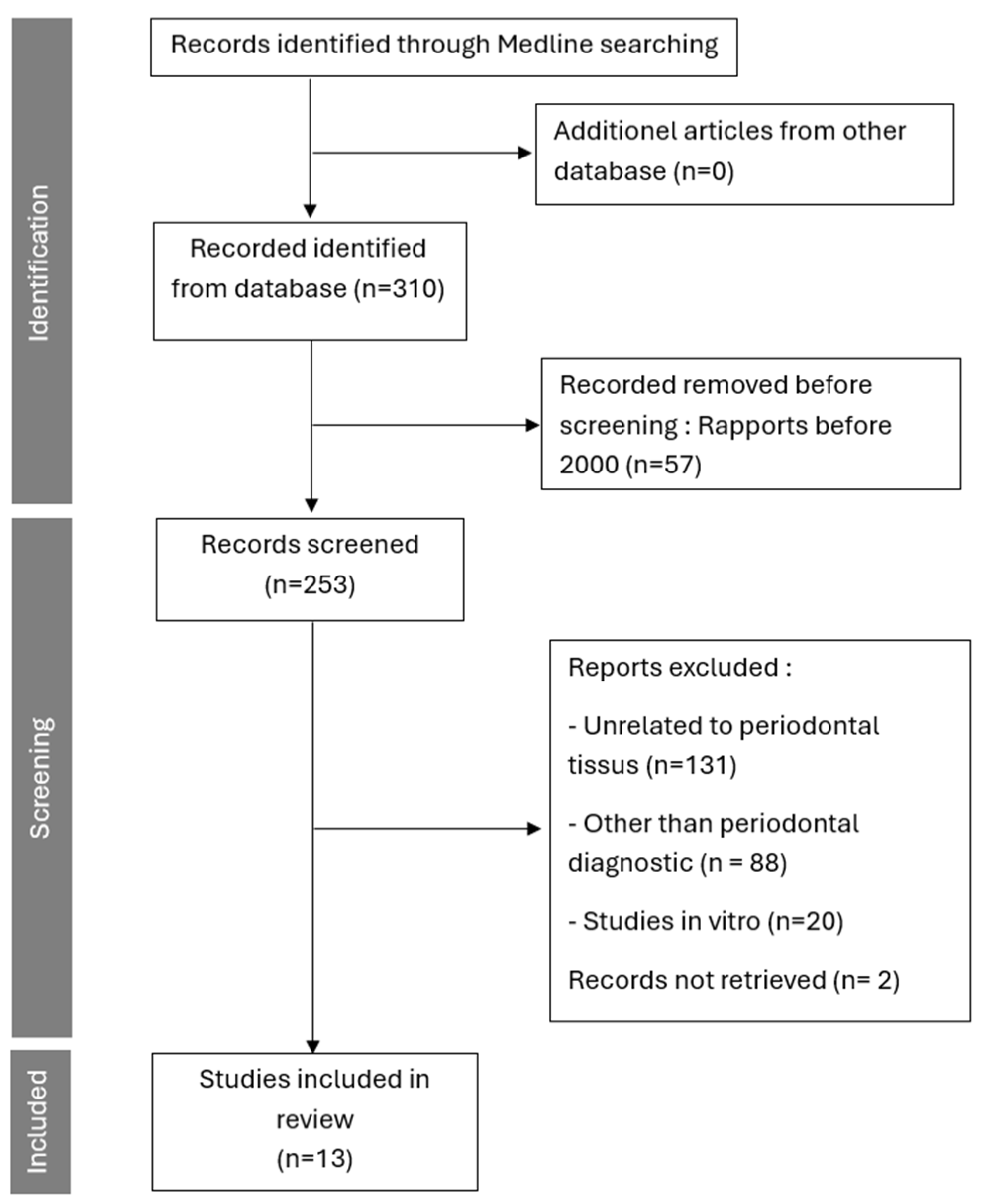
The search identified 310 published articles, 253 were published since the 1 January 2000. After applying the inclusion and exclusion criteria, 13 articles were included in this review (Figure 2). Among these articles, 7 focused on thermography, and 6 on spectroscopy as techniques for detecting periodontal inflammation, with a total of 712 patients.

3.1.1. Presentation of IR Thermography Studies

Regarding thermography, among the 7 studies included in the analysis (Table 1), we were able to identify three common themes: room temperature, camera distance, and the spectrum used by the camera. Studies included in the analysis were clinical trials using thermography technique in comparison with standard clinical examination. Thermography studies consistently showed temperature increases of 0.5–2 °C in inflamed sites compared to controls, with methodological variability in camera distances (20–50 cm) and room temperatures (20–25 °C).
Regarding spectroscopy, among the 6 studies included in the analysis (Table 2), two common themes were highlighted: the biophysical properties used, and the spectrum range of the camera. The studies included in the analysis were clinical trials based on spectroscopy technique and compared to standard clinical examination. Spectroscopy studies demonstrated reduced tissue oxygenation (e.g., 10–20% lower in inflamed vs. normal sites) and elevated total hemoglobin, with consistencies in NIR range use but discrepancies in patient subgroups (e.g., higher variability in diabetics). Across thermography and spectroscopy, similarities include non-invasive detection of vascular changes, but discrepancies involve measurement depth and quantitative vs. qualitative outputs.

3.1.2. Evolution of the Studies

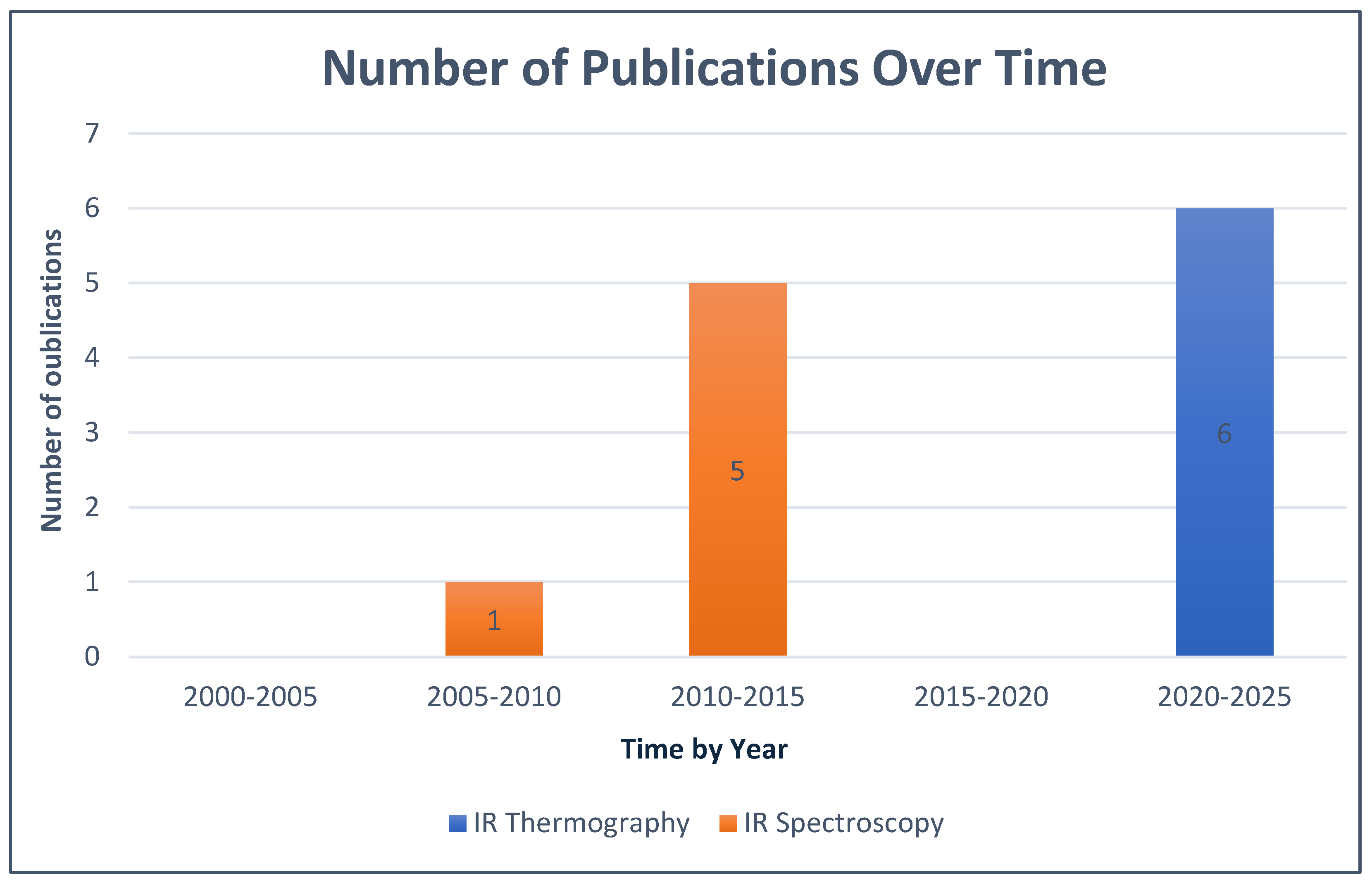
The application of IR spectroscopy and thermography for in vivo periodontal inflammation visualization was not widely practiced, and publications on this topic were limited. According to the publication periods used in this review, studies focusing on IR spectroscopy in this context only began in 2009, with no publications since 2015, while those on IR thermography in the same context only started later, in 2021. These data are presented below in Figure 3.

3.1.3. Presentation of Cameras Used for IR Thermography

This section provides a brief overview of the most important aspects of infrared cameras used for periodontal inflammation detection through thermography.
The literature presents various camera models and their characteristics (thermal resolution, infrared sensor, acquisition frequency) collected directly from articles data or camera’s technical specifications available on the manufacturer’s website [38] (The data collected outside of the scientific articles from the PubMed database are indicated with an asterisk* in the table.) It is worth noting that all the articles included in the review mentioned the type of camera used.
The collected characteristics, along with the image analysis software, are presented in Table 3 below. The image analysis and processing software are listed for reference, with various software packages being used, including ThermaCam and Researcher Pro, which were employed twice in two different studies by the same team with two different versions.

3.1.4. Overview of Devices Used for IR Spectroscopy

All spectroscopy studies utilized a miniaturized NIR device (e.g., Inspectra Spectrometer) with fiber-optic probes, measuring hemoglobin indices at 700–1000 nm, analyzed via custom software. All the studies included on infrared spectroscopy technology used the same diagnostic device because they were conducted by the same research team members, with the aim of validating the device on different subjects. The device and associated equipment used for performing the diagnosis and analyzing the results are presented in detail in Table 4 below.

4. Discussion

The host’s immune and inflammatory responses play a key role in the pathogenesis of periodontal disease. To formulate hypotheses and assess the feasibility of new periodontal diagnostic techniques, it is essential to first identify the clinical signs upon which we can rely to detect them in accordance with the technology used. In this review, two key signs were considered both reliable and potentially useful for improving periodontal diagnosis: edema and tissue temperature increase [39].
The heat emitted by tissues is a specific property detectable by infrared thermography [40]. In the context of periodontal inflammation, the increase in vascular dilation and permeability of the gingival tissues causes a higher heat production compared to normal state, which allows inflammation to be characterized using this technical criterion.
The included articles show that thermography can serve as an innovative diagnostic alternative or complement due to its non-invasive nature, particularly when radiological examination is contraindicated [25] such as in pregnant women during their first trimester. However, thermography has several limitations, such as limited tissue penetration [29], making it difficult to detect inflammation in the deeper tissues.
On a different note, IR spectroscopy primarily relies on edema to detect inflammation. Edema is a macroscopic sign resulting from the worsening of several microscopic processes related to hemodynamic changes during periodontal inflammation [37]. Among these changes, vascular dilation and increased permeability, as previously mentioned, are some of the most characteristic markers of gingival inflammation [35]. When infrared light passes through tissues, it selectively interacts with chromophores associated with oxygen, allowing for the real-time measurement of various tissue parameters like hemoglobin (oxygen carrier). This method offers a direct link to the oxygenation status and tissue perfusion. Most studies confirm that decreased tissue oxygenation is associated with an inflammatory state of the tissues, with a notable difference between periodontitis and gingivitis depending on the degree of inflammation. The more the inflammation progresses, the more tissue oxygenation decreases. In parallel, the tHB (total hemoglobin) index, which reflects regional blood volume, serves as an indicator of tissue perfusion status. Its elevation in periodontal areas, reflects altered vascularization, a characteristic of periodontal inflammation [32,33,34] as found in gingivitis or periodontitis.
Research teams have developed a miniaturized spectroscopy device suitable for periodontal use, based on near-infrared spectrum, the preferred choice for this technology [14]. This device has been validated by included studies investigating its applications on various subjects with different systemic conditions, such as diabetes, heart disease [32,33], or varying habits like smokers [34]. Overall, regarding infrared spectroscopy, the literature remains limited in its exploration beyond the use of a single device. This gap highlights the need for further research to expand its applications and compare them with other existing technologies for periodontal diagnosis. Compared to probing (invasive, subjective) and radiography (ionizing, late-stage detection), thermography offers real-time, non-contact heat mapping with high sensitivity for superficial inflammation, though limited penetration; spectroscopy provides quantitative hemodynamic data, superior for deeper assessment but requires device miniaturization. Advantages include safety and early detection, but challenges like cost and standardization persist. Thermography and spectroscopy share non-invasive feasibility but differ in depth (superficial vs. deeper); while technically viable, clinical applicability remains exploratory without validated thresholds, emphasizing need for larger trials over immediate use.
Because we focused on in vivo detection approach, the studies included in this review concentrate on periodontal inflammation tissue signs. However, this does not exclude the relevance for in vitro techniques, such as spectroscopy on saliva samples [41,42] or crevicular fluid [43,44], which remain potentially useful for early periodontal diagnosis.
In the same context, periodontal inflammation detection has been shown to be possible using near-infrared spectrum (near-infrared) through spectroscopy. However, there are other potential ways to exploit this light spectrum through similar innovative techniques, such as near-infrared Imaging (NIRI) technology, which is making its way into the oral healthcare field with the latest generation intraoral scanners [45,46]. Further studies are therefore needed to explore these new technologies through techniques already integrated into devices used in dental clinics.
Finally, to improve our review, it would be relevant to explore sources from other scientific disciplines, particularly databases used by biomedical imaging technicians and engineers, in order to obtain more detailed technical information on the technologies employed.
This review reveals the current literature on periodontal inflammation detection through the near and far IR spectrum. However, studies have shown the usefulness of other spectra on different tissues, such as skin tissue [14], Due to the histological similarity between mucosal and skin tissues, it is estimated that other spectra (mid-IR) and other techniques (Raman Spectroscopy) could be useful and exploitable for the detection of periodontal inflammation [43,47].
The use of medical imaging techniques in dental medicine is, to date, an underexplored field, which is why it represents an interesting research area.

5. Perspectives

The infrared spectrum, through these two approaches, shows promising potential that could contribute to improving the early diagnosis of periodontal diseases soon. However, further research is needed to identify the best practices for using this spectrum in this context.
With recent technological advancements in thermal cameras, infrared thermography is now a promising method for the detection of periodontal inflammation [30]. However, given these limitations, it is important to note that the clinical use of this technique still requires further in-depth studies to validate its effectiveness and improve its specific utility as an independent diagnostic tool.
Recent advancements, such as the integration of artificial intelligence with thermography, have shown enhanced accuracy in classifying gingival inflammation, particularly in specific populations like mouth breathers. This highlights the potential for AI to address limitations in manual analysis and improve diagnostic precision [31].
On the other hand, infrared spectroscopy, which has been used for years in the medical field, is making its way into periodontology through innovative devices designed by research teams [35,37]. This offers better prospects for reducing the size of spectrographs and facilitating their use by periodontists. This review highlights a promising start and the potential for exploiting this technique in the diagnosis of periodontal diseases, but there is still a long way to go before thermography becomes widely used in dental clinics.
Although no study has addressed this topic so far, a clinical study combining both techniques in a single device for periodontal diagnosis could be useful to maximize the effectiveness of these two techniques and achieve better diagnostic performance. This approach would allow one technique to offset the limitations of the other and/or simultaneously collect data from both techniques in order to achieve the best possible result through an expanded analysis of this data, facilitated by the introduction of artificial intelligence.

6. Conclusions

Numerous publications have demonstrated the potential of thermography and infrared spectroscopy (IR) for the in vivo detection of periodontal inflammation. These findings open new perspectives regarding the integration of these technologies into clinical periodontal practice in the near future. These non-invasive methods could allow a more precise and rapid assessment of the inflammatory status of periodontal tissues, providing a complementary tool to current diagnostic techniques. Although preliminary results are promising, this field remains largely underexplored, and several challenges must be addressed before widespread adoption. Further studies are needed to confirm the effectiveness, reliability, and reproducibility of these technologies in the daily practice of periodontists, considering the pathophysiological variables specific to each patient and the lack of standardized thresholds or validated metrics like sensitivity/specificity. These studies will help to determine the optimal conditions for their use and assess their impact on the early diagnosis of periodontal diseases in the future.

Supplementary Materials

The following supporting information can be downloaded at: https://www.mdpi.com/article/10.3390/diagnostics16020222/s1.

Author Contributions

Conceptualization, M.R. and G.Y.R.; Methodology, F.D., M.R. and G.Y.R.; Data curation, H.N.G. and M.R.; Writing—original draft preparation, H.N.G. and M.R.; Writing—review and editing, M.G. and G.Y.R.; Visualization, L.E. and V.R.; Supervision, M.R. and G.Y.R. All authors have read and agreed to the published version of the manuscript.

Funding

This research received no external funding.

Institutional Review Board Statement

Not applicable.

Informed Consent Statement

Not applicable.

Data Availability Statement

No new data were created or analyzed in this study. Data sharing is not applicable to this article.

Conflicts of Interest

The authors declare no conflicts of interest.

References

  1. Chapple, I.L.C.; Mealey, B.L.; Van Dyke, T.E.; Bartold, P.M.; Dommisch, H.; Eickholz, P.; Geisinger, M.L.; Genco, R.J.; Glogauer, M.; Goldstein, M.; et al. Periodontal health and gingival diseases and conditions on an intact and a reduced periodontium: Consensus report of workgroup 1 of the 2017 World Workshop on the Classification of Periodontal and Peri-Implant Diseases and Conditions. J. Clin. Periodontol. 2018, 45, S68–S77. [Google Scholar]
  2. Papapanou, P.N.; Sanz, M.; Buduneli, N.; Dietrich, T.; Feres, M.; Fine, D.H.; Flemmig, T.F.; Garcia, R.; Giannobile, W.V.; Graziani, F.; et al. Periodontitis: Consensus report of workgroup 2 of the 2017 World Workshop on the Classification of Periodontal and Peri-Implant Diseases and Conditions. J. Clin. Periodontol. 2018, 45, S162–S170. Available online: https://onlinelibrary.wiley.com/doi/10.1111/jcpe.12946 (accessed on 5 January 2026).
  3. Herrera, D.; Sanz, M.; Shapira, L.; Brotons, C.; Chapple, I.; Frese, T.; Graziani, F.; Hobbs, F.D.R.; Huck, O.; Hummers, E.; et al. Association between periodontal diseases and cardiovascular diseases, diabetes and respiratory diseases: Consensus report of the Joint Workshop by the European Federation of Periodontology (EFP) and the European arm of the World Organization of Family Doctors (WONCA Europe). J. Clin. Periodontol. 2023, 50, 819–841. [Google Scholar]
  4. Bui, F.Q.; Almeida-da-Silva, C.L.C.; Huynh, B.; Trinh, A.; Liu, J.; Woodward, J.; Asadi, H.; Ojcius, D.M. Association between periodontal pathogens and systemic disease. Biomed. J. 2019, 42, 27–35. [Google Scholar] [CrossRef] [Scilit] [PubMed]
  5. Preshaw, P.M.; Alba, A.L.; Herrera, D.; Jepsen, S.; Konstantinidis, A.; Makrilakis, K.; Taylor, R. Periodontitis and diabetes: A two-way relationship. Diabetologia 2012, 55, 21–31. [Google Scholar] [PubMed]
  6. Sanz, M.; Marco Del Castillo, A.; Jepsen, S.; Gonzalez-Juanatey, J.R.; D’Aiuto, F.; Bouchard, P.; Chapple, I.; Dietrich, T.; Gotsman, I.; Graziani, F.; et al. Periodontitis and cardiovascular diseases: Consensus report. J. Clin. Periodontol. 2020, 47, 268–288. [Google Scholar] [CrossRef] [Scilit]
  7. Caton, J.G.; Armitage, G.; Berglundh, T.; Chapple, I.L.C.; Jepsen, S.; Kornman, K.S.; Mealey, B.L.; Papapanou, P.N.; Sanz, M.; Tonetti, M.S. A New Classification Scheme for Periodontal and Peri-Implant Diseases and Conditions—Introduction and Key Changes from the 1999 Classification. J. Clin. Periodontol. 2018, 45, S1–S8. Available online: https://onlinelibrary.wiley.com/doi/10.1111/jcpe.12935 (accessed on 5 January 2026).
  8. Chen, I.D.S.; Yang, C.M.; Chen, M.J.; Chen, M.C.; Weng, R.M.; Yeh, C.H. Deep Learning-Based Recognition of Periodontitis and Dental Caries in Dental X-ray Images. Bioengineering 2023, 10, 911. [Google Scholar]
  9. Heo, J.S.; Ahn, K.H.; Park, J.S. Radiological screening of maternal periodontitis for predicting adverse pregnancy and neonatal outcomes. Sci. Rep. 2020, 10, 21266. [Google Scholar] [CrossRef] [Scilit] [PubMed]
  10. Benn, D.K.; Vig, P.S. Estimation of x-ray radiation related cancers in US dental offices: Is it worth the risk? Oral Surg. Oral Med. Oral Pathol. Oral Radiol. 2021, 132, 597–608. [Google Scholar] [CrossRef] [Scilit]
  11. Benavides, E.; Krecioch, J.R.; Connolly, R.T.; Allareddy, T.; Buchanan, A.; Spelic, D.; O’Brien, K.K.; Keels, M.A.; Mascarenhas, A.K.; Duong, M.L.; et al. Optimizing radiation safety in dentistry. J. Am. Dent. Assoc. 2024, 155, 280–293.e4. [Google Scholar] [CrossRef] [Scilit]
  12. American Dental Association Council on Scientific Affairs. The use of cone-beam computed tomography in dentistry: An advisory statement from the American Dental Association Council on Scientific Affairs. J. Am. Dent. Assoc. 2012, 143, 899–902. [Google Scholar]
  13. Tattersall, G.J. Infrared thermography: A non-invasive window into thermal physiology. Comp. Biochem. Physiol. Part A Mol. Integr. Physiol. 2016, 202, 78–98. [Google Scholar] [CrossRef] [Scilit]
  14. Gurjarpadhye, A.A.; Parekh, M.B.; Dubnika, A.; Rajadas, J.; Inayathullah, M. Infrared Imaging Tools for Diagnostic Applications in Dermatology. SM J. Clin. Med. Imaging 2015, 1, 1–5. [Google Scholar]
  15. Mapstone, R. Corneal thermal patterns in anterior uveitis. Br. J. Ophthalmol. 1968, 52, 917–921. [Google Scholar] [CrossRef] [Scilit] [PubMed]
  16. Paterson, J.; Watson, W.S.; Teasdale, E.; Evans, A.L.; Newman, P.; James, W.B.; Pitkeathly, D. Assessment of rheumatoid inflammation in the knee joint. A reappraisal. Ann. Rheum. Dis. 1978, 37, 48–52. [Google Scholar] [CrossRef] [Scilit] [PubMed]
  17. Bonmarin, M.; Le Gal, F. Chapitre 31—L’imagerie thermique en dermatologie. In L’imagerie en Dermatologie; Academic Press: Amsterdam, The Netherlands, 2016; pp. 437–454. [Google Scholar] [CrossRef] [Scilit]
  18. Liu, Q.; Li, M.; Wang, W.; Jin, S.; Piao, H.; Jiang, Y.; Li, N.; Yao, H. Infrared thermography in clinical practice: A literature review. Eur. J. Med. Res. 2025, 30, 33. [Google Scholar] [CrossRef] [Scilit] [PubMed]
  19. Kesztyüs, D.; Brucher, S.; Wilson, C.; Kesztyüs, T. Use of Infrared Thermography in Medical Diagnosis, Screening, and Disease Monitoring: A Scoping Review. Medicina 2023, 59, 2139. [Google Scholar] [CrossRef] [Scilit]
  20. Claßen, J.; Aupert, F.; Reardon, K.F.; Solle, D.; Scheper, T. Spectroscopic sensors for in-line bioprocess monitoring in research and pharmaceutical industrial application. Anal. Bioanal. Chem. 2017, 409, 651–666. [Google Scholar] [CrossRef] [Scilit]
  21. Delrue, C.; De Bruyne, S.; Speeckaert, M.M. The Potential Use of Near- and Mid-Infrared Spectroscopy in Kidney Diseases. Int. J. Mol. Sci. 2023, 24, 6740. [Google Scholar] [CrossRef] [Scilit]
  22. Wu, X.; Dai, Y.; Wang, L.; Peng, Y.; Lu, L.; Zhu, Y.; Shi, Y.; Zhuang, S. Diagnosis of methylglyoxal in blood by using far-infrared spectroscopy and o-phenylenediamine derivation. Biomed. Opt. Express 2020, 11, 960–973. [Google Scholar]
  23. Sakuma, S.; Inamoto, K.; Higuchi, N.; Ariji, Y.; Nakayama, M.; Izumi, M. Experimental pain in the gingiva and its impact on prefrontal cortical hemodynamics: A functional near-infrared spectroscopy study. Neurosci. Lett. 2014, 575, 74–79. [Google Scholar] [CrossRef] [Scilit]
  24. Page, M.J.; McKenzie, J.E.; Bossuyt, P.M.; Boutron, I.; Hoffmann, T.C.; Mulrow, C.D.; Shamseer, L.; Tetzlaff, J.M.; Akl, E.A.; Brennan, S.E.; et al. The PRISMA 2020 statement: An updated guideline for reporting systematic reviews. BMJ 2021, 10, n71. [Google Scholar] [CrossRef] [Scilit] [PubMed]
  25. Wziątek-Kuczmik, D.; Mrowiec, A.; Niedzielska, I.; Stanek, A.; Cholewka, A. Registration of thermal images of dead teeth to identify odontogenic infection foci. Sci. Rep. 2024, 14, 21405. [Google Scholar] [CrossRef] [Scilit]
  26. Bezerra De Melo, N.; Sobreira Duarte, L.N.; Maia Vieira Pereira, C.; Da Silva Barbosa, J.; Matos Gonçalves Da Silva, A.; De Souza Coelho Soares, R.; Bento, P.M. Thermographic examination of gingival phenotypes: Correlation between morphological and thermal parameters. Clin. Oral Investig. 2023, 27, 7705–7714. [Google Scholar] [CrossRef] [Scilit]
  27. Wziątek-Kuczmik, D.; Niedzielska, I.; Mrowiec, A.; Bałamut, K.; Handzel, M.; Szurko, A. Is Thermal Imaging a Helpful Tool in Diagnosis of Asymptomatic Odontogenic Infection Foci—A Pilot Study. Int. J. Environ. Res. Public Health 2022, 19, 16325. [Google Scholar] [CrossRef] [Scilit] [PubMed]
  28. Derruau, S.; Bogard, F.; Exartier-Menard, G.; Mauprivez, C.; Polidori, G. Medical Infrared Thermography in Odontogenic Facial Cellulitis as a Clinical Decision Support Tool. A Technical Note. Diagnostics 2021, 11, 2045. [Google Scholar] [CrossRef] [Scilit]
  29. Delarue, M.; Derruau, S.; Troyon, P.; Bogard, F.; Polidori, G.; Mauprivez, C. Medical infrared thermography in peri-operative management of peripheral ameloblastoma: A case report. Photodiagn. Photodyn. Ther. 2021, 34, 102167. [Google Scholar] [CrossRef] [Scilit] [PubMed]
  30. Aboushady, M.A.; Talaat, W.; Hamdoon, Z.; MElshazly, T.; Ragy, N.; Bourauel, C.; Talaat, S. Thermography as a non-ionizing quantitative tool for diagnosing periapical inflammatory lesions. BMC Oral Health 2021, 21, 260. [Google Scholar] [CrossRef] [Scilit]
  31. Çankaya, Z.T.; Koyuncu, A.; Gürbüz, S. Artificial Intelligence Assisted Thermal Imaging for Gingival Inflammation Assessment: A Novel Approach. J. Esthet. Restor. Dent. 2025. [Google Scholar] [CrossRef] [Scilit]
  32. Duarte, P.M.; Sowa, M.G.; Xiang, X.; Zhang, C.; Santos, V.R.; Miranda, T.S.; Reis, A.F.; Liu, K. Assessment of the hemodynamic profile in periodontal tissues of diabetic subjects with periodontitis by optical spectroscopy. J. Periodontal Res. 2015, 50, 594–601. [Google Scholar]
  33. Zhang, C.; Xiang, X.; Xu, M.; Fan, C.; Sowa, M.G.; Liu, K.Z. Assessment of tissue oxygenation of periodontal inflammation in patients with coronary artery diseases using optical spectroscopy. BMC Oral Health 2014, 14, 25. [Google Scholar] [CrossRef] [Scilit]
  34. Liu, K.; Duarte, P.M.; Santos, V.R.; Xiang, X.; Xu, M.; Miranda, T.S.; Fermiano, D.; Gonçalves, T.E.D.; Sowa, M.G. Assessment of tissue oxygenation of periodontal inflammation in smokers using optical spectroscopy. J. Clin. Periodontol. 2014, 41, 340–347. [Google Scholar] [CrossRef] [Scilit]
  35. Ge, Z.; Liu, K.Z.; Xiang, X.; Yang, Q.; Hui, J.; Kohlenberg, E.; Sowa, M.G. Assessment of local hemodynamics in periodontal inflammation using optical spectroscopy. J. Periodontol. 2011, 82, 1161–1168. [Google Scholar] [CrossRef] [Scilit]
  36. Nogueira-Filho, G.; Xiang, X.M.; Shibli, J.A.; Duarte, P.M.; Sowa, M.G.; Ferrari, D.S.; Onuma, T.; de Cardoso, L.A.G.; Liu, K.-Z. On site noninvasive assessment of peri-implant inflammation by optical spectroscopy. J. Periodontal Res. 2011, 46, 382–388. [Google Scholar] [CrossRef] [Scilit]
  37. Liu, K.Z.; Xiang, X.M.; Man, A.; Sowa, M.G.; Cholakis, A.; Ghiabi, E.; Singer, D.L.; Scott, D.A. In vivo determination of multiple indices of periodontal inflammation by optical spectroscopy. J. Periodontal Res. 2009, 44, 117–124. [Google Scholar] [CrossRef] [Scilit] [PubMed]
  38. FLIR T1020. Available online: https://www.flir.fr/products/t1020/ (accessed on 5 January 2026).
  39. Prasanth, C.S.; Betsy, J.; Jayanthi, J.L.; Nisha, U.G.; Prasantila, J.; Subhash, N. In vivo inflammation mapping of periodontal disease based on diffuse reflectance spectral imaging: A clinical study. J. Biomed. Opt. 2013, 18, 26019. [Google Scholar] [CrossRef] [Scilit]
  40. Bhargava, A.; Chanmugam, A.; Herman, C. Heat transfer model for deep tissue injury: A step towards an early thermographic diagnostic capability. Diagn. Pathol. 2014, 9, 36. [Google Scholar] [CrossRef] [Scilit] [PubMed]
  41. Xiang, X.; Sowa, M.G.; Iacopino, A.M.; Maev, R.G.; Hewko, M.D.; Man, A.; Liu, K.Z. An Update on Novel Non-Invasive Approaches for Periodontal Diagnosis. J. Periodontol. 2010, 81, 186–198. [Google Scholar] [CrossRef] [Scilit]
  42. Delrue, C.; De Bruyne, S.; Speeckaert, M.M. Unlocking the Diagnostic Potential of Saliva: A Comprehensive Review of Infrared Spectroscopy and Its Applications in Salivary Analysis. J. Pers. Med. 2023, 13, 907. [Google Scholar] [CrossRef] [Scilit] [PubMed]
  43. Xiang, X.; Duarte, P.M.; Lima, J.A.; Santos, V.R.; Gonçalves, T.D.; Miranda, T.S.; Liu, K. Diabetes-associated periodontitis molecular features in infrared spectra of gingival crevicular fluid. J Periodontol. 2013, 84, 1792–1800. [Google Scholar] [CrossRef] [Scilit]
  44. Seredin, P.; Litvinova, T.; Ippolitov, Y.; Goloshchapov, D.; Peshkov, Y.; Kashkarov, V.; Ippolitov, I.; Chae, B. A Study of the Association between Primary Oral Pathologies (Dental Caries and Periodontal Diseases) Using Synchrotron Molecular FTIR Spectroscopy in View of the Patient’s Personalized Clinical Picture (Demographics and Anamnesis). Int. J. Mol. Sci. 2024, 25, 6395. [Google Scholar] [CrossRef] [Scilit]
  45. Lin, W.S.; Alfaraj, A.; Lippert, F.; Yang, C.C. Performance of the caries diagnosis feature of intraoral scanners and near-infrared imaging technology-A narrative review. J. Prosthodont. 2023, 32, 114–124. [Google Scholar] [PubMed]
  46. Shmueli, A.; Fux-Noy, A.; Davidovich, E.; Ram, D.; Moskovitz, M. Comparing Images from Near-Infrared Light Reflection and Bitewing Radiography to Detect Proximal Caries in Primary Teeth. Children 2024, 11, 1455. [Google Scholar] [CrossRef] [Scilit] [PubMed]
  47. Albahri, J.; Allison, H.; Whitehead, K.A.; Muhamadali, H. The role of salivary metabolomics in chronic periodontitis: Bridging oral and systemic diseases. Metabolomics 2025, 21, 24. [Google Scholar] [CrossRef] [Scilit] [PubMed]
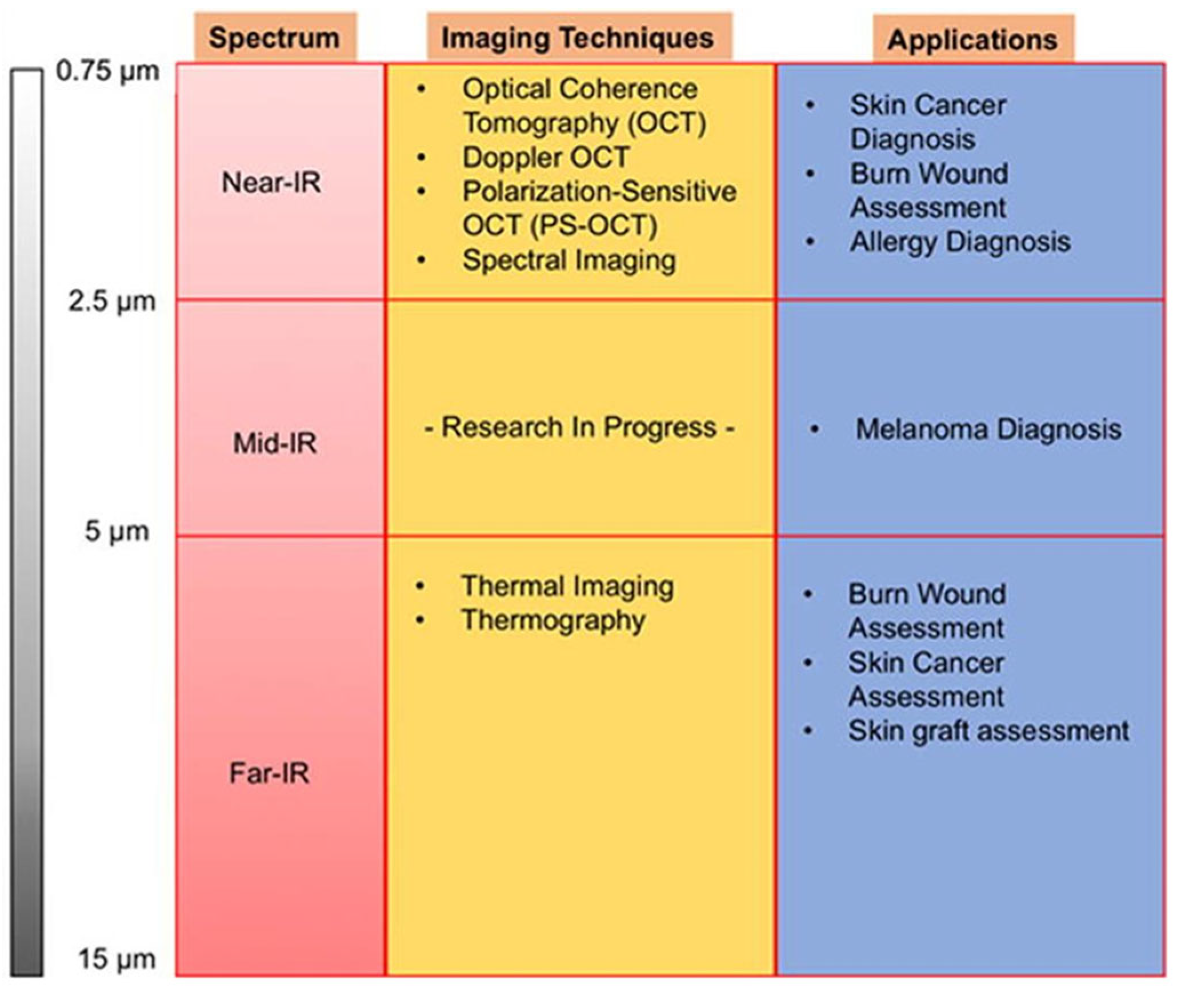
Figure 1. Summary of infrared imaging techniques across infrared spectra (adapted from [14]).
Diagnostics 16 00222 g001
Figure 2. Study Selection Flowchart.
Diagnostics 16 00222 g002
Figure 3. Evolution of Studies Over Time.
Diagnostics 16 00222 g003
Table 1. Overview of IR Thermography Studies.
ReferencesRegions of InterestNumber of PatientsRoom TemperatureObjectivesCamera DistanceSpectrumResults
Wziątek-Kuczmik D et al. 2024 [25]Periapical areas of dead teeth15023 ± 1 (°C)Examine the contribution of thermography in detecting asymptomatic infection sites in patients at high risk of systemic infections. D = 0.4 ± 0.05 mλ from 7.5 to 14 µmThe results showed a significant temperature difference between each patient group and the healthy group based on the measurement time.
Bezerra de Melo N et al. 2023 [26]Gums of the teeth33Between 20 and 25 °C (±1 °C)Analysis of the clinical and thermographic parameters of gingival morphology to detect if there is an exploitable difference, as well as other parameters related to periodontal diagnosis.D = 0.30 cmλ from 7.5 to 14 µmThe results show a significant correlation between thermographic features and clinical gingival parameters.
Wziątek-Kuczmik et al. 2022 [27]Periapical regions of the teeth823.0 ± 1 (°C)Determination of the effectiveness of infrared thermal imaging for detecting the inflammatory response of periapical regions.NCλ from 7.5 to 14 µmThe results demonstrate a difference between the temperatures of the periapical regions of suspect teeth and those of the corresponding regions of healthy teeth, with an average temperature increase ranging from 0.51 to 0.39 °C.
Derruau S et al. 2021 [28]Left buccal space2NCdentification of cellulitis in 2 patients through thermal imaging to validate its usefulness as a reliable diagnostic tool.D = 0.4 ± 0.05 mλ from 7.5 to 14 µmThe thermal results reveal a larger thermally activated area on the affected side compared to the healthy side, with a difference of more than 3 °C for Patient 1 and 2 °C for Patient 2.
Delarue M et al. 2021 [29]Periodontium of the posterior teeth of the mandibular arch1NCEvaluation of tumor margins of small masses and/or tumors (peripheral ameloblastoma) not detected by conventional imaging.NCλ from 7.5 to 14 µmThe results indicate that the peripheral ameloblastoma region was hotter than the surrounding healthy tissues, with an increase in the thermal gradient from 1.5 to 2.5 °C.
Aboushady et al. 2021 [30]Periapical regions of the teeth8020.0 ± 1 (°C)Evaluation of the validity of thermography for the diagnosis of periapical inflammatory lesions and the temperature ranges of acute pulpitis with apical periodontitis, acute and chronic periapical abscesses.Indirect Tec: D < 2 m, Direct Tec: D = 20 cmλ from 7.5 to 13 µmThe results demonstrate a significant difference between the average intra-oral thermal temperatures of the three diagnostic groups, with an increase in temperature in patients with acute pulpitis and apical periodontitis.
Turgut Çankaya Z et al. 2025 [31]Gingival tissues (thermal gingival images annotated and labeled based on bleeding on probing (BoP) and Gingival Index (GI) for inflammation severity)40 participants (stratified by periodontal status and breathing pattern: mouth or nasal breathing)Not specified (performed under standardized imaging conditions)To detect and classify gingival inflammation severity using AI-supported analysis of thermal gingival images in patients with mouth breathing habits, and to establish specific thermal thresholds for gingival health and disease in this populationNot specifiedNot specified (thermal imaging; typically far-infrared (FIR) in the 8–14 µm range for medical thermography, but no explicit details provided)XGBoost classification achieved an accuracy of 92.74%, precision of 92.95%, sensitivity of 92.74%, and F1 score of 92.78%; cross-validation confirmed reliability with mean test score of 88.28% and validation score of 89.43%
Table 2. Overview of IR Spectroscopy Studies.
Ref.Number of PatientsAverage AgeObjectivesKey Parameters MeasuredSpectrumResults
Duarte PM et al. 2015 [32]78Between 35 and 66 yearsEvaluate optical spectroscopy as a periodontal diagnostic method for patients with type 2 diabetes and chronic periodontitis, while documenting the local hemodynamic profile at the periodontal level in these subjects.The relative concentration of deoxygenated hemoglobin (Hb) and oxygenated hemoglobin (HbO2), the balance between oxygen supply and utilization in periodontal tissues.λ from 0.5 to1.1 µmIn diabetic patients, tissue oxygen saturation and HbO2 levels were significantly reduced in periodontitis sites compared to gingivitis sites (p < 0.01). Furthermore, tissue oxygenation in healthy sites was markedly higher in controls than in diabetic subjects (p < 0.01).
Zhang C et al. 2014 Maladie [33]121Between 33 and 71 yearsVerify the ability to identify periodontitis in patients with coronary artery disease using spectroscopy, with an instrument previously designed by the research team.In coronary disease patients, a variation in Hb and HbO2 levels was observed (p < 0.01), and oxygen saturation was reduced in periodontitis sites compared to healthy sites in the diseased patients. In contrast, no difference in saturation was noted between the healthy groups and those with coronary artery disease.
Liu KZ et al. 2014 [34]54Between 35 and 65 yearsAnalyze the effectiveness of spectroscopy in detecting periodontitis in smoking patients, using a device developed by the research team.In smoking patients, tissue oxygen saturation significantly decreased in gingivitis sites (p = 0.016) and periodontitis sites (p = 0.007) compared to healthy sites. A trend of initial increase followed by a decrease in HbO2 concentration was observed, moving from healthy sites to affected sites.
Ge Z et al. 2011 [35]51NCAnalysis of the hemodynamics of periodontal tissues during inflammation using optical spectroscopy.The results reveal that tissue oxygenation significantly decreases between healthy sites, gingivitis sites, and periodontitis sites. This is explained by a notable increase in deoxyhemoglobin between healthy and gingivitis sites, as well as a significant decrease in oxyhemoglobin between gingivitis and periodontitis sites.
Nogueira-Filho G et al. 2011 [36]64NCInvestigation of the diagnostic potential of optical spectroscopy in peri-implant inflammation in vivo.The results indicate that tissue oxygenation at peri-implant sites was reduced compared to healthy sites (p < 0.05) due to an increase in deoxyhemoglobin and a decrease in oxyhemoglobin. Furthermore, the tissue hydration index, calculated from the optical spectra, was significantly higher in cases of mucositis compared to the other groups (p < 0.05).
Liu KZ et al. 2009 [37]30Between 37 and 71 yearsAnalyze the ability of in vivo optical spectroscopy to simultaneously measure multiple inflammatory indices in periodontal tissues.Tissue oxygenation, total tissue hemoglobin, deoxyhemoglobin, oxygenated hemoglobin, and tissue edema.The results highlighted a decrease in oxygenation and an increase in deoxyhemoglobin at periodontitis sites, as well as a variation in the water index associated with electrolytes and temperature between the studied sites.
Table 3. Overview of Devices Used for IR Thermography.
ReferencesType of IR CameraImage ResolutionTemperature ResolutionAcquisition FrequencyImage Analysis Software
Wziątek-Kuczmik D et al. 2024 [25]FLIR T10201024 × 768 pixels *<0.02 °C *f = 30 Hz *ThermaCAM Researcher Pro 2.10
Bezerra de Melo N et al. 2023 [26]FLIR T650640 × 480 pixels0.05 °CNCFLIR Tools+™ 6.4
Wziątek Kuczmik et al. 2022 [27]FLIR T10201024 × 768 pixels *<0.02 °C *f = 30 Hz *ThermaCAM Researcher Pro 2.8 SR-3
Derruau S et al. 2021 [28]VarioCAM® HD1024 × 768 pixelsNCNCIRBIS® 3.1, InfraTec
Delarue M et al. 2021 [29]InfraTech VarioCAM HD1024 × 768 pixels0.03 °CNCNC
Aboushady et al. 2021 [30]FLIR E-5120 × 90 pixelsNCNCFLIR Thermal Analysis and Reporting
Table 4. Overview of Devices Used for IR Spectroscopy.
ReferencesDeviceProbePower of the Light SourceIntegration TimeSpectral RangeResolutionStatistical Analysis Software
Duarte PM et al. 2015 [32]Portable spectro-graph PDA512-ISACustom bifurcated optical fiber probe for oral use5 W0.03 sBetween 500 and 1100 nm5 nmStatistica 7.1
Zhang C et al. 2014 [33]
Liu KZ et al. 2014 [34]
Ge Z et al. 2011 [35]
Nogueira-F et al. 2011 [36]
Liu KZ et al. 2009 [37]
Disclaimer/Publisher’s Note: The statements, opinions and data contained in all publications are solely those of the individual author(s) and contributor(s) and not of MDPI and/or the editor(s). MDPI and/or the editor(s) disclaim responsibility for any injury to people or property resulting from any ideas, methods, instructions or products referred to in the content.