


Occult Neoplasms in Sudden Unexpected Death in Infancy: A Scoping Review of Post-Mortem Findings
 , , , , and
Abstract
1. Introduction
2. Materials and Methods
3. Results
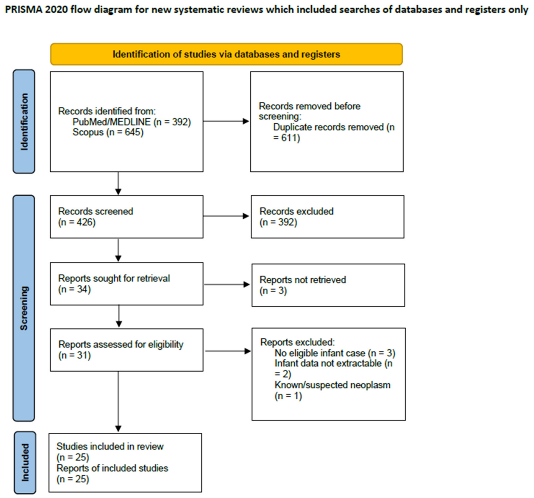
3.1. Study Selection
3.2. Characteristics of the Included Evidence
3.3. Demographic and Clinical Characteristics
3.4. Neoplasm Categories and Pathological Characteristics
3.5. Proposed Mechanisms of Death
3.6. Causal Role of the Neoplasm
3.7. Post-Mortem Diagnostic Investigations and Methodological Appraisal
4. Discussion
5. Conclusions
Supplementary Materials
Author Contributions
Funding
Institutional Review Board Statement
Informed Consent Statement
Data Availability Statement
Acknowledgments
Conflicts of Interest
Abbreviations
| PRISMA-ScR | Preferred Reporting Items for Systematic Reviews and Meta-Analyses Extension for Scoping Reviews |
| SUDI | Sudden unexpected death in infancy |
| SIDS | Sudden infant death syndrome |
| NGS | Next-generation sequencing |
| ECG | Electrocardiogram |
| WHO | World Health Organization |
References
- Garstang, J.; Ellis, C.; Sidebotham, P. An evidence-based guide to the investigation of sudden unexpected death in infancy. Forensic Sci. Med. Pathol. 2015, 11, 345–357. [Google Scholar] [CrossRef] [Scilit]
- Weber, M.A.; Pryce, J.W.; Ashworth, M.T.; Malone, M.; Sebire, N.J. Histological examination in sudden unexpected death in infancy: Evidence base for histological sampling. J. Clin. Pathol. 2012, 65, 58–63. [Google Scholar] [CrossRef] [Scilit]
- Cattermole, R.; Hutchinson, J.C.; Palm, L.; Sebire, N.J. Causes of sudden unexpected death in infants with and without pre-existing conditions: A retrospective autopsy study. BMJ Paediatr. Open 2024, 8, e002641. [Google Scholar] [CrossRef] [Scilit]
- Reyes, J.A.; Somers, G.R.; Chiasson, D.A. Sudden unexpected death in neonates: A clinico-pathological study. Pediatr. Dev. Pathol. 2018, 21, 528–536. [Google Scholar] [CrossRef] [Scilit]
- Camatti, J.; Bonasoni, M.P.; Santunione, A.L.; Cecchi, R.; Radheshi, E.; Carretto, E. Postmortem microbiology in forensic diagnostics: Interpretation of infectious causes of death and emerging applications. Diagnostics 2026, 16, 325. [Google Scholar] [CrossRef] [Scilit]
- Camatti, J.; Santunione, A.L.; Cecchi, R.; Radheshi, E.; Carretto, E.; Bonasoni, M.P. Hidden and under-recognized causes of sudden unexpected death in infancy (SUDI): A comprehensive review of autopsy findings. Diagnostics 2026, 16, 1730. [Google Scholar] [CrossRef] [Scilit]
- Camatti, J.; Santunione, A.L.; Cecchi, R.; Carretto, E.; Radheshi, E.; Bonasoni, M.P. Central nervous system infections in sudden unexpected death in infancy: A systematic review of autopsy-based pediatric pathology. Fetal Pediatr. Pathol. 2026, in press. [Google Scholar] [CrossRef] [Scilit]
- World Health Organization. WHO Recommendations for Conducting an External Inspection of a Body and Filling in the Medical Certificate of Cause of Death (MCCD); World Health Organization: Geneva, Switzerland, 2023. [Google Scholar]
- Ashworth, T.G. Pulmonary blastoma, a true congenital neoplasm. Histopathology 1983, 7, 585–594. [Google Scholar] [CrossRef] [Scilit]
- Ben Khelil, M.; Chkirbene, Y.; Mlika, M.; Haouet, S.; Hamdoun, M. Unexpected infant death secondary to a pulmonary infiltration due to acute myelocytic leukaemia. Malays. J. Pathol. 2017, 39, 193–196. [Google Scholar]
- Böhm, N.; Krebs, G. Solitary rhabdomyoma of the heart. Clinically silent case with sudden, unexpected death in an 11-month-old boy. Eur. J. Pediatr. 1980, 134, 167–172. [Google Scholar] [CrossRef] [Scilit]
- Bryant, V.A.; Booth, J.; Palm, L.; Ashworth, M.; Jacques, T.S.; Sebire, N.J. Childhood neoplasms presenting at autopsy: A 20-year experience. Pediatr. Blood Cancer 2017, 64, e26474. [Google Scholar] [CrossRef] [Scilit]
- Byard, R.W.; Jimenez, C.L.; Moore, L. Mechanisms of sudden death in patients with congenital teratoma. Pediatr. Surg. Int. 1992, 7, 464–467. [Google Scholar] [CrossRef] [Scilit]
- Cascini, F.; Longo, F.; Parenti, D.; Capelli, A. A case of sudden infant death due to a primary cardiac sarcoma. J. Forensic Sci. 2012, 57, 1368–1371. [Google Scholar] [CrossRef] [Scilit]
- Defraia, B.; Buccoliero, A.M.; Bianchi, I.; Faccioli, S.; Grifoni, R.; Focardi, M. Cardiac valve hemangioma in two cases of sudden infant death. Clin. Ter. 2025, 176, 8–13. [Google Scholar] [CrossRef]
- Dempers, J.; Wadee, S.A.; Boyd, T.; Wright, C.; Odendaal, H.J.; Sens, M.A.; Prenatal Alcohol and SIDS and Stillbirth (PASS) Network. Hepatic hemangioendothelioma presenting as sudden unexpected death in infancy: A case report. Pediatr. Dev. Pathol. 2011, 14, 71–74. [Google Scholar] [CrossRef] [Scilit]
- Dominguez, C.; Perkins, A.; Duque, A.; Bravo, V. Primary cardiac tumors in infancy: A case report and literature review. Acad. Forensic Pathol. 2017, 7, 112–118. [Google Scholar] [CrossRef] [Scilit]
- Grellner, W.; Henssge, C. Multiple cardiac rhabdomyoma with exclusively histological manifestation. Forensic Sci. Int. 1996, 78, 1–5. [Google Scholar] [CrossRef] [Scilit]
- Gupta, B.; Ghosh, S.; Kujur, M.; Khetan, K.; Kumar, T. Tricuspid valve hemangioma associated with hypoplastic left heart syndrome presenting as sudden infant death syndrome. Turk Patoloji Derg. 2019, 35, 55–57. [Google Scholar] [CrossRef] [Scilit]
- Irie, W.; Satoh, F.; Sasaki, C.; Ochiai, E.; Kondo, M.; Kamogawa, S.; Saito, Y. An autopsy case of sudden infant death caused by cardiac fibroma. Leg. Med. 2026, 80, 102760. [Google Scholar] [CrossRef] [Scilit]
- Izevbaye, I.; Sun, J.; Fazlollah, L. Numerous cortical tubers and rhabdomyomas in a case of sudden unexpected infant death. Am. J. Forensic Med. Pathol. 2011, 32, 331–335. [Google Scholar] [CrossRef] [Scilit]
- Jaffe, R. Neuroma in the region of the atrioventricular node. Hum. Pathol. 1981, 12, 375–376. [Google Scholar] [CrossRef] [Scilit]
- Kure, K.; Lingamfelter, D.; Taboada, E. Large multifocal cardiac myxoma causing the sudden unexpected death of a 2-month-old infant—A rapidly growing, acquired lesion versus a congenital process?: A case report. Am. J. Forensic Med. Pathol. 2011, 32, 166–168. [Google Scholar] [CrossRef] [Scilit]
- Lunetta, P.; Karikoski, R.; Penttilä, A.; Sajantila, A. Sudden death associated with a multifocal type II hemangioendothelioma of the liver in a 3-month-old infant. Am. J. Forensic Med. Pathol. 2004, 25, 56–59. [Google Scholar] [CrossRef] [Scilit]
- Mecchia, D.; Lavezzi, A.M.; Matturri, L. Primary cardiac fibroma and cardiac conduction system alterations in a case of sudden death of a 4-month-old infant. Open Cardiovasc. Med. J. 2013, 7, 47–49. [Google Scholar] [CrossRef] [Scilit]
- Mecklenburg, K.L.; Mitchell, E.K.; Prahlow, J.A. Cardiac rhabdomyoma: An uncommon culprit in sudden infant death. Forensic Sci. Med. Pathol. 2024, 20, 977–982. [Google Scholar] [CrossRef] [Scilit]
- Morota, K.; Shimizu, M.; Sugitate, R.; Ide, M.; Yamato, G.; Tomizawa, D.; Muramatsu, K.; Matsui, A. Sudden unexpected death caused by infantile acute lymphoblastic leukaemia. Oxf. Med. Case Rep. 2021, 2021, omab073. [Google Scholar] [CrossRef] [Scilit]
- Ottaviani, G.; Rossi, L.; Ramos, S.G.; Matturri, L. Pathology of the heart and conduction system in a case of sudden death due to a cardiac fibroma in a 6-month-old child. Cardiovasc. Pathol. 1999, 8, 109–112. [Google Scholar] [CrossRef] [Scilit]
- Ottaviani, G.; Matturri, L.; Rossi, L.; Lavezzi, A.M.; James, T.N. Multifocal cardiac Purkinje cell tumor in infancy. Europace 2004, 6, 138–141. [Google Scholar] [CrossRef] [Scilit]
- Pryce, J.; Kiho, L.; Scheimberg, I. Sudden unexpected death in infancy associated with an epithelial-type hepatoblastoma in a 6-month-old infant. Pediatr. Dev. Pathol. 2010, 13, 338–340. [Google Scholar] [CrossRef] [Scilit]
- Roesdahl, I.; Thomsen, J.L. Sudden infant death syndrome and lipoma: The presence of fat tissue in the spinal canal. Med. Sci. Law 1995, 35, 354–356. [Google Scholar] [CrossRef] [Scilit]
- Somers, G.R.; Smith, C.R.; Perrin, D.G.; Wilson, G.J.; Taylor, G.P. Sudden unexpected death in infancy and childhood due to undiagnosed neoplasia: An autopsy study. Am. J. Forensic Med. Pathol. 2006, 27, 64–69. [Google Scholar] [CrossRef] [Scilit]
- Whybourne, A.; Zillman, M.A.; Miliauskas, J.; Byard, R.W. Sudden and unexpected infant death due to occult lymphoblastic leukaemia. J. Clin. Forensic Med. 2001, 8, 160–162. [Google Scholar] [CrossRef] [Scilit]
- Page, M.J.; McKenzie, J.E.; Bossuyt, P.M.; Boutron, I.; Hoffmann, T.C.; Mulrow, C.D.; Shamseer, L.; Tetzlaff, J.M.; Akl, E.A.; Brennan, S.E.; et al. The PRISMA 2020 statement: An updated guideline for reporting systematic reviews. BMJ 2021, 372, n71. [Google Scholar] [CrossRef] [Scilit]
- Tricco, A.C.; Lillie, E.; Zarin, W.; O’Brien, K.K.; Colquhoun, H.; Levac, D.; Moher, D.; Peters, M.D.; Horsley, T.; Weeks, L.; et al. PRISMA Extension for Scoping Reviews (PRISMA-ScR): Checklist and Explanation. Ann. Intern. Med. 2018, 169, 467–473. [Google Scholar] [CrossRef] [Scilit]
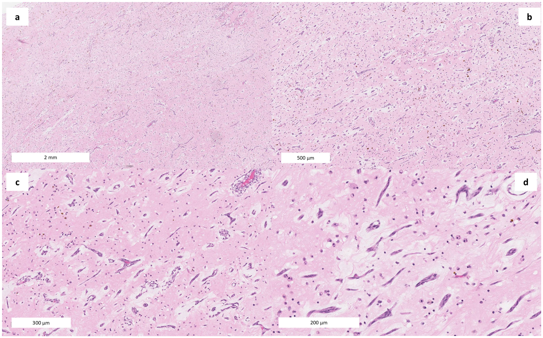
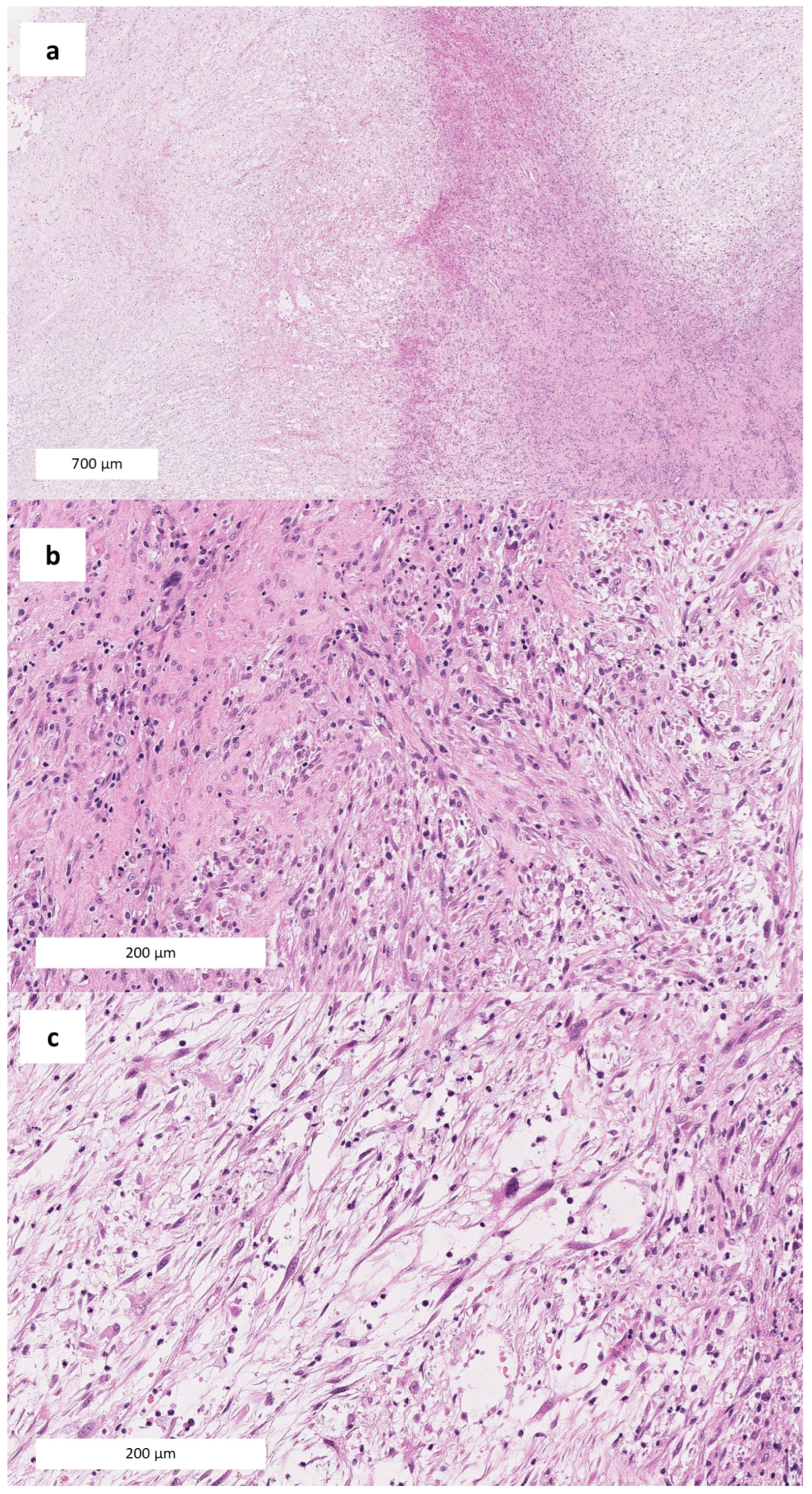
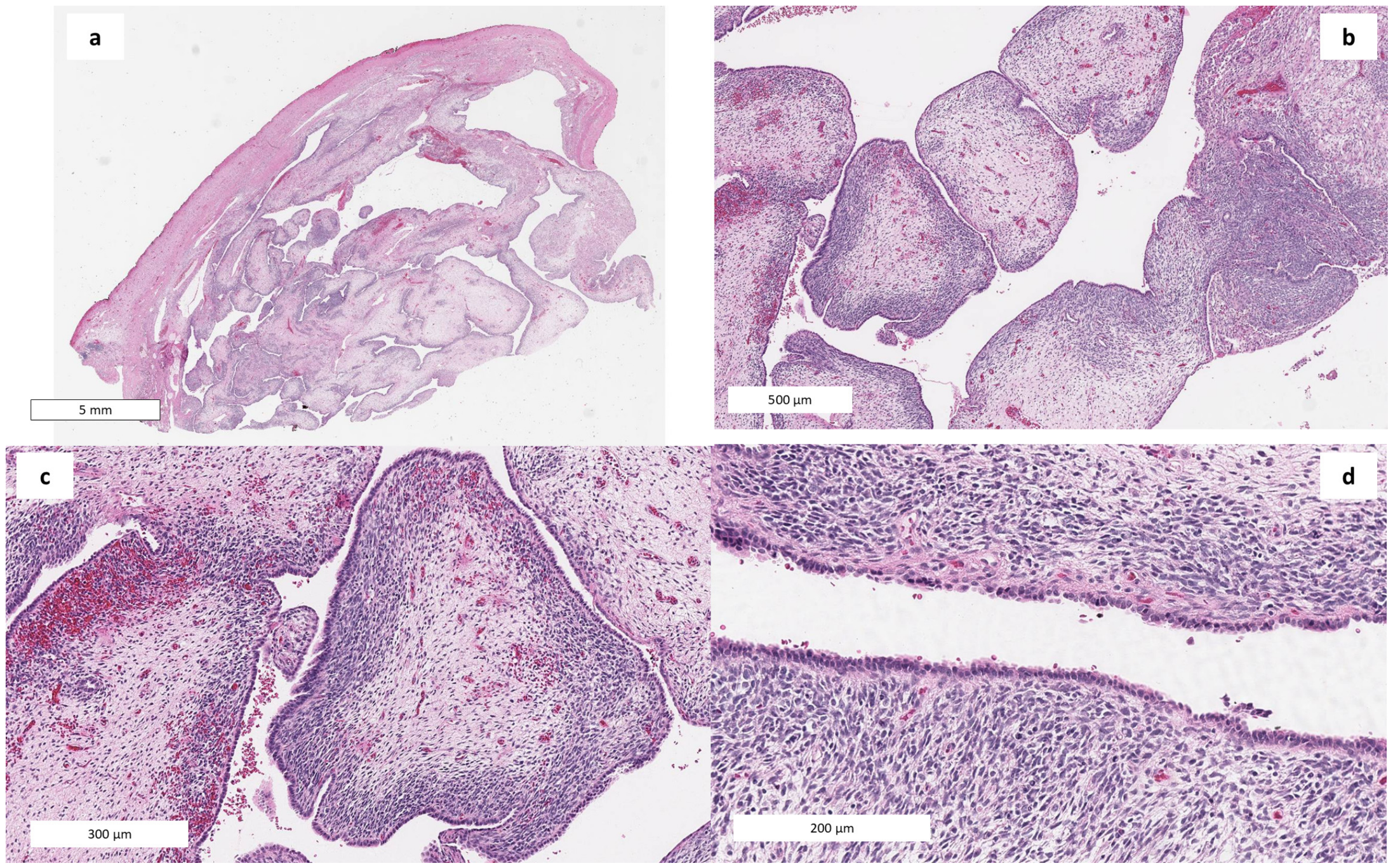
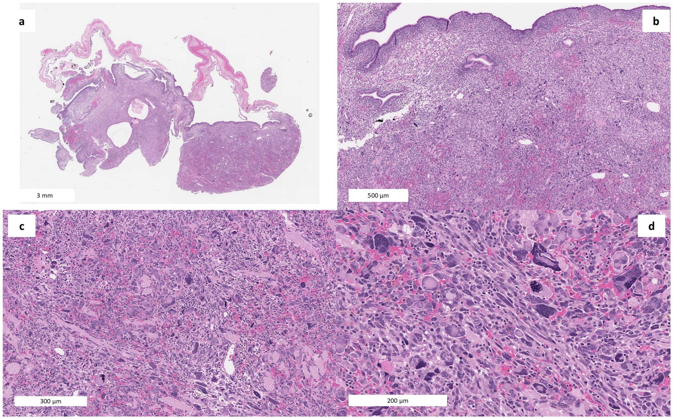
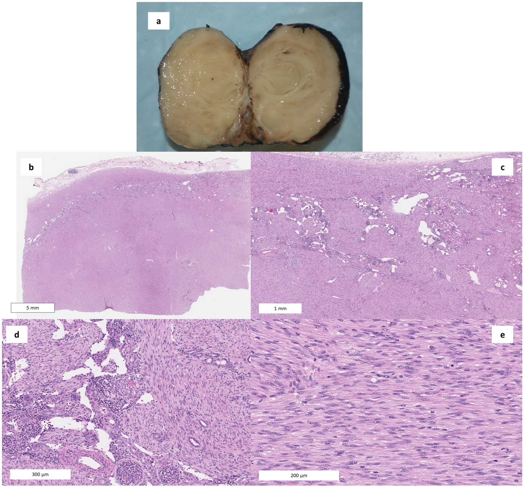
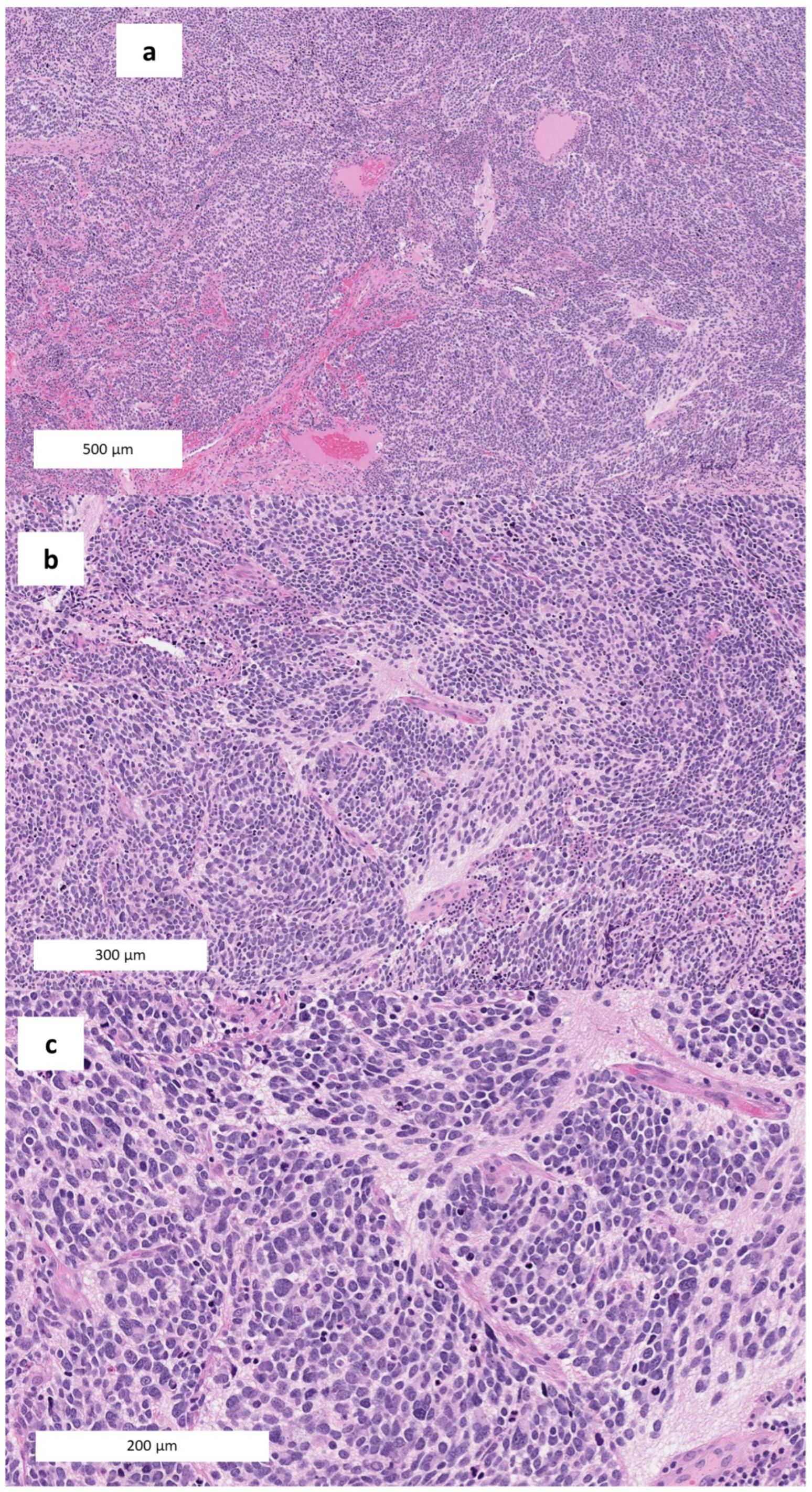
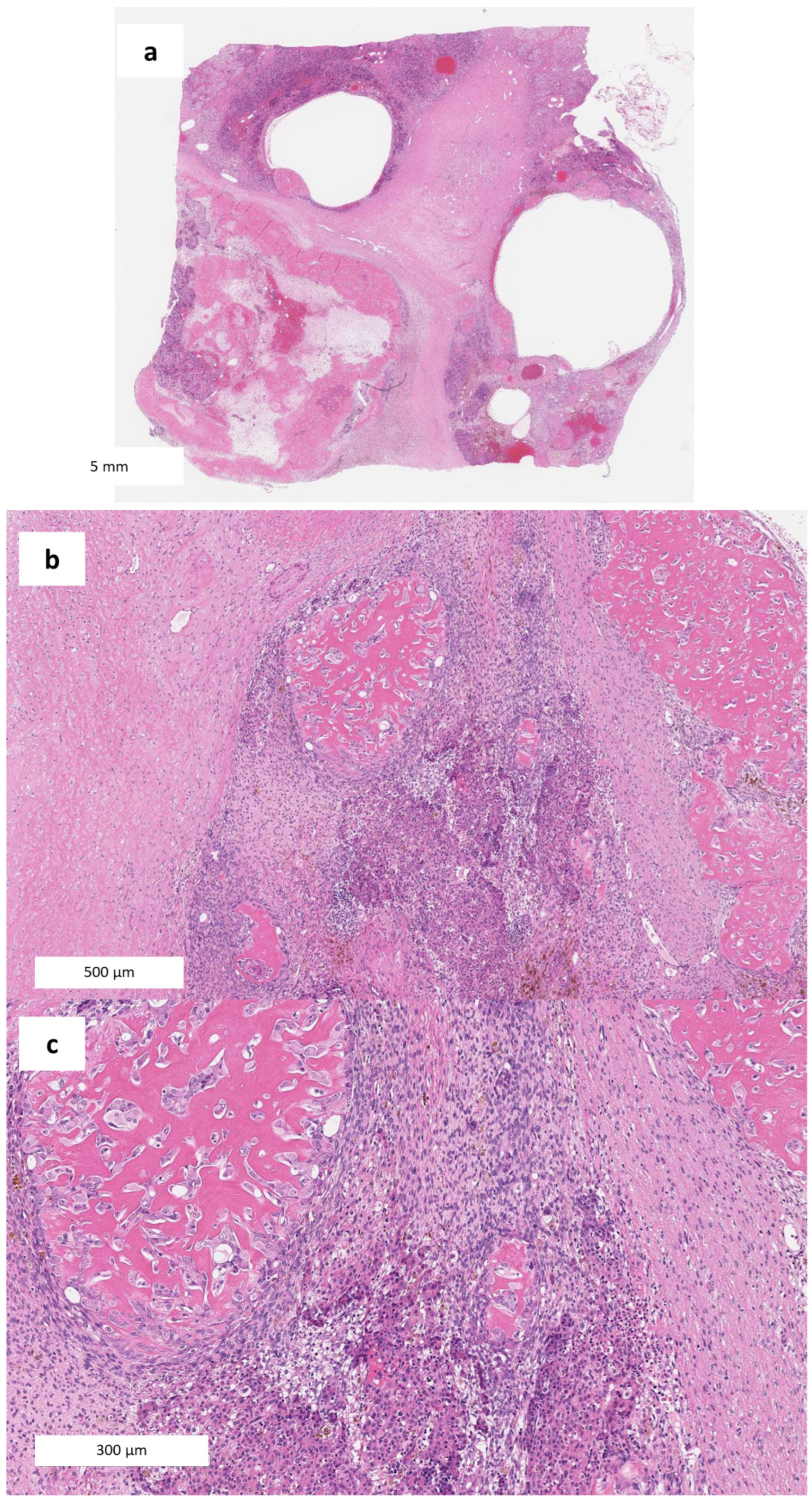
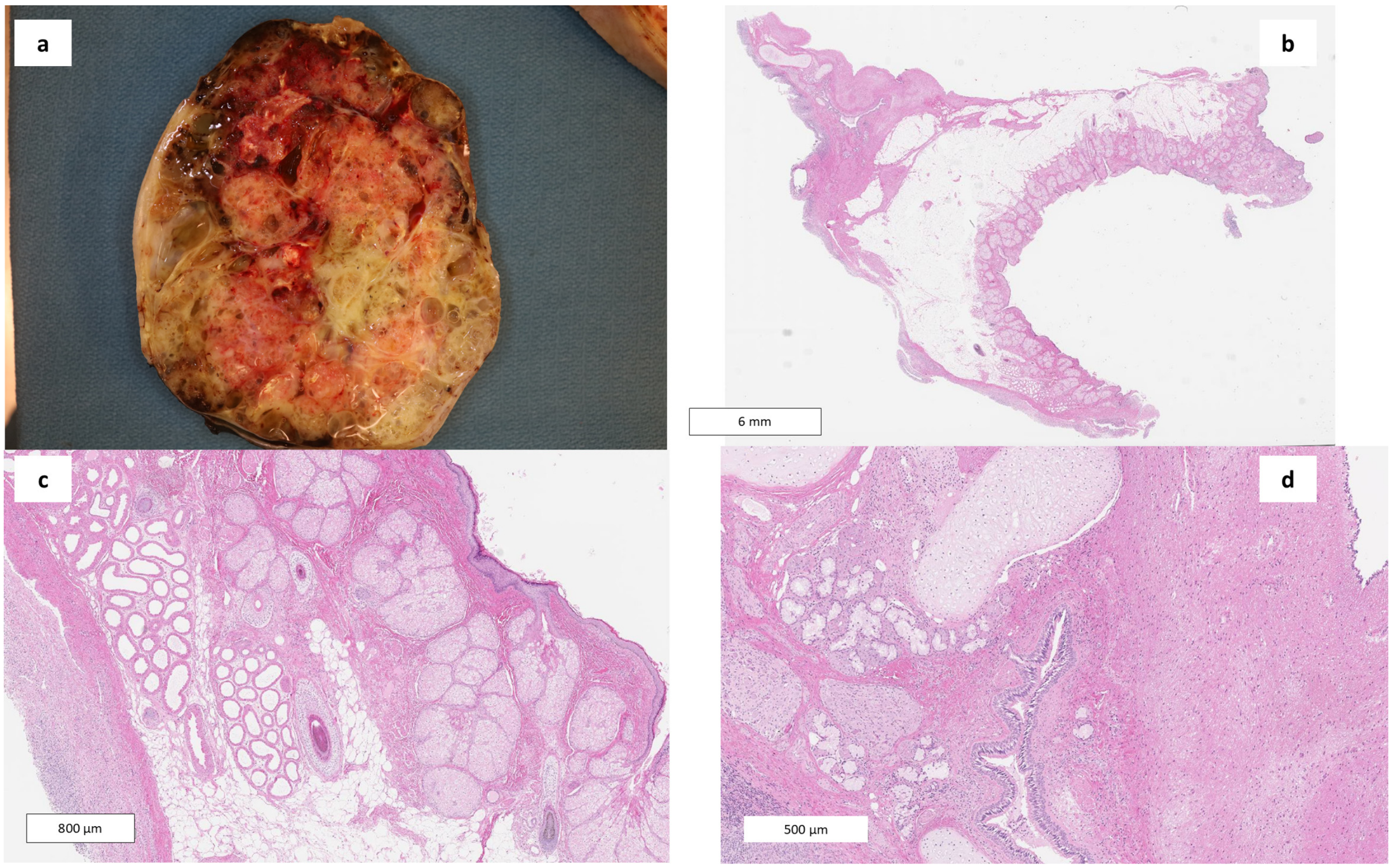

| Case | Study | Age/Sex | Presentation and Circumstances | Neoplasm, Site, and Size | Key Post-Mortem Findings and Proposed Mechanism | Causal Role in Death |
|---|---|---|---|---|---|---|
| 1 | [9] | 32 days, M | Persistent residual right-lung radiographic shadowing had been present since birth. Died suddenly 5 days after hospital discharge; exact location and circumstances were not reported. | Pulmonary blastoma (historical biphasic terminology); Right lung, lower lobe. | Clearly visible. Entire right lower lobe replaced; tumour extended to the visceral pleura without penetration; histological vascular invasion. Mechanism: Not explicitly stated; presumed acute respiratory compromise related to extensive replacement of the right lower lobe. | Causal |
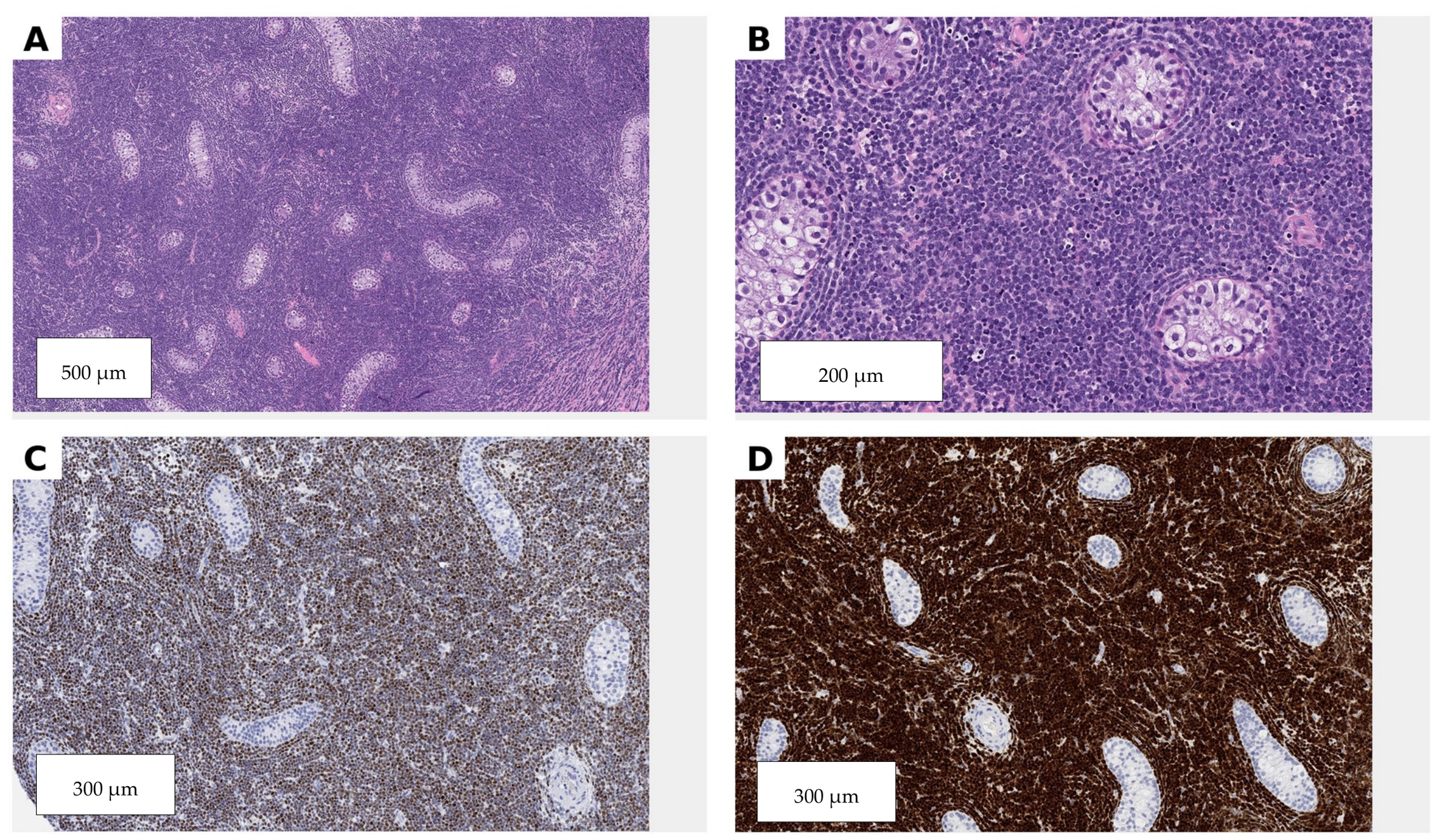
| 2 | [10] | 5 months, F | Three-day history of dyspnoea, sore throat and weakness, with worsening dyspnoea and hacking cough. Found unresponsive by a family member in supine position on her bed on the third day of illness. | Acute myeloid leukaemia with pulmonary leukostasis and extramedullary infiltration; systemic acute myeloid leukaemia with predominant bilateral pulmonary and hepatic infiltration; multifocal/disseminated. | Microscopic only. Bronchi, bronchioles, alveolar septa and lumina, pulmonary vascular lumina, and liver; severe bilateral pulmonary involvement. Mechanism: Acute respiratory failure due to diffuse pulmonary leukostasis and extramedullary myeloblastic infiltration. | Causal |
| 3 | [11] | 11 months, M | No preceding cardiac or systemic symptoms were reported. While crawling and playing on the floor under his mother’s observation, the infant suddenly collapsed; he was dead on arrival at hospital. | Solitary cardiac rhabdomyoma (cardiac hamartoma); anterior interventricular septum and anterolateral wall of the left ventricle; largest dimension 3 cm. | Clearly visible. Tumour located near the main bundle of His, protruding into the left ventricular cavity and appreciably narrowing the left ventricular outflow tract. Mechanism: Probable fatal conduction disturbance with sudden asystole due to proximity to the His bundle; left ventricular outflow obstruction with acute cardiac failure was also considered. | Causal |
| 4 | [12] | Newborn, F | Acute deterioration at 1 h of age with severe metabolic acidosis; treated as suspected sepsis despite normal inflammatory markers. Acute unexpected deterioration shortly after birth; required ventilation and died several hours later. | Congenital adrenal neuroblastoma; left adrenal gland; largest dimension 3 cm. | Clearly visible. Left adrenal gland; tumour reported as confined to the adrenal gland. Mechanism: Possible catecholamine or other tumour-mediated metabolic/haemodynamic effect mimicking sepsis. Competing findings: Sepsis was clinically suspected, but inflammatory markers were normal; incomplete perinatal information and absence of blood-pressure and placental data limited interpretation. | Contributory |
| 5 | [12] | 2.5 months, M | Swollen abdomen assessed by a general practitioner 13 days before death and considered a probable incidental umbilical hernia. Sudden collapse at home; confirmed dead on arrival at hospital. | Congenital mesoblastic nephroma; left kidney; largest dimension 10 cm. | Clearly visible. Large renal tumour with extensive intra-abdominal haemorrhage. Mechanism: Fatal intra-abdominal haemorrhage from a large necrotic renal tumour. | Causal |
| 6 | [12] | 4 months, F | Poor feeding and irritability for 3 days; fever, dyspnoea, bruising and oro-nasal bleeding on presentation. Rapid deterioration after presentation to the emergency department; death occurred within minutes. | Acute leukaemia, not otherwise specified; systemic; lymph nodes, liver and spleen; multifocal/disseminated. | Clearly visible. Widespread lymphadenopathy and hepatosplenomegaly. Mechanism: Not specifically stated; likely acute marrow failure with haemorrhagic and/or infectious complications. | Contributory |
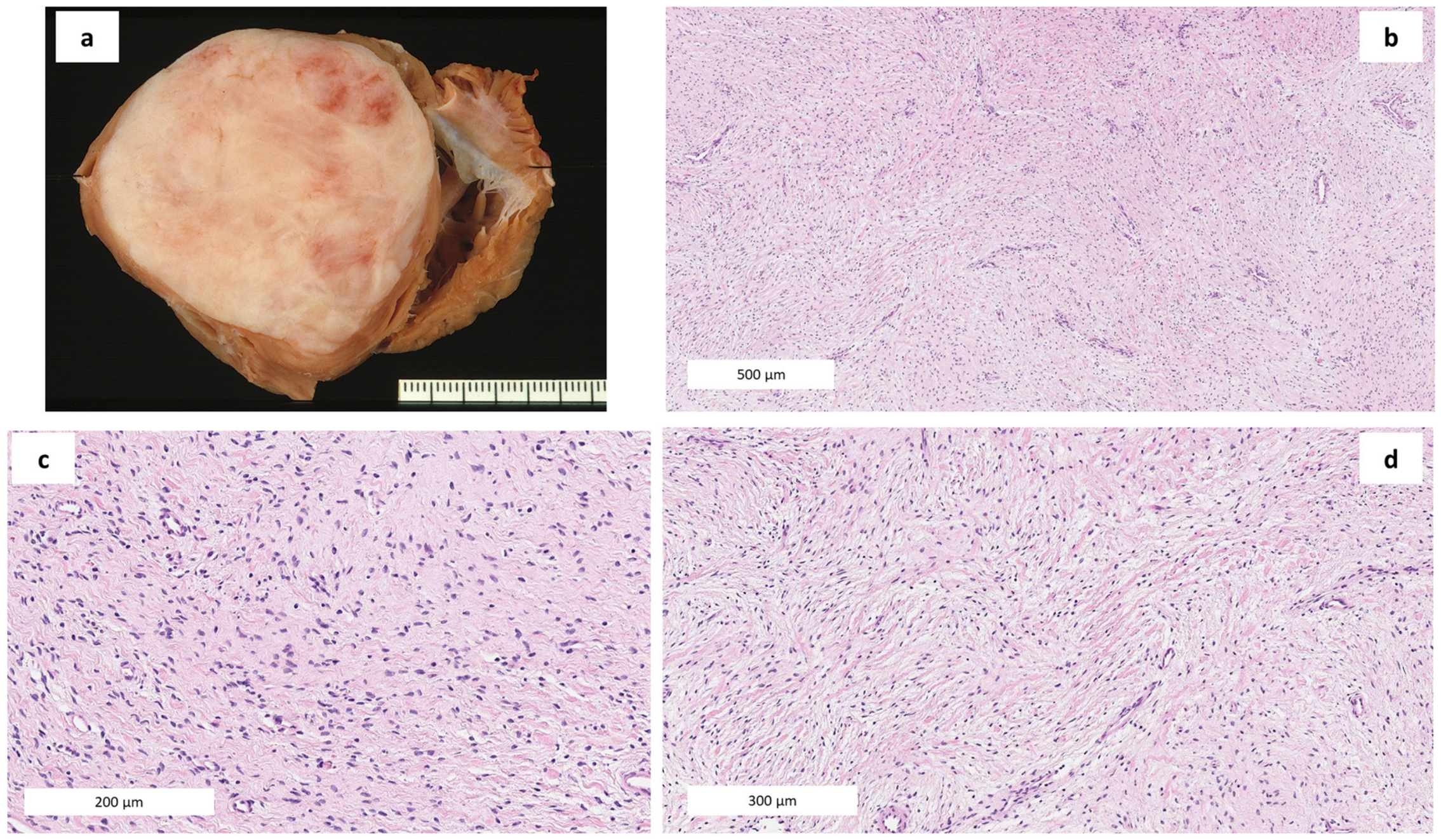
| 7 | [12] | 4.5 months, M | No preceding symptoms. Suddenly stopped breathing during a feed. | Cardiac fibroma; heart; left ventricular outflow tract; largest dimension 5 cm. | Clearly visible. Large intracardiac tumour obstructing the left ventricular outflow tract. Mechanism: Acute cardiac failure and/or fatal arrhythmia related to severe left ventricular outflow obstruction. | Causal |
| 8 | [13] | Newborn, M | Failed to establish spontaneous respiration immediately after birth. Severe cardiorespiratory compromise from birth; death certified after 1 h of unsuccessful resuscitation. | Congenital mature intrapericardial teratoma; intrapericardial, attached by a pedicle to the intrapericardial ascending aorta; largest dimension 7 cm. | Clearly visible. Pericardial cavity, heart and both lungs; major pulmonary and cardiac compression. Mechanism: Cardiorespiratory failure caused by severe cardiac and pulmonary compression, associated pericardial effusion and pulmonary hypoplasia/collapse. | Causal |
| 9 | [13] | Newborn, M | Immediate severe respiratory distress due to a large anterior cervical mass. Upper airway obstruction began immediately after birth; intubation and tracheotomy were unsuccessful and death occurred after approximately 1 h. | Congenital cervical teratoma with mature and immature elements; anterior cervical region with marked tracheal compression and deviation; largest dimension 10 cm. | Clearly visible. Upper airway and lungs; severe tracheal compression/kinking and pulmonary hypoplasia. Mechanism: Fatal upper-airway obstruction caused by severe tracheal compression and kinking, exacerbated by pulmonary hypoplasia. | Causal |
| 10 | [13] | Newborn, F | Profuse bleeding from a large sacrococcygeal mass during delivery, followed by cardiorespiratory collapse. Bleeding began during vaginal delivery; cardiac arrest occurred after hospital transfer and death occurred during attempted surgical excision. | Congenital sacrococcygeal teratoma with mature and immature elements; sacrococcygeal region, type I; largest dimension 10 cm. | Clearly visible. External sacrococcygeal soft tissues and major blood vessels; traumatic rupture with massive haemorrhage. Mechanism: Acute traumatic haemorrhage from the tumour during vaginal delivery, resulting in hypovolaemic arrest and death. | Causal |
| 11 | [14] | 3 months, F | No preceding symptoms. Suddenly collapsed in her mother’s arms at home after the evening feed; dead on arrival at hospital despite prolonged resuscitation. Routine paediatric examinations up to 2 months of age were normal. | Primary low-grade cardiac sarcoma, unclassifiable subtype; left ventricular free wall with diffuse myocardial and intracavitary extension; largest dimension 5 cm. | Clearly visible. Complete infiltration of the left ventricular myocardium and chamber, with mitral valve deformation and obstruction of left ventricular filling. Mechanism: Fatal ventricular fibrillation combined with tumour-related restrictive cardiomyopathy and obstruction of left ventricular filling. | Causal |
| 12 | [15] | 24 days, M | No symptoms were reported after discharge; the infant appeared healthy until death. Died suddenly and unexpectedly in the crib. | Multifocal capillary hemangiomas of the mitral and tricuspid valves; mitral and tricuspid valves. | Clearly visible. Mitral and tricuspid valve leaflets; no involvement of the conduction system. Mechanism: Not directly demonstrated; a possible arrhythmic or haemodynamic effect of multifocal valvular haemangiomas was implied. Competing findings: Mild septal hypertrophy and patent foramen ovale were known, but no obstructive lesion or conduction-system abnormality was found. | Contributory |
| 13 | [15] | 35 days, M | No preceding symptoms. Died suddenly and unexpectedly in the crib. | Solitary capillary hemangioma of the tricuspid valve; medial cusp of the tricuspid valve; largest dimension 1.5 mm. | Clearly visible. Tricuspid valve; no involvement of the cardiac conduction system. Mechanism: No specific fatal mechanism was demonstrated. Competing findings: Strong family history of unexplained infant death; the lesion was extremely small, with no obstruction or conduction-system involvement and no other convincing tumour-related lethal mechanism. | Incidental |
| 14 | [16] | 1 month, M | Runny nose, mild wheezing and diarrhoea during the 2 days before death; increased sleepiness and fussiness, but feeding remained satisfactory. Found dead in a shared adult bed after a sleep period. | Multifocal infantile hepatic hemangioma (historically infantile hemangioendothelioma type 1); liver, multifocal; largest dimension 2 cm. | Clearly visible. Diffuse hepatic involvement with marked hepatomegaly, high-flow vascular shunting, diaphragmatic elevation and secondary cardiopulmonary compromise. Mechanism: High-output congestive cardiac failure due to arteriovenous and arterioportal shunting, with a secondary restrictive respiratory effect from the enlarged liver and diaphragmatic incursion. | Contributory |
| 15 | [17] | 8 months, M | No preceding symptoms. Approximately 15 min after bottle feeding, the infant became cyanotic and lost consciousness. Sudden cyanosis and loss of consciousness at home shortly after bottle feeding; transported immediately to hospital and arrived without signs of life. | Cardiac fibroma; anterior and inferior free wall of the left ventricle; largest dimension 4.7 cm. | Clearly visible. Large intramural left-ventricular mass deforming the ventricular wall and compressing the ventricular cavity. Mechanism: Cardiogenic shock from severe ventricular-cavity compression; a fatal ventricular arrhythmia or conduction disturbance was also biologically plausible but not directly demonstrated. | Contributory |
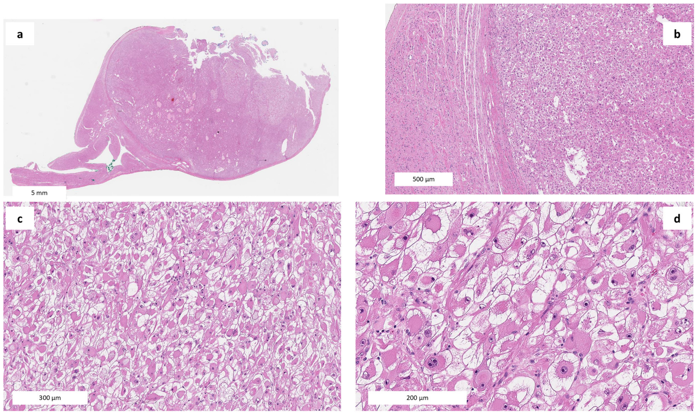
| 16 | [18] | 2.5 months, F | No acute preceding symptoms were reported. Found lifeless by her father in the parental bed, lying on a pillow beside her sleeping mother. Routine preventive medical examinations before death. | Multifocal microscopic cardiac rhabdomyoma; heart, multifocal microscopic intramyocardial foci; precise cardiac sites not specified. | Microscopic only. Possible involvement of the cardiac conduction system was proposed but not demonstrated. Mechanism: Acute dysrhythmia from presumed involvement of the cardiac conduction system. Competing findings: The infant was sleeping in an adult bed on a pillow beside her mother, creating a potentially hazardous sleep environment. | Contributory |
| 17 | [19] | 2 days, F | No preceding symptoms were reported; the neonate was unexpectedly found completely cyanosed during the night on the second postnatal day. Resuscitation was initiated but was unsuccessful. | Cavernous hemangioma of the tricuspid valve; tricuspid valve; largest dimension 8 mm. | Clearly visible. Tricuspid valve; possible aggravation of right-sided haemodynamics through tricuspid regurgitation was proposed but not demonstrated. Mechanism: Fatal circulatory failure caused by severe hypoplasia of the left-sided cardiac structures; the tricuspid haemangioma may have worsened haemodynamics by producing tricuspid regurgitation. Competing findings: Severe hypoplastic left heart syndrome was a sufficient and more convincing primary cause of death. | Contributory |
| 18 | [20] | 1 month, F | No preceding symptoms. The infant was suddenly found pale, unresponsive and not breathing while in a baby carrier. Shortly after the family entered a restaurant, the mother moved the baby carrier in the restroom and found the infant pale, unresponsive and apnoeic. | Cardiac fibroma; posterior wall of the left ventricle, extending to the ventricular septum and apex; largest dimension 4 cm. | Clearly visible. The tumour compressed and narrowed both ventricular outflow tracts and compressed the myocardium of the left bundle branch. Mechanism: Combined impairment of ventricular blood flow and fatal cardiac arrhythmia caused by overstretching and compression of the left bundle branch. | Causal |
| 19 | [21] | 5 months, F | No preceding respiratory, neurological or systemic symptoms were reported. Placed to sleep at approximately 22:00 and found unresponsive at 06:45 in a recently purchased playpen adapted as a crib. Routine paediatric follow-up had not identified tuberous sclerosis. | Tuberous sclerosis complex with multifocal cardiac rhabdomyomas and subependymal giant cell astrocytoma; heart and central nervous system; largest dimension 1.5 cm. | Clearly visible. Cardiac lesions involved intramural, subendocardial and subpericardial myocardium and could potentially affect the conduction system. Mechanism: Possible fatal arrhythmia from cardiac rhabdomyomas or fatal seizure related to extensive cortical tubers/subependymal tumour, with a possible concurrent positional or sleep-environment asphyxial mechanism. Competing findings: Prone sleeping and a soft sloping mattress represented relevant environmental factors. | Contributory |
| 20 | [22] | 3 months, M | Persistent cough associated with a recent upper respiratory tract infection; no other acute symptoms were reported. Fed at 08:30 and placed in his crib; found lifeless at 12:15. | Solitary microscopic cardiac neuroma/neurofibroma-like hamartomatous lesion; region of the atrioventricular node; largest dimension 0.75 mm. | Microscopic only. Located adjacent to the atrioventricular node; the atrioventricular node and bundle of His were structurally normal. Mechanism: Unexplained sudden infant death. No arrhythmic or obstructive mechanism attributable to the neural lesion was demonstrated. Competing findings: Recent upper respiratory tract infection with persistent cough and positive pleural-fluid culture without histological pneumonia. | Incidental |
| 21 | [23] | 2 months, M | A grade 5 holosystolic murmur radiating to the axilla and back was detected during a routine well-baby examination one day before death. The infant suddenly collapsed in his mother’s arms the following day. | Multifocal cardiac myxoma; multifocal cardiac involvement of both atria, aortic, mitral and tricuspid valves, left ventricular endocardium and membranous septum; largest dimension 2.3 cm. | Clearly visible. Near-complete aortic-valve obstruction with a residual orifice of 0.1 cm; marked mitral-valve obstruction; diffuse tricuspid-valve involvement. Mechanism: Mechanical and haemodynamic disturbance due to severe multivalvular obstruction; a conduction-related effect was also considered possible. Embolic death was excluded. | Causal |
| 22 | [24] | 3.5 months, F | None reported after discharge. Found dead by her father in a prone position in the parents’ bed at approximately 3.5 months of age. | Multifocal infantile hepatic vascular tumour, historically termed type II infantile hepatic hemangioendothelioma; liver, multifocal lesions involving both lobes; largest dimension 2 cm. | Clearly visible. Liver parenchyma; no direct compression of a vital structure and no evidence of cardiac failure. Mechanism: Unexplained sudden infant death; a possible degree of tumour-related cardiovascular compromise was discussed but not demonstrated. Competing findings: Prone sleeping in the parents’ bed, minor repeated milk aspiration changes, and a previously resolved systolic murmur. | Contributory |
| 23 | [25] | 4 months, F | Two episodes of vomiting two days before death; otherwise no illness or complaint was known. Sudden and unexpected death; the precise location and circumstances of discovery were not reported. | Microscopic septal cardiac fibroma involving the atrioventricular conduction region; cardiac fibrous body/interventricular septal region adjacent to and compressing the atrioventricular node. | Microscopic only. Atrioventricular node and adjacent conduction tissue. Mechanism: Possible fatal conduction disturbance or arrhythmia caused by compression of the atrioventricular node, potentially acting together with an accessory Mahaim fibre and other conduction-system abnormalities. | Contributory |
| 24 | [26] | 5 months, F | None reported. Found unresponsive during a nap in an adult bed at her caregiver’s home; she had been last known alive two hours earlier. | Multifocal microscopic left ventricular rhabdomyoma; multifocal microscopic involvement of the left ventricular wall, trabeculae and immediately subendocardial myocardium. | Microscopic only. Left ventricular subendocardial myocardium; a potential arrhythmic substrate was inferred, but direct conduction-system involvement was not documented. Mechanism: Presumed fatal cardiac arrhythmia caused by microscopic rhabdomyoma. | Contributory |
| 25 | [27] | 7 weeks, F | A skin nodule and abdominal distension developed after the 4-week check-up. On the day of death she suddenly developed gasping respiration while feeding, followed by respiratory and cardiac arrest at home. | KMT2A-MLLT1-rearranged infantile B-cell acute lymphoblastic leukaemia; systemic disease involving bone marrow, skin, liver, spleen, kidneys, pancreas, gastrointestinal tract, uterus, lymph nodes and vascular lumina of multiple organs including heart and lungs; multifocal/disseminated. | Clearly visible. Bone marrow and widespread visceral and intravascular infiltration, particularly pulmonary vascular involvement. Mechanism: Severe anaemia, hypoxaemia from widespread intravascular pulmonary blast infiltration and hyperkalaemia related to tumour lysis syndrome. | Causal |
| 26 | [28] | 6 months, F | None reported before the sudden collapse. Became unresponsive after crying; emergency services documented ventricular fibrillation. | Large septal cardiac fibroma; interventricular septum and left ventricle, extending from apex to base and affecting both ventricular outflow tracts and the atrioventricular conduction system; largest dimension 4.5 cm. | Clearly visible. Left ventricular cavity, right ventricular infundibulum, mitral valve, aortic outflow, atrioventricular node, His bundle and both bundle branches. Mechanism: Left ventricular outflow obstruction and severe conduction-system compression causing bradyarrhythmia/ventricular fibrillation and acute haemodynamic failure. | Causal |
| 27 | [29] | 2 months, F | None reported. Became unresponsive and was taken to an emergency department; the precise location and activity at collapse were not reported. | Multifocal cardiac Purkinje-cell tumour/histiocytoid cardiomyopathy; multifocal lesions in left and right endocardium, interatrial septum, both atrial walls and near the sinoatrial and atrioventricular nodes. | Microscopic only. Cardiac conduction system, including lesions close to the sinoatrial and atrioventricular nodes. Mechanism: Fatal cardiac electrical instability caused by diffuse involvement and compression of the specialised conduction system. | Contributory |
| 28 | [30] | 6 months, F | A slight cough occurred after feeding approximately one hour before she was found dead; otherwise, no concern had been reported. Found dead in her cot by her father approximately one hour after feeding. | Mixed fetal and embryonal epithelial hepatoblastoma; right hepatic lobe, occupying most of the lobe; largest dimension 12 cm. | Clearly visible. Liver and peritoneal cavity; extensive tumour necrosis and haemorrhage with 200 mL haemoperitoneum, although no rupture site was identified. Mechanism: Likely fatal infection in a markedly underweight infant with advanced malignancy; haemoperitoneum and tumour necrosis were additional possible contributors, but no bleeding source was demonstrated. Competing findings: Focal bilateral bronchopneumonia and chronic tracheal inflammation. | Contributory |
| 29 | [30] | 3.5 months, M | None reported. Found dead in a prone position in his pram approximately 30 min after breastfeeding and being put to sleep. | Thoracic extradural spinal lipoma with recent haemorrhage; thoracic extradural spinal canal, extending for approximately 7 cm below the cervical region; largest dimension 7 cm. | Clearly visible. Thoracic spinal canal adjacent to the spinal cord; no direct involvement of the phrenic nerve or demonstrable medullary compression. Mechanism: Unexplained sudden infant death. The authors considered but could not demonstrate increased intraspinal pressure acting with prone positioning to inhibit respiration. Competing findings: Prone sleeping position and recent haemorrhage within the lipoma. | Incidental |
| 30 | [31] | 1 month, F | Lethargy and poor feeding. Found unresponsive the morning after medical review; resuscitation attempts were unsuccessful. | Precursor B-cell acute lymphoblastic leukaemia; systemic leukaemia with distension of visceral and cerebral vessels by leukaemic cells; multifocal/disseminated. | Microscopic only. Visceral and cerebral microvasculature. Mechanism: Fatal microvascular obstruction from the extreme intravascular leukaemic burden. | Causal |
| 31 | [31] | 2 months, M | Tachycardia, tachypnoea and fever of 39.9 °C on the day of death. Cardiorespiratory arrest later the same day; resuscitation was unsuccessful. | Acute myeloid leukaemia with diffuse multiorgan and myocardial infiltration; systemic acute myelogenous leukaemia with hepatosplenomegaly, lymphadenopathy and extensive multiorgan infiltration including myocardium; multifocal/disseminated. | Clearly visible. Myocardium and systemic organs; cardiac infiltration caused tissue disruption, oedema, necrosis and haemorrhage. Mechanism: Diffuse myocardial infiltration and injury causing acute cardiac failure. | Causal |
| 32 | [32] | 16 weeks, M | No overt illness in the week before death. Home video taken within 24 h of death showed a prominent abdomen and subtle lower-chest intercostal recession. Found moribund in his crib by his father. | Precursor B-cell acute lymphoblastic leukaemia with diffuse multiorgan and myocardial infiltration; systemic disease involving bone marrow, heart, lungs, liver, kidneys, stomach, intestines, appendix, pancreas, thyroid, salivary gland, lymph nodes, spleen, adipose tissue and multiple vascular beds; multifocal/disseminated. | Clearly visible. Myocardium, coronary and systemic vessels, pulmonary interstitium and multiple abdominal organs. Mechanism: Probable fatal arrhythmia caused by diffuse myocardial infiltration, potentially compounded by pulmonary infiltration, restricted respiratory expansion from organomegaly, leukocytosis and hyperviscosity within coronary vessels. | Causal |
| Domain | Variable | Cases, n/N (%) or Summary |
|---|---|---|
| Demographic characteristics | Male | 15/32 (46.9%) |
| Female | 17/32 (53.1%) | |
| Age, median (range) | 2.5 months (birth–11 months) | |
| Neonatal (<28 days) | 6/32 (18.8%) | |
| 28 days to <3 months | 11/32 (34.4%) | |
| 3 to <6 months | 11/32 (34.4%) | |
| 6 to <12 months | 4/32 (12.5%) | |
| Clinical presentation and circumstances | No preceding symptoms or signs reported | 15/32 (46.9%) |
| Any preceding symptoms or signs reported | 17/32 (53.1%) | |
| Recent healthcare contact reported | 21/32 (65.6%) | |
| Found unresponsive, moribund, or dead | 15/32 (46.9%) | |
| Witnessed collapse or acute deterioration | 15/32 (46.9%) | |
| Collapse during or within 1 h after feeding | 6/32 (18.8%) | |
| Resuscitation attempted | 20/32 (62.5%) | |
| Neoplasm category | Cardiovascular/thoracic | 16/32 (50.0%) |
| Haematological/lymphoid | 6/32 (18.8%) | |
| Germ-cell/embryonal | 3/32 (9.4%) | |
| Hepatic/gastrointestinal | 3/32 (9.4%) | |
| Adrenal/endocrine | 1/32 (3.1%) | |
| Renal/genitourinary | 1/32 (3.1%) | |
| Nervous system | 1/32 (3.1%) | |
| Disseminated/multisystem | 1/32 (3.1%) | |
| Pathological characteristics | Benign | 16/32 (50.0%) |
| Intermediate/uncertain or tumour-like | 6/32 (18.8%) | |
| Malignant | 10/32 (31.3%) | |
| Multifocal or disseminated disease | 14/32 (43.8%) | |
| Clearly visible at gross examination | 25/32 (78.1%) | |
| Microscopic only | 7/32 (21.9%) | |
| Largest tumour dimension reported | 20/32 (62.5%) | |
| Largest dimension, median (range) | 42.5 mm (0.75–120 mm), n = 20 | |
| Proposed mechanism of death | Arrhythmia or conduction-system disturbance | 14/32 (43.8%) |
| Mechanical obstruction or compression | 9/32 (28.1%) | |
| Cardiac failure or haemodynamic compromise | 14/32 (43.8%) | |
| Haemorrhage, rupture, or exsanguination | 4/32 (12.5%) | |
| Respiratory failure or pulmonary involvement | 9/32 (28.1%) | |
| Systemic haematological effects or diffuse infiltration | 6/32 (18.8%) | |
| Neurological or seizure-related mechanism | 2/32 (6.3%) | |
| Infectious complication | 2/32 (6.3%) | |
| Mechanism uncertain or not demonstrated | 5/32 (15.6%) | |
| Review causal assessment | Causal | 16/32 (50.0%) |
| Contributory | 13/32 (40.6%) | |
| Incidental | 3/32 (9.4%) | |
| Ancillary diagnostic methods | Immunohistochemistry reported | 11/32 (34.4%) |
| Molecular or genetic testing reported | 3/32 (9.4%) | |
| Post-mortem imaging reported | 2/32 (6.3%) |
Disclaimer/Publisher’s Note: The statements, opinions and data contained in all publications are solely those of the individual author(s) and contributor(s) and not of MDPI and/or the editor(s). MDPI and/or the editor(s) disclaim responsibility for any injury to people or property resulting from any ideas, methods, instructions or products referred to in the content. |
© 2026 by the authors. Licensee MDPI, Basel, Switzerland. This article is an open access article distributed under the terms and conditions of the Creative Commons Attribution (CC BY) license.
Share and Cite
Camatti, J.; Bonasoni, M.P.; Nagy, A.; Cecchi, R.; Santunione, A.L.; Radheshi, E. Occult Neoplasms in Sudden Unexpected Death in Infancy: A Scoping Review of Post-Mortem Findings. Diagnostics 2026, 16, 3189. https://doi.org/10.3390/diagnostics16193189
Camatti J, Bonasoni MP, Nagy A, Cecchi R, Santunione AL, Radheshi E. Occult Neoplasms in Sudden Unexpected Death in Infancy: A Scoping Review of Post-Mortem Findings. Diagnostics. 2026; 16(19):3189. https://doi.org/10.3390/diagnostics16193189
Chicago/Turabian StyleCamatti, Jessika, Maria Paola Bonasoni, Anita Nagy, Rossana Cecchi, Anna Laura Santunione, and Erjon Radheshi. 2026. "Occult Neoplasms in Sudden Unexpected Death in Infancy: A Scoping Review of Post-Mortem Findings" Diagnostics 16, no. 19: 3189. https://doi.org/10.3390/diagnostics16193189
APA StyleCamatti, J., Bonasoni, M. P., Nagy, A., Cecchi, R., Santunione, A. L., & Radheshi, E. (2026). Occult Neoplasms in Sudden Unexpected Death in Infancy: A Scoping Review of Post-Mortem Findings. Diagnostics, 16(19), 3189. https://doi.org/10.3390/diagnostics16193189

