Assessment of Patellar Lateralization and Its Association with Patellofemoral Osteoarthritis Severity: A Retrospective MRI-Based Study

Abstract
1. Introduction
2. Materials and Methods
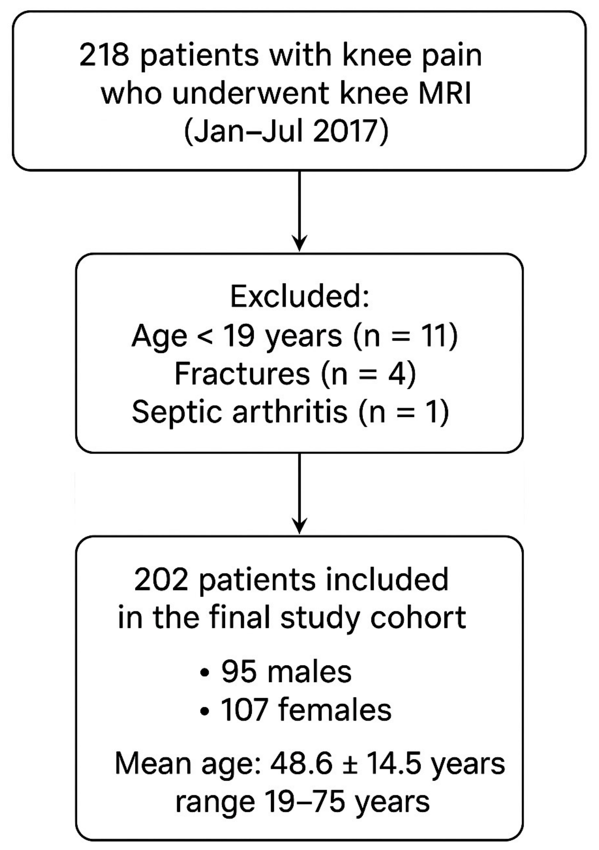
2.1. Case Selection
2.2. MRI Parameters
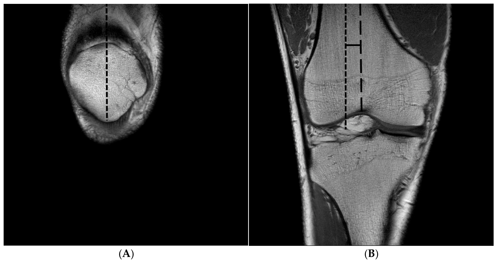
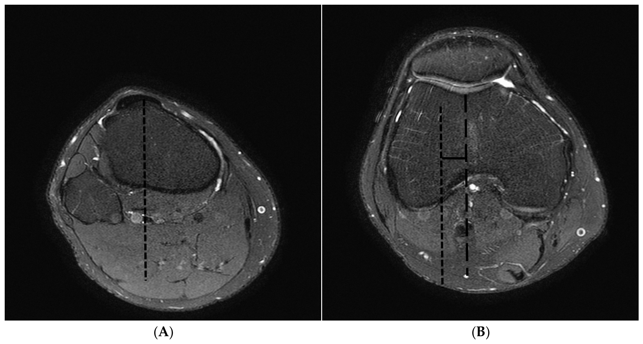
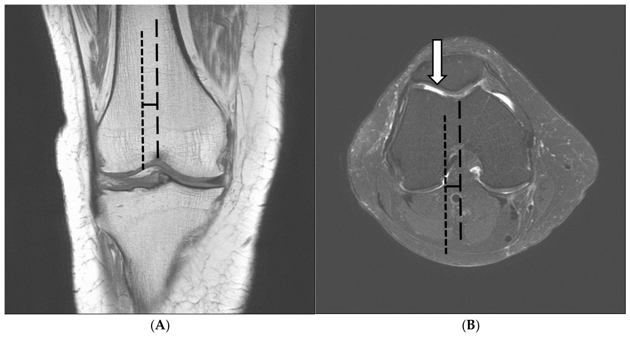
2.3. Measure of the Patellar Lateralization and TT-TG Distance
2.4. Image Analysis of the PFOA
2.5. Statistical Analysis
3. Results
4. Discussion
5. Conclusions
Author Contributions
Funding
Institutional Review Board Statement
Informed Consent Statement
Data Availability Statement
Conflicts of Interest
References
- Tsavalas, N.; Katonis, P.; Karantanas, A.H. Knee joint anterior malalignment and patellofemoral osteoarthritis: An MRI study. Eur. Radiol. 2012, 22, 418–428. [Google Scholar] [CrossRef] [Scilit] [PubMed]
- GBD 2021 Osteoarthritis Collaborators. Global, regional, and national burden of osteoarthritis, 1990–2020 and projections to 2050: A systematic analysis for the Global Burden of Disease Study 2021. Lancet Rheumatol. 2023, 21, e508–e522. [Google Scholar] [CrossRef] [Scilit] [PubMed]
- Bryliński, L.; Brylińska, K.; Woliński, F.; Sado, J.; Smyk, M.; Komar, O.; Karpiński, R.; Prządka, M.; Baj, J. Trace Elements-Role in Joint Function and Impact on Joint Diseases. Int. J. Mol. Sci. 2025, 26, 7493. [Google Scholar] [CrossRef] [Scilit] [PubMed]
- Karpiński, R.; Prus, A.; Baj, J.; Radej, S.; Prządka, M.; Krakowski, P.; Jonak, K. Articular Cartilage: Structure, Biomechanics, and the Potential of Conventional and Advanced Diagnostics. Appl. Sci. 2025, 15, 6896. [Google Scholar] [CrossRef] [Scilit]
- Hill, J.R.; Oei, E.H.G.; Crossley, K.M.; Menz, H.B.; Macri, E.M.; Smith, M.D.; Wyndow, N.; Maclachlan, L.R.; Ross, M.H.; Collins, N.J. Classification approaches used to grade radiographic patellofemoral osteoarthritis: A scoping review. Osteoarthr. Imaging 2024, 28, 100258. [Google Scholar] [CrossRef] [Scilit] [PubMed]
- Kobayashi, S.; Pappas, E.; Fransen, M.; Refshauge, K.; Simic, M. The prevalence of patellofemoral osteoarthritis: A systematic review and meta-analysis. Osteoarthr. Cartil. 2016, 24, 1697–1707. [Google Scholar] [CrossRef] [Scilit] [PubMed]
- Grelsamer, R.P.; Dejour, D.; Gould, J. The pathophysiology of patellofemoral arthritis. Orthop. Clin. N. Am. 2008, 39, 269–274. [Google Scholar] [CrossRef] [Scilit] [PubMed]
- Tsai, C.H.; Hsu, C.J.; Hung, C.H.; Hsu, H.C. Primary traumatic patellar dislocation. J. Orthop. Surg. Res. 2012, 7, 21. [Google Scholar] [CrossRef] [Scilit] [PubMed]
- Park, H.J.; Ahn, J.H.; Kim, S.S.; Lee, S.Y.; Choi, Y.J.; Chung, E.C.; Rho, M.H.; Kook, S.H. A new assessment of patellar instability using coronal magnetic resonance images of the patella superimposed on the femur and its clinical utility. J. Comput. Assist. Tomogr. 2013, 37, 470–474. [Google Scholar] [CrossRef] [Scilit] [PubMed]
- Earhart, C.; Patel, D.B.; White, E.A.; Gottsegen, C.J.; Forrester, D.M.; Matcuk, G.R., Jr. Transient lateral patellar dislocation: Review of imaging findings, patellofemoral anatomy, and treatment options. Emerg. Radiol. 2013, 20, 11–23. [Google Scholar] [CrossRef] [Scilit] [PubMed]
- Schoettle, P.B.; Zanetti, M.; Seifert, B.; Pfirrmann, C.W.A.; Fucentese, S.F.; Romero, J. The tibial tuberosity-trochlear groove distance; a comparative study between CT and MRI scanning. Knee 2006, 13, 26–31. [Google Scholar] [CrossRef] [Scilit] [PubMed]
- Park, H.J.; Kim, S.S.; Lee, S.Y.; Park, N.H.; Park, J.Y.; Choi, Y.J.; Jeon, H.J. A practical MRI grading system for osteoarthritis of the knee: Association with Kellgren-Lawrence radiographic scores. Eur. J. Radiol. 2013, 82, 112–117. [Google Scholar] [CrossRef] [Scilit] [PubMed]
- Britteberg, M.; Winalski, C.S. Evaluation of cartilage injuries and repair. J. Bone Jt. Surg. Am. 2003, 85-A, 58–69. [Google Scholar] [CrossRef] [Scilit] [PubMed]
- Koo, T.K.; Li, M.Y. A Guideline of Selecting and Reporting Intraclass Correlation Coefficients for Reliability Research. J. Chiropr. Med. 2016, 15, 155–163. [Google Scholar] [CrossRef] [Scilit] [PubMed]
- Cochran, W.G. Some Methods for Strengthening the Common χ2 Tests. Biometrics 1954, 10, 417–451. [Google Scholar] [CrossRef] [Scilit]
- Armitage, P. Tests for Linear Trends in Proportions and Frequencies. Biometrics 1955, 11, 375–386. [Google Scholar] [CrossRef] [Scilit]
- Bhattacharya, R.; Kumar, V.; Safawi, E.; Finn, P.; Hui, A.C. The skyline radiograph: Its usefulness in the diagnosis of patella-femoral osteoarthritis. Int. Orthop. 2007, 31, 247–252. [Google Scholar] [CrossRef] [Scilit] [PubMed][Green Version]
- Severyns, M.; Mallet, J.; Santoni, B.; Barnavon, T.; Germaneau, A.; Vendeuvre, T.; Drame, M. Radiographic measurement of the congruence angle according to Merchant: Validity, reproducibility, and limits. Knee Surg. Relat. Res. 2023, 35, 1. [Google Scholar] [CrossRef] [Scilit] [PubMed]
- Dixit, S.; Difiori, J.P.; Burton, M.; Mines, B. Management of patellofemoral pain syndrome. Am. Fam. Physician 2007, 75, 194–202. [Google Scholar] [PubMed]
- Platzer, W. Color Atlas of Human Anatomy, 5th ed.; Volume 1: Locomotor System; Thieme: New York, NY, USA, 2004. [Google Scholar]
- Noehren, B.; Duncan, S.; Lattermann, C. Radiographic parameters associated with lateral patella degeneration in young patients. Knee Surg. Sports Traumatol. Arthrosc. 2012, 20, 2385–2390. [Google Scholar] [CrossRef] [Scilit] [PubMed]
- Nomura, E.; Inoue, M. Cartilage lesions of the patella in recurrent patellar dislocation. Am. J. Sports Med. 2004, 32, 498–502. [Google Scholar] [CrossRef] [Scilit] [PubMed]
- Dejour, D.; Coultre, B.L. Osteotomies in patello-femoral instabilities. Sports Med. Arthrosc. Rev. 2007, 15, 39–46. [Google Scholar] [CrossRef] [Scilit] [PubMed]
- Schüttler, K.F.; Struewer, J.; Roessler, P.P.; Gesslein, M.; Rominger, M.B.; Ziring, E.; Efe, T. Patellofemoral osteoarthritis after Insall’s proximal realignment for recurrent patellar dislocation. Knee Surg. Sports Traumatol. Arthrosc. 2014, 22, 2623–2628. [Google Scholar] [CrossRef] [Scilit] [PubMed]
- Dai, Y.; Yin, H.; Xu, C.; Zhang, H.; Guo, A.; Diao, N. Association of patellofemoral morphology and alignment with the radiographic severity of patellofemoral osteoarthritis. J. Orthop. Surg. Res. 2021, 16, 548. [Google Scholar] [CrossRef] [Scilit] [PubMed]
- Sillanpää, P.J.; Mäenpää, H.M.; Mattila, V.M.; Visuri, T.; Pihlajamäki, H. Arthroscopic surgery for primary traumatic patellar dislocation: A prospective, nonrandomized study comparing patients treated with and without acute arthroscopic stabilization with a median 7-year follow-up. Am. J. Sports Med. 2008, 36, 2301–2309. [Google Scholar] [CrossRef] [Scilit] [PubMed]
- Otsuki, S.; Nakajima, M.; Okamoto, Y.; Oda, S.; Hoshiyama, Y.; Iida, G.; Neo, M. Correlation between varus knee malalignment and patellofemoral osteoarthritis. Knee Surg. Sports Traumatol. Arthrosc. 2016, 24, 176–181. [Google Scholar] [CrossRef] [Scilit] [PubMed]
- Powers, C.M. The influence of altered lower-extremity kinematics on patellofemoral joint dysfunction: A theoretical perspective. J. Orthop. Sports Phys. Ther. 2003, 33, 639–646. [Google Scholar] [CrossRef] [Scilit] [PubMed]
- Haj-Mirzaian, A.; Guermazi, A.; Hakky, M.; Sereni, C.; Zikria, B.; Roemer, F.W.; Tanaka, M.J.; Cosgarea, A.J.; Demehri, S. Tibial tuberosity to trochlear groove distance and its association with patellofemoral osteoarthritis-related structural damage worsening: Data from the osteoarthritis initiative. Eur. Radiol. 2018, 28, 4669–4680. [Google Scholar] [CrossRef] [Scilit] [PubMed]
- Macri, E.M.; Stefanik, J.J.; Khan, K.K.; Crossley, K.M. Is tibiofemoral or patellofemoral alignment or trochlear morphology associated with patellofemoral osteoarthritis? A systematic review. Arthritis Care Res. 2016, 68, 1453–1470. [Google Scholar] [CrossRef] [Scilit] [PubMed]
- Demehri, S.; Thawait, G.K.; Williams, A.A.; Kompel, A.; Elias, J.J.; Carrino, J.A.; Cosgarea, A.J. Imaging characteristics of contralateral asymptomatic patellofemoral joints in patients with unilateral instability. Radiology 2014, 273, 821–830. [Google Scholar] [CrossRef] [Scilit] [PubMed]
- Williams, A.A.; Elias, J.J.; Tanaka, M.J.; Thawait, G.K.; Demehri, S.; Carrino, J.A.; Cosgarea, A.J. The relationship between tibial tuberosity-trochlear groove distance and abnormal patellar tracking in patients with unilateral patellar instability. Arthroscopy 2016, 32, 55–61. [Google Scholar] [CrossRef] [Scilit] [PubMed]



| Imaging Parameter | Coronal T1 1 FSE | Coronal 2 FS T2 FSE | Sagittal FS 3 PD FSE | Sagittal T2 FSE | Axial FS T2 FSE |
|---|---|---|---|---|---|
| TR (msec) | 550–750 | 3800–9800 | 3100–5000 | 2900–4500 | 4400–7500 |
| TE (msec) | 10–15 | 65–75 | 30–36 | 72–100 | 65–80 |
| Flip angle (°) | 90 | 90 | 90 | 90 | 90 |
| Matrix size (pixel) | 256 × 346 | 352 × 224 | 320 × 240 | 356 × 280 | 320 × 238 |
| Field of view (mm) | 160 | 160 | 160 | 160 | 160 |
| Slice thickness (mm) | 3 | 3 | 3 | 3 | 3 |
| Inter-slice gap (mm) | 0.3 | 0.3 | 0.3 | 0.3 | 0.3 |
| Bandwidth (kHz) | 270 | 270 | 256 | 240 | 295 |
| Echo train length | 6 | 16 | 12 | 15 | 16 |
| Signal average | 1 | 1 | 1 | 1 | 1 |
| Scan time (min:s) | 2:50 | 3:00 | 2:30 | 2:40 | 2:30 |
| Grade | Cartilage Morphology on MRI |
|---|---|
| 0 | Normal |
| 1 | Abnormal increased signal with intact articular surface |
| 2 | Partial-thickness defect < 50% |
| 3 | Partial-thickness defect 50–99% |
| 4 | Full-thickness defect with exposure of subchondral bone and marrow edema |
| Non-PFOA (n = 155) | PFOA (n = 47) | 95% CI | p-Value | Cohen’s d | ||
|---|---|---|---|---|---|---|
| Reader 1 | TT-TG distance | 7.21 (±2.95, 4.26–10.16) | 7.98 (±3.44, 4.54–11.42) | [−1.659, 0.121] | 0.090 | 0.24 |
| Patellar lateralization | 4.74 (±2.81, 1.93–7.55) | 5.82 (±3.12, 2.7–8.94) | [−1.912, 0.254] | 0.011 | 0.37 | |
| Reader 2 | TT-TG distance | 7.36 (±3.06, 4.3–10.42) | 8.08 (±3.21, 4.87–11.29) | [−1.611, 0.164] | 0.110 | 0.41 |
| Patellar lateralization | 4.78 (±2.82, 1.96–7.6) | 6.17 (±3.15, 3.02–9.32) | [−2.234, −0.557] | 0.001 | 0.20 | |
| Interpreter | Group | Grades of the International Cartilage Repair Society | p-Value | Effect Size (Cramer’s V) | |||||
|---|---|---|---|---|---|---|---|---|---|
| 0 | I | II | III | IV | Total | ||||
| Reader 1 | No lateralization (%) | 96 (61.9) | 14 (9.0) | 19 (12.3) | 11 (7.1) | 15 (9.7) | 155 (100) | 0.009 | 0.19 |
| Patellar lateralization (%) | 21 (44.7) | 4 (8.5) | 7 (14.9) | 4 (8.5) | 11 (23.4) | 47 (100) | |||
| Total | 117 | 18 | 26 | 15 | 26 | 202 | |||
| Reader 2 | No lateralization (%) | 98 (65.8) | 8 (5.4) | 18 (12.1) | 13 (8.7) | 12 (8.1) | 149 (100) | 0.009 | 0.22 |
| Patellar lateralization (%) | 25 (47.2) | 4 (7.5) | 6 (11.3) | 6 (11.3) | 12 (22.6) | 53 (100) | |||
| Total | 123 | 12 | 24 | 19 | 24 | 202 | |||
| <PFOA Grade 2 (n = 135) | ≥PFOA Grade 2 (n = 67) | 95% CI | p-Value | Cohen’s d | ||
|---|---|---|---|---|---|---|
| Reader 1 | TT-TG distance | 7.20 (±3.04) | 8.23 (±3.37) | [−1.962, −0.104] | 0.029 | 0.33 |
| Patellar lateralization | 4.78 (±2.79) | 6.02 (±3.23) | [−2.110, −0.376] | 0.005 | 0.42 | |
| Reader 2 | TT-TG distance | 7.27 (±3.05) | 8.41 (±3.18) | [−2.051, −0.228] | 0.014 | 0.37 |
| Patellar lateralization | 4.87 (±2.81) | 6.23 (±3.25) | [−2.232, −0.488] | 0.002 | 0.46 | |
| Parameter | Reader | Adjusted OR * | 95% CI | p Value |
|---|---|---|---|---|
| TT-TG distance | 1 | 1.128 | 1.009–1.261 | 0.034 |
| TT-TG distance | 2 | 1.125 | 1.004–1.260 | 0.042 |
| Patellar lateralization | 1 | 1.245 | 1.083–1.432 | 0.002 |
| Patellar lateralization | 2 | 1.234 | 1.077–1.414 | 0.002 |
Disclaimer/Publisher’s Note: The statements, opinions and data contained in all publications are solely those of the individual author(s) and contributor(s) and not of MDPI and/or the editor(s). MDPI and/or the editor(s) disclaim responsibility for any injury to people or property resulting from any ideas, methods, instructions or products referred to in the content. |
© 2026 by the authors. Licensee MDPI, Basel, Switzerland. This article is an open access article distributed under the terms and conditions of the Creative Commons Attribution (CC BY) license.
Share and Cite
Ahn, D.Y.; Park, H.J.; Kim, J.N.; Kim, M.S. Assessment of Patellar Lateralization and Its Association with Patellofemoral Osteoarthritis Severity: A Retrospective MRI-Based Study. Diagnostics 2026, 16, 2997. https://doi.org/10.3390/diagnostics16182997
Ahn DY, Park HJ, Kim JN, Kim MS. Assessment of Patellar Lateralization and Its Association with Patellofemoral Osteoarthritis Severity: A Retrospective MRI-Based Study. Diagnostics. 2026; 16(18):2997. https://doi.org/10.3390/diagnostics16182997
Chicago/Turabian StyleAhn, Do Yeon, Hee Jin Park, Ji Na Kim, and Myung Sub Kim. 2026. "Assessment of Patellar Lateralization and Its Association with Patellofemoral Osteoarthritis Severity: A Retrospective MRI-Based Study" Diagnostics 16, no. 18: 2997. https://doi.org/10.3390/diagnostics16182997
APA StyleAhn, D. Y., Park, H. J., Kim, J. N., & Kim, M. S. (2026). Assessment of Patellar Lateralization and Its Association with Patellofemoral Osteoarthritis Severity: A Retrospective MRI-Based Study. Diagnostics, 16(18), 2997. https://doi.org/10.3390/diagnostics16182997