Hyoid Bone Position in Relation to Vertical Skeletal Pattern: A Systematic Review and Meta-Analysis
 , ,
, ,  , , and
Abstract
1. Introduction
2. Materials and Methods
2.1. Protocol and Registration
2.2. Eligibility Criteria
2.3. Information Sources and Search Strategy
2.4. Study Selection
2.5. Data Extraction
2.6. Risk of Bias Assessment
2.7. Statistical Analysis
2.8. Use of Generative AI
3. Results
3.1. Study Selection
3.2. Study Characteristics
3.3. Risk of Bias
3.4. Narrative Synthesis
3.4.1. Vertical Position of the Hyoid Bone (H-MP)
3.4.2. Anteroposterior Position of the Hyoid Bone (H-C3)
3.4.3. Additional Measurements
3.5. Meta-Analysis
3.5.1. H-MP—Hyperdivergent vs. Normodivergent
3.5.2. H-C3—Hyperdivergent vs. Normodivergent
4. Discussion
5. Conclusions
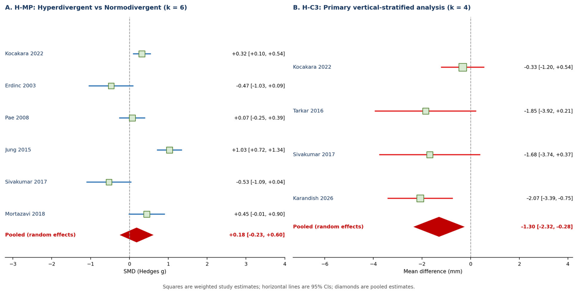
- The vertical position of the hyoid bone, measured as H-MP, did not differ significantly between hyperdivergent and normodivergent subjects in meta-analysis (SMD = 0.185; 95% CI: −0.231 to 0.601; p = 0.383), with very high heterogeneity (I2 = 87.2%). This may be partly attributable to the geometric dependence of H-MP on mandibular-plane inclination.
- The pooled H-C3 estimate indicated a more posterior hyoid position in groups classified as hyperdivergent (MD = −1.30 mm; 95% CI: −2.32 to −0.28; p = 0.013), with a similar estimate in the sensitivity analysis. Residual sagittal confounding prevents attribution of this association to vertical divergence alone, and its clinical significance remains uncertain.
- The effects of vertical and sagittal skeletal patterns on hyoid position are not independent; factorial and multivariate designs are needed to disentangle their contributions.
- Future studies should standardise hyoid measurement methods and use reference systems independent of mandibular inclination. Three-dimensional imaging should be used when clinically justified.
Supplementary Materials
Author Contributions
Funding
Institutional Review Board Statement
Informed Consent Statement
Data Availability Statement
Acknowledgments
Conflicts of Interest
References
- Bibby, R.E.; Preston, C.B. The hyoid triangle. Am. J. Orthod. 1981, 80, 92–97. [Google Scholar] [CrossRef] [Scilit]
- Opdebeeck, H.; Bell, W.H.; Eisenfeld, J.; Mishelevich, D. Comparative study between the SFS and LFS rotation as a possible morphogenic mechanism. Am. J. Orthod. 1978, 74, 509–521. [Google Scholar] [CrossRef] [Scilit]
- Page, M.J.; McKenzie, J.E.; Bossuyt, P.M.; Boutron, I.; Hoffmann, T.C.; Mulrow, C.D.; Shamseer, L.; Tetzlaff, J.M.; Akl, E.A.; Brennan, S.E.; et al. The PRISMA 2020 statement: An updated guideline for reporting systematic reviews. BMJ 2021, 372, n71. [Google Scholar] [CrossRef] [Scilit]
- National Heart, Lung, and Blood Institute. Study Quality Assessment Tools. Available online: https://www.nhlbi.nih.gov/health-topics/study-quality-assessment-tools (accessed on 4 June 2026).
- Pae, E.K.; Quas, C.; Quas, J.; Garrett, N. Can facial type be used to predict changes in hyoid bone position with age? A perspective based on longitudinal data. Am. J. Orthod. Dentofac. Orthop. 2008, 134, 792–797. [Google Scholar] [CrossRef] [Scilit]
- Kumar, S.; Arshad, F.; Nahin, J.; Lokesh, N.K.; Riyaz, K. Comparison of the changes in hyoid bone position in subjects with normodivergent and hyperdivergent growth patterns: A cephalometric study. APOS Trends Orthod. 2017, 7, 224–229. [Google Scholar] [CrossRef] [Scilit]
- DerSimonian, R.; Laird, N. Meta-analysis in clinical trials. Control. Clin. Trials 1986, 7, 177–188. [Google Scholar] [CrossRef] [Scilit] [PubMed]
- Erdinc, A.M.; Dincer, B.; Sabah, M.E. Evaluation of the position of the hyoid bone in relation to vertical facial development. J. Clin. Pediatr. Dent. 2003, 27, 347–352. [Google Scholar] [CrossRef] [Scilit]
- Kocakara, G.; Buyukcavus, M.H.; Orhan, H. Evaluation of pharyngeal airway dimensions and hyoid bone position according to craniofacial growth pattern. Cranio 2022, 40, 313–323. [Google Scholar] [CrossRef] [Scilit]
- Tarkar, J.S.; Parashar, S.; Gupta, G.; Bhardwaj, P.; Maurya, R.K.; Singh, A.; Singh, P. An evaluation of upper and lower pharyngeal airway width, tongue posture and hyoid bone position in subjects with different growth patterns. J. Clin. Diagn. Res. 2016, 10, ZC79–ZC83. [Google Scholar] [CrossRef] [Scilit]
- Sivakumar, A.; Azharuddin, M.; Sivakumar, I.; Sharan, J.; Kiran Raju, D.V.S.; Krishna, V. Assessment of hyoid bone position among different skeletal patterns. Orthod. Waves 2017, 76, 221–225. [Google Scholar] [CrossRef] [Scilit]
- Karandish, M.; Hasani, M.; Dialameh, S. Evaluation of the hyoid bone position based on the hyoid triangle in patients with different skeletal patterns: A lateral cephalometry cross-sectional study. Health Sci. Rep. 2026, 9, e72081. [Google Scholar] [CrossRef] [Scilit]
- Mortazavi, S.; Asghari-Moghaddam, H.; Dehghani, M.; Aboutorabzade, M.; Yaloodbardan, B.; Tohidi, E.; Hoseini-Zarch, S. Hyoid bone position in different facial skeletal patterns. J. Clin. Exp. Dent. 2018, 10, e346–e351. [Google Scholar] [CrossRef] [Scilit]
- Özen, G.Y.; Özen, A.K.; Bilge, N.H. Cephalometric assessment of airway-related hyoid position and velar morphology across skeletal malocclusions: A cross-sectional study. Diagnostics 2026, 16, 947. [Google Scholar] [CrossRef] [Scilit]
- Higgins, J.P.T.; Thomas, J.; Chandler, J.; Cumpston, M.; Li, T.; Page, M.J.; Welch, V.A. (Eds.) Cochrane Handbook for Systematic Reviews of Interventions, Version 6.4; Cochrane: London, UK, 2023. [Google Scholar]
- Zhong, Z.; Tang, Z.; Gao, X.; Zeng, X.L. A comparison study of upper airway among different skeletal craniofacial patterns in nonsnoring Chinese children. Angle Orthod. 2010, 80, 267–274. [Google Scholar] [CrossRef] [Scilit]
- Ucar, F.I.; Uysal, T. Orofacial airway dimensions in subjects with Class I malocclusion and different growth patterns. Angle Orthod. 2011, 81, 460–468. [Google Scholar] [CrossRef] [Scilit]
- Sana, F.; Mirza, O.; Kaleem, M.; Islam, M.N.; Minhas, M.R.; Khan, D.A. Comparison of pharyngeal airway dimensions in different skeletal patterns using cone beam computed tomography. APOS Trends Orthod. 2025, 15, 42–48. [Google Scholar]
- Ucar, F.I.; Ekizer, A.; Uysal, T. Comparison of craniofacial morphology, head posture and hyoid bone position with different breathing patterns. Saudi Dent. J. 2012, 24, 135–141. [Google Scholar] [CrossRef] [Scilit]
- Sayınsu, K.; Isik, F.; Arun, T. Sagittal airway dimensions following maxillary protraction: A pilot study. Eur. J. Orthod. 2006, 28, 184–189. [Google Scholar] [CrossRef] [Scilit]
- Haskell, J.A.; Haskell, B.S.; Spoon, M.E.; Feng, C. The relationship of vertical skeletofacial morphology to oropharyngeal airway shape using cone beam computed tomography: Possible implications for airway restriction. Angle Orthod. 2014, 84, 548–554. [Google Scholar] [CrossRef] [Scilit]
- Li, X.; Lan, Y.; Zeng, X. Three-dimensional analysis of upper airway in skeletal Class II patients with different vertical patterns using cone beam computed tomography. J. Craniofac. Surg. 2017, 28, e469–e474. [Google Scholar]
- Haralabakis, N.B.; Toutountzakis, N.M.; Yiagtzis, S.C. The hyoid bone position in adult individuals with open bite and normal occlusion. Eur. J. Orthod. 1993, 15, 265–271. [Google Scholar] [CrossRef] [Scilit]
- Jena, A.K.; Duggal, R. Hyoid bone position in subjects with different vertical jaw dysplasias. Angle Orthod. 2011, 81, 81–85. [Google Scholar] [CrossRef] [Scilit]
- Jung, S.K.; Kim, T.W. The relevance analysis of hyoid bone position to skeletal or dental open bite and dentofacial characteristics. Oral Surg. Oral Med. Oral Pathol. Oral Radiol. 2015, 120, 528–533. [Google Scholar] [CrossRef] [Scilit]
- Ribeiro, M.C. Posição do Osso Hióide em Pacientes Com Mordida Aberta Anterior. Master’s Thesis, Egas Moniz, Lisbon, Portugal, 2018. [Google Scholar]
- Nejaim, Y.; Aps, J.K.M.; Groppo, F.C.; Haiter Neto, F. Evaluation of pharyngeal space and its correlation with mandible and hyoid bone in patients with different skeletal classes and facial types. Am. J. Orthod. Dentofac. Orthop. 2018, 153, 825–833. [Google Scholar] [CrossRef] [Scilit]
- Chen, W.; Mou, H.E.; Qian, Y.; Qian, L. Evaluation of the position and morphology of tongue and hyoid bone in skeletal Class II malocclusion based on cone-beam computed tomography. BMC Oral Health 2021, 21, 475. [Google Scholar] [CrossRef] [Scilit]
- Soyoye, O.A.; Otuyemi, O.D.; Newman Nartey, M. Cephalometric evaluation of hyoid bone position in subjects with different vertical dental patterns. Niger. J. Clin. Pract. 2021, 24, 667–673. [Google Scholar] [CrossRef] [Scilit]
- Guo, R.; Wang, S.; Zhang, L.; Li, L.; Yu, Q.; Huang, Y.; Li, W. Oropharynx and hyoid bone changes in female extraction patients with distinct sagittal and vertical skeletal patterns: A retrospective study. Head Face Med. 2022, 18, 31. [Google Scholar] [CrossRef] [Scilit]
- Ghazala; Adil, S.; Islam, Z.U.; Jan, G.; Riaz, S.; Taj, L. The position of hyoid bone on lateral cephalographs in various vertical facial patterns. J. Postgrad. Med. Inst. 2023, 37, 269–273. [Google Scholar] [CrossRef] [Scilit]
- Mackevičiūtė, U.; Švalkauskienė, V.; Grinkevičius, P.; Grinkevičienė, D.; Vasiliauskas, A. Relationship between the hyoid triangle and skeletal open bite and deep bite in Class II patients. Sveik. Moksl. 2025, 35, 176–181. [Google Scholar] [CrossRef]
- Al-Somairi, M.A.A.; Zheng, B.; Almaqrami, B.S.; Al-Worafi, N.A.; Alyafrusee, E.S.; Al-Tayar, B.; Al-Rokhami, R.K.; Al-Warafi, L.A.; Alhashimi, N.; Xu, H.; et al. Correlation between the three-dimensional hyoid bone parameters and pharyngeal airway dimensions in different sagittal and vertical malocclusions. J. Stomatol. Oral Maxillofac. Surg. 2024, 125, 101994. [Google Scholar] [CrossRef] [Scilit]
- Domenyuk, D.A.; Davydov, B.N.; Domenyuk, S.D.; Ivanyuta, I.V.; Kokareva, A.V.; Ivanyuta, O.O. Cephalometric analysis of hyoid bone and tongue position in patients with different types of facial skeletal structure. Med. Alph. 2025, 30, 88–97. [Google Scholar] [CrossRef] [Scilit]
- Kang, J.H.; Kim, J.Y.; Jha, N.; Jung, S.K.; Lee, Y.S.; Kim, Y.J. Associations of tongue and hyoid position, tongue volume, and pharyngeal airway dimensions with various dentoskeletal growth patterns. PLoS ONE 2025, 20, e0326092. [Google Scholar] [CrossRef] [Scilit]


| First Author (Year) | Country | n | Age (y) | Design | Image | Classification | Key Outcomes |
|---|---|---|---|---|---|---|---|
| Haralabakis (1993) [23] | Greece | 82 | 15–27 | CS | Ceph | Open bite vs. CL I | H-PP ↑; H-Po-FH ↓ in OB |
| Erdinc (2003) [8] | Turkey | 75 | ~12 | CS | Ceph | SN-GoGn + Jarabak | H-SN, H-FH, H-PP ↓ in hyper *** |
| Pae (2008) [5] | USA | 163 | 46–61 | Cohort | Ceph | FMA | HYRGN hypo > normo > doli *** |
| Jena (2011) [24] | India | 71 | 15–25 | CS | Ceph | FMA + SN-MP | H-PTRper: hypo > normo *** |
| Jung (2015) [25] | Korea | 182 | ~25 | CS | Ceph | Björk sum | H-MP: skeletal > dental pattern *** |
| Tarkar (2016) [10] | India | 90 | 18–32 | CS | Ceph | Jarabak + FMA | H-RGn hypo > normo * (Bibby) |
| Kumar (2017) [6] | India | 40 | 20–27 | Pre/post observational | Ceph | FMA | No between-pattern difference; hyoid moved posteriorly/inferiorly after treatment |
| Sivakumar (2017) [11] | India | 100 | 18–25 | CS | Ceph | ANB + FMA | C3H: CII hyper < CI normo ** |
| Mortazavi (2018) [13] | Iran | 110 | ≥18 | CS | Ceph | ANB | H-C3: CI > CII *; H-PP: CI > CIII * |
| Ribeiro (2018) [26] | Portugal | 31 | NR | CS | Ceph | Overbite | H-MP vs. Ricketts norms * |
| Nejaim (2018) [27] | Brazil | 161 | 21–58 | CS | CBCT | VERT index + ANB | LL hyoid: doli > meso (morphology) |
| Chen (2021) [28] | China | 90 | ~16 | CS | CBCT | ANB + MP-FH | H-Me: hypo CL I > normo/hyper *** |
| Soyoye (2021) [29] | Nigeria | 174 | Adults | CS | Ceph | Overbite | C3-Hy: reduced > normal > deep * |
| Kocakara (2022) [9] | Turkey | 611 | ≤17 | CS | Ceph | ANB + SN-GoGn | All hyoid measures NS |
| Guo (2022) [30] | China | 160 | 20–50 | CS | CBCT | ANB + SN-MP | H-X: CII hyper < all others ** |
| Ghazala (2023) [31] | Pakistan | 75 | Adults | CS | Ceph | SN-MP | H-MP, H-axis-MP: hyper > normo * |
| Mackevičiūtė (2025) [32] | Lithuania | 60 | Adults | CS | Ceph | Overbite in CII | C3-RGn: open > deep bite * |
| Al-Somairi (2024) [33] | China | 368 | Adults | CS | CBCT | FMA | H-VP: hypo > normo > hyper *** |
| Karandish (2026) [12] | Iran | 296 | Adults | CS | Ceph | ANB + FMA | C3-H, H-RGn: CII < CI *** |
| Özen (2026) [14] | Turkey | 120 | 18–30 | CS | Ceph | ANB | H-RGn: CIII > CI, CII * |
| Domenyuk (2025) [34] | Russia | 106 | Adults | CS | Ceph + CBCT | Vertical/neutral/horizontal | H-Me: horizontal > others * (median) |
| Kang (2025) [35] | Korea | 185 | ~29 | CS | CBCT | Continuous (ANB, ODI, SN-GoGn) | Hyoid x: class III (sagittal) only |
| Bibby & Preston (1981) [1] † | S. Africa | 54 | ~13 | CS | Ceph | CL I (normative) | Normative values: C3-H = 31.8 ± 2.9 mm |
| Study | k | Effect (95% CI) | CI low | CI High | p | I2 (%) | τ | RoB | Sig. |
|---|---|---|---|---|---|---|---|---|---|
| ANALYSIS 1: H-MP—Hyperdivergent vs. Normodivergent (SMD, Hedges g; positive = hyoid more inferior in hyperdivergent) | |||||||||
| Kocakara 2022 [9] | 0.322 (0.105, 0.538) | 0.105 | 0.538 | GOOD | |||||
| Erdinc 2003 [8] † | −0.472 (−1.034, 0.090) | −1.034 | 0.090 | FAIR | |||||
| Pae 2008 [5] | 0.073 (−0.248, 0.393) | −0.248 | 0.393 | FAIR | |||||
| Jung 2015 [25] | 1.031 (0.721, 1.341) | 0.721 | 1.341 | FAIR | |||||
| Sivakumar 2017 [11] | −0.527 (−1.091, 0.036) | −1.091 | 0.036 | FAIR | |||||
| Mortazavi 2018 [13] | 0.448 (−0.005, 0.901) | −0.005 | 0.901 | FAIR | |||||
| POOLED (RE) | 6 | 0.185 (−0.231, 0.601) | −0.231 | 0.601 | 0.383 | 87.2 | 0.475 | — | NS |
| PRIMARY ANALYSIS: H-C3—Studies reporting a vertical-pattern contrast (residual sagittal confounding remains) | |||||||||
| Kocakara 2022 [9] | −0.33 (−1.20, 0.54) | −1.20 | 0.54 | GOOD | |||||
| Tarkar 2016 [10] | −1.85 (−3.92, 0.21) | −3.92 | 0.21 | FAIR | |||||
| Sivakumar 2017 [11] | −1.68 (−3.74, 0.37) | −3.74 | 0.37 | FAIR | |||||
| Karandish 2026 [12] | −2.07 (−3.39, −0.75) | −3.39 | −0.75 | FAIR | |||||
| POOLED (RE) | 4 | −1.30 (−2.32, −0.28) | −2.32 | −0.28 | 0.013 | 48.6 | 0.717 | — | * |
| SENSITIVITY ANALYSIS: H-C3—Extended pool including two predominantly sagittal-proxy studies | |||||||||
| Kocakara 2022 [9] | −0.33 (−1.20, 0.54) | −1.20 | 0.54 | GOOD | |||||
| Tarkar 2016 [10] | −1.85 (−3.92, 0.21) | −3.92 | 0.21 | FAIR | |||||
| Sivakumar 2017 [11] | −1.68 (−3.74, 0.37) | −3.74 | 0.37 | FAIR | |||||
| Mortazavi 2018 [13] | −3.54 (−5.61, −1.47) | −5.61 | −1.47 | FAIR | |||||
| Karandish 2026 [12] | −2.07 (−3.39, −0.75) | −3.39 | −0.75 | FAIR | |||||
| Özen 2026 [14] | 0.25 (−1.86, 2.36) | −1.86 | 2.36 | GOOD | |||||
| POOLED (RE) | 6 | −1.45 (−2.51, −0.38) | −2.51 | −0.38 | 0.008 | 60.8 | 1.002 | — | ** |
Disclaimer/Publisher’s Note: The statements, opinions and data contained in all publications are solely those of the individual author(s) and contributor(s) and not of MDPI and/or the editor(s). MDPI and/or the editor(s) disclaim responsibility for any injury to people or property resulting from any ideas, methods, instructions or products referred to in the content. |
© 2026 by the authors. Licensee MDPI, Basel, Switzerland. This article is an open access article distributed under the terms and conditions of the Creative Commons Attribution (CC BY) license.
Share and Cite
Pérez-Sánchez, D.; Ibor-Miguel, M.; Aura-Tormos, J.I.; Marqués-Martínez, L.; Guinot-Barona, C.; García-Miralles, E. Hyoid Bone Position in Relation to Vertical Skeletal Pattern: A Systematic Review and Meta-Analysis. Diagnostics 2026, 16, 2826. https://doi.org/10.3390/diagnostics16172826
Pérez-Sánchez D, Ibor-Miguel M, Aura-Tormos JI, Marqués-Martínez L, Guinot-Barona C, García-Miralles E. Hyoid Bone Position in Relation to Vertical Skeletal Pattern: A Systematic Review and Meta-Analysis. Diagnostics. 2026; 16(17):2826. https://doi.org/10.3390/diagnostics16172826
Chicago/Turabian StylePérez-Sánchez, Davinia, Marta Ibor-Miguel, Juan Ignacio Aura-Tormos, Laura Marqués-Martínez, Clara Guinot-Barona, and Esther García-Miralles. 2026. "Hyoid Bone Position in Relation to Vertical Skeletal Pattern: A Systematic Review and Meta-Analysis" Diagnostics 16, no. 17: 2826. https://doi.org/10.3390/diagnostics16172826
APA StylePérez-Sánchez, D., Ibor-Miguel, M., Aura-Tormos, J. I., Marqués-Martínez, L., Guinot-Barona, C., & García-Miralles, E. (2026). Hyoid Bone Position in Relation to Vertical Skeletal Pattern: A Systematic Review and Meta-Analysis. Diagnostics, 16(17), 2826. https://doi.org/10.3390/diagnostics16172826