Case Report

Not So Crystal Clear: Pulmonary Crystal-Storing Histiocytosis Without Haematological Disease: A Unique Radiological Manifestation and Review of the Literature

1
Department of Respiratory Medicine, Galway University Hospital, H91 YR71 Galway, Ireland
2
Department of Histopathology, Galway University Hospital, H91 YR71 Galway, Ireland
3
Respiratory Department, Cavan & Monaghan Hospital & Chronic Disease Management Hub, HSE & RCSI University of Medicine, H12 Y7W1 Cavan, Ireland
4
Royal College of Surgeons in Ireland, University of Medicine & Health Sciences, D02 YN77 Dublin, Ireland
5
Department of Haematology, Galway University Hospital, H91 YR71 Galway, Ireland
*
Author to whom correspondence should be addressed.
Diagnostics 2026, 16(15), 2410; https://doi.org/10.3390/diagnostics16152410

Submission received: 23 June 2026 / Revised: 23 July 2026 / Accepted: 27 July 2026 / Published: 31 July 2026

Abstract

Background and Clinical Significance: Pulmonary crystal-storing histiocytosis (CSH) without an associated haematological malignancy, lymphoproliferative disorder, plasma cell disorder, or other identifiable underlying condition is exceptionally rare. Long-term radiological data from published cases remain limited, and this case, contextualised by a narrative review of the literature, may expand the recognised imaging spectrum of localised pulmonary CSH. Case Presentation: We report the case of a 59-year-old man with incidental multiple pulmonary lesions identified on CT thorax imaging. The lesions demonstrated an unusual combination of cystic change, cavitation, and surrounding ground-glass opacities. His medical history was significant for bipolar disorder treated with lithium and a 50-pack-year smoking history. Interval imaging showed progression, prompting further investigation and ultimately right upper lobectomy. Histopathological analysis confirmed pulmonary CSH; lesional cells contained crystalloid material and showed CD68 and PAS positivity, with dual kappa and lambda expression on immunohistochemistry. Markers for other differential diagnoses, including Congo red, birefringence, Langerin, and CD1a were negative. Following diagnosis and resection, serial imaging demonstrated fluctuating yet slowly progressive pulmonary abnormalities. No lymphoproliferative or plasma cell disorder has emerged after more than seven years of post-diagnostic surveillance and more than ten years since the initial imaging abnormality. Conclusions: This case demonstrates that the radiological spectrum of localised pulmonary CSH may include cystic, cavitary and ground-glass abnormalities, with subsequent fluctuating yet slowly progressive post-resection evolution. Pulmonary CSH should be considered in the differential diagnosis of unexplained or atypical pulmonary nodules, particularly when histiocyte-rich pathology with intracytoplasmic crystalloid material is identified. Long-term multidisciplinary surveillance is warranted.

1. Introduction

Crystal-Storing Histiocytosis (CSH) is an exceptionally rare disorder characterised by the intracytoplasmic accumulation of crystallised materials, most commonly immunoglobulin, within histiocytes [1]. More than 95% of reported cases are associated with lymphoproliferative or plasma cell disorders (LP-PCDs) including multiple myeloma, plasmacytoma, and low-grade B-cell lymphomas [1]. Less frequently, CSH has been described in association with autoimmune disease, chronic infection, occupational exposure or medication use, often involving non-immunoglobulin crystalline material [2,3].
CSH may present in a generalised form, involving multiple organs or in a localised form confined to a single anatomical region [3]. Pulmonary CSH represents a rare subtype of the localised form with fewer than 30 English-language clinicopathological cases identified. Most published cases of Pulmonary CSH are associated with either LP-PCD or other identifiable secondary conditions [1,3]. To the best of our knowledge, only one prior English-language clinicopathological case has been reported without an identifiable associated condition [4].
Here, we report a rare case of pulmonary CSH presenting as multiple pulmonary lesions initially suspected to represent lung cancer. The diagnosis of pulmonary CSH was established histologically following surgical resection, with no evidence of an associated LP-PCD or alternative underlying condition despite extensive evaluation. The patient has undergone over a decade of longitudinal clinical, biochemical and radiological surveillance, providing unique insight into the long-term natural history and radiological evolution of this rare entity. To contextualise our findings, we also present a narrative review of published cases of localised pulmonary CSH.

2. Case Report

2.1. Clinical History

A 59-year-old Irish man undergoing evaluation for unintentional weight loss was incidentally found to have multiple bilateral pulmonary lesions on CT of the thorax, abdomen and pelvis (CT TAP). His past medical history included bipolar affective disorder managed with long-term lithium therapy and a 50-pack-year smoking history. He denied respiratory symptoms, constitutional complaints other than weight loss, and any features suggestive of rheumatological disease. His weight at initial review was 53 kg, corresponding to a body mass index of 17.7 kg/m2. Clinical examination was unremarkable.
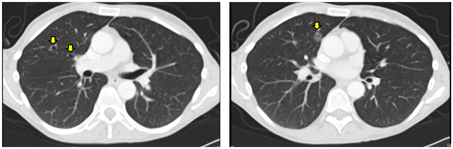
His initial CT imaging revealed multiple thin-walled pulmonary cavities, the largest measuring 12 mm in the right upper lobe, with surrounding patchy ground-glass opacification (Figure 1). Mediastinal and hilar lymph nodes remained within normal limits. No additional abnormalities were identified elsewhere on the CT. Based on these radiological findings, the differential diagnosis included primary lung malignancy, post-infectious or granulomatous disease such as tuberculosis, autoimmune lung disease and cystic lung disorders, including Pulmonary Langerhans Cell Histiocytosis (PLCH).
Initial laboratory testing, including full blood count with eosinophil differential, Quantiferon TB testing, and a comprehensive autoimmune/vasculitic screen such as ANA, ANCA, RF, anti-CCP, connective tissue disease (CTD) and myositis panel, were all within normal limits. Viral serology for HIV, hepatitis B and hepatitis C was negative. Pulmonary function tests (PFTs) demonstrated preserved lung function (FEV1 3.34 L, FVC 4.42 L, FEV1/FVC 75%, DLCO 83%). Sputum samples, including three specimens submitted for microscopy and culture for bacteria, mycobacteria and acid-fast bacilli (AFB), were negative.
Bronchoscopy was initially recommended for further evaluation, however, the patient declined the procedure, as he remained clinically well and asymptomatic. A period of radiological surveillance was therefore adopted, during which the lesions remained stable over the subsequent year. However, after this period, he had a hospital admission for respiratory failure secondary to pneumonia and pulmonary embolism. CT pulmonary angiogram performed during that admission showed additional infective changes, and a repeat CT scan was planned several months after discharge to allow interval resolution.
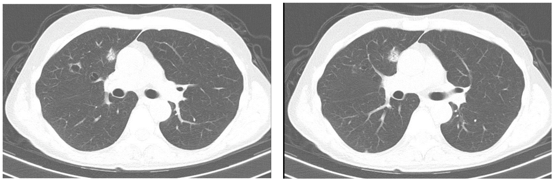
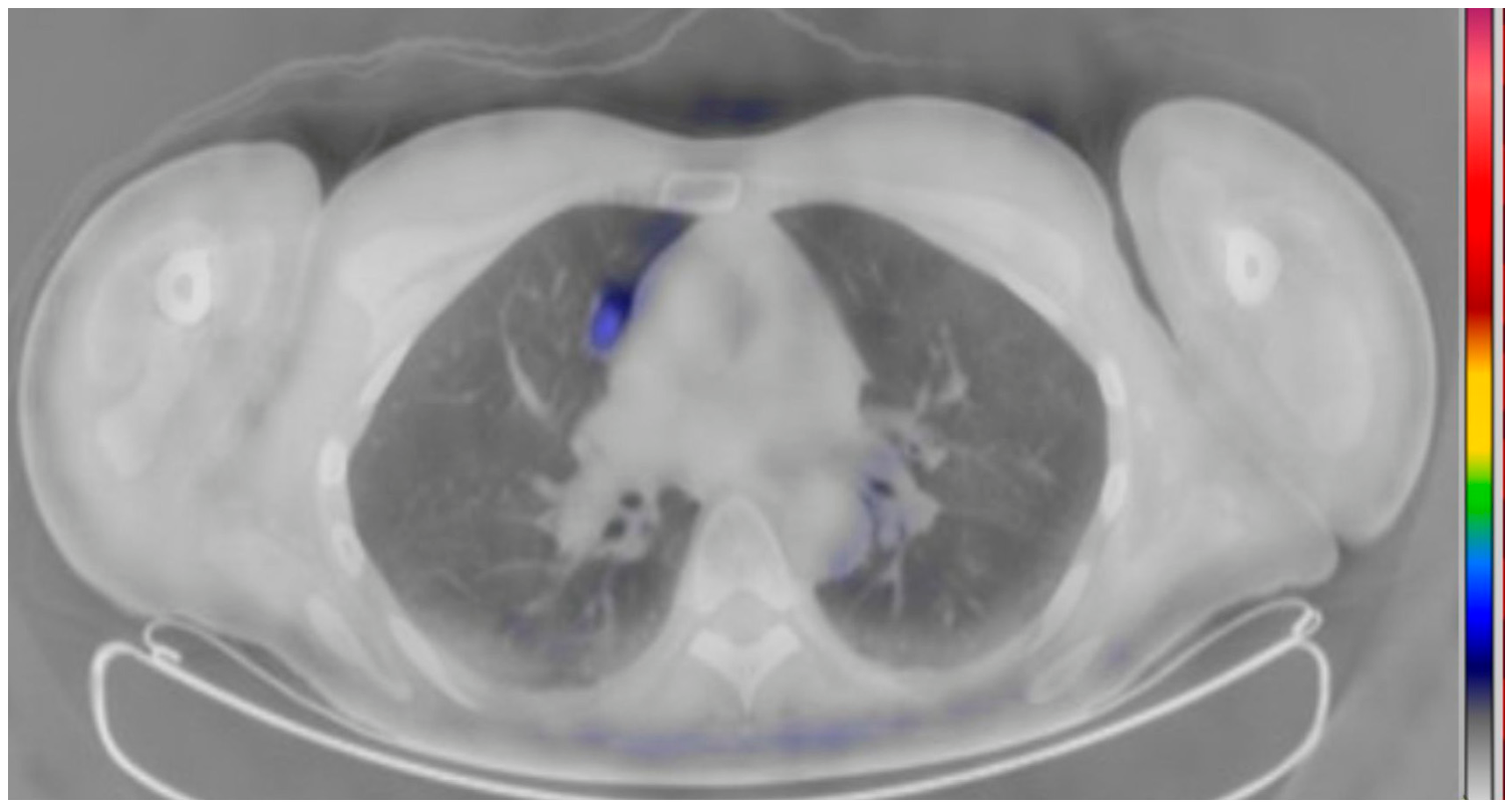
The repeat CT scan demonstrated interval enlargement of the dominant lesion from 12 mm to 16 mm, with resolution of the infective changes seen on previous imaging (Figure 2). Given this progression, a whole-body PET-CT was performed, revealing low-grade fluorodeoxyglucose (FDG) uptake confined to the right upper lobe lesion (Figure 3). Although the metabolic activity was modest, malignancy could not be excluded. After multidisciplinary discussion, the patient underwent a right upper lobectomy via video-assisted thoracoscopy surgery (VATS). Lymph node stations 2, 4, 7, 9, 10 and 11 were also dissected. Post-operatively, he managed to stop smoking successfully.

2.2. Histology and Immunohistochemistry

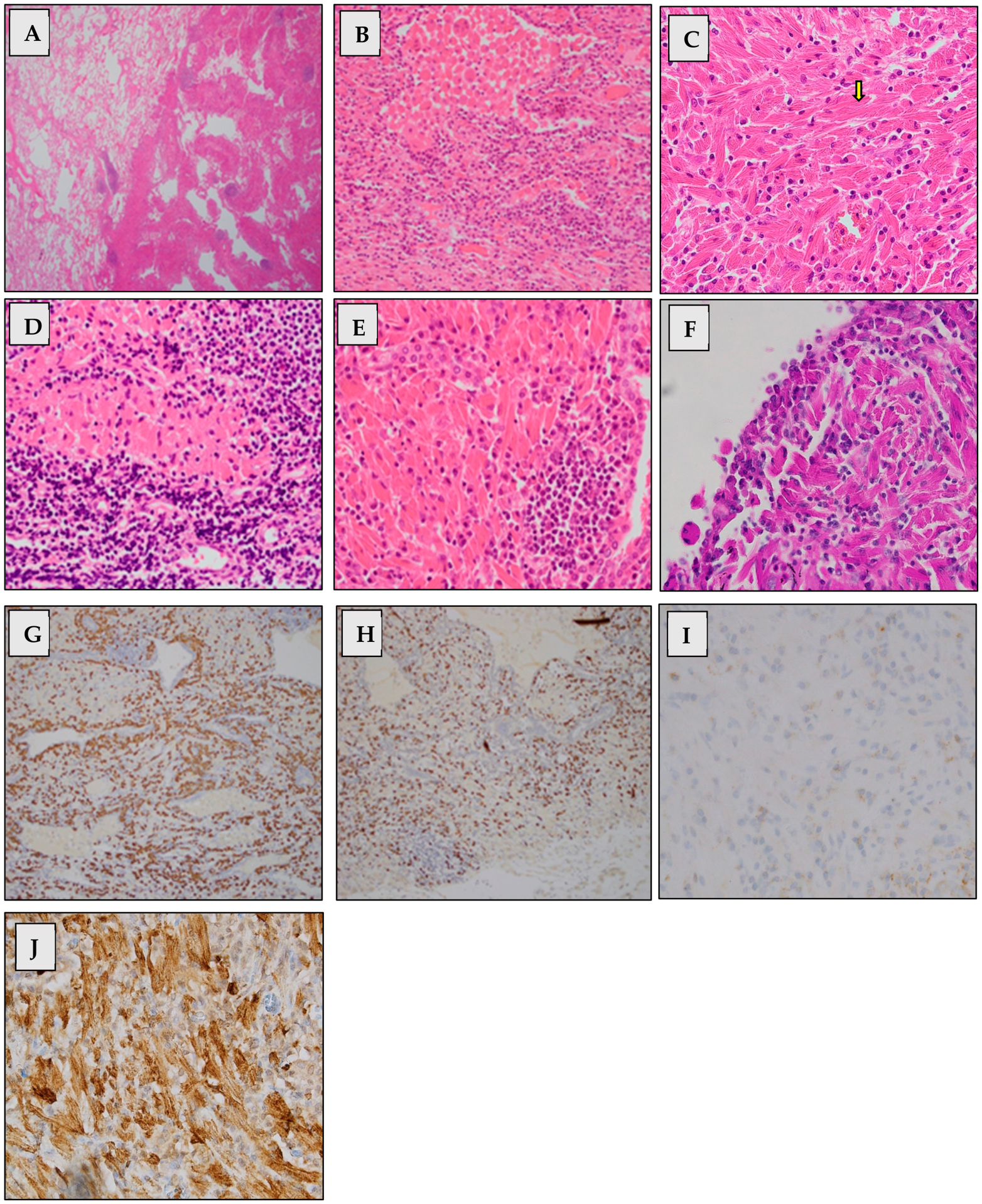
Gross pathological examination identified two distinct homogeneous, cream-coloured, firm lesions measuring 23 mm and 11 mm within the parenchyma. The nodules were relatively well-circumscribed and demonstrated an admixed lymphoid population with numerous lymphoid follicles. Microscopically, both lesions were composed of enlarged, rounded-to-angulated cells with abundant eosinophilic cytoplasm containing fine-needle-like crystalloid material, admixed with lymphocytes and plasma cells (Figure 4A–E). These crystalloid structures were subtle on light microscopy and challenging to highlight clearly. Resection margins and visceral pleura were free of involvement. Notably, some of lymph nodes from stations 11, 7, 4 and 2 showed similar features to the pulmonary nodules. This appearance prompted consideration of CSH diagnosis.
Immunohistochemical analysis further supported this diagnosis. The lesional cells demonstrated CD68 positivity, confirming macrophage/histiocytic lineage, while PAS positivity supported the presence of intracytoplasmic crystalloid material. Within the accompanying lymphoid component, CD138 selectively highlighted plasma cells, assisting in the evaluation of plasma cell population (Figure 4F–J). Importantly, immunostaining for kappa and lambda light chains demonstrated a dual expression pattern, indicating the absence of light-chain restriction (Figure 4G,H). A broad panel of additional markers was performed to exclude alternative diagnoses. Negative staining for AE1/3, CD1a, desmin, MyoD1, HMB45, S100, Congo red stain and birefringence helped exclude other differential diagnoses, including epithelial neoplasm, Langerhans cell histiocytosis, myogenic tumor, melanocytic lesion, S100-positive histiocytic processes and amyloidosis. The combined immunohistochemical findings, including histiocyte markers (CD68, PAS positivity), polytypic plasma cells and a broad panel negative for other similar differentials and competing mimics, strongly support the diagnosis of CSH.

2.3. Haematology Work-Up

Comprehensive haematological evaluation, including serum protein electrophoresis and serum free light-chain analysis, demonstrated no monoclonal band and no light-chain abnormality. Beta-2 microglobulin and lactate dehydrogenase levels were also within normal limits. A urine Bence Jones protein test was not performed, as its sensitivity is inferior to serum free light-chain testing [5,6]. Bone marrow biopsy was considered but ultimately deferred due to the absence of clinical, biochemical, or radiological features suggestive of an underlying clonal haematological disorder. Immunoglobulin heavy-chain gene rearrangement testing was likewise not pursued, given the lack of morphological or immunophenotypic evidence of lymphoma. The patient’s limited medication list was reviewed, and no agents known to induce crystal-storing histiocytosis were identified; in particular, lithium was not recognised among reported drug-associated CSH triggers in the literature review [1,3]. Autoimmune screening remained negative throughout the evaluation.

2.4. Diagnosis and Follow-Up

A diagnosis of pulmonary CSH was established at Galway University Hospital in Galway, Ireland. There was no evidence of an associated lymphoproliferative or plasma cell disorder or any other identifiable underlying condition. The patient remained clinically stable throughout follow-up. His initial weight loss subsequently improved and was later attributed to difficulties with mastication related to denture problems, which resolved following dental assessment and management by the maxillofacial team. His weight has since remained stable at approximately 65 kg.
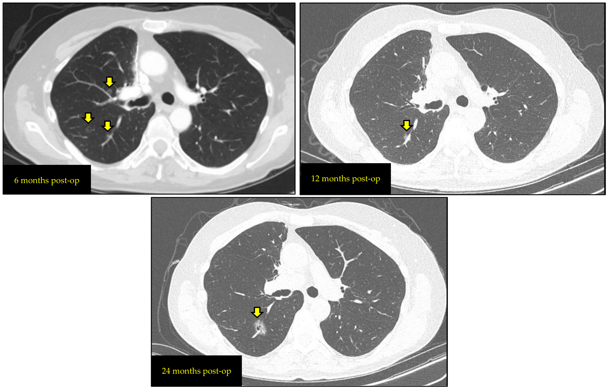
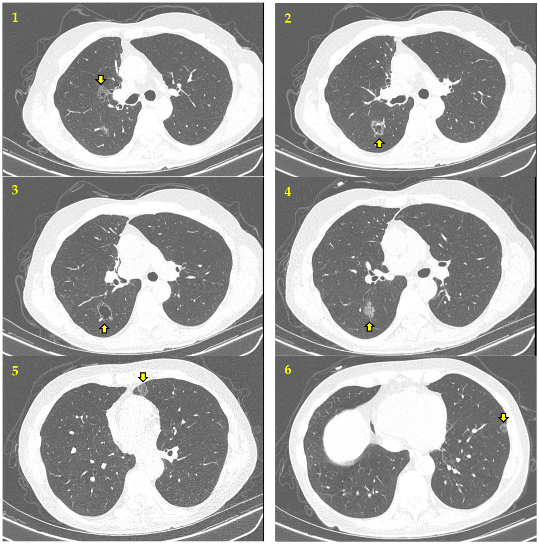
The patient was enrolled in long-term radiological and biochemical surveillance at six to twelve-months intervals. Follow-up imaging performed six months after the lobectomy demonstrated new bilateral pulmonary lesions with similar morphological characteristics to the original lesions. Several of these lesions regressed or disappeared on subsequent scans, while the right lower lobe lesion showed gradual interval enlargement (Figure 5). Over time, the right lower lobe nodule continued to demonstrate slow but persistent growth, whereas other lesions remained stable, as seen on the most recent CT scan, performed ten years after the initial CT and seven years post-lobectomy (Figure 6). The evolving radiological findings remained morphologically similar to the previously resected CSH. PFTs almost 10 years after baseline demonstrated slightly reduced FEV1 and DLCO, while his FVC has improved (FEV1 2.95 L, FVC 4.56 L, FEV1/FVC 65%, DLCO 71%). These changes have remained stable over recent years and partly may reflect post-lobectomy status and smoking cessation.
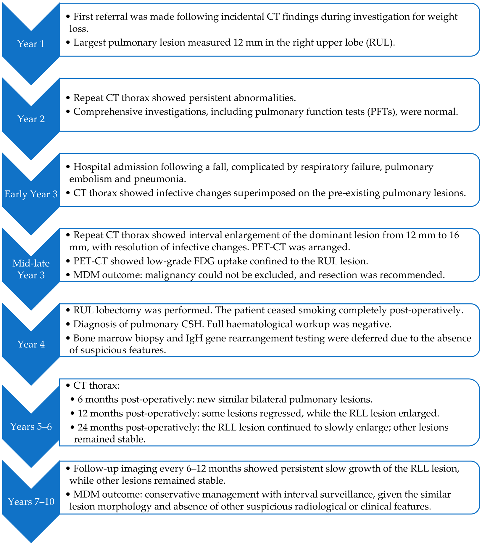
The multidisciplinary team review concluded that conservative management remained appropriate in view of the patient’s clinical stability and the absence of suspicious radiological features or systemic abnormalities. Despite more than seven years of clinical, biochemical, and radiological surveillance post-diagnosis and over 10 years since initial imaging, no associated lymphoproliferative disorder, inflammatory disease, medication-related cause, or other underlying condition has been identified. The case timeline is summarised in Figure 7, and the diagnostic considerations and associated work-up are presented in Table 1.

3. Discussion

Pulmonary crystal-storing histiocytosis (CSH) is a rare, localised manifestation of CSH, with fewer than 30 cases reported to date (Table 1). Diagnosis in the absence of an associated condition is particularly uncommon. LP-PCD may precede or occur concurrently with pulmonary CSH, highlighting the need for long-term surveillance even when initial investigations are negative [1]. Although localised CSH is generally considered histologically benign, reported follow-up durations remain limited. Generalised CSH, in contrast, carries a markedly poor prognosis owing to its strong association with haematological malignancy; among six reported cases with pulmonary involvement, only one patient survived [7,8,9,10,11,12].
We performed a narrative review of published English-language reports of localised pulmonary CSH. Cases with pulmonary involvement as part of generalised CSH were excluded owing to their multisystem disease burden and distinct clinical trajectory. Similarly, cases with predominantly extra-parenchymal involvement, such as tracheal or pleural CSH, were excluded due to their differing anatomical distribution and radiologic characteristics. The available literature was heterogeneous; most reports were conventional case reports or small case series, one was published as a conference abstract only [13], and two were brief case descriptions [14,15]. Older references were retained when they represented original published cases of localised pulmonary CSH required for completeness of the literature review. One additional spatial transcriptomic study of a confirmed pulmonary CSH case was identified but was not included in the quantitative clinicopathological summary because conventional clinical association, systemic work-up, and follow-up data were not sufficiently reported [16].
Including our case, a total of 26 cases of localised pulmonary CSH were identified and included (Table 2). These comprised 12 men and 14 women, with a median age of 60 years (range, 12–89 years). Only one paediatric case was identified, occurring in a child with common variable immunodeficiency (CVID) and pulmonary mucosa-associated lymphoid tissue (MALT) lymphoma [17].
The 26 cases were reported from geographically diverse institutions. The largest number were reported from the USA (n = 9), followed by Italy (n = 4), Japan (n = 3), and India (n = 2). Single cases were reported from Canada, Australia, South Korea, Taiwan, New Zealand, China, and Ireland. One additional report involved author affiliations in both the Netherlands and the United Kingdom, although the country in which the patient was diagnosed was not specified. These data describe the location of the principal diagnosing or reporting institution only and should not be interpreted as reflecting patient ethnicity, ancestry, race, or country of origin. Ethnicity, race, and ancestry were not systematically documented in the published pulmonary CSH cases and therefore could not be analysed.
Sixteen cases of pulmonary CSH were associated with LP-PCD, eight had non-LP-PCD associations or secondary contexts, and only two, including our case, had no clearly identifiable associated condition. Pulmonary CSH has a heterogeneous spectrum of associated conditions. Among the LP-PCD-associated cases listed in Table 2, pulmonary marginal zone lymphoma/MALT lymphoma was the most frequent association (n = 10), followed by MGUS (n = 2), plasmacytoma (n = 2), lymphoplasmacytoid lymphoma (n = 1), and small B-cell non-Hodgkin lymphoma (n = 1). Non-LP-PCD-associated cases included autoimmune disease such as rheumatoid arthritis and Sjögren syndrome, occupational or inflammatory contexts including asbestos exposure and silicosis, anti-tuberculosis treatment or clofazimine exposure, and plasma cell granuloma. One case occurred in a patient with remote gastric DLBCL in complete remission. Even though the lung lesion showed a minor B-cell population with IgH rearrangement and slight kappa predominance, the authors considered the findings insufficient for a diagnosis of active LP-PCD and classified the case as pulmonary localised CSH without underlying LP-PCD [18]. Chen et al. described a confirmed pulmonary CSH case using spatial transcriptomic profiling. Although the study provided valuable molecular insight into the PCSH lesion microenvironment, it was tabulated separately and excluded from quantitative clinicopathological association analysis because conventional systemic work-up and, most importantly, association status were not sufficiently reported [16].
Clinical presentation was most commonly incidental or asymptomatic, with lesions detected on imaging in 13 of 26 cases. When symptoms were reported, they were non-specific and included cough or chronic cough (n = 6), shortness of breath (n = 3), chest pain (n = 3), fever (n = 2), weight loss (n = 2), haemoptysis (n = 1), and fatigue (n = 1). Some patients had more than one symptom. One case had no presenting symptoms documented. Three cases were in the context of active or recent Tuberculosis (TB) treatment. This findings suggested that pulmonary CSH lacks distinctive clinical features and is rarely suspected prior to histologic diagnosis. In our case, CT imaging was prompted by weight loss; however, this subsequently resolved and was later considered unrelated to the pulmonary lesions.
Radiologically, pulmonary CSH most commonly presents as nodules, masses or mass-like lesions, reported in 22 of 26 cases. Lesions were solitary in 13 cases and multiple in 11 cases. Lesion number was not documented in one case and not applicable in another case presenting as consolidation. Reported lesion size ranged from 11 to 52 mm among cases with available measurements. Less common radiological patterns included opacity, consolidation, calcification, cystic change, and occasional cavitation. Cystic nodules had previously been described in LP-PCD-associated cases, including one case with MGUS and coexisting nonspecific interstitial pneumonia, and another with recurrent marginal zone lymphoma [19]. In contrast, our patient demonstrated a distinctive combination of ground-glass opacity, cystic change, and cavitation in the absence of an identifiable LP-PCD or other secondary association.
Although mixed ground-glass opacity, cystic change, and cavitation have not been widely recognised as radiological features of pulmonary CSH, several pathological descriptions provide a biologically plausible basis for these appearances. Ionescu et al. described a nodular mass of large histiocytic cells expanding alveoli and infiltrating alveolar septa, a pattern that could produce partial airspace filling and ground-glass attenuation [20]. The same report described bronchiolar destruction and a desquamative interstitial pneumonia-like pattern, both of which may contribute to ground-glass change. Mucostasis within bronchioles, with luminal obstruction by mucus, desquamated epithelial cells, pigmented macrophages, and crystal-laden macrophages, could also create a ball-valve effect, promoting distal air trapping, and parenchymal overdistention. This mechanism is hypothesised to predispose to cyst formation [21].
Jones et al. similarly described crystal-containing histiocytes expanding the lung interstitium and extending into alveolar spaces, accompanied by lymphoplasmacytic inflammation [22]. Such changes may distort parenchymal architecture and plausibly contribute to cystic or cavitary transformation and may also produce interstitial thickening and ground-glass opacities. Taken together, these pathological findings strongly support the biological plausibility of mixed ground-glass opacity and cystic and cavitary abnormalities observed in our patient, although this precise radiological combination has not previously been documented in pulmonary CSH (Table 2).
The existing literature provides little information on radiological evolution over time, with most reports limited to short clinical follow-up and without serial long-term imaging descriptions (Table 2). Jones et al. documented recurrence of a histologically similar pulmonary lesion 10 years after resection, but interval imaging evolution was not described [22]. Our case therefore represents a unique longitudinal course, characterised by fluctuating yet progressively evolving pulmonary lesions over more than 10 years. To our knowledge, this is the longest radiologically documented follow-up of localised pulmonary CSH without an identifiable associated condition. Importantly, unlike previously reported cystic cases, our patient showed no evidence of LP-PCD or systemic disease despite prolonged clinical, biochemical, and radiological surveillance. These observations suggest that although pulmonary CSH is histologically benign, its pulmonary manifestations may not always remain radiologically static. Instead, the disease may follow a slow, progressive trajectory, potentially driven by ongoing deposition of crystallised immunoglobulin within histiocytes or by a subtle clonal process below current diagnostic thresholds, or an unrecognised environmental or inflammatory trigger.
Management strategies were primarily driven by diagnostic concern for malignancy rather than by disease aggressiveness. Surgical resection was the most frequent approach, performed in 18 cases. Other diagnostic approaches included surgical biopsy without formal resection (n = 2), CT-guided FNA or percutaneous aspiration biopsy (n = 2), CT-guided core biopsy (n = 1), endobronchial biopsy with repeat lung biopsy (n = 1), BAL cytology (n = 1), and autopsy diagnosis (n = 1). BAL diagnosis was reported in a clofazimine-associated case in a patient treated for multidrug-resistant tuberculosis [15]. Systemic therapy was documented in five cases and was directed at associated haematological or inflammatory disease rather than pulmonary CSH itself. For example, steroids and azathioprine were commenced in a patient diagnosed with pulmonary CSH in the setting of MGUS and active NSIP [19]. Another example was a patient that was started on chemotherapy for concurrent diagnosis of recurrent marginal cell lymphoma [19]. In our case, a right upper lobectomy was performed because malignancy was the primary initial concern. As no associated underlying disorder was identified, no additional therapy was administered beyond close clinical and radiologic surveillance.
Overall prognosis was favourable in most cases with available follow-up. Three deaths were reported, all attributable to unrelated causes rather than pulmonary CSH progression: sepsis in an autopsy-diagnosed case, myocardial infarction, and interstitial lung disease [4,19]. Seven cases have no documentation of prognosis. Long-term follow-up of two years or longer was available in six cases, including one recurrent pulmonary case reported by Jones et al., which recurred after 10 years [22], and the present case, which demonstrated fluctuating but slowly progressive radiological evolution over more than 10 years. These findings support the view that localised pulmonary CSH is typically an indolent process but may recur or evolve radiologically, particularly when long-term imaging is available.
Histopathologic examination remains the diagnostic cornerstone of pulmonary CSH. Across cases in Table 2, lesions were characterised by aggregates of histiocytes containing abundant eosinophilic cytoplasm filled with refractile crystals. Crystal morphology was variably described. Needle-shaped or needle-like crystals were the most frequent morphology, either as a pure pattern or as part of a mixed pattern. Pure needle-shaped or needle-like morphology was reported in 11 cases, while mixed needle-shaped and polygonal morphology (which include rhomboid, trapezoid and rectangular forms) was reported in five cases. Other described morphology included spindle-shaped, and rod-shaped forms. Crystal morphology was not documented in seven cases. No consistent relationship was apparent between crystal morphology, radiological pattern, or underlying disease association.
Immunoglobulin and clonality findings were heterogeneous, although kappa light-chain involvement predominated across the reported cases. Kappa restriction or kappa predominance was particularly frequent in LP-PCD-associated pulmonary CSH, consistent with the strong association between CSH and clonal lymphoplasmacytic or plasma-cell proliferations. In contrast, polyclonal or polytypic kappa and lambda expression was more often observed in non-LP-PCD-associated cases and in cases without a clearly identifiable underlying condition, although exceptions were noted, including LP-PCD-associated cases with polytypic staining but molecular evidence of clonality. One drug-associated case represented non-immunoglobulin crystal storage related to clofazimine exposure.
The mechanism of immunoglobulin crystal accumulation remains incompletely understood. However, the predominance of kappa light-chain involvement supports the established concept that kappa light chains may be particularly prone to crystallisation. This has been attributed to conformational alteration, including amino acid substitutions, which may reduce solubility and increase resistance to lysosomal degradation compared with lambda light chains [23]. Nevertheless, the occurrence of pulmonary CSH in patients without demonstrable systemic LP-PCD indicates that localised pulmonary disease may also arise in non-neoplastic, inflammatory, drug-related or currently unexplained contexts.
The differential diagnosis of pulmonary CSH is broad and includes several entities with eosinophilic cytoplasmic inclusions or histiocyte-rich lesions. In our case, Pulmonary Langerhans Cell Histiocytosis (PLCH) was an important consideration, as it typically affects young or middle-aged smokers and may demonstrate small intralobular nodules, cavitating nodules, tree-in-bud opacities and thin- or thick-walled cysts [24]. Some of these features overlap with our case; however, the absence of CD1a- and Langerin-positive cells, together with lack of radiological improvement following smoking cessation, made this diagnosis less likely. Pulmonary amyloidosis was also considered, as it can present with nodules, consolidation, and cystic change [25]. However, the strictly intracytoplasmic nature of the deposits, absence of fibrillar morphology, negative Congo red staining, and lack of systemic involvement argued strongly against amyloidosis, despite rare reports of coexistent amyloid and CSH [12,19]. Other differentials, such as Rosai–Dorfman disease, rhabdomyoma and granular cell tumour, were excluded by the absence of appropriate immunophenotypic markers, including S100, desmin, and myoglobin.
Although our patient’s clinical course has remained indolent, continued vigilance is warranted given his smoking history and the ability of CSH to mimic or obscure other pulmonary pathology. Both localised and generalised pulmonary CSH have been reported in association with lung adenocarcinoma, supporting the need for multidisciplinary review and ongoing surveillance when imaging changes evolve [8,26].
To our knowledge, this case represents only the second reported instance in English language of localised pulmonary CSH without a clearly identifiable associated condition, and the first to demonstrate fluctuating but slowly progressive radiological evolution over more than 10 years. Despite more than seven years of surveillance after diagnosis, and more than 10 years since the initial imaging abnormality, no lymphoproliferative disorder, inflammatory condition, medication-related trigger, or alternative systemic disease has emerged. This case reinforces that isolated localised pulmonary CSH with this radiological pattern, while histologically benign, may display unpredictable and slowly progressive radiologic behaviour, highlighting the importance of long-term, potentially lifelong, multidisciplinary follow-up as understanding of this rare entity continues to evolve.
Table 2. Clinicopathological and radiological features of reported English-language localised pulmonary crystal-storing histiocytosis cases, categorised by underlying disease association (Abbreviations: N/A = not applicable; N/D = not documented; LLL = left lower lobe; LUL = left upper lobe; RML = right middle lobe; RLL = right lower lobe; RUL = right upper lobe; BAL = bronchoalveolar lavage; CVID = common variable immunodeficiency; MI = myocardial infarction; ILD = interstitial lung disease; MALT = mucosa-associated lymphoid tissue; MZL = Marginal Zone Lymphoma; GGO = ground-glass opacities; FNA = fine-needle aspiration; Ig = immunoglobulin; MDR-TB = multidrug-resistant tuberculosis; MGUS = monoclonal gammopathy of undetermined significance; LC-MS/MS = liquid chromatography-tandem mass spectrometry). * = Only conference abstract, ** = short case description, ◊ = spatial transcriptomic study; included in the table as confirmed pulmonary CSH case but not included in the quantitative analysis because it was not a conventional clinicopathological case report and did not provide sufficient conventional association work-up or follow-up data. Polyclonal/polytypic indicates both kappa and lambda light chains were detected without light-chain restriction. α The authors were affiliated with institutions in the Netherlands and the United Kingdom; however, the publication did not specify in which country the patient was diagnosed or treated. β The series was reported predominantly by Italian institutions, with specialist pathology input from a US-affiliated coauthor. The specific diagnosing institution was not stated for each case.
LP-PCD-Associated Pulmonary CSH
No.Author,
Year
Primary Reporting CountryAge/SexClinical
Presentation
Site of LungRadiological PatternNumber of LesionsMax Size,
mm
Diagnostic Method/InterventionCrystal
Morphology
Ig/ClonalityAssociationPrognosis/
Recurrence
1Kazzaz, 1992 [27]Netherlands/UK α60/MIncidentalRMLOpacity/nodule Multiple20Surgical resection (lobectomy)N/DKappa-restrictedPlasmacytomaNo recurrence after 9 years
2Prasad, 1998 [28]USA72/FIncidentalRLL Solid noduleSolitary22Surgical resection (segmentectomy)Polygonal (rhomboid, trapezoid, rectangular)Kappa-restricted (IgM)Lymphoplasmacytoid lymphomaNo recurrence after 20 months
3Sun, 2003 [29]USA59/MChest pain, weight lossBothSolid noduleMultiple20Surgical resection (RML lobectomy and LLL wedge resection)Mixed (polygonal, spindle-shaped)Polyclonal/polytypic kappa, lambda.
IgG, IgM positive.
IgH rearrangement
MZL (pulmonary, extranodal, MALT)No recurrence after 9 months
4Fairweather, 2006 [30]Australia69/FIncidental LLLSolid noduleSolitary25Surgical resection (segmentectomy)Mixed (polygonal, needle-shaped)Kappa-restricted
IgH rearrangement
MZL (pulmonary, extranodal, MALT)No recurrence after 4 months
5Todd, 2010 [31]USA75/FChronic cough RLLSolid noduleSolitary11CT-guided FNANeedle-shapedKappa-restricted (IgG)MGUSStable over 2 years
6Ko, 2012 [26]Canada64/MIncidentalBothSolid noduleMultipleN/DFNA cytology and surgical resection (wedge)Rod-shapedKappa-restrictedMZL (pulmonary, extranodal, MALT)N/D
7Urisman, 2012 (Case 1) [4]USA77/MIncidental (at autopsy)BothSolid nodules with occasional cavitationMultiple20Autopsy diagnosisMixed (polygonal, needle-shaped)Kappa-restrictedMZL (pulmonary, extranodal, MALT)Died of other cause (sepsis) pre-diagnosis
8Zhang, 2013 [32]USA54/FIncidentalRUL/RMLSolid noduleSolitary19Surgical resection (bilobectomy)N/DKappa-restrictedMZL (pulmonary, extranodal, MALT)N/D
9Rossi, 2013 [19]Italy β54/FSOBRMLCystic noduleSolitaryN/DSurgical resection (wedge) steroids, azathioprineNeedle-shapedKappa-restrictedMGUSDied of other cause (ILD at 3 months)
1089/FFeverBothSolid nodulesMultipleN/DSurgical resection (wedge), steroidsNeedle-shapedKappa-restrictedMZL (pulmonary, extranodal, MALT)Died of other cause (MI, at 35 months)
1150/FIncidentalBothCystic nodule with calcificationMultipleN/DSurgical resection (wedge), chemotherapyNeedle-shapedKappa-restrictedMZL (pulmonary, extranodal, MALT, recurrent)Alive/stable at 3 months
1263/MChest pain, haemoptysisLULCalcified noduleSolitaryN/DSurgical resection (lobectomy), chemotherapyNeedle-shapedN/DPlasmacytomaNo recurrence after 3 months
13Kelemen, 2013 [17]USA12/FFever, SOB, confusionBothSolid Nodule/massMultiple33Surgical resection
(LLL wedge resection)
Needle-shapedKappa-restricted (IgM)MZL (pulmonary, extranodal, MALT)
and CVID
No recurrence after 13 months
14Kokuho, 2017 [33]Japan38/FIncidentalBothSolid nodule/massMultiple42Transbronchial biopsy followed by VATS surgical biopsyN/DKappa-restricted
IgH rearrangement
MZL (pulmonary, extranodal, MALT)N/D
15Chahal, 2020 [13] *New Zealand54/MIncidentalRightSolid noduleSolitaryN/DSurgical resectionN/DIgH rearrangementMZL (pulmonary, extranodal, MALT)N/D
16Osama, 2026 [34]India51/MChronic cough, fatigueBothSolid nodule/mass, with calcificationMultipleN/DEndobronchial biopsy, repeat lung biops, chemotherapyN/DKappa-restricted
(IgM)
B-cell non-Hodgkin lymphomaNo recurrence after 6 months
Non-LP-PCD-Associated Pulmonary CSH
1Jones, 1999 (case 12) [22]USA54/FIncidentalN/DSolid nodule/massSolitary30Surgical resection (wedge)Mixed (polygonal, needle-shaped)Polyclonal/polytypicPlasma cell granulomaRecurred after 10 years, no further disease 48 months after recurrence
2Ionescu, 2005 [20]USA50/FIncidentalLULSolid noduleSolitary19Surgical resection (wedge)Needle-shapedPolyclonal/polytypic kappa, lambda.
IgM staining
Rheumatoid arthritisN/D
3 Lee, 2009 [35]South Korea64/MIncidentalRMLConsolidationN/A45Surgical biopsyMixed (polygonal, needle-shaped)Polyclonal/polytypic
kappa, lambda
IgG, IgA, IgM positive
Asbestos exposure,
recent anti-TB treatment.
Stable over 2 months
4Kawano, 2013 [18]Japan80/MN/DLULSolid nodule/massSolitary52Surgical resection (wedge)N/DKappa (weakly),
IgH rearrangement
Remote gastric DLBCL in complete remission, no active LP-PCD identifiedNo recurrence after 23 months
5Divate, 2020 [15] **India37/MChronic coughN/DN/DN/DN/DBAL cytologyNeedle-shaped
(bright red birefringent crystals on polarising microscopy)
Non-immunoglobulin/drug crystalsClofazimine exposure for MDR TB. N/D
6Wu, 2020 [36]Taiwan60/FChronic cough LLLSolid noduleSolitary15Surgical resection (wedge)Mixed (polygonal, needle-like)Kappa-restrictedSjogren, no active LP-PCD identified No recurrence after 3 years
7Kiya, 2021 [37]Japan64/FChronic coughRLLSolid noduleSolitary12Surgical resection (segmentectomy)N/DPolyclonal/polytypic; kappa, lambda
IgG, IgM positive
SjogrenNo recurrence after 6 months
8Tang, 2024 [14] **China69/MIncidental LULSolid noduleSolitary12CT-guided percutaneous aspiration biopsyNeedle-shapedKappa positive; clonality unclear On TB treatment,
history of silicosis
N/D
No Clear Identifiable Association
1Urisman, 2012 (Case 2) [4]USA78/FChest pain, SOB, coughBothSolid nodules, one partially calcifiedMultiple25CT-guided core biopsyNeedle-shapedPolyclonal/polytypic, (LC-MS/MS: IgG kappa)No identifiable associated conditionStable over 4 months
2 Our present case Ireland59/MWeight lossRUL, later bothMixed GGO, cystic, cavitary lesionsMultiple16Surgical resection (lobectomy, with lymph node sampling) Fine needle-likePolyclonal/polytypic kappa, lambda. No light-chain restriction. No identifiable associated conditionFluctuating but progressive over more than 10 years
Additional Pulmonary CSH Case Not Included in Quantitative Association Analysis
1Chen, 2026 [16] China61/MN/DLLLSolid noduleSolitary32Surgical resection (sleeve lobectomy)Polygonal (rectangular)Kappa-chain predominant by spatial transcriptomics. Conventional clonality not established.Not classifiable—insufficient conventional clinical association work-up reportedN/D

4. Conclusions

Pulmonary crystal-storing histiocytosis is a rare entity with an incompletely characterised clinical and radiological spectrum. In the context of the limited published literature, this case adds to current understanding by demonstrating an unusual combination of cystic change, cavitation and ground-glass opacity in the absence of an identifiable associated condition, with post-resection surveillance showing fluctuating yet slowly progressive pulmonary abnormalities over prolonged follow-up. Pulmonary CSH should be considered in the differential diagnosis of atypical pulmonary nodules or cystic, cavitary or ground-glass features, particularly when histiocyte-rich pathology with intracytoplasmic crystalloid material is identified. Given the potential for indolent progression and occult or late-recognised systemic associations, long-term multidisciplinary surveillance is warranted.

Author Contributions

Conceptualisation: D.H., D.A. and A.O.; Data curation: D.H.; Investigation: D.H., E.M., D.A., R.S., J.K. and A.O.; Resources: D.H., E.M., D.A., R.S., J.K. and A.O.; Visualisation: D.H., E.M., D.A. and R.S.; Validation: D.H., E.M., D.A., R.S., J.K. and A.O.; Writing—original draft: D.H.; Writing—review and editing: E.M., D.A., R.S., J.K. and A.O.; Supervision: A.O. All authors have read and agreed to the published version of the manuscript.

Funding

This research received no external funding.

Institutional Review Board Statement

Formal institutional ethics approval was not sought for this manuscript, as it describes a single-patient case report with an accompanying narrative review of previously published literature and did not involve a prospective research intervention, experimental treatment, or additional procedures performed for research purposes. The case was prepared using retrospective clinical information obtained during routine care.

Informed Consent Statement

Written informed consent was obtained from the patient for publication of the clinical details and accompanying radiological and histopathological images. All patient information has been anonymised as far as possible.

Data Availability Statement

The original contributions presented in this study are included in the article. Further inquiries can be directed to the corresponding author.

Acknowledgments

The authors would like to thank the patient for providing written informed consent for publication of this case and accompanying images. We also acknowledge the multidisciplinary team involved in the patient’s clinical care, radiological surveillance, histopathological assessment, and haematological evaluation. The authors confirm that all figures used in the manuscript are original and were prepared by the authors. The radiological and histopathological images were obtained during routine clinical care and have been anonymised. Written informed consent was obtained from the patient for publication of the case details and accompanying images. No third-party copyrighted figures or images have been reproduced or adapted in this manuscript.

Conflicts of Interest

The authors declare no conflict of interest.

References

  1. Mobarki, M.; Papoudou-Bai, A.; Dumollard, J.M.; Alhazmi, A.H.; Musawi, S.; Madkhali, M.A.; Muqri, K.Y.; Péoc’h, M.; Karpathiou, G. Crystal-Storing Histiocytosis: The Iceberg of More Serious Conditions. Diagnostics 2023, 13, 271. [Google Scholar] [CrossRef] [PubMed]
  2. Beltran, M.; Khurana, S.; Gil, Y.; Lewis, J.T.; Kumar, R.; Foran, J.M. Nonimmunoglobulin Crystal-Storing Histiocytosis (CSH): Case Report and Literature Review. Case Rep. Hematol. 2020, 2020, 8856411. [Google Scholar] [CrossRef] [PubMed]
  3. Dogan, S.; Barnes, L.; Cruz-Vetrano, W.P. Crystal-storing histiocytosis: Report of a case, review of the literature (80 cases) and a proposed classification. Head Neck Pathol. 2012, 6, 111–120. [Google Scholar] [CrossRef] [PubMed]
  4. Urisman, A.; Jones, K.D. Pulmonary Crystal-Storing Histiocytosis: Two Cases and Review of the Literature. AJSP Rev. Rep. 2012, 17, 186–191. [Google Scholar] [CrossRef]
  5. Dejoie, T.; Corre, J.; Caillon, H.; Hulin, C.; Perrot, A.; Caillot, D.; Boyle, E.; Chretien, M.L.; Fontan, J.; Belhadj, K.; et al. Serum free light chains, not urine specimens, should be used to evaluate response in light-chain multiple myeloma. Blood 2016, 128, 2941–2948. [Google Scholar] [CrossRef] [PubMed]
  6. Dispenzieri, A.; Kyle, R.; Merlini, G.; Miguel, J.S.; Ludwig, H.; Hajek, R.; Palumbo, A.; Jagannath, S.; Blade, J.; Lonial, S.; et al. International Myeloma Working Group guidelines for serum-free light chain analysis in multiple myeloma and related disorders. Leukemia 2009, 23, 215–224. [Google Scholar] [CrossRef] [PubMed]
  7. Baird, S.M.; Kenealy, M.K.; Hoy, R. Complete remission of Waldenström’s associated generalized crystal-storing histiocytosis of IgM lambda subtype with bortezomib-based combination chemotherapy. Leuk. Lymphoma 2015, 56, 3233–3235. [Google Scholar] [CrossRef] [PubMed]
  8. Papla, B.; Spólnik, P.; Rzenno, E.; Zduńczyk, A.; Rudzki, Z.; Okoń, K.; Szczepański, W.; Dąbroś, W.; Stachura, J. Generalized crystal-storing histiocytosis as a presentation of multiple myeloma: A case with a possible pro-aggregation defect in the immunoglobulin heavy chain. Virchows Arch. 2004, 445, 83–89. [Google Scholar] [PubMed]
  9. Lebeau, A.; Zeindl-Eberhart, E.; MülLer, E.-C.; MülLer-HöcKer, J.; Jungblut, P.R.; Emmerich, B.; LöhRs, U. Generalized crystal-storing histiocytosis associated with monoclonal gammopathy: Molecular analysis of a disorder with rapid clinical course and review of the literature. Blood 2002, 100, 1817–1827. [Google Scholar] [CrossRef] [PubMed]
  10. García, J.F.; Sánchez, E.; Lloret, E.; Martín, J.; Piris, M.A. Crystal-storing histiocytosis and immunocytoma associated with multifocal fibrosclerosis. Histopathology 1998, 33, 459–464. [Google Scholar] [CrossRef] [PubMed]
  11. Terashima, K.; Takahashi, K.; Kojima, M.; Imai, Y.; Tsuchida, S.; Migita, S.; Ebina, S.; Itoh, C. Kappa-type light chain crystal storage histiocytosis. Acta Pathol. Jpn. 1978, 28, 111–138. [Google Scholar] [CrossRef] [PubMed]
  12. Takahashi, K.; Naito, M.; Takatsuki, K.; Kono, F.; Chitose, M.; Ooshima, S.; Mori, N.; Sakuma, H.; Uchino, F. Multiple myeloma, IgA kappa type, accompanying crystal-storing histiocytosis and amyloidosis. Acta Pathol. Jpn. 1987, 37, 141–154. [Google Scholar] [CrossRef] [PubMed]
  13. Chahal, S.; Serfontein, J.J.; Kingston, N. Localised pulmonary crystal-storing histiocytosis with underlying extranodal marginal zone lymphoma: A case report. Pathology 2020, 52, S68. [Google Scholar] [CrossRef]
  14. Tang, Y.; Li, X.-H. Crystal-storing histiocytosis of the lungs: A rare case report. Asian J. Surg. 2024, 47, 4178–4179. [Google Scholar] [CrossRef] [PubMed]
  15. Divate, S.; Sharma, S. Pulmonary clofazimine-induced crystal-storing histiocytosis: Bronchoalveolar lavage cytological diagnosis. Indian J. Med. Res. 2020, 152, S169. [Google Scholar] [CrossRef] [PubMed]
  16. Chen, Y.; Huang, Z.; Wang, G.; Wu, M.; Chen, X.; Sun, X.; Gao, L.; Xie, M. Spatial transcriptomic characterization of the pathologic niche in a patient with pulmonary crystal-storing histiocytosis. Genes Dis. 2026, 13, 101904. [Google Scholar] [CrossRef] [PubMed]
  17. Kelemen, K.; Burgesser, M.V.; Bottom, K.; Leslie, K.O. Crystal-storing histiocytosis and extranodal marginal zone lymphoma of the lung in a child with common variable immunodeficiency. J. Hematop. 2013, 6, 219–224. [Google Scholar] [CrossRef]
  18. Kawano, N.; Beppu, K.; Oyama, M.; Himeji, D.; Yoshida, S.; Kuriyama, T.; Ono, N.; Masuyama, H.; Yamashita, K.; Yamaguchi, K.; et al. Successful surgical treatment for pulmonary crystal-storing histiocytosis following the onset of gastric non-hodgkin lymphoma. J. Clin. Exp. Hematop. 2013, 53, 241–245. [Google Scholar] [CrossRef] [PubMed][Green Version]
  19. Rossi, G.; De Rosa, N.; Cavazza, A.; Mengoli, M.C.; Della Casa, G.; Nannini, N.; Colby, T.V. Localized pleuropulmonary crystal-storing histiocytosis: 5 cases of a rare histiocytic disorder with variable clinicoradiologic features. Am. J. Surg. Pathol. 2013, 37, 906–912. [Google Scholar] [CrossRef] [PubMed]
  20. Ionescu, D.N.; Pierson, D.M.; Qing, G.; Li, M.; Colby, T.V.; Leslie, K.O. Pulmonary crystal-storing histiocytoma. Arch. Pathol. Lab. Med. 2005, 129, 1159–1163. [Google Scholar] [CrossRef] [PubMed]
  21. Elia, D.; Torre, O.; Cassandro, R.; Caminati, A.; Harari, S. Ultra-rare cystic disease. Eur. Respir. Rev. 2020, 29, 190163. [Google Scholar] [CrossRef] [PubMed]
  22. Jones, D.; Bhatia, V.K.; Krausz, T.; Pinkus, G.S. Crystal-storing histiocytosis: A disorder occurring in plasmacytic tumors expressing immunoglobulin kappa light chain. Hum. Pathol. 1999, 30, 1441–1448. [Google Scholar] [CrossRef] [PubMed]
  23. Hou, B.; Zhu, N.; Yao, J.; Yu, J.; Zhang, L.; Tan, Y.; Hou, Y.; Ge, X. Crystal-storing histiocytosis associated with MALT Lymphoma: A case report and literature review. Ann. Med. Surg. 2025, 87, 4536–4542. [Google Scholar] [CrossRef]
  24. Radzikowska, E. Update on Pulmonary Langerhans Cell Histiocytosis. Front. Med. 2020, 7, 582581. [Google Scholar]
  25. Riehani, A.; Soubani, A.O. The spectrum of pulmonary amyloidosis. Respir. Med. 2023, 218, 107407. [Google Scholar] [CrossRef] [PubMed]
  26. Ko, H.M.; Santos, G.d.C.; Boerner, S.L.; Bailey, D.J.; Geddie, W.R. Negative images of crystalline immunoglobulin in crystal storing histiocytosis: A potential cytologic mimic of mycobacteria in smears. Diagn. Cytopathol. 2012, 40, 916–919. [Google Scholar] [PubMed]
  27. Kazzaz, B.; Dewar, A.; Corrin, B. An unusual pulmonary plasmacytoma. Histopathology 1992, 21, 285–287. [Google Scholar] [CrossRef] [PubMed]
  28. Prasad, M.L.; Charney, D.A.; Sarlin, J.; Keller, S.M. Pulmonary immunocytoma with massive crystal storing histiocytosis: A case report with review of literature. Am. J. Surg. Pathol. 1998, 22, 1148–1153. [Google Scholar] [CrossRef] [PubMed]
  29. Sun, Y.; Tawfiqul, B.; Valderrama, E.; Kline, G.; Kahn, L.B. Pulmonary crystal-storing histiocytosis and extranodal marginal zone B-cell lymphoma associated with a fibroleiomyomatous hamartoma. Ann. Diagn. Pathol. 2003, 7, 47–53. [Google Scholar] [CrossRef] [PubMed]
  30. Fairweather, P.M.; Williamson, R.; Tsikleas, G. Pulmonary extranodal marginal zone lymphoma with massive crystal storing histiocytosis. Am. J. Surg. Pathol. 2006, 30, 262–267. [Google Scholar] [CrossRef] [PubMed]
  31. Todd, W.U.; Drabick, J.J.; Benninghoff, M.G.; Frauenhoffer, E.E.; Zander, D.S. Pulmonary crystal-storing histiocytosis diagnosed by computed tomography-guided fine-needle aspiration. Diagn. Cytopathol. 2010, 38, 274–278. [Google Scholar] [PubMed]
  32. Zhang, C.; Myers, J.L. Crystal-storing histiocytosis complicating primary pulmonary marginal zone lymphoma of mucosa-associated lymphoid tissue. Arch. Pathol. Lab. Med. 2013, 137, 1199–1204. [Google Scholar] [CrossRef] [PubMed]
  33. Kokuho, N.; Terasaki, Y.; Kunugi, S.; Onda, N.; Urushiyama, H.; Terasaki, M.; Hino, M.; Gemma, A.; Hatori, T.; Shimizu, A. Localized pulmonary crystal-storing histiocytosis complicating pulmonary mucosa-associated lymphoid tissue lymphoma presenting with multiple mass lesions. Hum. Pathol. 2017, 65, 180–186. [Google Scholar] [CrossRef] [PubMed]
  34. Osama, M.A.; Pal, A.; Phom, M.H.; Bai, M.; Mittal, S.; Thirunavukkarasu, B.; Malik, P.S.; Nambirajan, A.; Jain, D. All That Glitters Is Not Gold: Crystal-Storing Histiocytosis Presenting as a Lung Mass. Int. J. Surg. Pathol. 2026, 34, 805–811. [Google Scholar] [PubMed]
  35. Lee, W.S.; Kim, S.R.; Moon, H.; Choe, Y.H.; Park, S.J.; Lee, H.B.; Lee, Y.C.; Jin, G.Y.; Chung, M.J. Pulmonary crystal-storing histiocytoma in a patient without a lymphoproliferative disorder. Am. J. Med. Sci. 2009, 338, 421–424. [Google Scholar] [CrossRef] [PubMed]
  36. Wu, P.S. Pulmonary crystal-storing histiocytosis: A case report and literature review. Int. J. Clin. Exp. Pathol. 2020, 13, 2169–2172. [Google Scholar] [PubMed]
  37. Kiya, S.; Morino, S.; Iwasaki, K.; Nakamura, A. Surgical resection of pulmonary crystal-storing histiocytosis with Sjögren’s syndrome: A case report. Int. J. Surg. Case Rep. 2021, 85, 106196. [Google Scholar] [CrossRef] [PubMed]
Figure 1. Axial images from the initial CT thorax demonstrating multiple incidental thin-walled cavitary pulmonary lesions in the right upper lobe, indicated by the yellow arrows, with surrounding patchy ground-glass opacification.
Diagnostics 16 02410 g001
Figure 2. Follow-up CT thorax demonstrating increasing size of the right upper lobe pulmonary lesions.
Diagnostics 16 02410 g002
Figure 3. Axial fused PET/CT image demonstrating low-grade FDG uptake within the right upper-lobe lesion (SUV max 3.0) The colour bar represents increasing relative FDG uptake from blue to red.
Diagnostics 16 02410 g003
Figure 4. (A) Low-power view of interface between normal lung and nodule of CSH. (B) Low-power view of histiocytes with abundant eosinophilic cytoplasm containing crystals within alveolar space. (C) High-power view of histiocytes with eosinophilic cytoplasm containing elongated, parallel, fine eosinophilic structures consistent with needle-like crystalloid material. Yellow arrow points to one of the crystals. (D,E) High-power view of histiocytes with eosinophilic cytoplasm containing needle-like crystalloid materials with surrounding lymphocytes and plasma cells. (F,I,J) PAS, CD 138 and CD 68 stain, respectively. (G,H) Immunohistochemistry for lambda light chain and kappa light chain, respectively. Dual staining pattern. Objective magnifications: (A,B) ×4; (CE) ×40; and (FJ) ×10.
Diagnostics 16 02410 g004
Figure 5. Representative axial CT images obtained at 6 months postoperatively, demonstrating newly developed pulmonary nodules. These lesions resembled the initial abnormalities, with ground-glass change and cystic/cavitary morphology. Several had regressed on imaging at 12 months postoperatively, whereas a residual right lower-lobe lesion subsequently enlarged on later follow-up CT. Yellow arrows indicate the newly developed nodules at 6 months and the residual right lower-lobe lesion at 12 and 24 months.
Diagnostics 16 02410 g005
Figure 6. Representative axial thoracic CT images from the most recent surveillance scan, performed ten years after the index CT and seven years after surgical resection and smoking cessation. The right lower-lobe nodule, shown in (24), demonstrates gradual interval enlargement compared with earlier postoperative imaging shown in Figure 5. Other lesions, shown in (1,5,6), remained radiologically stable during long-term follow-up. The yellow arrows in (24) identify the same enlarging right lower-lobe nodule at different axial levels, whereas those in (1,5,6) indicate separate pulmonary lesions that remained stable.
Diagnostics 16 02410 g006
Figure 7. Summary of the patient’s clinical course, diagnostic evaluation, surgical management, and radiological follow-up over 10 years, from the incidental detection of pulmonary lesions to right upper lobectomy and subsequent long-term surveillance.
Diagnostics 16 02410 g007
Table 1. Summary of diagnostic considerations and work-up across three stages: pre-biopsy clinical and radiological assessment, post-biopsy histopathological assessment, and evaluation for conditions associated with pulmonary crystal-storing histiocytosis.
StageDiagnostic
Consideration
Why ConsideredFindings Used in Assessment
Pre-biopsyPrimary lung malignancy
Smoking history
Unintentional weight loss
Enlarging RUL lesion
FDG uptake on PET-CT
PET-CT demonstrated low-grade FDG uptake in the RUL lesion, contributing to the decision to proceed with lobectomy
Metastatic malignancy
Multiple bilateral pulmonary lesions raised the possibility of metastatic disease
Patient had no known primary cancer
CT TAP and PET-CT showed no extra-thoracic malignancy
TB or mycobacterial infection
Cavitary lesions
GGO lesions
Weight loss
Quantiferon-TB was negative
Three sputum samples were negative for mycobacteria and AFB
Patient remained clinically well, and weight loss improved.
PLCH
Active cigarette smoker
Thin-walled cystic/cavitary lesions
Considered radiologically
Later excluded by histology/IHC
Autoimmune/vasculitic lung disease
Multiple lesions with cavitation and surrounding GGO
ANA, ANCA, RF, anti-CCP, CTD and myositis panel was negative
No rheumatological symptoms
Post-biopsyPulmonary CSH
Histiocytes containing fine-needle-like crystalloid material
Compatible histological morphology
CD68 and PAS positivity
Intracytoplasmic crystalloid material
PLCH
Cystic/cavitary lesions
Active smoker
Histiocytic process
CD1a was negative
Langerin was negative
Amyloidosis
Pulmonary nodules and cystic changes
Lymphoplasmacytic background
Congo red was negative
Birefringence was negative
Deposits were intracytoplasmic rather than amyloid-type extracellular deposits
Granular cell tumour
Eosinophilic granular cytoplasm can mimic histiocytic lesions
S100 was negative
Histiocytic lesion with intracytoplasmic crystalloid material
Rosai–Dorfman disease
Histiocyte-rich lesion
Eosinophilic cytoplasm
Lymphoplasmacytic background
S100 was negative
No supportive morphology described
Association work-upLymphoproliferative disorder associatedMost reported cases of CSH are associated with underlying clonal B-cell or lymphoproliferative disorder
No morphological or immunophenotypic evidence of lymphoma
No light-chain restriction
IgH rearrangement was deferred because no suspicious features
Plasma cell disorder/monoclonal gammopathyCSH often reflects monoclonal immunoglobulin deposition and may accompany a plasma cell disorder or monoclonal gammopathy
Kappa/lambda dual expression without restriction
SPEP and SFLC was normal
β2-microglobulin and LDH was normal
Drug-related CSHRare non-immunoglobulin CSH cases are drug-associated, especially clofazimine
Medication review found no recognised trigger
Lithium was not identified as a reported trigger among the reviewed pulmonary CSH cases
Autoimmune-associated CSHPulmonary CSH has occasionally been reported in association with autoimmune disease
Autoimmune screen was negative;
No clinical rheumatological features
Disclaimer/Publisher’s Note: The statements, opinions and data contained in all publications are solely those of the individual author(s) and contributor(s) and not of MDPI and/or the editor(s). MDPI and/or the editor(s) disclaim responsibility for any injury to people or property resulting from any ideas, methods, instructions or products referred to in the content.