Systematic Review

The Association Between Neutrophil-to-Lymphocyte Ratio and Histological Tumor Differentiation in Solid Malignancies: A Systematic Review


by
Paul Șiancu
1,2,†,
Adina Emilia Croitoru
3,4,†,
Cosmin Adrian Teodoru
5,*,
Gabriela Boța
2,
Monica Pătran
1,
Denisa Tănăsescu
6,
Alexandra-Kristine Tonch-Cerbu
6,7,
Lilioara-Alexandra Oprinca-Muja
8,
George-Călin Oprinca
8,
Călin-Ilie Mohor
8,
Vicențiu-Vasile Vereș
8,
Maria-Emilia Cerghedean-Florea
5 and
Ciprian Tănăsescu
5,9
1
Oncology Department, Sibiu County Emergency Clinical Hospital, 550245 Sibiu, Romania
2
Department of Dentistry and Nursing, Faculty of Medicine, Lucian Blaga University of Sibiu, 550169 Sibiu, Romania
3
Oncology Department, Fundeni Clinical Institute, 022238 Bucharest, Romania
4
Department of Oncology, Faculty of Medicine, Carol Davila University of Medicine and Pharmacy, 050474 Bucharest, Romania
5
Surgical Clinical Department, Faculty of Medicine, Lucian Blaga University of Sibiu, 550169 Sibiu, Romania
6
Medical Clinical Department, Faculty of Medicine, Lucian Blaga University of Sibiu, 550169 Sibiu, Romania
7
Cardiology Department, Sibiu County Emergency Clinical Hospital, 550245 Sibiu, Romania
8
Preclinical Department, Faculty of Medicine, Lucian Blaga University of Sibiu, 550169 Sibiu, Romania
9
Surgical Department, Sibiu County Emergency Clinical Hospital, 550245 Sibiu, Romania
*
Author to whom correspondence should be addressed.
These authors contributed equally to this work.
Diagnostics 2026, 16(14), 2291; https://doi.org/10.3390/diagnostics16142291
Submission received: 24 June 2026 / Revised: 18 July 2026 / Accepted: 21 July 2026 / Published: 22 July 2026
(This article belongs to the Special Issue Biomarker-Guided Advances in Diagnostic Medicine)

Abstract

Background: The neutrophil-to-lymphocyte ratio (NLR) has been the focus of extensive research in recent years as an inexpensive inflammatory biomarker with prognostic value in oncology, but its relationship with tumor grade across solid malignancies remains uncertain. Methods: This systematic review evaluated original clinical studies reporting the association between peripheral blood NLR and histological tumor grade in solid tumors. PubMed and Web of Science were searched using Boolean strategies, and eligible full-text studies were synthesized qualitatively because of heterogeneity in tumor type, grading system, NLR threshold, and statistical reporting. Results: The qualitative synthesis included 13 retrospective primary studies comprising 5394 patients. Eight studies reported statistically significant associations between higher NLR and higher tumor grade or poorer differentiation, most consistently in bladder cancer and in single studies of prostate cancer, soft tissue sarcoma, renal cell carcinoma, pancreatic cancer, breast cancer, and colorectal cancer with synchronous liver metastases. Four studies reported no statistically significant grade-related association, mainly in heterogeneous breast cancer and ovarian cancer cohorts, whereas a small colorectal adenocarcinoma cohort study showed a non-significant positive trend. Conclusions: The available evidence suggests a possible relationship between systemic inflammation and tumor grade in selected malignancies, but the evidence remains preliminary, tumor-specific, retrospective, and vulnerable to confounding. NLR should not be interpreted as a surrogate for histological grade.

1. Introduction

Cancer represents a significant global public health problem, and the development of effective control strategies, targeted therapies, and accessible biomarkers remains a priority in contemporary medical practice [1].
Inflammation is a fundamental component of tissue defense and repair; however, in oncology it has a dual biological role. Acute inflammatory responses may support antitumor immunity, whereas chronic inflammation can promote tumor initiation, immune escape, angiogenesis, stromal remodeling, invasion, metastatic dissemination, and therapeutic resistance [2,3].
A distinguishing hallmark of solid tumors is the presence of a complex inflammatory tumor microenvironment composed of malignant epithelial or mesenchymal cells, immune cells, stromal cells, extracellular matrix elements, vascular structures, and soluble inflammatory mediators. This microenvironment exerts a substantial influence on tumor progression and the efficacy of therapy. The tumor microenvironment is a dynamic system in which neoplastic cells constantly interact with stromal and immune components, as well as with the extracellular matrix and inflammatory mediators. Therefore, the tumor can remodel this environment by stimulating angiogenesis and inducing immune tolerance. Concurrently, infiltrating immune cells, including neutrophils, monocytes, and lymphocytes, actively participate in modulating tumor growth and progression [3,4,5].
Neutrophils, which represent the predominant circulating leukocyte population, are functionally heterogeneous. In the context of oncological diseases, they may promote angiogenic signaling, extracellular matrix remodeling, immunosuppressive networks, and neutrophil extracellular trap formation, while also retaining context-dependent cytotoxic activity [6,7]. Lymphocytes are similarly heterogeneous. Cytotoxic CD8+ T-cell infiltration is generally associated with an effective antitumor immune response, whereas regulatory T cells may suppress cytotoxic immunity and contribute to immune tolerance. Peripheral lymphopenia may therefore represent a systemic correlate of impaired antitumor immunosurveillance [8,9].
In medical practice, biomarkers provide clinically relevant information for diagnosis, prognosis, prediction of treatment response, and disease monitoring [10,11,12]. The neutrophil-to-lymphocyte ratio (NLR) is one such biomarker. It integrates two opposing components of systemic inflammation: circulating neutrophilia, reflecting innate inflammatory activation, and relative lymphopenia, reflecting impaired adaptive antitumor immunity. NLR is inexpensive, rapid, reproducible from routine complete blood counts, and extensively investigated as a prognostic marker in non-hematologic malignancies [9,13,14]. Nonetheless, prognostic association and histological grade are distinct endpoints. A marker associated with recurrence or survival does not necessarily reflect tumor differentiation [15,16]. This conceptual distinction is essential for the present review.
Investigating the association between NLR and tumor grade is of potential clinical relevance. If a reproducible association existed, a preoperative complete blood count could provide an early, cost-effective indicator of aggressive histobiology before definitive surgical pathology, potentially accelerating multidisciplinary risk assessment, staging intensity, and treatment-planning discussions [17,18,19].
Histopathological examination remains the diagnostic reference standard for most solid tumors. Beyond confirming malignancy, histological characterization provides prognostically relevant information through tumor architecture, glandular or tissue-lineage differentiation, nuclear pleomorphism, mitotic activity, necrosis, stromal response, tumor budding, and other tumor-specific grading elements. While tumor grade is a morphologic expression of biological aggressiveness, grading systems are not uniform across organs [20,21,22,23].
The research question of this systematic review was: among patients with solid malignancies, is peripheral blood NLR associated with histological tumor grading? The objective was to synthesize available primary clinical evidence, evaluate the direction and consistency of association across tumor types, and identify methodological barriers that currently limit clinical interpretation.

2. Materials and Methods

This article was conducted as a systematic review and follows the conceptual structure of the PRISMA 2020 statement [24]. The protocol was not prospectively registered because the project was initially developed as a narrative review and subsequently converted into a systematic review during revision. This is reported transparently as a methodological limitation. A retrospective registration was subsequently completed in the Open Science Framework [25]. Due to the substantial variation among the eligible studies in terms of tumor type, grading scheme, NLR timing, cutoff derivation, statistical model, and effect reporting, a qualitative synthesis was conducted instead of a meta-analysis.
PubMed and Web of Science were searched between 4 February 2026 and 28 March 2026. The Boolean search strategies used for the revised submission were as follows:
PubMed: (“neutrophil-to-lymphocyte ratio” [Title/Abstract]) AND (“tumor grading in malignant cancer” [Title/Abstract]) OR (“histopathological grade in malignant tumors” [Title/Abstract]) OR (“tumor differentiation in malignant cancer” [Title/Abstract]).
Web of Science: (“neutrophil-to-lymphocyte ratio” AND (“tumor grading in malignant cancer” OR “histopathological grade in malignant tumors” OR “tumor differentiation in malignant cancer”)).
Two authors jointly conducted the literature search and screened the retrieved records at both the title/abstract and full-text stages using the predefined eligibility criteria. Any disagreements or uncertainties regarding study eligibility were resolved through discussion and consensus; when uncertainty remained, a third author independently reassessed the relevant article and determined whether it met the inclusion criteria. Data extraction was performed collaboratively by two authors using a predefined extraction framework rather than as independent duplicate extraction. The completed dataset was subsequently verified by a third author against the source articles, and any discrepancies were resolved by consensus.
Eligible studies were original clinical studies of patients with histologically confirmed solid malignancies that reported peripheral blood NLR and directly evaluated its association with tumor grade, histological grade, Gleason grade group, low- versus high-grade classification, or histological differentiation (Table 1). Studies were excluded if they were reviews, systematic reviews, meta-analyses, editorials, commentaries, letters, conference abstracts, book chapters, animal studies, hematologic malignancy studies, case reports, studies without full text in English, or studies focusing only on prognosis, survival, recurrence, radiological response, or treatment outcome without grade-specific NLR analyses.
Titles and abstracts were screened for relevance, followed by full-text assessment of potentially eligible articles against the prespecified criteria. Only articles available in full text were included, because detailed extraction of grade-specific analyses, NLR timing, cutoffs, and confounder handling was required for this review (Figure 1). For each eligible study, the following items were extracted: first author, publication year, cancer type, sample size, sex distribution when available, disease stage or grade distribution when available, timing of NLR measurement, NLR analysis method, cutoff value, grading system and statistical result for the NLR-grade association (Table 2).
The Newcastle–Ottawa Scale (NOS) was used for observational studies. The NOS evaluates three domains: selection of study groups, comparability of groups, and ascertainment of exposure or outcome. Scores range from 0 to 9. For interpretive purposes, studies scoring 7–9 were considered high quality, 5–6 moderate quality, and 0–4 low quality [26,27]. Individual NOS domain scores are reported as selection/comparability/outcome, together with the total score in Table 3. Across the 13 included studies, NOS scores ranged from 5 to 8, with a median of 7; nine studies were rated as high quality and four as moderate quality.
Because of marked clinical and methodological heterogeneity, no pooled effect estimate was calculated. Synthesis was performed by direction of association and tumor type. Associations were classified as statistically significant positive association, non-significant positive trend, no significant association, or discordant direction. Ambiguous qualitative descriptors were avoided. Where available, exact p-values, correlation coefficients, AUC values, or cutoff values were reported rather than qualitative descriptors.
Table 2. Study characteristics and NLR measurement details. NLR, neutrophil-to-lymphocyte ratio; M, male; F, female; ROC, receiver operating characteristic. “Not reported” indicates that the item could not be extracted from the source article.
StudyYearTumor Type/SettingNumber of Patients (n)Sex (M/F)NLR TimingNLR Analysis/CutoffCutoff Method
[28] Nomelini et al.2019Ovarian cancer72Female-only cohortBaseline/pretreatmentCategorical, <3 vs. ≥3Fixed threshold 3.0
[29] Arora et al.2023Invasive breast carcinoma73Female-only cohortBaseline/pretreatmentCategorical, <3 vs. ≥3Fixed threshold 3.0
[30] De La Cruz-Ku et al.2020Metastatic triple-negative breast cancer118Female-only cohortAt diagnosis/before systemic therapyLow versus high NLRStudy-defined dichotomy
[31] Mano et al.2015Non-muscle-invasive bladder cancer10791/16PreoperativeMedian NLR 2.85; cutoff > 2.41Study-defined cutoff
[32] Jadoon et al.2023Invasive breast cancer2050Not reportedBaseline/pretreatmentMedian NLR 2.14; cutoff 2.5ROC-derived threshold
[33] Tang et al.2017Bladder cancer302Not reportedPreoperativeCategorical by NLR 2.5Study-defined threshold
[34] Oh et al.2016Prostate cancer after transrectal ultrasound-guided biopsy1106Male-only cohortPeri-biopsy baselineNLR higher in high Gleason tumors; cutoff/value 1.83Comparative analysis
[35] Chan et al.2018Soft tissue sarcoma712346/366At diagnosis, before therapy/surgeryMedian NLR 4.36 vs. 2.85Study-defined comparison
[36] Chandrasekaran et al.2022Renal cell carcinoma150111/39PreoperativeCutoff 2.55 for high gradeROC-derived threshold
[37] Nafissi et al.2025Breast cancer114Not reportedBaseline/pretreatmentCutoff 2.15ROC-derived threshold
[38] Ali et al.2022Colorectal adenocarcinoma46Not reportedNot reportedMean NLR increased from well to poor differentiationComparative analysis
[39] Kawahara et al.2024Pancreatic cancer461Not reportedPreoperativeCutoff 3.2ROC-derived threshold
[40] Kim et al.2019Colorectal cancer with synchronous liver metastasis8362/21PreoperativeCutoff 1.94ROC-derived threshold
Table 3. NOS = Newcastle–Ottawa Scale. Maximum score: 9 points (Selection = 4, Comparability = 2, Outcome/Exposure = 3). Studies were classified as high quality (7–9 points), moderate quality (5–6 points), and low quality (0–4 points).
StudySelection (Max 4)Comparability (Max 2)Outcome/Exposure (Max 3)Total NOS (Max 9)Quality
[28] Nomelini et al.3126Moderate
[29] Arora et al.3126Moderate
[30] De La Cruz-Ku et al.3227High
[31] Mano et al.3227High
[32] Jadoon et al.3126Moderate
[33] Tang et al.3227High
[34] Oh et al.3227High
[35] Chan et al.4228High
[36] Chandrasekaran et al.3227High
[37] Nafissi et al.3227High
[38] Ali et al.2125Moderate
[39] Kawahara et al.3227High
[40] Kim et al.3227High

3. Results

3.1. Individual Study Findings

Nomelini et al. (2019) evaluated the association between NLR and histological tumor grade in ovarian cancer (n = 72) [28]. The findings did not demonstrate a statistically significant correlation between NLR values and the histological grade. Arora et al. (2023) evaluated the association between NLR and tumor grade in breast cancer (n = 73) [29]. No statistically significant relationship was found between NLR and tumor grade. De La Cruz-Ku et al. (2020) evaluated the association between NLR and tumor grade in breast cancer (n = 118) [30]. Patients with lower NLR values exhibited higher histological grades; however, this association was not statistically significant (p = 0.374). Mano et al. (2015) investigated this association in patients with bladder cancer (n = 107) [31]. Elevated NLR values were associated with higher tumor grade. This relationship was statistically significant (p = 0.028). Jadoon et al. evaluated the association in a cohort of 2050 patients with breast cancer [32]. The median NLR was 2.14 and the cutoff value determined by ROC analysis was 2.5. No statistically significant association was found between NLR and tumor grade (p = 0.694). Tang et al. investigated this relationship in patients with bladder cancer (n = 302) [33]. Patients with high-grade tumors had significantly higher NLR values than those with low-grade tumors median NLR 4.42 vs. 3.42, p < 0.001). Oh et al. (2016) investigated this association in prostate cancer in a retrospective study including 1106 patients [34]. Higher NLR values were observed in patients with high Gleason score tumors (≥4 + 3) than in those with low-grade tumors or negative biopsies, supporting an association with more aggressive disease (p < 0.001). Chan et al. (2018) investigated this relationship in a cohort of 712 patients with soft tissue sarcoma [35]. Elevated NLR values were significantly associated with high-grade tumors and with metastatic disease at diagnosis (p < 0.0001). Chandrasekaran et al. (2022) reported a significant association between elevated NLR values and high tumor grade in renal cell carcinoma in a cohort of 150 patients (p < 0.05) [36]. Nafissi et al. (2025) evaluated the role of multiple peripheral inflammatory biomarkers in relation to tumor grade in breast cancer in a cohort of 114 patients [37]. Elevated NLR values were significantly associated with poorer tumor differentiation (p = 0.027). Ali et al. (2022) evaluated the association between NLR and tumor grade in 46 patients with colorectal adenocarcinoma [38]. Mean NLR values increased across tumor grades; however, the observed trend did not reach statistical significance (p = 0.4). Kawahara et al. (2024) evaluated the association between NLR and tumor grade in patients with pancreatic cancer (n = 461) [39]. Elevated NLR values (≥3.2) were significantly associated with poorer histological differentiation (p = 0.002). Kim et al. (2019) analyzed this relationship in colorectal cancer with synchronous liver metastases (n = 83) [40]. Elevated NLR values were significantly associated with poorly differentiated tumors (p = 0.048).

3.2. Study Characteristics

The final qualitative synthesis included 13 primary retrospective studies comprising 5394 patients.
The included studies evaluated ovarian cancer [28], invasive breast carcinoma [29,32], metastatic triple-negative breast cancer [30], non-muscle-invasive bladder cancer [31], prostate cancer [34], soft tissue sarcoma [35], renal cell carcinoma [36], colorectal adenocarcinoma [38], pancreatic cancer [39], and colorectal cancer with synchronous liver metastases [40]. All studies used peripheral blood NLR, but NLR timing, cutoff derivation, and grade definitions varied substantially (Table 2).
According to the Newcastle–Ottawa Scale, nine of the 13 included studies were classified as high quality, whereas four were rated as moderate quality. No studies were classified as low quality. NOS scores ranged from 5 to 8, with a median of 7 (Table 3).

3.3. Direction of Association Between NLR and Tumor Grade

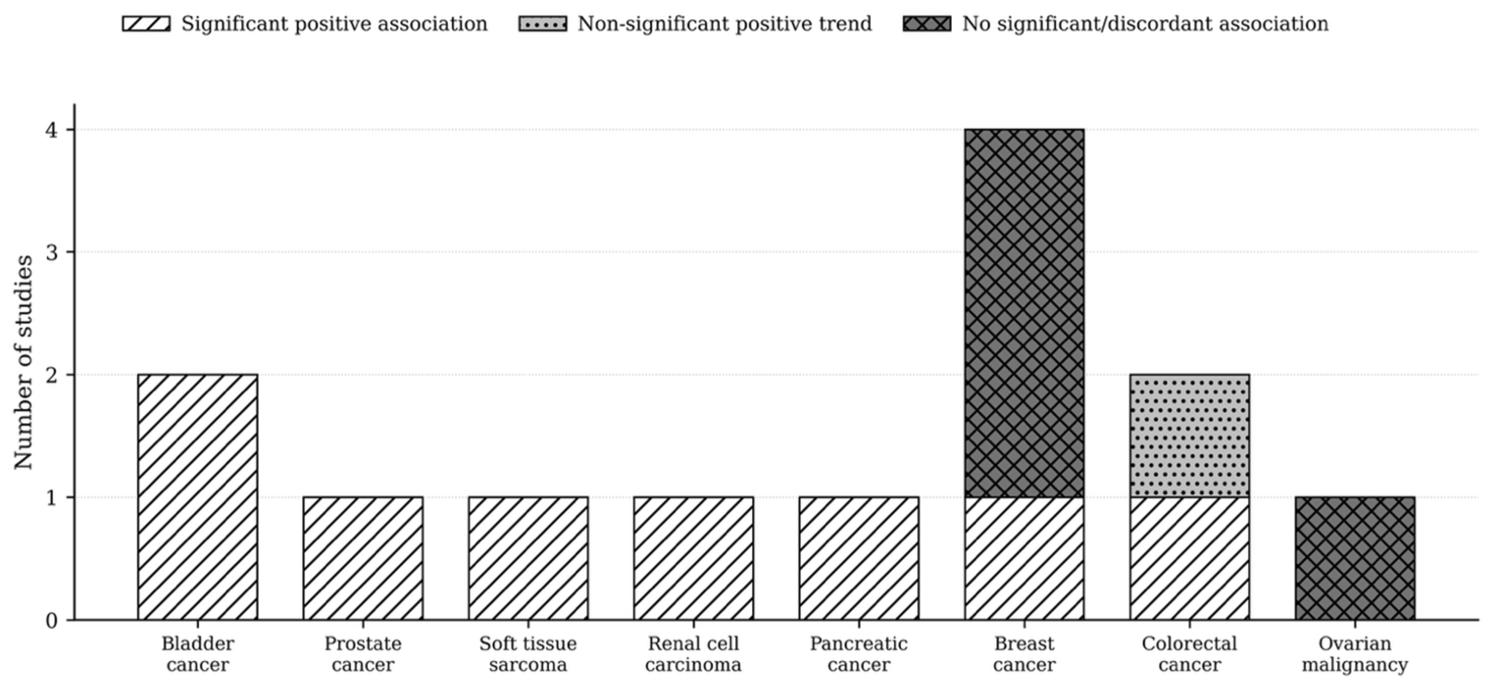
Eight of the 13 included studies demonstrated statistically significant associations between elevated NLR and higher tumor grade or poorer histological differentiation [31,33,34,35,36,37,39,40]. The most consistent evidence was observed for bladder cancer, with both available studies reporting statistically significant positive associations [31,33]. Single studies in prostate cancer, soft tissue sarcoma, renal cell carcinoma, pancreatic cancer, breast cancer, and colorectal cancer with synchronous liver metastases also reported statistically significant positive associations [34,35,36,37,39,40] (Table 4).
The available evidence in breast cancer was heterogeneous. Across the four included studies, one demonstrated a statistically significant association between elevated NLR and poorer histological differentiation [37], two found no significant association [29,32], and one study of metastatic triple-negative breast cancer reported a non-significant inverse trend, with higher tumor grade observed in the lower-NLR group [30]. These findings do not support a consistent association between NLR and tumor grade in breast cancer.
The available evidence for colorectal cancer was similarly inconsistent. In a small cohort of patients with colorectal adenocarcinoma, mean NLR values increased with higher tumor grades; however, the observed trend did not reach statistical significance (p = 0.4) [38]. In contrast, a study of patients with colorectal cancer and synchronous liver metastases reported a statistically significant association between elevated preoperative NLR and poor histological differentiation (p = 0.048) [40]. The metastatic setting is relevant because tumor burden, hepatic involvement, systemic inflammation, and perioperative physiological stress may influence NLR independently of tumor differentiation.
Ovarian malignancy evidence was limited to one small study reporting no significant grade-related association [28].

3.4. Visual Synthesis

A harvest-style direction-of-effect synthesis was used to summarize tumor-specific patterns without implying statistical pooling (Figure 2) (Table 5).

4. Discussion

4.1. Principal Findings

This systematic review indicates that an association between elevated NLR and higher tumor grade is biologically plausible in selected solid malignancies, but the overall evidence remains heterogeneous and methodologically fragile. The most consistent evidence was observed in bladder cancer, whereas findings in breast and colorectal cancer were heterogeneous and context-dependent. Because all included studies were retrospective and varied substantially in clinical setting, grading system, NLR measurement, cutoff derivation, and confounder adjustment, NLR cannot currently be considered a reliable surrogate marker for tumor grade.

4.2. Tumor-Specific Interpretation

The available evidence was most consistent for bladder cancer. Across studies including patients with non-muscle-invasive and broader bladder cancer cohorts, elevated NLR was consistently associated with high-grade disease [31,33]. This is clinically plausible because high-grade urothelial carcinoma is characterized by architectural disorganization, nuclear pleomorphism, mitotic activity, necrosis, and a greater propensity for invasion and recurrence [41,42]. Nonetheless, bladder cancer studies remain limited in number, and the association may also reflect stage, tumor burden, occult inflammation, or perioperative confounders.
Breast cancer studies were heterogeneous and should be interpreted cautiously. Breast cancer grade is influenced by tubule formation, nuclear pleomorphism, and mitotic count [21]. However, peripheral NLR may be affected by tumor subtype, menopausal status, body mass index, systemic treatment, metastatic burden, and concurrent inflammation [9,32,43]. The discordant findings among invasive breast cancer, metastatic triple-negative breast cancer, and biomarker-focused cohorts suggest that breast cancer should not be treated as a single biological entity when evaluating NLR-grade relationships [29,30,32,37].
The available evidence in colorectal cancer highlights the importance of clinical context. In a small cohort of patients with localized colorectal adenocarcinoma, mean NLR values increased with higher tumor grades, although the observed trend did not reach statistical significance [38]. By contrast, the study including patients with colorectal cancer and synchronous liver metastases demonstrated a statistically significant association between elevated preoperative NLR and poor histological differentiation [40]. In the metastatic setting, hepatic tumor burden may amplify NLR independently of primary tumor grade, limiting causal interpretation [40].
Single studies in prostate cancer, soft tissue sarcoma, renal cell carcinoma, and pancreatic cancer reported positive associations [34,35,36,39]. Each has a distinct grading logic. Gleason grading reflects glandular architectural dedifferentiation [44]; FNCLCC sarcoma grading integrates differentiation, mitotic activity, and necrosis [45]; WHO/ISUP renal grading emphasizes nucleolar prominence and nuclear anaplasia [46]; pancreatic cancer differentiation is embedded in a desmoplastic, immunosuppressive tumor microenvironment [47]. Therefore, a shared NLR signal across these entities may reflect systemic inflammation accompanying aggressive tumor biology rather than a universal grade-specific mechanism.

4.3. Relationship to the Broader NLR Literature

The broader oncology literature supports NLR as a prognostic systemic inflammatory marker in many non-hematologic cancers [13,14,17]. However, prognosis and tumor grade are not interchangeable endpoints [48]. A high NLR may reflect tumor burden, occult infection, treatment-related inflammation, cachexia, metastatic disease, host immune status, or comorbid inflammatory conditions rather than intrinsic histological differentiation [9]. The present review therefore extends the current understanding of NLR beyond its established prognostic role by evaluating its potential association with tumor grade, although the available evidence remains inconclusive.

4.4. Biological Plausibility

The available evidence suggests a biologically plausible association between elevated NLR and high-grade tumor morphology. Poorly differentiated tumors often display necrosis, hypoxia, proliferative stress, stromal remodeling, genomic instability, and inflammatory cytokine signaling. These processes can recruit neutrophils, expand granulopoiesis, and suppress lymphocyte-mediated antitumor immunity [2,3,4,5,6,7]. Conversely, lymphopenia may mark reduced cytotoxic immune surveillance, whereas neutrophil-dominant inflammation may contribute to angiogenesis, extracellular matrix degradation, immune evasion, and metastatic competence [2,3,5,6,49]. Nonetheless, these mechanisms are not grade-specific. They may also reflect stage, tumor volume, infection, medication exposure, or systemic host factors [9]. This non-specificity is central to the cautious interpretation of NLR.

4.5. Clinical Implications

NLR has practical advantages: it is inexpensive, rapid, and available from routine blood counts [50]. However, its clinical applicability remains limited by the absence of a universally accepted pathological cutoff value. Although most studies suggest that NLR values above 3.0 are associated with adverse oncological outcomes, substantial variability exists across the literature, with reported thresholds ranging from approximately 2.5 to 5.0. This lack of standardization hampers direct comparisons across studies and limits the routine clinical implementation of NLR. Consequently, despite its potential as an adjunctive biomarker, NLR should currently be interpreted with caution and integrated with established clinical, pathological, and radiological findings rather than used in isolation [9,51,52].
The study of biomarkers remains a major area of interest in oncology. Although histopathology and clinical staging have traditionally formed the basis for treatment decisions and have provided a practical and effective framework for decades, it has been shown that these factors fail to fully reflect the heterogeneity of the disease and the variability in patients’ clinical course [53,54].
Although modern and emerging biomarkers such as circulating tumor DNA (ctDNA) offer a superior capacity for disease characterization, their use remains limited by cost and availability [55,56]. In this context, the investigation of readily available biomarkers such as NLR is of particular clinical relevance, given their potential for rapid implementation in routine clinical practice. The association between biomarkers and tumor grade may support both diagnostic and therapeutic decision-making. However, the oncological care pathway remains complex and is influenced by multiple factors, including differences between the information provided and the patient’s understanding, as well as the need for close multidisciplinary collaboration among radiologists, pathologists, surgeons, oncologists, and other members of the multidisciplinary team to ensure accurate diagnosis and optimal clinical decision-making. Furthermore, despite ongoing advances in diagnostic techniques, challenges remain that warrant the continued refinement and integration of emerging approaches into routine clinical practice [57,58,59].

4.6. Limitations

Several limitations must be emphasized. First, all included studies were retrospective, which introduces selection bias, incomplete confounder control, and variability in clinical data capture. Second, the review included highly heterogeneous tumor types and grading systems, making a pooled meta-analysis inappropriate without harmonized effect measures. Third, NLR thresholds varied substantially across studies, including fixed cutoffs, ROC-derived thresholds, medians, and continuous analyses. Fourth, NLR is non-specific and may be influenced by age, obesity, infection, diabetes, emotional stress, corticosteroid exposure, hematologic disorders, cytotoxic treatment, granulocyte colony-stimulating factor, tumor burden, and metastatic disease [9,13,14]. Fifth, sex distribution, stage distribution, treatment timing, and inflammatory-confounder exclusion were incompletely reported in several source articles and were therefore reported conservatively rather than imputed. Finally, only English-language full-text articles were included, which may introduce language and availability bias.

5. Conclusions

The current evidence suggests a possible association between elevated NLR and higher tumor grade in selected solid malignancies, particularly where tumor-specific studies reported consistent positive findings. However, the totality of evidence remains preliminary, retrospective, heterogeneous, and vulnerable to confounding. NLR should not currently be interpreted as a reliable surrogate for histological differentiation or tumor grade. Future studies should be prospective, tumor-specific, and standardized regarding NLR timing, cutoff derivation, inflammatory exclusion criteria, grading definitions, and multivariable adjustment. Until such evidence is available, NLR is best considered a low-cost systemic inflammatory marker of investigational value rather than a clinically actionable marker of tumor grade.

Author Contributions

P.Ș. Conceptualization, Methodology, Software, Validation, Formal analysis, Investigation, Data curation, Writing—original draft, Writing—review & editing, Visualization, Supervision, Project administration, Resources; A.E.C. Conceptualization, Methodology, Software, Validation, Formal analysis, Investigation, Data curation, Writing—original draft, Writing—review & editing, Visualization, Supervision, Project administration, Resources; P.Ș. and A.E.C. contributed equally to this manuscript and share first authorship; G.B. Conceptualization, Validation, Formal analysis, Investigation, Writing—review & editing, Visualization, Supervision, Project administration, Resources; M.P. Conceptualization, Methodology, Validation, Formal analysis, Writing—review & editing, Visualization, Supervision, Resources; D.T. Methodology, Software, Validation, Formal analysis, Investigation, Data curation, Writing—original draft, Writing—review & editing, Visualization, Supervision, Project administration, Resources; A.-K.T.-C. Methodology, Software, Formal analysis, Writing—original draft, Resources; L.-A.O.-M. Methodology, Formal analysis, Writing—original draft, Resources; G.-C.O. Methodology, Formal analysis, Writing—original draft, Resources; C.-I.M. Project administration, Supervision, Validation, Writing—review & editing; V.-V.V. Project administration, Supervision, Validation, Writing—review & editing; M.-E.C.-F. Methodology, Formal analysis, Writing—original draft, Resources; C.A.T. Project administration, Supervision, Validation, Writing—review & editing; C.T. Conceptualization, Methodology, Software, Validation, Formal analysis, Investigation, Data curation, Writing—review & editing, Visualization, Supervision, Project administration, Resources. All authors have read and agreed to the published version of the manuscript.

Funding

This research received no external funding.

Institutional Review Board Statement

Not applicable.

Informed Consent Statement

Not applicable.

Data Availability Statement

No new data were created or analyzed in this study. Data sharing is not applicable to this article.

Conflicts of Interest

The authors declare no conflicts of interest.

Abbreviations

The following abbreviations are used in this manuscript:
AUCArea under the curve
ctDNAcirculating tumor DNA (deoxyribonucleic acid)
FNCLCCFederation Nationale des Centres de Lutte Contre le Cancer
NLRNeutrophil-to-lymphocyte ratio
NOSNewcastle–Ottawa Scale
PICOSPopulation, Exposure/Intervention, Comparison, Outcome, Study design
PRISMAPreferred Reporting Items for Systematic Reviews and Meta-Analyses
RCCRenal cell carcinoma
ROCReceiver operating characteristic
TNBCTriple-negative breast cancer
WHO/ISUPWorld Health Organization/International Society of Urological Pathology

References

  1. Wu, Z.; Xia, F.; Lin, R. Global Burden of Cancer and Associated Risk Factors in 204 Countries and Territories, 1980–2021: A Systematic Analysis for the GBD 2021. J. Hematol. Oncol. 2024, 17, 119. [Google Scholar] [CrossRef] [PubMed]
  2. Nishida, A.; Andoh, A. The Role of Inflammation in Cancer: Mechanisms of Tumor Initiation, Progression, and Metastasis. Cells 2025, 14, 488. [Google Scholar] [CrossRef] [PubMed]
  3. Akkız, H.; Şimşek, H.; Balcı, D.; Ülger, Y.; Onan, E.; Akçaer, N.; Delik, A. Inflammation and Cancer: Molecular Mechanisms and Clinical Consequences. Front. Oncol. 2025, 15, 1564572. [Google Scholar] [CrossRef] [PubMed]
  4. Xie, J.; Guo, Z.; Zhu, Y.; Ma, M.; Jia, G. Peripheral Blood Inflammatory Indexes in Breast Cancer: A Review. Medicine 2023, 102, e36315. [Google Scholar] [CrossRef] [PubMed]
  5. Bakrim, S.; Fessikh, M.E.; Elhrech, H.; Omari, N.E.; Amanullah, M.; Ming, L.C.; Moshawih, S.; Bouyahya, A. Targeting Inflammation in Cancer Therapy: From Mechanistic Insights to Emerging Therapeutic Approaches. J. Transl. Med. 2025, 23, 588. [Google Scholar] [CrossRef] [PubMed]
  6. Zhou, W.; Cao, X.; Xu, Q.; Qu, J.; Sun, Y. The Double-edged Role of Neutrophil Heterogeneity in Inflammatory Diseases and Cancers. MedComm 2023, 4, e325. [Google Scholar] [CrossRef] [PubMed]
  7. Zhou, J.; Liu, H.; Jiang, S.; Wang, W. Role of Tumor-associated Neutrophils in Lung Cancer (Review). Oncol. Lett. 2022, 25, 2. [Google Scholar] [CrossRef] [PubMed]
  8. Obeagu, E.I.; Obeagu, G.U. Lymphocyte Infiltration in Breast Cancer: A Promising Prognostic Indicator. Medicine 2024, 103, e40845. [Google Scholar] [CrossRef] [PubMed]
  9. Buonacera, A.; Stancanelli, B.; Colaci, M.; Malatino, L. Neutrophil to Lymphocyte Ratio: An Emerging Marker of the Relationships between the Immune System and Diseases. IJMS 2022, 23, 3636. [Google Scholar] [CrossRef] [PubMed]
  10. Das, S.; Dey, M.K.; Devireddy, R.; Gartia, M.R. Biomarkers in Cancer Detection, Diagnosis, and Prognosis. Sensors 2023, 24, 37. [Google Scholar] [CrossRef] [PubMed]
  11. Passaro, A.; Al Bakir, M.; Hamilton, E.G.; Diehn, M.; André, F.; Roy-Chowdhuri, S.; Mountzios, G.; Wistuba, I.I.; Swanton, C.; Peters, S. Cancer Biomarkers: Emerging Trends and Clinical Implications for Personalized Treatment. Cell 2024, 187, 1617–1635. [Google Scholar] [CrossRef] [PubMed]
  12. Sarhadi, V.K.; Armengol, G. Molecular Biomarkers in Cancer. Biomolecules 2022, 12, 1021. [Google Scholar] [CrossRef] [PubMed]
  13. Koc, D.C.; Mănescu, I.B.; Mănescu, M.; Dobreanu, M. A Review of the Prognostic Significance of Neutrophil-to-Lymphocyte Ratio in Nonhematologic Malignancies. Diagnostics 2024, 14, 2057. [Google Scholar] [CrossRef] [PubMed]
  14. Heshmat-Ghahdarijani, K.; Sarmadi, V.; Heidari, A.; Falahati Marvasti, A.; Neshat, S.; Raeisi, S. The Neutrophil-to-Lymphocyte Ratio as a New Prognostic Factor in Cancers: A Narrative Review. Front. Oncol. 2023, 13, 1228076. [Google Scholar] [CrossRef] [PubMed]
  15. Altman, D.G.; McShane, L.M.; Sauerbrei, W.; Taube, S.E. Reporting Recommendations for Tumor Marker Prognostic Studies (REMARK): Explanation and Elaboration. PLoS Med. 2012, 9, e1001216. [Google Scholar] [CrossRef] [PubMed]
  16. Surinova, S.; Radová, L.; Choi, M.; Srovnal, J.; Brenner, H.; Vitek, O.; Hajdúch, M.; Aebersold, R. Non-invasive Prognostic Protein Biomarker Signatures Associated with Colorectal Cancer. EMBO Mol. Med. 2015, 7, 1153–1165. [Google Scholar] [CrossRef] [PubMed]
  17. Templeton, A.J.; McNamara, M.G.; Šeruga, B.; Vera-Badillo, F.E.; Aneja, P.; Ocaña, A.; Leibowitz-Amit, R.; Sonpavde, G.; Knox, J.J.; Tran, B.; et al. Prognostic Role of Neutrophil-to-Lymphocyte Ratio in Solid Tumors: A Systematic Review and Meta-Analysis. J. Natl. Cancer Inst. 2014, 106, dju124. [Google Scholar] [CrossRef] [PubMed]
  18. Guthrie, G.J.K.; Charles, K.A.; Roxburgh, C.S.D.; Horgan, P.G.; McMillan, D.C.; Clarke, S.J. The Systemic Inflammation-Based Neutrophil–Lymphocyte Ratio: Experience in Patients with Cancer. Crit. Rev. Oncol. Hematol. 2013, 88, 218–230. [Google Scholar] [CrossRef] [PubMed]
  19. Li, X.; Dai, D.; Chen, B.; Tang, H.; Xie, X.; Wei, W. The Value of Neutrophil-to-Lymphocyte Ratio for Response and Prognostic Effect of Neoadjuvant Chemotherapy in Solid Tumors: A Systematic Review and Meta-Analysis. J. Cancer 2018, 9, 861–871. [Google Scholar] [CrossRef] [PubMed]
  20. Martins-Filho, S.N.; Paiva, C.; Azevedo, R.S.; Alves, V.A.F. Histological Grading of Hepatocellular Carcinoma—A Systematic Review of Literature. Front. Med. 2017, 4, 193. [Google Scholar] [CrossRef] [PubMed]
  21. Van Dooijeweert, C.; Van Diest, P.J.; Ellis, I.O. Grading of Invasive Breast Carcinoma: The Way Forward. Virchows Arch. 2022, 480, 33–43. [Google Scholar] [CrossRef] [PubMed]
  22. Endo, T.; Crane, J.; Trivedi, J.; Black, M.; Fox, M.; Van Berkel, V. Does Grade Matter? Evaluation of Histologic Grade in Patients with Stage 1 Non–Small Cell Lung Cancer Using the National Cancer Database. JTCVS Open 2025, 25, 393–405. [Google Scholar] [CrossRef] [PubMed]
  23. Crețu, O.I.; Stepan, A.E.; Simionescu, C.E.; Marinescu, D.; Stepan, M.D. Classification and Grading Systems in Gastric Adenocarcinomas. Curr. Health Sci. J. 2022, 48, 284. [Google Scholar] [CrossRef] [PubMed]
  24. Page, M.J.; McKenzie, J.E.; Bossuyt, P.M.; Boutron, I.; Hoffmann, T.C.; Mulrow, C.D.; Shamseer, L.; Tetzlaff, J.M.; Akl, E.A.; Brennan, S.E.; et al. The PRISMA 2020 Statement: An Updated Guideline for Reporting Systematic Reviews. BMJ 2021, 372, n71. [Google Scholar] [CrossRef] [PubMed]
  25. Șiancu, P.; Croitoru, A.E.; Teodoru, A.C.; Boța, G.; Pătran, M.; Tănăsescu, D.; Tonch-Cerbu, A.-K.; Oprinca-Muja, L.-A.; Oprinca, G.-C.; Mohor, C.-I.; et al. The Association Between Neutrophil-to-Lymphocyte Ratio and Histological Tumor Differentiation in Solid Malignancies: A Systematic Review. Available online: https://osf.io/rdcn2 (accessed on 16 July 2026).
  26. De Oliveira Almeida, K.; Nogueira Alves, I.G.; De Queiroz, R.S.; De Castro, M.R.; Gomes, V.A.; Santos Fontoura, F.C.; Brites, C.; Neto, M.G. A Systematic Review on Physical Function, Activities of Daily Living and Health-Related Quality of Life in COVID-19 Survivors. Chronic Illn. 2023, 19, 279–303. [Google Scholar] [CrossRef] [PubMed]
  27. Gualdi-Russo, E.; Zaccagni, L. The Newcastle–Ottawa Scale for Assessing the Quality of Studies in Systematic Reviews. Publications 2026, 14, 4. [Google Scholar] [CrossRef]
  28. Nomelini, R.; Carrijo Chiovato, A.; Abdulmassih, F.F.; Da Silva, R.D.C.; Tavares-Murta, B.; Murta, E.C. Neutrophil-to-Lymphocyte Ratio and Platelet Count as Prognostic Factors in Ovarian Malignancies. J. Can. Res. Ther. 2019, 15, 1226–1230. [Google Scholar] [CrossRef] [PubMed]
  29. Arora, R.; Alam, F.; Zaka-ur-Rab, A.; Maheshwari, V.; Alam, K.; Hasan, M. Peripheral Neutrophil to Lymphocyte Ratio (NLR), a Cogent Clinical Adjunct for Ki-67 in Breast Cancer. J. Egypt. Natl. Cancer Inst. 2023, 35, 43. [Google Scholar] [CrossRef] [PubMed]
  30. De La Cruz-Ku, G.; Chambergo-Michilot, D.; Torres-Roman, J.S.; Rebaza, P.; Pinto, J.; Araujo, J.; Morante, Z.; Enriquez, D.; Flores, C.; Luque, R.; et al. Neutrophil-to-Lymphocyte Ratio Predicts Early Mortality in Females with Metastatic Triple-Negative Breast Cancer. PLoS ONE 2020, 15, e0243447. [Google Scholar] [CrossRef] [PubMed]
  31. Mano, R.; Baniel, J.; Shoshany, O.; Margel, D.; Bar-On, T.; Nativ, O.; Rubinstein, J.; Halachmi, S. Neutrophil-to-Lymphocyte Ratio Predicts Progression and Recurrence of Non–Muscle-Invasive Bladder Cancer. Urol. Oncol. Semin. Orig. Investig. 2015, 33, 67.e1–67.e7. [Google Scholar] [CrossRef] [PubMed]
  32. Jadoon, S.K.; Soomro, R.; Ahsan, M.N.; Ijaz Khan, R.M.; Iqbal, S.; Yasmin, F.; Najeeb, H.; Saleem, N.; Cho, N.; Resham; et al. Association of Neutrophil-to-Lymphocyte Ratio with Clinical, Pathological, Radiological, Laboratory Features and Disease Outcomes of Invasive Breast Cancer Patients: A Retrospective Observational Cohort Study. Medicine 2023, 102, e33811. [Google Scholar] [CrossRef] [PubMed]
  33. Tang, X.; Wang, S.; An, C.; Du, P.; Yang, Y. Preoperative High Neutrophil–to–Lymphocyte Ratio Is Associated with High-Grade Bladder Cancer. AR 2017, 37, 4659–4663. [Google Scholar] [CrossRef] [PubMed]
  34. Oh, J.; Kwon, O.; Lee, J.; Byun, S.-S.; Lee, S.; Lee, S.; Hong, S. Association of the Neutrophil-to-Lymphocyte Ratio and Prostate Cancer Detection Rates in Patients via Contemporary Multi-Core Prostate Biopsy. Asian J. Androl. 2016, 18, 937–941. [Google Scholar] [CrossRef] [PubMed]
  35. Chan, J.Y.; Zhang, Z.; Chew, W.; Tan, G.F.; Lim, C.L.; Zhou, L.; Goh, W.L.; Poon, E.; Somasundaram, N.; Selvarajan, S.; et al. Biological Significance and Prognostic Relevance of Peripheral Blood Neutrophil-to-Lymphocyte Ratio in Soft Tissue Sarcoma. Sci. Rep. 2018, 8, 11959. [Google Scholar] [CrossRef] [PubMed]
  36. Chandrasekaran, D.; Sundaram, S.; Maheshkumar, K.; Kathiresan, N.; Padmavathi, R. Preoperative Neutrophil–Lymphocyte Ratio/Platelet–Lymphocyte Ratio: A Potential and Economical Marker for Renal Cell Carcinoma. J. Cancer Res. Ther. 2022, 18, 1635–1639. [Google Scholar] [CrossRef] [PubMed]
  37. Nafissi, N.; Rezazadeh, M.; Kheradpishe, A.; Radkhah, H.; Bayani, M.; Amiri, B.S.; Tavakoli, G.; Hasheminezhad, M.M.; Khalili, M.; Mousavi, A. Diagnostic Value of Peripheral Blood Inflammatory Indices for Breast Cancer Grade and Immunohistochemical Markers: A Retrospective Observational Study. BMC Cancer 2025, 25, 1886. [Google Scholar] [CrossRef] [PubMed]
  38. Ali, S.; Shahab, S.; Rauf, M.; Riaz, S.K.; Sheikh, A.K.; Tariq, J. Neutrophil to lymphocyte ratio as a predictor of severity in colorectal adenocarcinoma. J. Ayub Med. Coll. Abbottabad 2022, 34, 431–437. [Google Scholar] [CrossRef] [PubMed]
  39. Kawahara, S.; Aoyama, T.; Murakawa, M.; Kanemoto, R.; Takahashi, D.; Kamioka, Y.; Hashimoto, I.; Kamiya, M.; Kato, A.; Maezawa, Y.; et al. Clinical Impact of Preoperative Neutrophil-to-Lymphocyte Ratio in Surgical Patients with Pancreatic Cancer. Anticancer. Res. 2024, 44, 221–228. [Google Scholar] [CrossRef] [PubMed]
  40. Kim, H.; Jung, H.I.; Kwon, S.H.; Bae, S.H.; Kim, H.C.; Baek, M.-J.; Lee, M.S. Preoperative Neutrophil-Lymphocyte Ratio and CEA Is Associated with Poor Prognosis in Patients with Synchronous Colorectal Cancer Liver Metastasis. Ann. Surg. Treat. Res. 2019, 96, 191. [Google Scholar] [CrossRef] [PubMed]
  41. Raspollini, M.R.; Comperat, E.M.; Lopez-Beltran, A.; Montironi, R.; Cimadamore, A.; Tsuzuki, T.; Netto, G.J. News in the Classification of WHO 2022 Bladder Tumors. Pathologica 2023, 115, 32. [Google Scholar] [CrossRef] [PubMed]
  42. Downes, M.R.; Van Der Kwast, T.H.; Lopez-Beltran, A.; Cheng, L. Grading of Bladder Cancer: Updates, Controversies and Practical Solutions. Histopathology 2026, 88, 86–107. [Google Scholar] [CrossRef] [PubMed]
  43. Islam, M.M.; Satici, M.O.; Eroglu, S.E. Unraveling the Clinical Significance and Prognostic Value of the Neutrophil-to-Lymphocyte Ratio, Platelet-to-Lymphocyte Ratio, Systemic Immune-Inflammation Index, Systemic Inflammation Response Index, and Delta Neutrophil Index: An Extensive Literature Review. Turk. J. Emerg. Med. 2024, 24, 8–19. [Google Scholar] [CrossRef] [PubMed]
  44. Van Leenders, G.J.L.H.; Van Der Kwast, T.H.; Grignon, D.J.; Evans, A.J.; Kristiansen, G.; Kweldam, C.F.; Litjens, G.; McKenney, J.K.; Melamed, J.; Mottet, N.; et al. The 2019 International Society of Urological Pathology (ISUP) Consensus Conference on Grading of Prostatic Carcinoma. Am. J. Surg. Pathol. 2020, 44, e87–e99. [Google Scholar] [CrossRef] [PubMed]
  45. Coindre, J.-M. Grading of Soft Tissue Sarcomas: Review and Update. Arch. Pathol. Lab. Med. 2006, 130, 1448–1453. [Google Scholar] [CrossRef] [PubMed]
  46. Browning, L.; Colling, R.; Verrill, C. WHO/ISUP Grading of Clear Cell Renal Cell Carcinoma and Papillary Renal Cell Carcinoma; Validation of Grading on the Digital Pathology Platform and Perspectives on Reproducibility of Grade. Diagn. Pathol. 2021, 16, 75. [Google Scholar] [CrossRef] [PubMed]
  47. Li, Z.-Y.; Peng, S.-Y.; Li, F.-L.; Cai, H.-Q. Immunosuppressive Tumor Microenvironment Shape Pancreatic Cancer Unresponsive to Current Immunotherapies. World J. Clin. Oncol. 2026, 17, 114423. [Google Scholar] [CrossRef] [PubMed]
  48. Jo, S.J.; Kim, K.D.; Lim, S.H.; Kim, J.; Kim, M.J.; Park, J.B.; Lee, K.W. Analysis of the Effect of Tumor-Grade Change on the Prognosis of Retroperitoneal Sarcoma. Cancers 2022, 14, 3020. [Google Scholar] [CrossRef] [PubMed]
  49. Wang, M.; Xiao, Y.; Miao, J.; Zhang, X.; Liu, M.; Zhu, L.; Liu, H.; Shen, X.; Wang, J.; Xie, B.; et al. Oxidative Stress and Inflammation: Drivers of Tumorigenesis and Therapeutic Opportunities. Antioxidants 2025, 14, 735. [Google Scholar] [CrossRef] [PubMed]
  50. Burlacu, A.; Namolovan, C.A.; Brinza, C.; Covic, A.; Floria, M.; Voroneanu, L.; Covic, A. Neutrophil-to-Lymphocyte Ratio (NLR)—Independent Prognostic Marker of Renal Function Decline in Chronic Kidney Disease: A Systematic Review and Meta-Analysis. JCM 2025, 14, 6822. [Google Scholar] [CrossRef] [PubMed]
  51. Zahorec, R. Neutrophil-to-Lymphocyte Ratio, Past, Present and Future Perspectives. BLL 2021, 122, 474–488. [Google Scholar] [CrossRef] [PubMed]
  52. Kusumanto, A.; Zucha, M.A.; Salima, S.; Harsono, A.B.; Kurniadi, A. Diagnostic Value of NLR and PLR for Malignancy in Ovarian Tumor: Feasible Markers in Low-Resource Setting. J. South Asian Fed. Obstet. Gynaecol. 2025, 16, S229–S233. [Google Scholar] [CrossRef]
  53. Sutera, P.; Skinner, H.; Witek, M.; Mishra, M.; Kwok, Y.; Davicioni, E.; Feng, F.; Song, D.; Nichols, E.; Tran, P.T.; et al. Histology Specific Molecular Biomarkers: Ushering in a New Era of Precision Radiation Oncology. Semin. Radiat. Oncol. 2023, 33, 232–242. [Google Scholar] [CrossRef] [PubMed]
  54. Liu, H.; Karsidag, I.; Golin, R.; Wu, G. Bridging Discovery and Treatment: Cancer Biomarker. Cancers 2025, 17, 3720. [Google Scholar] [CrossRef] [PubMed]
  55. Kramer, A.; Greuter, M.J.E.; Schraa, S.J.; Vink, G.R.; Phallen, J.; Velculescu, V.E.; Meijer, G.A.; Van Den Broek, D.; Koopman, M.; Roodhart, J.M.L.; et al. Early Evaluation of the Effectiveness and Cost-Effectiveness of ctDNA-Guided Selection for Adjuvant Chemotherapy in Stage II Colon Cancer. Ther. Adv. Med. Oncol. 2024, 16, 17588359241266164. [Google Scholar] [CrossRef] [PubMed]
  56. Bartolomucci, A.; Nobrega, M.; Ferrier, T.; Dickinson, K.; Kaorey, N.; Nadeau, A.; Castillo, A.; Burnier, J.V. Circulating Tumor DNA to Monitor Treatment Response in Solid Tumors and Advance Precision Oncology. npj Precis. Oncol. 2025, 9, 84. [Google Scholar] [CrossRef] [PubMed]
  57. Pulumati, A.; Pulumati, A.; Dwarakanath, B.S.; Verma, A.; Papineni, R.V.L. Technological Advancements in Cancer Diagnostics: Improvements and Limitations. Cancer Rep. 2023, 6, e1764. [Google Scholar] [CrossRef] [PubMed]
  58. Serban, D.; Spataru, R.; Vancea, G.; Balasescu, S.; Socea, B.; Tudor, C.; Dascalu, A. Informed consent in all surgical specialties: From legal obligation to patient satisfaction. RJLM 2020, 28, 317–321. [Google Scholar] [CrossRef]
  59. Serban, D.; Smarandache, A.; Cristian, D.; Tudor, C.; Duta, L.; Dascalu, A. Medical errors and patient safety culture—Shifting the healthcare paradigm in Romanian hospitals. RJLM 2020, 28, 195–201. [Google Scholar] [CrossRef]
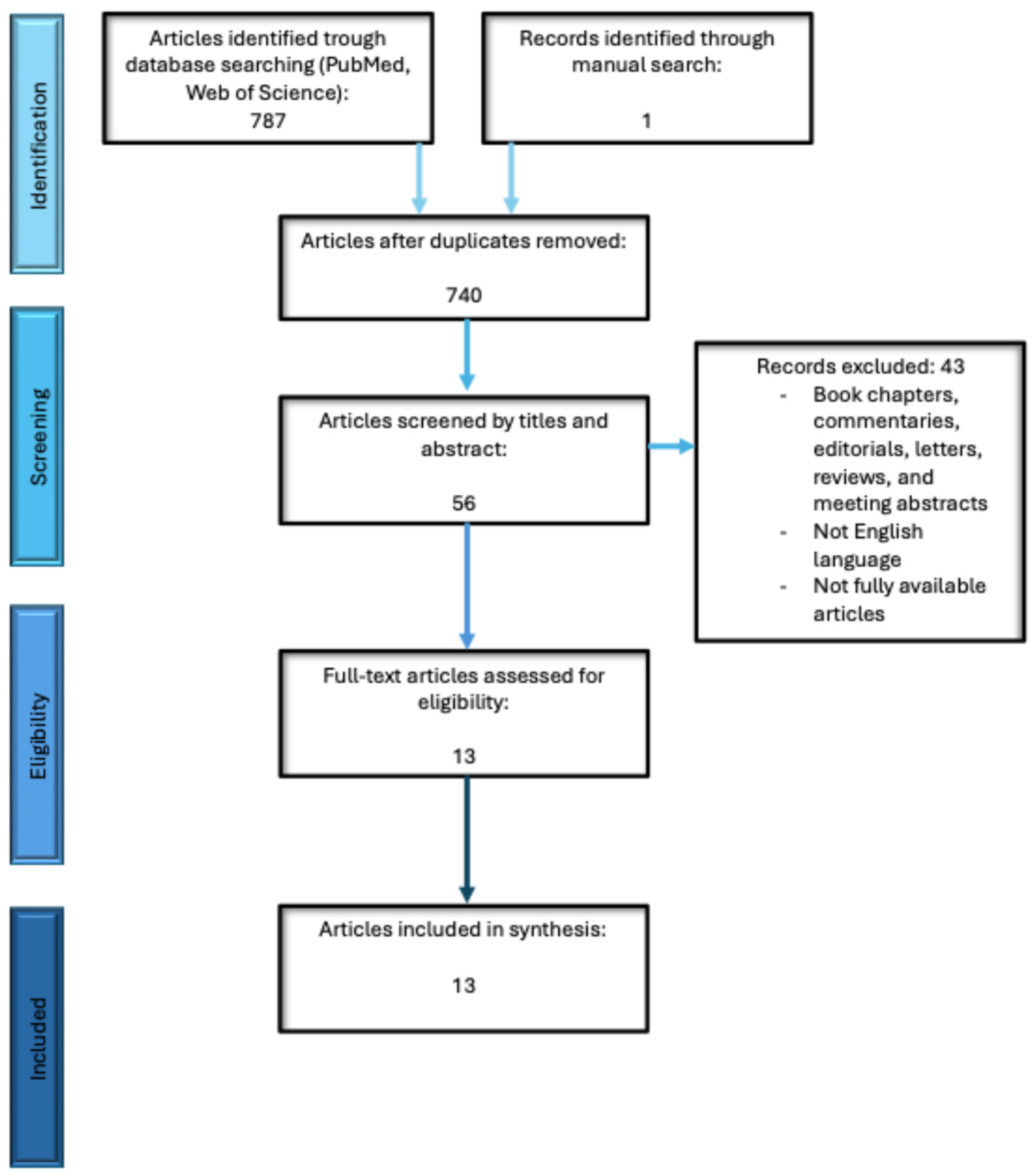
Figure 1. Flowchart illustrating the selection process for the articles included in the review.
Diagnostics 16 02291 g001
Figure 2. Harvest-style direction-of-effect synthesis of the 13 included primary studies. The figure summarizes the direction and statistical significance of the reported associations according to tumor type and reflects a qualitative synthesis of the available evidence rather than a pooled quantitative effect estimate.
Diagnostics 16 02291 g002
Table 1. PICOS framework used for study eligibility.
PICOS ElementDefinition Used in This Review
PopulationPatients with histologically confirmed solid malignancies
IndexPeripheral blood NLR, analyzed as a continuous variable or as a categorical variable using study-defined thresholds
ComparisonNLR levels compared across tumor-grade or differentiation groups; alternatively, frequency of high-grade tumors compared between low- and high-NLR groups
OutcomeStatistically reported association between NLR and histological tumor grade or differentiation
Study designOriginal clinical observational studies or interventional cohorts with extractable baseline NLR-grade data
Table 4. Grade-related NLR results and Newcastle–Ottawa Scale assessment. NOS domains are reported as selection/comparability/outcome.
StudyGrade DefinitionDirectionKey StatisticsSignificantAdjustment/Confounder NotesNOS Score
[28] Nomelini et al.Histological gradeNo significant grade-related associationp = 0.568NoStage-related NLR signal; grade association not statistically significant3/1/2 = 6 (moderate)
[29] Arora et al.Histological grade/Ki-67 contextNo significant grade-related associationp > 0.05NoSmall breast cancer cohort; focused on Ki-67/NLR relationship3/1/2 = 6 (moderate)
[30] De La Cruz-Ku et al.Histological grade in metastatic TNBCDiscordant/non-significant directionp = 0.374NoPrimarily early mortality study; grade analysis secondary3/2/2 = 7 (high)
[31] Mano et al.Low- vs. high-grade NMIBCHigher NLR associated with higher gradeR = 0.21, p = 0.028YesHematologic malignancy excluded; retrospective cohort3/2/2 = 7 (high)
[32] Jadoon et al.Tumor grade in invasive breast cancerNo significant grade-related associationp = 0.694NoLarge retrospective breast cancer cohort; outcome-focused study3/1/2 = 6 (moderate)
[33] Tang et al.Low-grade vs. high-grade bladder cancerHigh NLR associated with high-grade tumorsp < 0.001YesPreoperative NLR; retrospective bladder cancer cohort3/2/2 = 7 (high)
[34] Oh et al.Gleason score groupsHigher NLR associated with higher Gleason gradep < 0.001YesMale biopsy cohort; grade represented by Gleason classification3/2/2 = 7 (high)
[35] Chan et al.FNCLCC low- vs. high-grade sarcomaHigher NLR associated with high tumor gradep < 0.0001YesBlood sampled before therapy; infection/hematologic disorders excluded4/2/2 = 8 (high)
[36] Chandrasekaran et al.WHO/ISUP low- vs. high-grade RCCHigh NLR associated with high-grade tumorsp = 0.001; AUC 0.79YesPreoperative cohort; ROC-derived high-grade cutoff3/2/2 = 7 (high)
[37] Nafissi et al.High/moderate/poor breast cancer gradeHigh NLR associated with poorer differentiationp = 0.027; AUC 0.652YesPreoperative inflammatory index analysis; ROC cutoff 2.153/2/2 = 7 (high)
[38] Ali et al.Well/moderate/poor colorectal differentiationNon-significant positive trendMean NLR 4.5 → 5.0 → 6.0; p = 0.4NoVery small cohort; limited statistical precision2/1/2 = 5 (moderate)
[39] Kawahara et al.Well vs. poor pancreatic cancer differentiationHigh NLR associated with poorer differentiationp = 0.002YesPreoperative surgical cohort; prognostic study with grade analysis3/2/2 = 7 (high)
[40] Kim et al.Poorly vs. well/moderately differentiated CRC with synchronous liver metastasisHigh NLR associated with poorer differentiationp = 0.048YesMetastatic surgical cohort; NLR cutoff 1.943/2/2 = 7 (high)
Table 5. Direction-of-effect synthesis by tumor type.
Tumor TypeNo. StudiesSignificant Positive AssociationNon-Significant Positive TrendNo Significant/Discordant AssociationInterpretive Note
Bladder cancer2200Consistent positive direction in both studies
Prostate cancer1100Grade represented by Gleason classification
Soft tissue sarcoma1100Single heterogeneous mesenchymal tumor cohort
Renal cell carcinoma1100Single preoperative surgical cohort
Pancreatic cancer1100Single surgical cohort
Breast cancer4103Most heterogeneous tumor-specific evidence
Colorectal cancer2110Discordance likely influenced by stage and metastatic setting
Ovarian malignancies1001No association in one small cohort
Disclaimer/Publisher’s Note: The statements, opinions and data contained in all publications are solely those of the individual author(s) and contributor(s) and not of MDPI and/or the editor(s). MDPI and/or the editor(s) disclaim responsibility for any injury to people or property resulting from any ideas, methods, instructions or products referred to in the content.