


Abstract
Renal dysfunction is highly prevalent among patients with cirrhosis and has major implications for management, prognostication, and liver transplant decision-making. Both acute kidney injury (AKI) and chronic kidney disease (CKD) occur frequently in this population and are driven by a complex interplay of hemodynamic derangements, systemic inflammation, and intrinsic or functional renal injury. Accurate assessment of kidney function is essential, as serum creatinine and estimated glomerular filtration rate (eGFR) calculations directly influence MELD-based transplant allocation, eligibility for simultaneous liver–kidney transplantation (SLKT), and clinical decisions such as drug dosing and peri-transplant immunosuppression strategies. However, creatinine-based assessments are fundamentally limited in cirrhosis due to reduced creatinine production, sarcopenia, expanded volume of distribution, increased tubular secretion, and assay interference from hyperbilirubinemia, resulting in frequent overestimation of true GFR. Alternative methods, including cystatin C-based equations, combined creatinine–cystatin C equations, timed urine clearances, and direct GFR measurement using exogenous filtration markers (iohexol, iothalamate), offer potential advantages but also carry important limitations, particularly in decompensated disease with ascites or inflammation. Newer cirrhosis-specific eGFR equations such as the GRAIL and Royal Free Hospital formulas attempt to address these shortcomings, yet validation studies demonstrate inconsistent accuracy across patient cohorts. This review summarizes the pathophysiologic contributors to renal dysfunction in cirrhosis, evaluates the performance and pitfalls of existing kidney function assessment tools, and highlights the ongoing need for reliable, validated methods tailored to this unique population. Until improved approaches are widely accessible, serum creatinine remains the most practical—albeit imperfect—marker guiding clinical decision-making in cirrhotic patients.
1. Introduction
Renal dysfunction is frequently experienced by cirrhotic patients and it is frequently associated with the characteristic abnormal hemodynamic changes specific to cirrhotic patients [1], with or without kidney parenchymal pathology, and can manifest as acute kidney injury (AKI) or chronic kidney disease (CKD) [2,3]. Both AKI and CKD shape long-term outcomes, influence transplant candidacy, and complicate the management of patients with advanced cirrhosis.
AKI is a common complication of decompensated cirrhosis, and its incidence increases as liver disease progress, with almost 50% of cirrhotic patients with ascites developing AKI as a complication at some point [4].
CKD is also becoming increasingly common among cirrhotic patients. According to a recent estimate, the global prevalence of metabolic dysfunction-associated fatty liver disease (MAFLD) is around 30% [5]. In patients with cirrhosis, CKD is associated with worse outcomes for those on the transplant waitlist [6].
Accurately characterizing kidney function in this setting is of central importance in this population for several reasons. Estimated GFR (eGFR) and serum creatinine are not only embedded in prognostic models, inform transplant priority and simultaneous liver–kidney transplant (SLKT) listing, but also guide decisions about appropriate drug dosing [1,7]. Consequently, inaccurate measurement of kidney function can lead to inappropriate drug dosing, under-recognition of high-risk patients, and delayed access to combined organ transplantation. In cirrhotic patients waiting for liver transplantation, renal dysfunction is notably associated with worse outcomes with higher waitlist mortality and inferior post-liver-transplant outcomes [6].
Current methods for evaluating kidney function include serum creatinine- and cystatin C-based estimated glomerular filtration rate (eGFR) equations, timed creatinine clearance, direct glomerular filtration rate (GFR) measurement using exogenous filtration markers as well as a growing number of emerging biomarkers that reflect kidney damage [8,9]. However, none of these is ideal for patients with cirrhosis. In the cirrhotic population, factor such as reduced creatinine production, abnormal tubular creatinine secretion, expanded volume of distribution, hyperbilirubinemia, systemic inflammation, and hemodynamic alterations may lead to significant systematic error in the assessment of kidney function [9,10]. The present review will highlight the importance of accurate estimation of kidney function in cirrhosis, summarize the available methods for estimation of kidney functions and will provide the key advantages, limitations and pitfalls of each method.
2. Hemodynamic Abnormalities in Cirrhosis
Patients with cirrhosis experience a series of hemodynamic derangements and neuro-humoral activation that starts early with cirrhosis and progress in parallel to the progression of the liver disease. These abnormalities can culminate into kidney injury.
In cirrhosis, arterial vasodilation is mediated by several potent vasodilators, with nitric oxide (NO) playing the major role [11]. NO production rises in the splanchnic circulation due to shear stress-induced upregulation of endothelial NO synthase (eNOS) and endotoxin-related activation of eNOS [12,13]. Additional mediators, including calcitonin gene-related peptide (CGRP), substance P, carbon monoxide, endocannabinoids, and adrenomedullin, may further contribute to this vasodilated state [14,15,16,17,18]. Marked vascular remodeling develops within the liver and splanchnic circulation, driven by pro-angiogenic factors such as vascular endothelial growth factor (VEGF) and platelet-derived growth factor (PDGF), promoting an extensive network of portosystemic collaterals [19,20].
Portal hypertension drives profound alterations in gut microbiota composition and function, including gut bacterial overgrowth, delayed intestinal transit, and disruption of tight junction proteins. These alterations facilitate translocation of bacteria, bacterial products, and endotoxins from the gut lumen into the splanchnic and then systemic circulation [21].
The inflammatory cascade initiated by bacterial translocation has dual consequences for the kidney: First, pathogen-associated molecular patterns (PAMPs) and proinflammatory cytokines (TNF-α, IL-6) exacerbate splanchnic and systemic vasodilation, compounding the hemodynamic insult and worsening renal hypoperfusion [22]. Second, gut-derived inflammatory mediators cause direct renal injury through microcirculatory dysfunction, microthrombi formation, renal tubular oxidative stress, and tubular damage [22]. In health, the liver acts as a barrier between the gut and the systemic circulation, clearing bacteria and endotoxins via the reticuloendothelial system. Cirrhosis disrupts this protection through both hepatocellular dysfunction and portosystemic shunting, allowing gut-derived products to bypass hepatic clearance [23].
Importantly, disruption of the gut–liver axis confounds kidney function estimation in this population. Gut-derived endotoxemia and systemic inflammation lower creatinine production and render creatinine-based GFR estimates unreliable. Low-grade endotoxemia from increased gut permeability, impaired hepatic lipopolysaccharide clearance, and portosystemic shunting promotes chronic systemic inflammation, which in turn accelerates muscle protein degradation [24,25]. Thus, the gut–liver axis simultaneously drives true kidney injury and influences the biomarkers used to detect it.
Targeting the gut–liver axis represents a potential therapeutic avenue to mitigate kidney injury in cirrhosis. Intestinal decontamination with rifaximin has shown conflicting results: one study in alcohol-related cirrhosis demonstrated improved systemic hemodynamics, reduced endotoxin and cytokine levels, and increased GFR after 4 weeks of treatment [26]. Another retrospective study found that long-term rifaximin use was associated with decreased incidence of AKI and hepatorenal syndrome [27]. However, a randomized, double-blind, placebo-controlled trial found no effect of rifaximin on hepatic venous pressure gradient, systemic hemodynamics, GFR, or vasoactive hormones in patients with decompensated cirrhosis [28]. Fecal microbiota transplantation (FMT) has shown promising early results in patients with cirrhosis [29,30], though its effects on kidney function specifically remain to be established. These therapeutic considerations are relevant to the present review because interventions that modulate the gut–liver axis may alter both true GFR and the biomarkers used to estimate it, further complicating serial kidney function assessment.
The net effect is a fall in systemic vascular resistance and a reduction in effective circulating volume, even though total plasma and blood volumes may be increased. Invasive hemodynamic studies in large cirrhotic cohorts support this physiology, showing low systemic vascular resistance, reduced effective circulating blood volume, and increased splanchnic pooling [31,32].
Reduction in effective arterial blood volume compromises renal perfusion which results in kidney injury [33].
Furthermore, as effective arterial volume declines, compensatory systems are triggered: renin–angiotensin–aldosterone system (RAAS) activation, unloading of high-pressure baroreceptors in the carotid body and aortic arch with subsequent sympathetic nervous system (SNS) activation, and non-osmotic antidiuretic hormone (ADH) release. Together, these responses promote intense renal vasoconstriction and a decline in GFR. As cirrhosis worsens, splanchnic vasodilation becomes even more pronounced, perpetuating a vicious cycle of neurohormonal activation and progressive renal vasoconstriction [11]. Figure 1 summarizes the hemodynamic and other systemic derangements that contribute to renal dysfunction in cirrhotic patients.
Figure 1.
Liver cirrhosis is the initial driver of physiological derangements leading to kidney dysfunction. Central graph depicting cirrhosis with ascites and sarcopenia, sympathetic response-mediated renal vasoconstriction in the left lower quadrant of the graph, and renal venous congestion graph in the right lower corner were created using generative AI (artificial intelligence). The scientific content and directional relationships of the graph were manually created by the authors. Boxes with red margins represent additional contributing factors. Red upward arrows represent an increase in the corresponding factor. Abbreviations: NO, nitric oxide; SNS, sympathetic nervous system; RAAS, renin–angiotensin–aldosterone system; Na, sodium; ADH, antidiuretic hormone; HOHF, high-output heart failure; LVP, large-volume paracentesis; NSBB, nonselective beta-blockers.
These derangements give rise to a state of volume overload and ‘third spacing’ of fluid which can dilute traditional markers of kidney function, such as serum creatinine, leading to erroneous determination of both measured and estimated GFR. Moreover, circulating levels of creatinine may be influenced by several factors such as type of assay (false low values in the presence of elevated bilirubin), nutritional status and muscle mass as well as reduced synthesis of precursor creatine in liver. Newer biomarkers such as cystatin C have been proposed to overcome these limitations but their levels can also be influenced by infections and concurrent use of certain medications.
To overcome these limitations, novel biomarkers such as neutrophil gelatinase-associated lipocalin (NGAL), interleukin 18 (IL-18), kidney injury molecule-1 (KIM-1) and liver-type fatty acid-binding protein (L-FABP) have recently generated much interest. However, it should be noted that these are markers of tubular injury and cannot be utilized for estimation of kidney function. Moreover, their levels can also be confounded by infections and inflammation [34].
3. AKI and CKD in Cirrhosis: Causes and Implications
3.1. Acute Kidney Injury
AKI in cirrhosis is typically classified as prerenal (due to hypoperfusion), intrinsic or parenchymal renal injury, and postrenal obstruction. Prerenal AKI is the most common and often arises from common triggers in this population: aggressive diuretic use, large-volume paracentesis without adequate albumin replacement, diarrhea from lactulose use, and reduced oral fluid intake [11,35]. Hepatorenal syndrome (HRS-AKI, previously called HRS type 1) is a functional form of AKI that typically occurs in the setting of advanced cirrhosis. The defining feature of HRS-AKI is marked renal vasoconstriction, which begins early in the course of liver disease, often months before rise in creatinine becomes clinically apparent [36]. Vasoconstriction progressively intensifies leading to continuous decline in GFR and eventually increase in serum creatinine level and development of AKI [35,37]. Table 1 describes the most common etiologies of AKI in cirrhosis and the key diagnostic features of each one. Desai et al., using a nationally representative dataset, characterized the trends in incidence, healthcare burden and outcomes of hospitalized patients with cirrhosis with and without AKI. In over 3.6 million admissions, 22% had a diagnosis of AKI but this increased from 15% in 2004 to 30% in 2016. This is especially relevant, as AKI admissions are more costly and have a longer length of stay than non-AKI cirrhosis admissions. Additionally, AKI admissions were 3.75 times more likely to result in patient death and were also 3.75 times more likely to result in CKD [38]. A retrospective cohort multicenter study from the HRS-HARMONY consortium analyzed 2063 consecutive hospital admissions in 2019 with cirrhosis and AKI. The most common etiology of AKI was prerenal/hypovolemic AKI (44%), followed by acute tubular necrosis (ATN, 30%) and HRS-AKI (12%). Notably, the 90-day mortality approached 37%, with significant variation by etiology, with the lowest mortality in prerenal AKI (22%), with a substantially higher mortality in HRS-AKI and ATN (49 to 53%). Overall, higher AKI stage and lack of response to therapy were independent predictors of increased mortality, with patients requiring renal replacement therapy having the worse outcomes. Of note, more than one-third of patients at risk developed de novo CKD 3 months after discharge, with similar rates across different etiologies of AKI [39]. Taken together, AKI in patients with cirrhosis carries significant implications, both in terms of economic burden as well as increased risk of subsequent morbidity and mortality.
Table 1.
Different etiologies for AKI in cirrhosis and key diagnostic features. AKI: acute kidney injury; ATN, acute tubular necrosis, SBP: spontaneous bacterial peritonitis; POCUS: point-of-care ultrasound; GI: gastrointestinal; HRS-AKI: hepatorenal syndrome–acute kidney injury; IgA: immunoglobulin A; IVC: inferior vena cava; RAAS; renin–aldosterone–angiotensin system; SNS: sympathetic nervous system, AIN: acute interstitial nephritis; HCV: hepatitis C virus ribonucleic acid; HBV: hepatitis B virus deoxynucleic acid; PCR: polymerase chain reaction; GN: glomerulonephritis.
3.2. Chronic Kidney Disease
As mentioned previously, the burden of CKD among patients with cirrhosis has risen in parallel with the growing prevalence of MAFLD and the metabolic syndrome in the general population. These conditions are closely linked to long-standing diabetes mellitus and hypertension, both well-established drivers of chronic kidney disease [40]. MAFLD is increasingly being recognized as a leading indication for liver transplantation (LT), further underscoring the intersection between metabolic liver disease and renal dysfunction [41]. As MAFLD has become more prevalent among LT candidates, the proportion of waitlisted patients with underlying CKD has increased over the past 15 years and now approaches 15% [42]. Although less common than MAFLD in the current era, chronic hepatitis B and C infections remain clinically relevant causes of CKD due to their association with immune-mediated glomerulopathy. Indeed, kidney biopsy studies indicate that nearly two-thirds of LT candidates with otherwise unexplained renal dysfunction have histologic evidence of glomerulonephritis, along with moderate to severe glomerulosclerosis and interstitial fibrosis [43].
Beyond intrinsic kidney disease, portal hypertension itself can contribute to chronic kidney dysfunction, particularly in patients with refractory ascites. Previously termed type 2 HRS, this more indolent form of functional kidney impairment occurs in the absence of structural renal disease and reflects sustained hemodynamic derangements in advanced cirrhosis. Under contemporary definitions, these patients are categorized according to duration as HRS–acute kidney disease (HRS-AKD, <3 months) or HRS–CKD (≥3 months) [44].
Pre-LT CKD has important implications for post-transplant outcomes. In an analysis of 39,719 LT recipients using United Network for Organ Sharing (UNOS) data, pre-LT CKD (GFR < 60 mL/min for ≥90 days or kidney replacement therapy for ≥72 days) was associated with a 16% increased risk of post-LT mortality. Notably, SLKT did not mitigate this risk [42]. These findings underscore the importance of close monitoring of kidney function in cirrhosis, both to identify progressive dysfunction early and to implement strategies that may limit future renal complications.
4. The Importance of Accurately Assessing Kidney Function in Cirrhosis
4.1. Model for End-Stage Liver Disease (MELD) and Liver Transplant Candidacy
Since 2002, the Model for End-Stage Liver Disease (MELD) was implemented as the main tool to prioritize liver transplantation for liver transplant candidates in the USA. Serum creatinine has been incorporated in the MELD score due to its ability to independently predict short-term mortality in cirrhosis which underscores the pivotal role of kidney dysfunction in prioritizing patients for liver transplantation [45,46].
Accurate estimation of kidney function is also essential for patients undergoing liver transplantation evaluation, as renal dysfunction can markedly influence complications and survival both in the pre- and postoperative periods [47,48].
In 2016, MELD was updated to MELD-Na to incorporate hyponatremia, which is often seen in cirrhotic patients and is also associated with AKI. The intent was to improve the probability of getting transplanted among waitlisted patients [46,49]. However, incorporation of serum creatinine carried a disadvantage for females who have lower skeletal muscle mass and patients with ascites and sarcopenia who also have decreased hepatic creatinine production and poor nutrition [50,51,52]. To mitigate some of these disadvantages, MELD 3.0 was introduced, which incorporated gender and albumin. This has helped to address the disparities that women had experienced with the previous system, marking a step forward in organ allocation [40].
4.2. Simultaneous Liver–Kidney Transplantation Allocation Depends on eGFR
The accurate assessment of renal function is central to guide transplantation strategies, both for LT alone and for SLKT, because the Organ Procurement and Transplantation Network (OPTN) criteria depend on GFR/eGFR thresholds sustained over time. Therefore, misclassification can inappropriately drive toward dual-organ listing or, conversely, deny SLKT when it is truly indicated. To standardize the eligibility for SLKT, OPTN established new allocation criteria in 2017 [53]. Under current OPTN guidelines, patients are eligible for SLKT if they have CKD for at least 90 consecutive days as defined by GFR < 60 mL/min and GFR ≤ 30 mL/min at the time of listing or having persistent AKI for at least 6 weeks with either dialysis once every 7 days or GFR < 25 mL/min for 6 consecutive weeks [53].
4.3. Adequate Drug Dosing
Many commonly used drugs require adjustment for kidney function, and this is typically obtained with creatinine-based equations. These equations frequently overestimate GFR in patients with cirrhosis and sarcopenia or volume overload, leading to potential drug-related toxicity. By contrast, cystatin C-based equations may underestimate kidney function in this population [8,9]. These implications also extend into the immediate peri-transplant period when selecting the appropriate immunosuppression induction strategy is critical, as renal-sparing approaches such as basiliximab with delayed calcineurin inhibitor initiation, or reduced-dose tacrolimus regimens have been associated with better renal outcomes in liver transplant recipients at higher risk of kidney dysfunction in the post-transplant period [54,55].
4.4. Limitations and Complexities of Assessing Renal Function in Cirrhosis
Assessment of renal function in patients with cirrhosis is challenging and carries numerous pitfalls. In the following sections, we will review currently available methods, describing their applications, challenges, and pitfalls.
4.4.1. Measured GFR with Iothalamate or Iohexol Clearance
This method is the current gold standard for measuring the kidney function, also known as measured GFR. This method utilizes the clearance of an exogenous tracer such as iohexol or iothalamate. It yields an absolute, non-body surface area (BSA) indexed value (mL/min) and therefore reflects the individual filtration capacity independent of body-sized indexing, which is especially relevant in patients at the extremes of BSA such as in cirrhosis. This method is also not affected by multiple factors influencing the serum levels of creatinine and cystatin C which are commonly encountered in cirrhotic patients [56].
However, tracer methods are not universally available, have elevated cost, and require specialized workflows. Furthermore, in cirrhotic patients, ascites and associated volume overload can lead to increased volume of distribution, allowing filtration markers to distribute into third space fluid compartments, creating a dilutional effect. As a result, these methods may overestimate GFR in such patients [1,8,57]. A practical way to minimize this error is to estimate clearance after a large-volume paracentesis, to mitigate the effect of third spacing [8,58].
Other direct measurement strategies include isotopic tracers such as 99mTc-DTPA, and 51Cr-EDTA [59,60,61,62]. In practice, these tests are not widely available, and none has been adequately validated in cirrhotic populations for routine GFR determination, although they may still be useful in selected cases [63,64,65,66].
4.4.2. Twenty-Four-Hour Urine for Creatinine and/or Urea Clearance
In routine practice, a 24 h urine collection to calculate creatinine clearance is commonly used as a surrogate for GFR. However, in patients with cirrhosis this approach substantially overestimates true GFR. This discrepancy has long been well known. In a seminal study from 1987 than included 23 patients with cirrhosis with ascites found a major discrepancy between serum creatinine and creatinine clearance, where creatinine often remained “normal” even when creatinine clearance was markedly reduced [64]. This was confirmed in a subsequent meta-analysis including 193 patients with cirrhosis, where measured creatinine clearance substantially overestimated true GFR, surpassing inulin clearance by +13 mL/min/1.73 m2, with wide confidence intervals (+60 to −34 mL/min/1.73 m2). This was most pronounced in patients with lower GFR with mean overestimation of 18% in those with GFR ≥ 60 mL/min/1.73 m2 and 49% in those with GFR < 60 mL/min/1.73 m2. Notably, 14% of patients with measured creatinine clearance ≥60 mL/min/1.73 m2 had an inulin clearance <30 mL/min/1.73 m2, reflecting severe misclassification [65]. This is largely because creatinine is secreted by renal tubules and that secretory component becomes proportionally more pronounced as true GFR falls [8]. Even so, measured creatinine clearance performs better than serum creatinine alone when estimating GFR [64,66]. Measuring urea clearance may help to mitigate some of the issues with creatinine clearance as urea clearance underestimates and creatinine clearance overestimates the true GFR. This composite method which utilizes the mean of both clearances has been proven to outperform estimated GFR using different equations and creatinine clearance alone in patients with GFR < 60 mL/min in patients without cirrhosis [35,67]. However, it suffers from errors in collection and remains vulnerable to shifts in volume status and third spacing, especially because patients with cirrhosis often have frequent bowel movements (or even diarrhea) in the context of lactulose use, making the accurate collection of 24 h urine samples prone to collection error [8].
4.4.3. Serum Creatinine and eGFR Using Creatinine-Based Equations
Equations that calculate GFR are widely employed to estimate renal function in the general population. Depending on the formula, they consider patient characteristics such as age, sex, weight, ethnicity, and laboratory values including serum creatinine, serum urea nitrogen, and serum albumin to generate a predicted GFR. The commonly used Cockcroft-Gault equation, which estimates creatinine clearance and not GFR, has been shown to overestimate GFR in patients with advanced liver disease when compared against inulin clearance (the reference standard), with the greatest overestimation seen when inulin clearance is below 70 mL/min [64,68,69]. Several other equations have also been evaluated in liver transplant candidates. In one report that compared different equations in liver transplant candidates, even the best-performing equation, the model Modification of Diet in Renal Disease-6 (MDRD-6) achieved only 66% of estimates within 30% of GFR measured by I125 iothalamate with lower precision observed in the MDRD-4, MDRD-5 and the Nankivell equations, highlighting clinically significant imprecision at the individual level [68]. The CKD-EPI (Chronic Kidney Disease Collaboration) creatinine equation has generally performed better than MDRD at higher levels of GFR in a review of 12 studies, although most of those studies did not include patients with cirrhosis [70]. However, in cohorts with liver disease CKD-EPI did not consistently outperform MDRD [71,72].
It is important to understand why serum creatinine and creatinine-based eGFR equations which are useful markers of kidney function assessment in the general population, are unreliable in patients with cirrhosis. Serum creatinine is heavily influenced by factors unrelated to glomerular filtration, potentially demonstrating “normal” serum levels even when true GFR is significantly reduced, frequently overestimating kidney function. Moreover, creatine, the precursor of creatinine, is primarily synthesized in the liver. There is good evidence to show that its production is reduced by half in patients with cirrhosis [8,64]. This in turn reduces the production of creatinine in muscles and other tissues. Additionally, sarcopenia and poor nutritional status are common in cirrhosis, further lowering creatinine production [73]. Finally, tubular secretion of creatinine is increased in these patients, particularly in the context of renal dysfunction, which may result in lower serum creatinine concentrations. All these factors contribute to overestimation of GFR in these patients [74,75,76]. Furthermore, some creatinine assays can report falsely low values when bilirubin is markedly elevated or when there are protein-related interferences, although this effect can vary across laboratories [66,77]. Table 2 summarizes the key mechanisms that make serum creatinine and creatinine-based eGFR equations unreliable estimates of true GFR in cirrhotic patients.
Table 2.
Key mechanisms that make creatinine and creatinine-based eGFR unreliable in cirrhosis (reduced creatinine generation, third spacing/dilution, increased tubular secretion, and assay interference), resulting in a creatinine to GFR discordance. Abbreviations: GFR, glomerular filtration rate; eGFR, estimated glomerular filtration rate.
Considering all these limitations, serum creatinine can be misleading in cirrhosis. In one report, 37% of patients with cirrhosis with normal creatinine had a measured GFR < 50 mL/min, while another 31% had a GFR between 50 and 80 mL/min [78]. This suggests poor correlation between serum creatinine and true GFR in cirrhosis. The discrepancy is especially pronounced in women, likely due to their lower muscle mass [66,79]. Unfortunately, the discrepancies between estimated and measured GFR have also been illustrated in a large cohort of liver transplant candidates, demonstrating equation-based eGFR often misclassified and overestimated kidney function, which is essential for transplant decisions [80].
4.4.4. Cystatin C and eGFR Using Cystatin C-Based Equations
Cystatin C is produced by all nucleated cells and is cleared primarily by glomerular filtration. Circulating levels are generally not affected by age, sex, muscle mass, and bilirubin [63,79,81,82,83]. Hence, it has been proposed as an alternative marker to serum creatinine, especially in populations with low muscle mass such as cirrhotic patients. Several studies have suggested that cystatin C reflects GFR more accurately than creatinine in patients with cirrhosis, especially in the context of significant GFR decline, and advanced, decompensated cirrhosis [69,82,84,85,86,87,88].
Moreover, cystatin C has been consistently linked to clinically meaningful outcomes, supporting its role beyond a filtration surrogate. In a prospective study by Maiwall et al. [89] cystatin C appeared to better capture risk than creatinine alone, correlating with recurrent AKI and improving mortality prediction when incorporated into MELD components. In patients listed for liver transplant, Mauro et al. reported that elevated cystatin C (≥1.5 mg/L), when considered alongside sarcopenia, identified patients at higher risk for developing acute-on-chronic liver failure (ACLF) and mortality, underscoring its potential value [90]. Similarly, in patients hospitalized with acutely decompensated cirrhosis, Markwardt et al. found that plasma cystatin C predicted subsequent renal dysfunction, ACLF, and 90-day transplant-free mortality, suggesting that cystatin C may reflect integrated systemic risk in addition to kidney filtration [91]. Consistent with this, a multicenter prospective study by Kim et al. showed that cystatin C correlated well with AKI development or progression and with mortality among patients with decompensated cirrhosis, reinforcing its potential utility for short-term risk stratification in hospitalized patients with cirrhosis [92]. Despite its multiple advantages, cystatin C has not been adopted universally to assess kidney function mainly because of practical barriers, such as cost and availability [8]. Importantly, levels can be influenced by factors like infection and by certain medications, such as corticosteroids and calcineurin inhibitors [93]. This is of significant implication for liver transplant recipients.
4.4.5. Estimated GFR Using a Combination of Cystatin C and Creatinine
In cirrhosis, an equation combining creatinine and cystatin C would be especially appealing considering it could potentially “balance out” the limitations that affect each marker [9]. cystatin C-based formulas, including CKD-EPI-Cystatin C and the combined CKD-EPI creatinine and cystatin C equation, may estimate GFR more accurately than creatinine-only equations in cirrhotic patients [9,83,94,95,96,97,98,99,100,101]. Prospective studies in patients with cirrhosis comparing combined creatinine and cystatin C equations against measured GFR demonstrated better performance in contrast to creatinine-only estimates [102]. As mentioned earlier, this is especially relevant in cirrhosis because transplant decisions as well as medication dosing are based on specific thresholds [103]. More recently, race-neutral equations have been adopted in the general population. Although their bias, accuracy, and precision in cirrhosis remain uncertain, a recent study reported lower eGFR values when race-neutral creatinine and cystatin C-based equations were applied and suggested that their use would qualify more patients for simultaneous liver–kidney transplant (SLKT) listing [104].
In summary, within the equation-based methods, the combined creatinine–cystatin C CKD-EPI equation has shown better diagnostic performance than many conventional equations using creatinine or cystatin C alone when compared against measured GFR in cirrhosis, although the accuracy in this population is still significantly affected when compared to its performance in non-cirrhotic individuals and in the setting of ascites and volume overload [8,9,32,58].
4.4.6. Novel Biomarkers for Assessing Kidney Function
Advances in the field of proteomics have led to the emergence of novel biomarkers such as NGAL, IL18, KIM-1, and L-FABP. The use of these biomarkers carries much promise as they permit detection of kidney injury within hours of onset, differentiate prerenal causes of AKI from intrinsic renal injury, and help in prognostication and prediction of long-term outcomes [33].
In a multicenter, prospective cohort study of patients with cirrhosis, analysis of urinary biomarkers of kidney injury such as NGAL, KIM-1, IL18 and L-FABP helped to differentiate between structural and functional causes of AKI (kidney biomarkers and differential diagnosis of patients with cirrhosis and acute kidney injury) [105]. In another study, elevated levels of urinary biomarkers were associated with worse prognosis in patients with cirrhosis and AKI [106]. Elevated urinary levels of these biomarkers may also help to predict short-term mortality, as concluded by a systemic review and meta-analysis of five studies [107].
Of all the biomarkers, NGAL is the most extensively studied in patients with cirrhosis [33]. In a prospective study of 320 consecutive cases of decompensated liver cirrhosis with AKI, urinary NGAL was an independent predictive factor of AKI progression [108].
Although us of these novel biomarkers carries much potential, their use is limited by their lack of widespread availability, validation and standardization. Moreover, studies involving these biomarkers lack homogeneity [109]. Finally, they are markers of tubular injury and provide information about structural kidney damage but not functional capacity. Hence, they have not yet become standard of care [110]. Table 3 depicts a comparison of the performance of creatinine, cystatin C, and novel kidney injury biomarkers across different clinical scenarios in cirrhosis.
Table 3.
Comparison of creatinine, cystatin C and novel biomarkers for assessment of kidney function in patients with cirrhosis. Abbreviations: GFR, glomerular filtration rate; NGAL, neutrophil gelatinase-associated lipocalin; KIM-1, kidney injury molecule-1; AKI, acute kidney injury; MELD, Model for End-Stage Liver Disease; ATN, acute tubular necrosis; HRS-AKI, hepatorenal syndrome–acute kidney injury; HRS, hepatorenal syndrome.
4.4.7. Cirrhosis-Specific eGFR Equations
As both creatinine and cystatin C-based equations perform poorly in the presence of ascites and volume overload [8,9], Mindikoglu et al. developed a cirrhosis-specific equation for patients with diuretic refractory ascites and it appeared to outperform CKD-EPI cystatin C and CKD-EPI creatinine–cystatin C in that specific phenotype [98]. However, a more recent study comparing multiple equations head to head reported that even within cirrhosis-specific equations, significant inconsistencies and misclassification occur, with notable discrepancies when different methods were applied to the same patient [57].
In response, two cirrhosis-specific models were developed precisely to reduce the systematic bias that shows up when you apply general population equations to patients with liver dysfunction, ascites, edema, and sarcopenia: the GRAIL (GFR Assessment in Liver Disease) equation and Royal Free Hospital Cirrhosis GFR equation [103,111]. These equations were explicitly developed and tested in the population where serum creatinine correlates poorly with renal function [103,111].
The GRAIL equation was developed with the intention of improving GFR estimation in cirrhotics [103]. The same research group then tested whether substituting serum creatinine with an eGFR obtained using the mentioned equation could improve waitlist mortality risk prediction (MELD-GRAIL-Na model). However, while the MELD-GRAIL-Na model showed better mortality prediction in women and patients with MELD scores above 25, its use has not been validated in other patient groups [112]. Further, it was devised primarily to improve prediction of waitlist mortality in liver transplantation and its use in other aspects of cirrhosis remains unclear. Even in this context, MELD-GRAIL-Na reclassified only about 16% of waitlist patients, suggesting limited impact. Finally, it involves complex calculations which may not be practical.
The Royal Free Hospital (RFH) cirrhosis GFR equation was introduced in 2017 and then validated in an independent cohort as a cirrhosis-specific alternative. Unlike other conventional equations, the RFH model was derived from patients with cirrhosis with a broad spectrum of disease severity (MELD 6–44) and incorporated seven clinical variables (serum creatinine, serum urea, INR, age, serum sodium, sex [male vs. female coefficient], ascites severity [specifically moderate/severe ascites as a coefficient]) [111]. In the initial validation study, the RFH equation demonstrated the highest accuracy for predicting measured GFR, achieving a P30 (precision within 30%) of 89% compared to P30 of 27–75% for MDRD-4, MDRD-6, and CKD-EPI-creatinine. This was consistent across various stages of kidney impairment and liver dysfunction [111].
The RFH score was developed for cirrhosis patients admitted to ICU and may not be applicable to ambulatory patients or patients who are less critically ill [113]. The score also did not account for cardiovascular disease, sarcopenia, frailty or other comorbidities, which limits its prognostic predictiveness. Finally, it performed only marginally better than MELD and could not be validated by other cohorts. In a study that used a Hispanic cohort, the RFH equation outperformed creatinine-only equations. However, its performance was comparable to CKD-EPI-cystatin C. Notably, both RFH and CKD-EPI-cystatin C equations tended to underestimate GFR when it was >60 mL/min/1.73 m2 and overestimated it when it was <60 mL/min/1.73 m2 [114]. Another retrospective study of 134 cirrhotic patients who had measured GFR by 51Cr-EDTA reported that the RFH equation had significantly lower accuracy than CKD-EPI, MDRD-4, and MDRD-6, highlighting that the performance of the RFH equation may be cohort- and method-dependent and that further prospective validation of this equation is still warranted [115]. A distinctive feature of RFH is that it incorporates the degree of ascites, which addresses a unique feature of cirrhosis but also introduces a subjectivity component that could limit reproducibility [111].
In summary, the GRAIL equation and the RFH score were developed to overcome limitations of creatinine in high-risk subgroups but have limited generalizability, modest incremental benefit, and insufficient external validation. Overall, these models are not yet robust or practical enough to replace conventional approaches in routine clinical practice.
4.4.8. Practical Guide for Clinicians
Until both estimation equations and direct measurement tools are better validated and widely available in cirrhosis, expert opinion continues to support serum creatinine as the most practical marker for the assessment of renal dysfunction, particularly because it is part of the current MELD score and because no alternative strategy has consistently proven to be superior for assessing renal function in this population [116]. Creatinine-based eGFR remains the first-line screening tool but systematically overestimates true GFR due to reduced creatinine production from sarcopenia, malnutrition, and impaired hepatic synthesis—an effect that worsens with advanced disease, female sex, and low GFR. When creatinine is unreliable, serum cystatin C should be added, and the combined CKD-EPI creatinine–cystatin C equation is the least biased estimating equation overall. Measured GFR (using iohexol or iothalamate clearance) is not widely used because of practical difficulties and lack of validation. Cirrhosis-specific scores like GRAIL and RFH may complement standard equations in the transplant setting but lack widespread adoption and have mixed external validation. Finally, novel biomarkers like NGAL and KIM-1 may help in early detection of acute kidney injury, but it should be remembered that they are markers of tubular injury and do not help in determination of GFR. Table 4 includes practical recommendations for the use of eGFR equations and biomarkers for the assessment of kidney function in cirrhosis.
Table 4.
Biomarkers and methods to assess kidney function in cirrhosis: A Practical Guide for Clinicians. Abbreviations: eGFR, estimated glomerular filtration rate; CKD-EPI-Cr, Chronic Kidney Disease Epidemiology Collaboration creatinine equation; MDRD, Modification of Diet in Renal Disease; CKD-EPI-CysC, Chronic Kidney Disease Epidemiology Collaboration cystatin C equation; CKD-EPI-Cr-CysC, Chronic Kidney Disease Epidemiology Collaboration creatinine–cystatin C equation; GFR, glomerular filtration rate; mGFR, measured glomerular filtration rate; AKI, acute kidney injury; ACLF, acute-on-chronic liver failure; HRS, hepatorenal syndrome; HRS-AKI, hepatorenal syndrome–acute kidney injury; BUN, blood urea nitrogen; LT, liver transplantation; GRAIL, GFR Assessment in Liver Disease; RFH, Royal Free Hospital; INR, international normalized ratio; NGAL, neutrophil gelatinase-associated lipocalin; ATN, acute tubular necrosis; AUROC, area under the receiver operating characteristic curve; KIM-1, kidney injury molecule-1.
4.4.9. Etiology- and Age-Specific Considerations in Kidney Function Assessment in Cirrhosis
Although the hemodynamic mechanisms underlying renal dysfunction are shared across various etiologies of liver cirrhosis, the spectrum of intrinsic kidney disease and the magnitude of GFR estimation error vary by cause and also by patient age. Alcohol-associated cirrhosis carries a high prevalence of secondary IgA nephropathy and is associated with significant sarcopenia, amplifying creatinine-based GFR overestimation [24,117]. HCV-related cirrhosis is linked to immune complex-mediated membranoproliferative glomerulonephritis, usually in the context of type II mixed cryoglobulinemia, while HBV-related cirrhosis is most commonly associated with membranous nephropathy [118,119].
NAFLD is independently associated with a two-fold increased prevalence of CKD, and the sarcopenic obesity phenotype common in these patients can cause creatinine-based equations to either over- or underestimate GFR depending on the relative contributions of preserved body mass and underlying muscle wasting [120,121].
Patient age introduces additional complexity, as age-related sarcopenia compounds cirrhosis-related muscle loss, further lowering creatinine production and widening the gap between estimated and true GFR. In older patients, cystatin C-based or combined creatinine–cystatin C equations become particularly important, though cystatin C may itself be affected by the higher burden of systemic inflammation in elderly individuals [122]. Conversely, younger patients with NAFLD cirrhosis and sarcopenic obesity may have relatively preserved creatinine levels that mask significant renal impairment. Taken together, these etiology- and age-specific nuances underscore the need for individualized kidney function assessment rather than a uniform approach, although the literature does not yet support distinct GFR estimation algorithms stratified by etiology or age.
5. Conclusions
In conclusion, accurate assessment of kidney function in individuals with advanced liver disease remains a critical yet unresolved challenge. Current estimation methods are limited by substantial physiological and methodological constraints, frequently resulting in an imprecise reflection of true renal function. Therefore, continued investigation is needed to develop, refine, and validate more reliable assessment tools that can better support clinical decision-making and improve outcomes in this high-risk population. Meanwhile, expert opinion continues to support serum creatinine as the most practical marker for the assessment of renal dysfunction, particularly due to its incorporation into the MELD score and due to its wide base availability. Future studies should focus on further validation of newer cirrhosis-specific GFR equations and increase the use of isotopic and non-isotopic GFR measurement methods for more accurate GFR assessment and ultimately better care of patients with cirrhosis.
Author Contributions
Conceptualization, H.M.W. and F.G.-L.; methodology, H.M.W. and F.G.-L.; software, F.G.-L.; validation, F.G.-L., N.P., M.M., R.C. and H.M.W.; formal analysis, H.M.W. and F.G.-L.; investigation, H.M.W. and F.G.-L.; resources, H.M.W. and F.G.-L.; data curation, F.G.-L., N.P.and H.M.W.; writing—original draft preparation, F.G.-L., N.P.and H.M.W..; writing—review and editing, F.G.-L., N.P.and H.M.W.; visualization, M.M., R.C.; supervision, H.M.W. All authors have read and agreed to the published version of the manuscript.
Funding
This research received no external funding.
Institutional Review Board Statement
Not applicable.
Informed Consent Statement
Not applicable.
Data Availability Statement
The original contributions presented in this study are included in the article. Further inquiries can be directed to the corresponding author.
Acknowledgments
Generative artificial intelligence (Chat GPT 5.4) was used to create selected graphical elements in Figure 1, including the central illustration depicting cirrhosis with ascites and sarcopenia, the left lower quadrant illustration of sympathetic response-mediated renal vasoconstriction, and the right lower corner illustration of renal venous congestion. The scientific content, interpretation, and final figure design were reviewed and approved by the authors.
Conflicts of Interest
The authors declare no conflict of interest.
References
- Nadim, M.K.; Garcia-Tsao, G. Acute Kidney Injury in Patients with Cirrhosis. N. Engl. J. Med. 2023, 388, 733–745. [Google Scholar] [CrossRef] [Scilit] [PubMed]
- Wadei, H.M.; Geiger, X.J.; Cortese, C.; Mai, M.L.; Kramer, D.J.; Rosser, B.G.; Keaveny, A.P.; Willingham, D.L.; Ahsan, N.; Gonwa, T.A. Kidney allocation to liver transplant candidates with renal failure of undetermined etiology: Role of percutaneous renal biopsy. Am. J. Transplant. 2008, 8, 2618–2626. [Google Scholar] [CrossRef] [Scilit] [PubMed]
- Wadei, H.M.; Heckman, M.G.; Rawal, B.; Taner, C.B.; Mai, M.L.; Cortese, C.; Rosser, B.G.; Gonwa, T.A.; Keaveny, A.P. Renal outcomes of liver transplant recipients who had pretransplant kidney biopsy. Transplantation 2014, 98, 1323–1330. [Google Scholar] [CrossRef] [Scilit] [PubMed]
- Montoliu, S.; Balleste, B.; Planas, R.; Álvarez, M.A.; Rivera, M.; Miquel, M.; Masnou, H.; Cirera, I.; Morillas, R.M.; Coll, S.; et al. Incidence and prognosis of different types of functional renal failure in cirrhotic patients with ascites. Clin. Gastroenterol. Hepatol. 2010, 8, 616–622. [Google Scholar] [CrossRef] [Scilit] [PubMed]
- Le, M.H.; Yeo, Y.H.; Li, X.; Li, J.; Zou, B.; Wu, Y.; Ye, Q.; Huang, D.Q.; Zhao, C.; Zhang, J.; et al. 2019 Global NAFLD Prevalence: A Systematic Review and Meta-analysis. Clin. Gastroenterol. Hepatol. 2021, 20, 2809–2817. [Google Scholar] [CrossRef] [Scilit] [PubMed]
- Cullaro, G.; Verna, E.C.; Lai, J.C. Association Between Renal Function Pattern and Mortality in Patients with Cirrhosis. Clin. Gastroenterol. Hepatol. 2019, 17, 2364–2370. [Google Scholar] [CrossRef] [Scilit] [PubMed]
- St Hillien, S.A.; Robinson, J.E.; Ouyang, T.; Patidar, K.R.; Belcher, J.M.; Cullaro, G.; Regner, K.R.; Chung, R.T.; Ufere, N.; Velez, J.C.Q.; et al. Acute Kidney Injury in Patients with Cirrhosis and Chronic Kidney Disease: Results from the HRS-HARMONY Consortium. Clin. Gastroenterol. Hepatol. 2025, 23, 1755–1765.e4. [Google Scholar] [CrossRef] [Scilit] [PubMed]
- Campion, D.; Rizzi, F.; Bonetto, S.; Giovo, I.; Roma, M.; Saracco, G.M.; Alessandria, C. Assessment of glomerular filtration rate in patients with cirrhosis: Available tools and perspectives. Liver Int. 2022, 42, 2360–2376. [Google Scholar] [CrossRef] [Scilit] [PubMed]
- Singapura, P.; Ma, T.W.; Sarmast, N.; Gonzalez, S.A.; Durand, F.; Maiwall, R.; Nadim, M.K.; Fullinwider, J.; Saracino, G.; Francoz, C.; et al. Estimating Glomerular Filtration Rate in Cirrhosis Using Creatinine-Based and Cystatin C-Based Equations: Systematic Review and Meta-Analysis. Liver Transplant. 2021, 27, 1538–1552. [Google Scholar] [CrossRef] [Scilit] [PubMed]
- Attieh, R.M.; Wadei, H.M. Acute Kidney Injury in Liver Cirrhosis. Diagnostics 2023, 13, 2361. [Google Scholar] [CrossRef] [Scilit] [PubMed]
- Schrier, R.W.; Arroyo, V.; Bernardi, M.; Epstein, M.; Henriksen, J.H.; Rodes, J. Peripheral arterial vasodilation hypothesis: A proposal for the initiation of renal sodium and water retention in cirrhosis. Hepatology 1988, 8, 1151–1157. [Google Scholar] [CrossRef] [Scilit] [PubMed]
- Martin, P.Y.; Gines, P.; Schrier, R.W. Nitric oxide as a mediator of hemodynamic abnormalities and sodium and water retention in cirrhosis. N. Engl. J. Med. 1998, 339, 533–541. [Google Scholar] [CrossRef] [Scilit] [PubMed]
- Mitchell, J.A.; Kohlhaas, K.L.; Sorrentino, R.; Warner, T.D.; Murad, F.; Vane, J.R. Induction by endotoxin of nitric oxide synthase in the rat mesentery: Lack of effect on action of vasoconstrictors. Br. J. Pharmacol. 1993, 109, 265–270. [Google Scholar] [CrossRef] [Scilit] [PubMed]
- Bendtsen, F.; Schifter, S.; Henriksen, J.H. Increased circulating calcitonin gene-related peptide (CGRP) in cirrhosis. J. Hepatol. 1991, 12, 118–123. [Google Scholar] [CrossRef] [Scilit] [PubMed]
- Guevara, M.; Gines, P.; Jimenez, W.; Sort, P.; Fernández-Esparrach, G.; Escorsell, A.; Bataller, R.; Bosch, J.; Arroyo, V.; Rivera, F.; et al. Increased adrenomedullin levels in cirrhosis: Relationship with hemodynamic abnormalities and vasoconstrictor systems. Gastroenterology 1998, 114, 336–343. [Google Scholar] [CrossRef] [Scilit] [PubMed]
- Fernandez-Rodriguez, C.M.; Prieto, J.; Quiroga, J.; Zozoya, J.M.; Andrade, A.; Núñez, M.; Sangro, B.; Penas, J. Plasma levels of substance P in liver cirrhosis: Relationship to the activation of vasopressor systems and urinary sodium excretion. Hepatology 1995, 21, 35–40. [Google Scholar] [CrossRef] [Scilit] [PubMed]
- De las Heras, D.; Fernandez, J.; Gines, P.; Cárdenas, A.; Ortega, R.; Navasa, M.; Barberá, J.A.; Calahorra, B.; Guevara, M.; Bataller, R.; et al. Increased carbon monoxide production in patients with cirrhosis with and without spontaneous bacterial peritonitis. Hepatology 2003, 38, 452–459. [Google Scholar] [CrossRef] [Scilit] [PubMed]
- Caraceni, P.; Viola, A.; Piscitelli, F.; Giannone, F.; Berzigotti, A.; Cescon, M.; Domenicali, M.; Petrosino, S.; Giampalma, E.; Riili, A.; et al. Circulating and hepatic endocannabinoids and endocannabinoid-related molecules in patients with cirrhosis. Liver Int. 2010, 30, 816–825. [Google Scholar] [CrossRef] [Scilit] [PubMed]
- Fernandez, M.; Semela, D.; Bruix, J.; Colle, I.; Pinzani, M.; Bosch, J. Angiogenesis in liver disease. J. Hepatol. 2009, 50, 604–620. [Google Scholar] [CrossRef] [Scilit]
- Paternostro, C.; David, E.; Novo, E.; Parola, M. Hypoxia, angiogenesis and liver fibrogenesis in the progression of chronic liver diseases. World J. Gastroenterol. 2010, 16, 281–288. [Google Scholar] [CrossRef] [Scilit] [PubMed]
- Albillos, A.; de Gottardi, A.; Rescigno, M. The gut-liver axis in liver disease: Pathophysiological basis for therapy. J. Hepatol. 2020, 72, 558–577. [Google Scholar] [CrossRef] [Scilit] [PubMed]
- Gines, P.; Schrier, R.W. Renal failure in cirrhosis. N. Engl. J. Med. 2009, 361, 1279–1290. [Google Scholar] [CrossRef] [Scilit] [PubMed]
- Piano, S.; Bunchorntavakul, C.; Marciano, S.; Rajender Reddy, K. Infections in cirrhosis. Lancet Gastroenterol. Hepatol. 2024, 9, 745–757. [Google Scholar] [CrossRef] [Scilit] [PubMed]
- Lai, J.C.; Tandon, P.; Bernal, W.; Tapper, E.B.; Ekong, U.; Dasarathy, S.; Carey, E.J. Malnutrition, Frailty, and Sarcopenia in Patients with Cirrhosis: 2021 Practice Guidance by the American Association for the Study of Liver Diseases. Hepatology 2021, 74, 1611–1644. [Google Scholar] [CrossRef] [Scilit] [PubMed]
- Maslennikov, R.; Alieva, A.; Poluektova, E.; Zharikov, Y.; Suslov, A.; Letyagina, Y.; Vasileva, E.; Levshina, A.; Kozlov, E.; Ivashkin, V. Sarcopenia in cirrhosis: Prospects for therapy targeted to gut microbiota. World J. Gastroenterol. 2023, 29, 4236–4251. [Google Scholar] [CrossRef] [Scilit] [PubMed]
- Kalambokis, G.N.; Mouzaki, A.; Rodi, M.; Pappas, K.; Fotopoulos, A.; Xourgia, X.; Tsianos, E.V. Rifaximin improves systemic hemodynamics and renal function in patients with alcohol-related cirrhosis and ascites. Clin. Gastroenterol. Hepatol. 2012, 10, 815–818. [Google Scholar] [CrossRef] [Scilit] [PubMed]
- Dong, T.; Aronsohn, A.; Gautham Reddy, K.; Te, H.S. Rifaximin Decreases the Incidence and Severity of Acute Kidney Injury and Hepatorenal Syndrome in Cirrhosis. Dig. Dis. Sci. 2016, 61, 3621–3626. [Google Scholar] [CrossRef] [Scilit] [PubMed]
- Kimer, N.; Pedersen, J.S.; Busk, T.M.; Gluud, L.L.; Hobolth, L.; Krag, A.; Møller, S.; Bendtsen, F.; Copenhagen Rifaximin (CoRif) Study Group. Rifaximin has no effect on hemodynamics in decompensated cirrhosis: A randomized, double-blind, placebo-controlled trial. Hepatology 2017, 65, 592–603. [Google Scholar] [CrossRef] [Scilit] [PubMed]
- Ichim, C.; Boicean, A.; Todor, S.B.; Boeras, I.; Anderco, P.; Birlutiu, V. Clinical and MicroRNA Responses to Fecal Microbiota Transplantation in Patients with Alcohol-Related Cirrhosis: A Pilot Study. Diagnostics 2026, 16, 846. [Google Scholar] [CrossRef] [Scilit] [PubMed]
- Ichim, C.; Boicean, A.; Todor, S.B.; Anderco, P.; Birlutiu, V. Fecal Microbiota Transplantation in Patients with Alcohol-Associated Cirrhosis: A Clinical Trial. J. Clin. Med. 2025, 14, 5981. [Google Scholar] [CrossRef] [Scilit] [PubMed]
- Moller, S.; Hobolth, L.; Winkler, C.; Bendtsen, F.; Christensen, E. Determinants of the hyperdynamic circulation and central hypovolaemia in cirrhosis. Gut 2011, 60, 1254–1259. [Google Scholar] [CrossRef] [Scilit] [PubMed]
- Kiszka-Kanowitz, M.; Henriksen, J.H.; Moller, S.; Bendtsen, F. Blood volume distribution in patients with cirrhosis: Aspects of the dual-head gamma-camera technique. J. Hepatol. 2001, 35, 605–612. [Google Scholar] [CrossRef] [Scilit] [PubMed]
- Allegretti, A.S.; Sola, E.; Gines, P. Clinical Application of Kidney Biomarkers in Cirrhosis. Am. J. Kidney Dis. 2020, 76, 710–719. [Google Scholar] [CrossRef] [Scilit] [PubMed]
- Fagundes, C.; Pepin, M.N.; Guevara, M.; Barreto, R.; Casals, G.; Solà, E.; Pereira, G.; Rodríguez, E.; Garcia, E.; Prado, V.; et al. Urinary neutrophil gelatinase-associated lipocalin as biomarker in the differential diagnosis of impairment of kidney function in cirrhosis. J. Hepatol. 2012, 57, 267–273. [Google Scholar] [CrossRef] [Scilit] [PubMed]
- Nadim, M.K.; Kellum, J.A.; Forni, L.; Francoz, C.; Asrani, S.K.; Ostermann, M.; Allegretti, A.S.; Neyra, J.A.; Olson, J.C.; Piano, S.; et al. Acute kidney injury in patients with cirrhosis: Acute Disease Quality Initiative (ADQI) and International Club of Ascites (ICA) joint multidisciplinary consensus meeting. J. Hepatol. 2024, 81, 163–183. [Google Scholar] [CrossRef] [Scilit] [PubMed]
- Gonwa, T.A.; Wadei, H.M. Kidney disease in the setting of liver failure: Core curriculum 2013. Am. J. Kidney Dis. 2013, 62, 1198–1212. [Google Scholar] [CrossRef] [Scilit] [PubMed]
- Kastelan, S.; Ljubicic, N.; Kastelan, Z.; Ostojic, R.; Uravic, M. The role of duplex-doppler ultrasonography in the diagnosis of renal dysfunction and hepatorenal syndrome in patients with liver cirrhosis. Hepatogastroenterology 2004, 51, 1408–1412. [Google Scholar] [PubMed]
- Desai, A.P.; Knapp, S.M.; Orman, E.S.; Ghabril, M.S.; Nephew, L.D.; Anderson, M.; Ginès, P.; Chalasani, N.P.; Patidar, K.R. Changing epidemiology and outcomes of acute kidney injury in hospitalized patients with cirrhosis—A US population-based study. J. Hepatol. 2020, 73, 1092–1099. [Google Scholar] [CrossRef] [Scilit] [PubMed]
- Patidar, K.R.; Belcher, J.M.; Regner, K.R.; Hillien, S.A.S.; Simonetto, D.A.; Asrani, S.K.; Neyra, J.A.; Sharma, P.; Velez, J.C.Q.; Wadei, H.; et al. Incidence and outcomes of acute kidney injury including hepatorenal syndrome in hospitalized patients with cirrhosis in the US. J. Hepatol. 2023, 79, 1408–1417. [Google Scholar] [CrossRef] [Scilit] [PubMed]
- Kim, W.R.; Mannalithara, A.; Heimbach, J.K.; Kamath, P.S.; Asrani, S.K.; Biggins, S.W.; Wood, N.L.; Gentry, S.E.; Kwong, A.J. MELD 3.0: The Model for End-Stage Liver Disease Updated for the Modern Era. Gastroenterology 2021, 161, 1887–1895.e4. [Google Scholar] [CrossRef] [Scilit] [PubMed]
- Younossi, Z.M.; Stepanova, M.; Ong, J.; Trimble, G.; AlQahtani, S.; Younossi, I.; Ahmed, A.; Racila, A.; Henry, L. Nonalcoholic Steatohepatitis Is the Most Rapidly Increasing Indication for Liver Transplantation in the United States. Clin. Gastroenterol. Hepatol. 2021, 19, 580–589.e5. [Google Scholar] [CrossRef] [Scilit] [PubMed]
- Cullaro, G.; Verna, E.C.; Lee, B.P.; Lai, J.C. Chronic Kidney Disease in Liver Transplant Candidates: A Rising Burden Impacting Post-Liver Transplant Outcomes. Liver Transplant. 2020, 26, 498–506. [Google Scholar] [CrossRef] [Scilit] [PubMed]
- Wadei, H.M.; Abader, P.; Alsaad, A.A.; Croome, K.; Cortese, C.; Geiger, X.J.; Khouzam, S.; Mai, M.L.; Taner, C.B.; Keaveny, A.P. Arterial Blood Pressure at Liver Transplant Evaluation Predicts Renal Histology in Candidates with Renal Dysfunction. Liver Transplant. 2019, 25, 1756–1767. [Google Scholar] [CrossRef] [Scilit] [PubMed]
- Angeli, P.; Garcia-Tsao, G.; Nadim, M.K.; Parikh, C.R. News in pathophysiology, definition and classification of hepatorenal syndrome: A step beyond the International Club of Ascites (ICA) consensus document. J. Hepatol. 2019, 71, 811–822. [Google Scholar] [CrossRef] [Scilit] [PubMed]
- Wiesner, R.; Edwards, E.; Freeman, R.; Harper, A.; Kim, R.; Kamath, P.; Kremers, W.; Lake, J.; Howard, T.; Merion, R.M.; et al. Model for end-stage liver disease (MELD) and allocation of donor livers. Gastroenterology 2003, 124, 91–96. [Google Scholar] [CrossRef] [Scilit] [PubMed]
- Kim, W.R.; Biggins, S.W.; Kremers, W.K.; Wiesner, R.H.; Kamath, P.S.; Benson, J.T.; Edwards, E.; Therneau, T.M. Hyponatremia and mortality among patients on the liver-transplant waiting list. N. Engl. J. Med. 2008, 359, 1018–1026. [Google Scholar] [CrossRef] [Scilit] [PubMed]
- Nair, S.; Verma, S.; Thuluvath, P.J. Pretransplant renal function predicts survival in patients undergoing orthotopic liver transplantation. Hepatology 2002, 35, 1179–1185. [Google Scholar] [CrossRef] [Scilit] [PubMed]
- Hung, T.H.; Tsai, C.C.; Tseng, K.C.; Tseng, C.W.; Hsieh, Y.H.; Tsai, C.C.; Lee, H.F. High Mortality of Cirrhotic Patients with End-Stage Renal Disease. Medicine 2016, 95, e3057. [Google Scholar] [CrossRef] [Scilit] [PubMed]
- Nagai, S.; Chau, L.C.; Schilke, R.E.; Safwan, M.; Rizzari, M.; Collins, K.; Yoshida, A.; Abouljoud, M.S.; Moonka, D. Effects of Allocating Livers for Transplantation Based on Model for End-Stage Liver Disease-Sodium Scores on Patient Outcomes. Gastroenterology 2018, 155, 1451–1462.e3. [Google Scholar] [CrossRef] [Scilit] [PubMed]
- Ooi, P.H.; Hager, A.; Mazurak, V.C.; Dajani, K.; Bhargava, R.; Gilmour, S.M.; Mager, D.R. Sarcopenia in Chronic Liver Disease: Impact on Outcomes. Liver Transplant. 2019, 25, 1422–1438. [Google Scholar] [CrossRef] [Scilit] [PubMed]
- Locke, J.E.; Shelton, B.A.; Olthoff, K.M.; Pomfret, E.A.; Forde, K.A.; Sawinski, D.; Gray, M.; Ascher, N.L. Quantifying Sex-Based Disparities in Liver Allocation. JAMA Surg. 2020, 155, e201129. [Google Scholar] [CrossRef] [Scilit] [PubMed]
- Allen, A.M.; Heimbach, J.K.; Larson, J.J.; Mara, K.C.; Kim, W.R.; Kamath, P.S.; Therneau, T.M. Reduced Access to Liver Transplantation in Women: Role of Height, MELD Exception Scores, and Renal Function Underestimation. Transplantation 2018, 102, 1710–1716. [Google Scholar] [CrossRef] [Scilit] [PubMed]
- Organ Procurement and Transplantation Network (OPTN). Liver Policy. OPTN Website. Available online: https://optn.transplant.hrsa.gov/media/3idbp5vq/policy-guid-change_impr-liv-alloc-meld-peld-sta-1a-sta-1b_liv.pdf (accessed on 28 May 2026).
- Boyd, A.; Brown, A.; Patel, J.; Nightingale, P.; Perera, M.T.P.; Ferguson, J.; Neuberger, J.; Rajoriya, N. Basiliximab with Delayed Tacrolimus Improves Short-Term Renal Outcomes Post-Liver Transplantation-a Real-World Experience. Transplant. Proc. 2021, 53, 1541–1547. [Google Scholar] [CrossRef] [Scilit] [PubMed]
- Cederborg, A.; Noren, A.; Barten, T.; Lindkvist, B.; Bennet, W.; Herlenius, G.; Castedal, M.; Marschall, H.-U.; Åberg, F. Renal function after liver transplantation: Real-world experience with basiliximab induction and delayed reduced-dose tacrolimus. Dig. Liver Dis. 2022, 54, 1076–1083. [Google Scholar] [CrossRef] [Scilit] [PubMed]
- Schaeffner, E.; Ebert, N.; White, C.A. Direct Measurement of GFR: Who, When, and How?: A Practical Approach Emphasizing Iohexol Plasma Clearance. J. Am. Soc. Nephrol. 2026, 37, 840–849. [Google Scholar] [CrossRef] [Scilit] [PubMed]
- Gonzalez-Alayon, C.; Porrini, E.; Luis-Lima, S.; Negrín-Mena, N.; Moreno, M.; Morales-Arráez, D.; González-Rinne, F.; Díaz-Martín, L.; Gaspari, F.; González-Delgado, A.; et al. Estimated glomerular filtration rate by formulas in patients with cirrhosis: An unreliable procedure. Liver Int. 2022, 42, 884–895. [Google Scholar] [CrossRef] [Scilit] [PubMed]
- Stammler, F.; Derain-Dubourg, L.; Lemoine, S.; Meeusen, J.W.; Dasari, S.; Lieske, J.C.; Robertson, A.; Schiffer, E. Impact of race-independent equations on estimating glomerular filtration rate for the assessment of kidney dysfunction in liver disease. BMC Nephrol. 2023, 24, 83. [Google Scholar] [CrossRef] [Scilit] [PubMed]
- Gaspari, F.; Perico, N.; Remuzzi, G. Application of newer clearance techniques for the determination of glomerular filtration rate. Comparative Study. Curr. Opin. Nephrol. Hypertens. 1998, 7, 675–680. [Google Scholar] [CrossRef] [Scilit] [PubMed]
- Levey, A.S.; Bosch, J.P.; Lewis, J.B.; Greene, T.; Rogers, N.; Roth, D. A more accurate method to estimate glomerular filtration rate from serum creatinine: A new prediction equation. Modification of Diet in Renal Disease Study Group. Ann. Intern. Med. 1999, 130, 461–470. [Google Scholar] [CrossRef] [Scilit] [PubMed]
- Perrone, R.D.; Steinman, T.I.; Beck, G.J.; Skibinski, C.I.; Royal, H.D.; Lawlor, M.; Hunsicker, L.G. Utility of radioisotopic filtration markers in chronic renal insufficiency: Simultaneous comparison of 125I-iothalamate, 169Yb-DTPA, 99mTc-DTPA, and inulin. Am. J. Kidney Dis. Off. J. Natl. Kidney Found. 1990, 16, 224–235. [Google Scholar] [CrossRef] [Scilit] [PubMed]
- Wilson, D.M.; Bergert, J.H.; Larson, T.S.; Liedtke, R.R. GFR determined by nonradiolabeled iothalamate using capillary electrophoresis. Am. J. Kidney Dis. Off. J. Natl. Kidney Found. 1997, 30, 646–652. [Google Scholar] [CrossRef] [Scilit] [PubMed]
- Francoz, C.; Glotz, D.; Moreau, R.; Durand, F. The evaluation of renal function and disease in patients with cirrhosis. J. Hepatol. 2010, 52, 605–613. [Google Scholar] [CrossRef] [Scilit] [PubMed]
- Papadakis, M.A.; Arieff, A.I. Unpredictability of clinical evaluation of renal function in cirrhosis. Am. J. Med. 1987, 82, 945–952. [Google Scholar] [CrossRef] [Scilit] [PubMed]
- Proulx, N.L.; Akbari, A.; Garg, A.X.; Rostom, A.; Jaffey, J.; Clark, H.D. Measured creatinine clearance from timed urine collections substantially overestimates glomerular filtration rate in patients with liver cirrhosis: A systematic review and individual patient meta-analysis. Nephrol. Dial. Transplant. 2005, 20, 1617–1622. [Google Scholar] [CrossRef] [Scilit] [PubMed]
- Sherman, D.S.; Fish, D.N.; Teitelbaum, I. Assessing renal function in cirrhotic patients: Problems and pitfalls. Review. Am. J. Kidney Dis. Off. J. Natl. Kidney Found. 2003, 41, 269–278. [Google Scholar] [CrossRef] [Scilit]
- Selistre, L.; de Souza, V.; Nicola, C.; Juillard, L.; Lemoine, S.; Derain-Dubourg, L. Average creatinine-urea clearance: Revival of an old analytical technique? Clin. Kidney J. 2023, 16, 1298–1306. [Google Scholar] [CrossRef] [Scilit] [PubMed]
- Gonwa, T.A.; Jennings, L.; Mai, M.L.; Stark, P.C.; Levey, A.S.; Klintmalm, G.B. Estimation of glomerular filtration rates before and after orthotopic liver transplantation: Evaluation of current equations. Liver Transplant. 2004, 10, 301–309. [Google Scholar] [CrossRef] [Scilit] [PubMed]
- Orlando, R.; Mussap, M.; Plebani, M.; Piccoli, P.; De Martin, S.; Floreani, M.; Padrini, R.; Palatini, P. Diagnostic value of plasma cystatin C as a glomerular filtration marker in decompensated liver cirrhosis. Clin. Chem. 2002, 48, 850–858. [Google Scholar] [CrossRef] [Scilit]
- Earley, A.; Miskulin, D.; Lamb, E.J.; Levey, A.S.; Uhlig, K. Estimating equations for glomerular filtration rate in the era of creatinine standardization: A systematic review. Ann. Intern. Med. 2012, 156, 785–795. [Google Scholar] [CrossRef] [Scilit] [PubMed]
- Chen, Y.W.; Chen, H.H.; Wang, T.E.; Chang, C.W.; Wu, C.J. Difference between CKD-EPI and MDRD equations in calculating glomerular filtration rate in patients with cirrhosis. Evaluation Studies. World J. Gastroenterol. WJG 2011, 17, 4532–4538. [Google Scholar] [CrossRef] [Scilit] [PubMed]
- Gerhardt, T.; Poge, U.; Stoffel-Wagner, B.; Palmedo, H.; Sauerbruch, T.; Woitas, R.P. Creatinine-based glomerular filtration rate estimation in patients with liver disease: The new Chronic Kidney Disease Epidemiology Collaboration equation is not better. Comparative Study Evaluation Studies. Eur. J. Gastroenterol. Hepatol. 2011, 23, 969–973. [Google Scholar] [CrossRef] [Scilit] [PubMed]
- Gecelter, G.R.; Comer, G.M. Nutritional support during liver failure. Review. Crit. Care Clin. 1995, 11, 675–683. [Google Scholar] [CrossRef] [Scilit]
- Levey, A.S. Measurement of renal function in chronic renal disease. Kidney Int. 1990, 38, 167–184. [Google Scholar] [CrossRef] [Scilit] [PubMed]
- Roy, L.; Legault, L.; Pomier-Layrargues, G. Glomerular filtration rate measurement in cirrhotic patients with renal failure. Clin. Nephrol. 1998, 50, 342–346. [Google Scholar] [PubMed]
- Sansoe, G.; Ferrari, A.; Castellana, C.N.; Bonardi, L.; Villa, E.; Manenti, F. Cimetidine administration and tubular creatinine secretion in patients with compensated cirrhosis. Clin. Sci. 2002, 102, 91–98. [Google Scholar]
- Kuster, N.; Bargnoux, A.S.; Pageaux, G.P.; Cristol, J.P. Limitations of compensated Jaffe creatinine assays in cirrhotic patients. Clin. Biochem. 2012, 45, 320–325. [Google Scholar] [CrossRef] [Scilit] [PubMed]
- Amarapurkar, D.N.; Dhawan, P.; Kalro, R.H. Role of routine estimation of creatinine clearance in patients with liver cirrhosis. Indian J. Gastroenterol. 1994, 13, 79–82. [Google Scholar] [PubMed]
- Cholongitas, E.; Shusang, V.; Marelli, L.; Nair, D.; Thomas, M.; Patch, D.; Burns, A.; Sweny, P.; Burroughs, A.K. Review article: Renal function assessment in cirrhosis—Difficulties and alternative measurements. Aliment. Pharmacol. Ther. 2007, 26, 969–978. [Google Scholar] [CrossRef] [Scilit] [PubMed]
- Francoz, C.; Nadim, M.K.; Baron, A.; Prié, D.; Antoine, C.; Belghiti, J.; Valla, D.; Moreau, R.; Durand, F. Glomerular filtration rate equations for liver-kidney transplantation in patients with cirrhosis: Validation of current recommendations. Hepatology 2014, 59, 1514–1521. [Google Scholar] [CrossRef] [Scilit] [PubMed]
- Filler, G.; Bokenkamp, A.; Hofmann, W.; Le Bricon, T.; Martinez-Bru, C.; Grubb, A. Cystatin C as a marker of GFR--history, indications, and future research. Clin. Biochem. 2005, 38, 1–8. [Google Scholar] [CrossRef] [Scilit] [PubMed]
- Gerbes, A.L.; Gulberg, V.; Bilzer, M.; Vogeser, M. Evaluation of serum cystatin C concentration as a marker of renal function in patients with cirrhosis of the liver. Gut 2002, 50, 106–110. [Google Scholar] [CrossRef] [Scilit] [PubMed]
- Xirouchakis, E.; Marelli, L.; Cholongitas, E.; Manousou, P.; Calvaruso, V.; Pleguezuelo, M.; Guerrini, G.P.; Maimone, S.; Kerry, A.; Hajjawi, M.; et al. Comparison of cystatin C and creatinine-based glomerular filtration rate formulas with 51Cr-EDTA clearance in patients with cirrhosis. Clin. J. Am. Soc. Nephrol. 2011, 6, 84–92. [Google Scholar] [CrossRef] [Scilit] [PubMed]
- Demirtas, S.; Bozbas, A.; Akbay, A.; Yavuz, Y.; Karaca, L. Diagnostic value of serum cystatin C for evaluation of hepatorenal syndrome. Clin. Chim. Acta 2001, 311, 81–89. [Google Scholar] [CrossRef] [Scilit] [PubMed]
- Mazur, M.J.; Heilman, R.L. Early detection of acute renal failure by serum cystatin C: A new opportunity for a hepatologist. Liver Transplant. 2005, 11, 705–707. [Google Scholar] [CrossRef] [Scilit] [PubMed]
- Randers, E.; Ivarsen, P.; Erlandsen, E.J.; Hansen, E.F.; Aagaard, N.K.; Bendtsen, F.; Vilstrup, H. Plasma cystatin C as a marker of renal function in patients with liver cirrhosis. Scand. J. Clin. Lab. Investig. 2002, 62, 129–134. [Google Scholar] [CrossRef] [Scilit] [PubMed]
- Ustundag, Y.; Samsar, U.; Acikgoz, S.; Cabuk, M.; Kiran, S.; Kulah, E.; Aydemir, S. Analysis of glomerular filtration rate, serum cystatin C levels, and renal resistive index values in cirrhosis patients. Clin. Chem. Lab. Med. 2007, 45, 890–894. [Google Scholar] [CrossRef] [Scilit] [PubMed]
- Woitas, R.P.; Stoffel-Wagner, B.; Flommersfeld, S.; Poege, U.; Schiedermaier, P.; Klehr, H.-U.; Spengler, U.; Bidlingmaier, F.; Sauerbruch, T. Correlation of serum concentrations of cystatin C and creatinine to inulin clearance in liver cirrhosis. Clin. Chem. 2000, 46, 712–715. [Google Scholar] [CrossRef] [Scilit]
- Maiwall, R.; Kumar, A.; Bhardwaj, A.; Kumar, G.; Bhadoria, A.S.; Sarin, S.K. Cystatin C predicts acute kidney injury and mortality in cirrhotics: A prospective cohort study. Liver Int. 2018, 38, 654–664. [Google Scholar] [CrossRef] [Scilit] [PubMed]
- Mauro, E.; Crespo, G.; Martinez-Garmendia, A.; Gutierrez-Acevedo, M.N.; Diaz, J.M.; Saidman, J.; Bermudez, C.; Ortiz-Patron, J.; Garcia-Olveira, L.; Zalazar, F.; et al. Cystatin C and Sarcopenia Predict Acute on Chronic Liver Failure Development and Mortality in Patients on the Liver Transplant Waiting List. Transplantation 2020, 104, e188–e198. [Google Scholar] [CrossRef] [Scilit] [PubMed]
- Markwardt, D.; Holdt, L.; Steib, C.; Benesic, A.; Bendtsen, F.; Bernardi, M.; Moreau, R.; Teupser, D.; Wendon, J.; Nevens, F.; et al. Plasma cystatin C is a predictor of renal dysfunction, acute-on-chronic liver failure, and mortality in patients with acutely decompensated liver cirrhosis. Hepatology 2017, 66, 1232–1241. [Google Scholar] [CrossRef] [Scilit] [PubMed]
- Kim, T.H.; Seo, Y.S.; Kang, S.H.; Kim, M.Y.; Kim, S.G.; Lee, H.Y.; Lee, J.; Lee, Y.; Kim, J.H.; Jeong, S.W.; et al. Prognosis predictability of serum and urine renal markers in patients with decompensated cirrhosis: A multicentre prospective study. Liver Int. 2020, 40, 3083–3092. [Google Scholar] [CrossRef] [Scilit] [PubMed]
- Teaford, H.R.; Barreto, J.N.; Vollmer, K.J.; Rule, A.D.; Barreto, E.F. Cystatin C: A Primer for Pharmacists. Pharmacy 2020, 8, 35. [Google Scholar] [CrossRef] [Scilit] [PubMed]
- Hoek, F.J.; Kemperman, F.A.; Krediet, R.T. A comparison between cystatin C, plasma creatinine and the Cockcroft and Gault formula for the estimation of glomerular filtration rate. Nephrol. Dial. Transpl. 2003, 18, 2024–2031. [Google Scholar] [CrossRef] [Scilit] [PubMed]
- Larsson, A.; Malm, J.; Grubb, A.; Hansson, L.O. Calculation of glomerular filtration rate expressed in mL/min from plasma cystatin C values in mg/L. Scand. J. Clin. Lab. Investig. 2004, 64, 25–30. [Google Scholar]
- Cholongitas, E.; Ioannidou, M.; Goulis, I.; Chalevas, P.; Ntogramatzi, F.; Athanasiadou, Z.; Notopoulos, A.; Alevroudis, M.; Sinakos, E.; Akriviadis, E. Comparison of creatinine and cystatin formulae with (51) Chromium-ethylenediaminetetraacetic acid glomerular filtration rate in patients with decompensated cirrhosis. J. Gastroenterol. Hepatol. 2017, 32, 191–198. [Google Scholar] [CrossRef] [Scilit] [PubMed]
- Inker, L.A.; Schmid, C.H.; Tighiouart, H.; Eckfeldt, J.H.; Feldman, H.I.; Greene, T.; Kusek, J.W.; Manzi, J.; Van Lente, F.; Zhang, Y.L.; et al. Estimating glomerular filtration rate from serum creatinine and cystatin C. N. Engl. J. Med. 2012, 367, 20–29. [Google Scholar] [CrossRef] [Scilit] [PubMed]
- Mindikoglu, A.L.; Dowling, T.C.; Magder, L.S.; Christenson, R.H.; Weir, M.R.; Seliger, S.L.; Hutson, W.R.; Howell, C.D. Estimation of Glomerular Filtration Rate in Patients with Cirrhosis by Using New and Conventional Filtration Markers and Dimethylarginines. Clin. Gastroenterol. Hepatol. 2016, 14, 624–632.e2. [Google Scholar] [CrossRef] [Scilit] [PubMed]
- Poge, U.; Gerhardt, T.; Stoffel-Wagner, B.; Klehr, H.U.; Sauerbruch, T.; Woitas, R.P. Calculation of glomerular filtration rate based on cystatin C in cirrhotic patients. Nephrol. Dial. Transpl. 2006, 21, 660–664. [Google Scholar] [CrossRef] [Scilit]
- De Souza, V.; Hadj-Aissa, A.; Dolomanova, O.; Rabilloud, M.; Rognant, N.; Lemoine, S.; Radenne, S.; Dumortier, J.; Chapuis-Cellier, C.; Beyerle, F.; et al. Creatinine- versus cystatine C-based equations in assessing the renal function of candidates for liver transplantation with cirrhosis. Hepatology 2014, 59, 1522–1531. [Google Scholar] [CrossRef] [Scilit] [PubMed]
- Krones, E.; Fickert, P.; Zitta, S.; Artinger, K.; Reibnegger, G.; Durchschein, F.; Wagner, D.; Stojakovic, T.; Stadlbauer, V.; Fauler, G.; et al. The chronic kidney disease epidemiology collaboration equation combining creatinine and cystatin C accurately assesses renal function in patients with cirrhosis. BMC Nephrol. 2015, 16, 196. [Google Scholar] [CrossRef] [Scilit] [PubMed]
- Mindikoglu, A.L.; Dowling, T.C.; Weir, M.R.; Seliger, S.L.; Christenson, R.H.; Magder, L.S. Performance of chronic kidney disease epidemiology collaboration creatinine-cystatin C equation for estimating kidney function in cirrhosis. Hepatology 2014, 59, 1532–1542. [Google Scholar] [CrossRef] [Scilit] [PubMed]
- Asrani, S.K.; Jennings, L.W.; Trotter, J.F.; Levitsky, J.; Nadim, M.K.; Kim, W.; Gonzalez, S.A.; Fischbach, B.; Bahirwani, R.; Emmett, M.; et al. A Model for Glomerular Filtration Rate Assessment in Liver Disease (GRAIL) in the Presence of Renal Dysfunction. Hepatology 2019, 69, 1219–1230. [Google Scholar] [CrossRef] [Scilit] [PubMed]
- Panchal, S.; Serper, M.; Bittermann, T.; Asrani, S.K.; Goldberg, D.S.; Mahmud, N. Impact of Race-Adjusted Glomerular Filtration Rate Estimation on Eligibility for Simultaneous Liver-Kidney Transplantation. Liver Transplant. 2022, 28, 959–968. [Google Scholar] [CrossRef] [Scilit]
- Belcher, J.M.; Sanyal, A.J.; Peixoto, A.J.; Perazella, M.A.; Lim, J.; Thiessen-Philbrook, H.; Ansari, N.; Coca, S.G.; Garcia-Tsao, G.; Parikh, C.R.; et al. Kidney biomarkers and differential diagnosis of patients with cirrhosis and acute kidney injury. Hepatology 2014, 60, 622–632. [Google Scholar] [CrossRef] [Scilit] [PubMed]
- Belcher, J.M.; Garcia-Tsao, G.; Sanyal, A.J.; Thiessen-Philbrook, H.; Peixoto, A.J.; Perazella, M.A.; Ansari, N.; Lim, J.; Coca, S.G.; Parikh, C.R. Urinary biomarkers and progression of AKI in patients with cirrhosis. Clin. J. Am. Soc. Nephrol. 2014, 9, 1857–1867. [Google Scholar] [CrossRef] [Scilit] [PubMed]
- Puthumana, J.; Ariza, X.; Belcher, J.M.; Graupera, I.; Gines, P.; Parikh, C.R. Urine Interleukin 18 and Lipocalin 2 Are Biomarkers of Acute Tubular Necrosis in Patients with Cirrhosis: A Systematic Review and Meta-analysis. Clin. Gastroenterol. Hepatol. 2017, 15, 1003–1013.e3. [Google Scholar] [CrossRef] [Scilit] [PubMed]
- Huelin, P.; Sola, E.; Elia, C.; Solé, C.; Risso, A.; Moreira, R.; Carol, M.; Fabrellas, N.; Bassegoda, O.; Juanola, A.; et al. Neutrophil Gelatinase-Associated Lipocalin for Assessment of Acute Kidney Injury in Cirrhosis: A Prospective Study. Hepatology 2019, 70, 319–333. [Google Scholar] [CrossRef] [Scilit] [PubMed]
- Asrani, S.K.; Shankar, N.; da Graca, B.; Nadim, M.K.; Cardenas, A. Role of Novel Kidney Biomarkers in Patients with Cirrhosis and After Liver Transplantation. Liver Transplant. 2022, 28, 466–482. [Google Scholar] [CrossRef] [Scilit]
- Flamm, S.L.; Wong, F.; Ahn, J.; Kamath, P.S. AGA Clinical Practice Update on the Evaluation and Management of Acute Kidney Injury in Patients with Cirrhosis: Expert Review. Clin. Gastroenterol. Hepatol. 2022, 20, 2707–2716. [Google Scholar] [CrossRef] [Scilit]
- Kalafateli, M.; Wickham, F.; Burniston, M.; Cholongitas, E.; Theocharidou, E.; Garcovich, M.; O’Beirne, J.; Westbrook, R.; Leandro, G.; Burroughs, A.K.; et al. Development and validation of a mathematical equation to estimate glomerular filtration rate in cirrhosis: The royal free hospital cirrhosis glomerular filtration rate. Hepatology 2017, 65, 582–591. [Google Scholar] [CrossRef] [Scilit] [PubMed]
- Asrani, S.K.; Jennings, L.W.; Kim, W.R.; Kamath, P.S.; Levitsky, J.; Nadim, M.K.; Testa, G.; Leise, M.D.; Trotter, J.F.; Klintmalm, G. MELD-GRAIL-Na: Glomerular Filtration Rate and Mortality on Liver-Transplant Waiting List. Hepatology 2020, 71, 1766–1774. [Google Scholar] [CrossRef] [Scilit] [PubMed]
- Theocharidou, E.; Pieri, G.; Mohammad, A.O.; Cheung, M.; Cholongitas, E.; Banwari, A.; Burroughs, A.K. The Royal Free Hospital score: A calibrated prognostic model for patients with cirrhosis admitted to intensive care unit. Comparison with current models and CLIF-SOFA score. Am. J. Gastroenterol. 2014, 109, 554–562. [Google Scholar] [CrossRef] [Scilit] [PubMed]
- Tejeda-Maldonado, J.; Nino-Cruz, J.A.; Cruz-Rivera, C.; Torre, A.; Aguirre-Valadez, J. Evaluation of the Royal Free Hospital Cirrhosis Glomerular Filtration Rate Formula in Hispanic Patients with Cirrhosis: An External Validation. Rev. Investig. Clin. 2019, 71, 195–203. [Google Scholar] [CrossRef] [Scilit] [PubMed]
- Protopapas, A.A.; Papagiouvanni, I.; Fragkou, N.; Alevroudis, E.; Sinakos, E.; Goulis, I. Estimation of glomerular filtration rate in patients with cirrhosis: Evaluation of equations currently used in clinical practice and validation of Royal Free Hospital cirrhosis glomerular filtration rate. Eur. J. Gastroenterol. Hepatol. 2022, 34, 84–91. [Google Scholar] [CrossRef] [Scilit] [PubMed]
- Nadim, M.K.; Kellum, J.A.; Davenport, A.; Wong, F.; Davis, C.; Pannu, N.; Tolwani, A.; Bellomo, R.; Genyk, Y.S.; ADQI Workgroup. Hepatorenal syndrome: The 8th international consensus conference of the Acute Dialysis Quality Initiative (ADQI) Group. Crit. Care 2012, 16, R23. [Google Scholar] [CrossRef] [Scilit] [PubMed]
- Ronsin, C.; Braud, P.; Kandel-Aznar, C.; Dujardin, A.; Petit, C.; Larmet, D.; Garandeau, C.; Deltombe, C.; Le Clech, A.; Leman, C.; et al. Clinical Presentation, Pathological Spectrum, and Outcomes of Alcoholic Cirrhosis-Related Immunoglobulin A Nephropathy. Kidney Int. Rep. 2024, 9, 1369–1378. [Google Scholar] [CrossRef] [Scilit] [PubMed]
- Jadoul, M.; Awan, A.; Berenguer, M.; Bruchfeld, A.; Fabrizi, F.; Goldberg, D.; Jia, J.; Kamar, N.; Mohamed, R.; Pessôa, M.; et al. KDIGO 2022 Clinical Practice Guideline FOR the Prevention, Diagnosis, Evaluation, and Treatment of Hepatitis C in Chronic Kidney Disease. Kidney Int. 2022, 102, S129–S205. [Google Scholar] [CrossRef] [Scilit] [PubMed]
- Lin, Z.; Zhang, X.; Lai, Y.; Tong, X.; Duan, T.; Du, Y.; Bao, K.; Liang, B.; Liu, B. Glomerulonephritis, the Most Common Extrahepatic Manifestation Caused by Hepatitis B Virus Infection: Possible Pathogenesis and Therapeutic Approaches. J. Med. Virol. 2025, 97, e70731. [Google Scholar] [CrossRef] [Scilit] [PubMed]
- Rinella, M.E.; Neuschwander-Tetri, B.A.; Siddiqui, M.S.; Abdelmalek, M.F.; Caldwell, S.; Barb, D.; Kleiner, D.E.; Loomba, R. AASLD Practice Guidance on the clinical assessment and management of nonalcoholic fatty liver disease. Hepatology 2023, 77, 1797–1835. [Google Scholar] [CrossRef] [Scilit] [PubMed]
- Yoo, J.J.; Kim, S.G.; Kim, Y.S.; Lee, B.; Lee, M.H.; Jeong, S.W.; Jang, J.Y.; Lee, S.H.; Kim, H.S.; Kim, Y.D.; et al. Estimation of renal function in patients with liver cirrhosis: Impact of muscle mass and sex. J. Hepatol. 2019, 70, 847–854. [Google Scholar] [CrossRef] [Scilit] [PubMed]
- Stevens, P.E.; Ahmed, S.B.; Carrero, J.J.; Foster, B.; Francis, A.; Hall, R.K.; Herrington, W.G.; Hill, G.; Inker, L.A.; Kazancıoğlu, R.; et al. KDIGO 2024 Clinical Practice Guideline for the Evaluation and Management of Chronic Kidney Disease. Kidney Int. 2024, 105, S117–S314. [Google Scholar] [CrossRef] [Scilit] [PubMed]
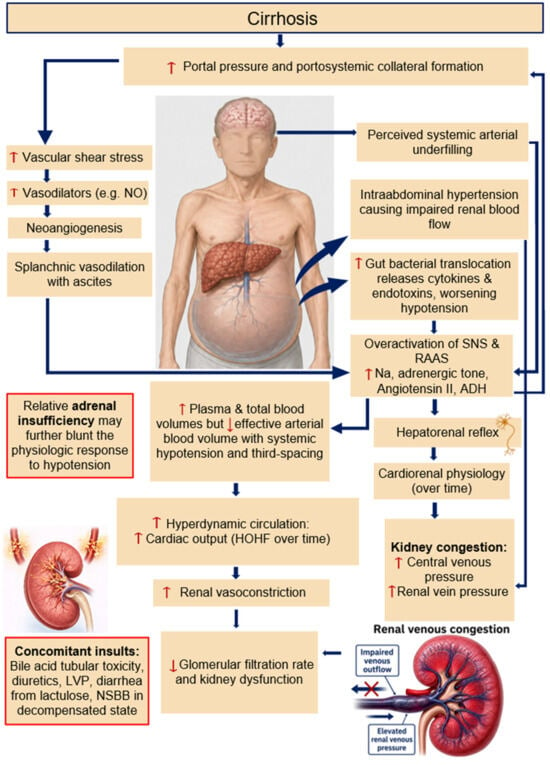
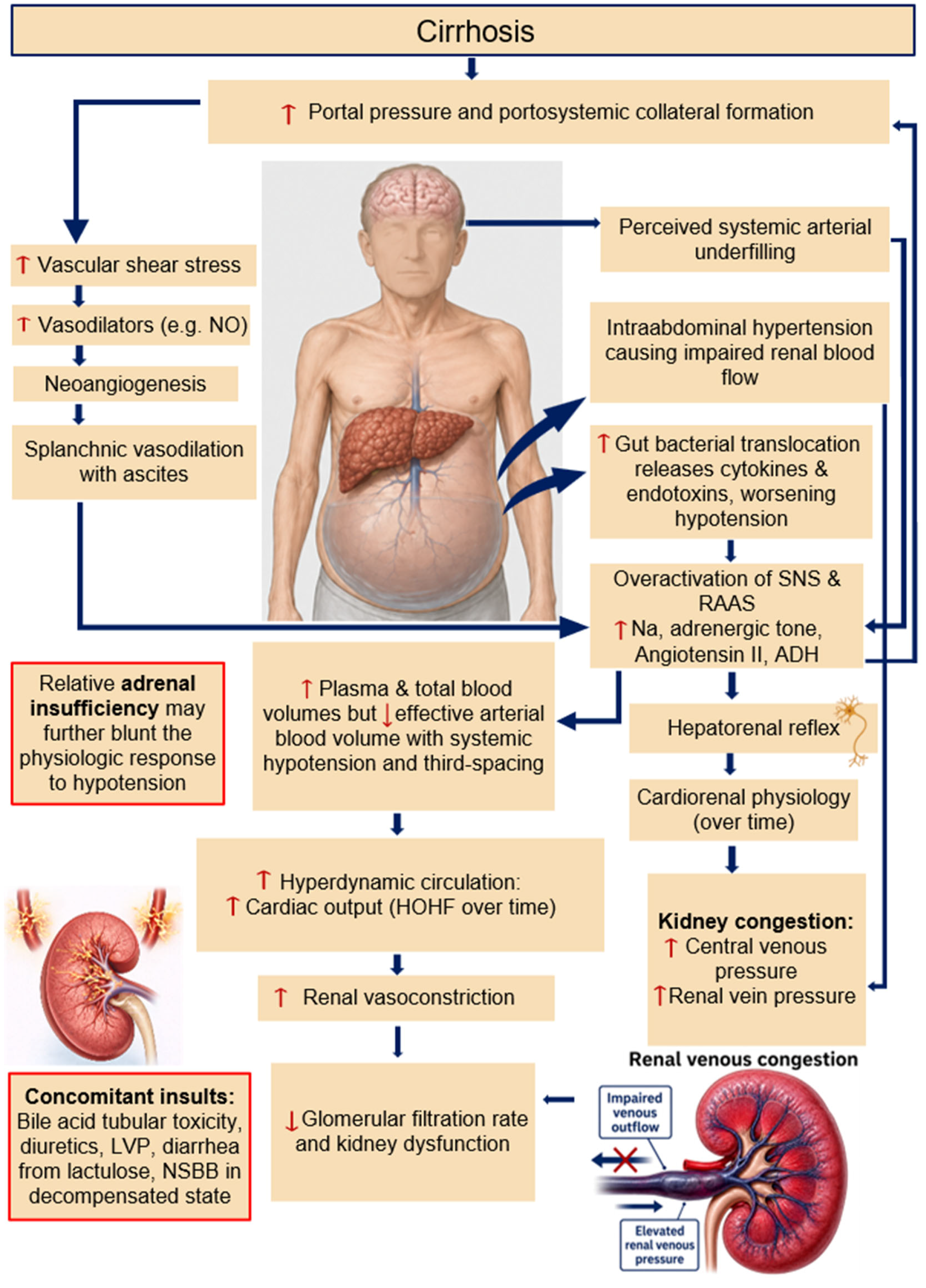

Disclaimer/Publisher’s Note: The statements, opinions and data contained in all publications are solely those of the individual author(s) and contributor(s) and not of MDPI and/or the editor(s). MDPI and/or the editor(s) disclaim responsibility for any injury to people or property resulting from any ideas, methods, instructions or products referred to in the content. |
© 2026 by the authors. Licensee MDPI, Basel, Switzerland. This article is an open access article distributed under the terms and conditions of the Creative Commons Attribution (CC BY) license.