Determinants of Pneumothorax and Alveolar Hemorrhage After CT-Guided Lung Biopsy

 ,
,  ,
,  ,
,  ,
,
Abstract
1. Introduction
2. Materials and Methods
2.1. Study Design
2.2. Procedure Technique
2.3. Variables and Definitions
2.3.1. Patient-Related Variables
2.3.2. Lesion-Related Variables
2.3.3. Procedural Variables
2.3.4. Histopathological Results
2.4. Complications
2.5. Outcomes
2.6. Statistical Analysis
3. Results
3.1. Study Population and Diagnostic Yield
3.2. Incidence of Complications

| Complication | n (%) | Clinical Severity (SIR-Based) | Management |
|---|---|---|---|
| Pneumothorax—conservative | 65 (27.1) | Mild | Clinical observation |
| Pneumothorax requiring drainage | 14 (5.8) | Moderate | Chest tube drainage |
| Alveolar hemorrhage | 49 (20.4) | Mild | Conservative |
| Hemoptysis | 13 (5.4) | Mild | Conservative |
| Hemothorax | 3 (1.2) | Mild | Conservative |
3.3. Determinants of PNX
| Characteristics | No PNX (n = 161) | PNX (n = 79) | p-Value |
|---|---|---|---|
| Patient characteristics | |||
| Age (years), median [IQR] | 67 [56–72] | 69 [63–74] | 0.019 |
| Sex—male, n (%) | 107 (66.5%) | 58 (73.4%) | 0.302 |
| BMI (kg/m2), median [IQR] | 26.7 [23.3–29.3] | 25.5 [23.0–29.4] | 0.641 |
| Smoking status, n (%) | |||
| Ever smoked | 121 (75.2%) | 58 (73.4%) | 0.755 |
| Pack–years (ever-smokers), median [IQR] | 30 [0–40] | 30 [0–40] | 0.733 |
| Modified coagulation times (%) | 29.2% | 31.9% | 0.665 |
| INR, median [IQR] | 1.0 [0.9–1.1] | 1.0 [0.9–1.1] | 0.649 |
| Lesion characteristics | |||
| Maximum diameter (mm), median [IQR] | 49 [28–73.5] | 39.5 [28.2–56] | 0.027 |
| Pleura-to-lesion distance (mm), median [IQR] | 0 [0–22] | 22 [12–33.5] | <0.001 |
| Lesion morphology, n (%) | |||
| Solid nodule | 140 (87.0%) | 68 (86.1%) | 0.959 |
| Spiculation | 119 (73.9%) | 65 (85.5%) | 0.109 |
| Central necrosis | 51 (33.1%) | 28 (33.3%) | 1.000 |
| Parenchymal background, n (%) | |||
| Emphysema | 72 (44.7%) | 45 (57.0%) | 0.074 |
| Carcinomatous lymphangitis | 61 (36.7%) | 18 (24.7%) | 0.074 |
| Pleural effusion | 75 (38.3%) | 4 (9.1%) | <0.001 |
| Procedural characteristics | |||
| Biopsy approach, n (%) | |||
| Posterior | 81 (50.3%) | 38 (48.1%) | 0.784 |
| Lateral | 36 (22.4%) | 28 (35.4%) | 0.043 |
| Anterior | 44 (27.3%) | 13 (16.5%) | 0.076 |
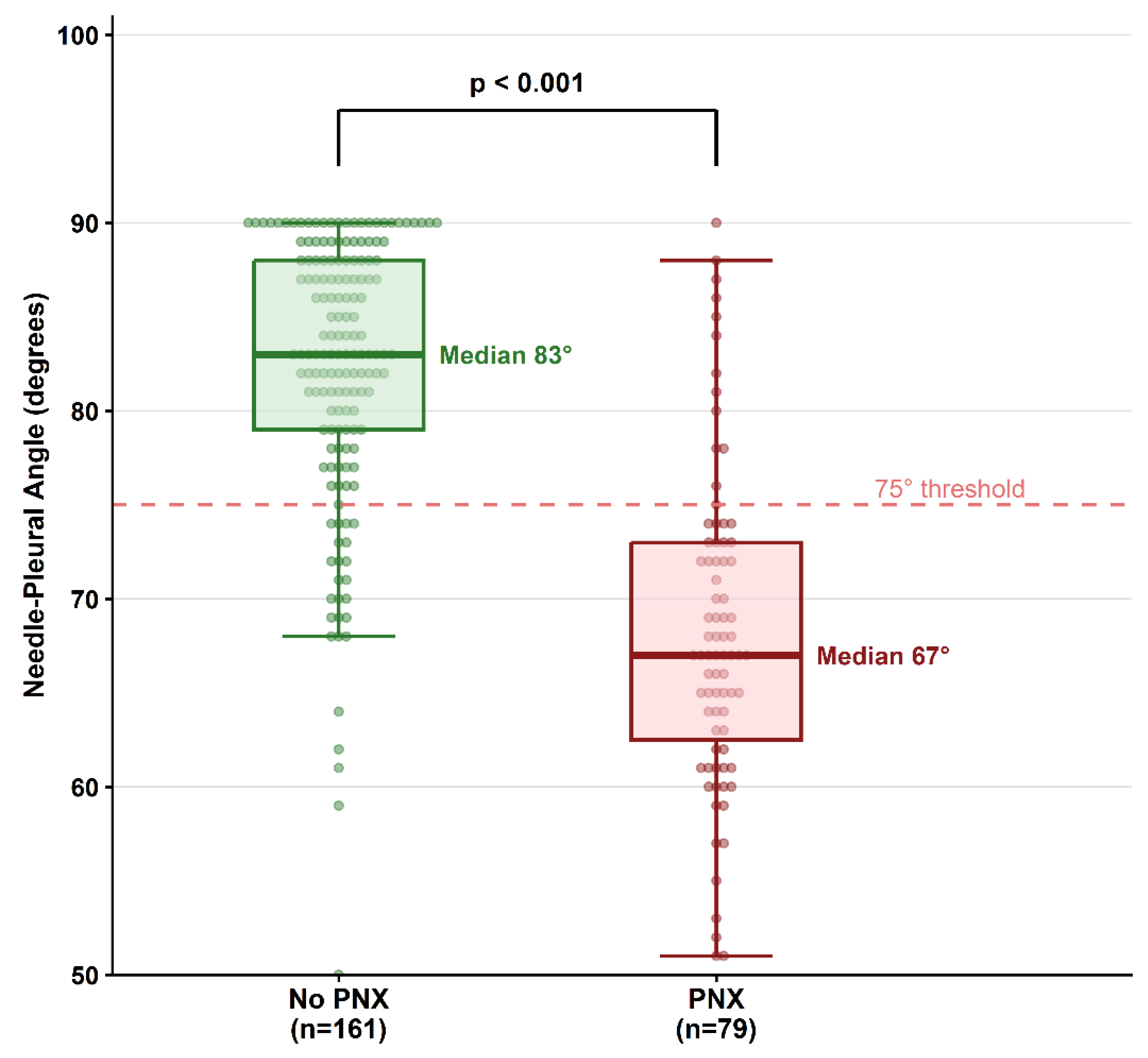
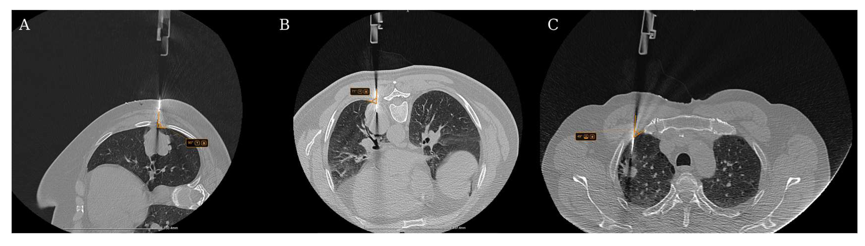
| Needle–pleural angle (°), median [IQR] | 83 [79–88] | 67 [63–73] | <0.001 |
| Needle–pleural angle < 75°, n (%) | 26 (16.1%) | 67 (84.8%) | <0.001 |
| Needle gauge, n (%) | |||
| 14 gauge | 9 (5.6%) | 3 (3.8%) | 0.755 |
| 16 gauge | 71 (44.1%) | 31 (39.2%) | 0.491 |
| 18 gauge | 81 (50.3%) | 45 (57.0%) | 0.340 |
| Needle length (mm), median [IQR] | 120 [100–120] | 120 [100–120] | 0.344 |
| Fissure traversal, n (%) | 61 (27.7%) | 17 (89.5%) | <0.001 |
| DLP (mGy·cm2), median [IQR] | 788 [502–1304] | 1003 [537–1344] | 0.269 |

| Variable | OR | 95% CI | p-Value |
|---|---|---|---|
| PNX (n = 240; events = 79; AUC = 0.927; Nagelkerke R2 = 0.656) | |||
| Needle–pleural angle (per 1° increase) | 0.837 | 0.795–0.881 | <0.001 |
| Pleura-to-lesion distance (per 10 mm) | 1.675 | 1.260–2.226 | <0.001 |
| Fissure traversal | 26.718 | 3.074–232.185 | 0.003 |
| Patient age (per 1 year) | 1.076 | 1.026–1.128 | 0.002 |
| Pleural effusion | 0.385 | 0.122–1.206 | 0.101 |
3.4. PNX Rim Progression (Explanatory, EPV = 7.5)
3.5. PNX Requiring Chest Tube Drainage (Explanatory, EPV = 7)
3.6. Determinants of Alveolar Hemorrhage
| Characteristics | No Hemorrhage (n = 191) | Hemorrhage (n = 49) | p-Value |
|---|---|---|---|
| Patient characteristics | |||
| Age (years), median [IQR] | 67 [59–72] | 67 [56–74] | 0.669 |
| Sex—male, n (%) | 129 (67.5%) | 36 (73.5%) | 0.492 |
| BMI (kg/m2), median [IQR] | 26.7 [23.3–29.5] | 24.8 [22.5–28.8] | 0.288 |
| Smoking status, n (%) | |||
| Ever smoked | 89 (46.6%) | 25 (51.0%) | 0.140 |
| Pack–years (ever-smokers), median [IQR] | 30 [0–40] | 30 [20–40] | 0.044 |
| Modified coagulation times, n (%) | 61 (31.9%) | 10 (20.4%) | 0.160 |
| INR, median [IQR] | 1.0 [0.9–1.1] | 1.0 [0.9–1.1] | 0.432 |
| Lesion characteristics | |||
| Maximum diameter (mm), median [IQR] | 49 [30–70] | 36 [26–54] | 0.018 |
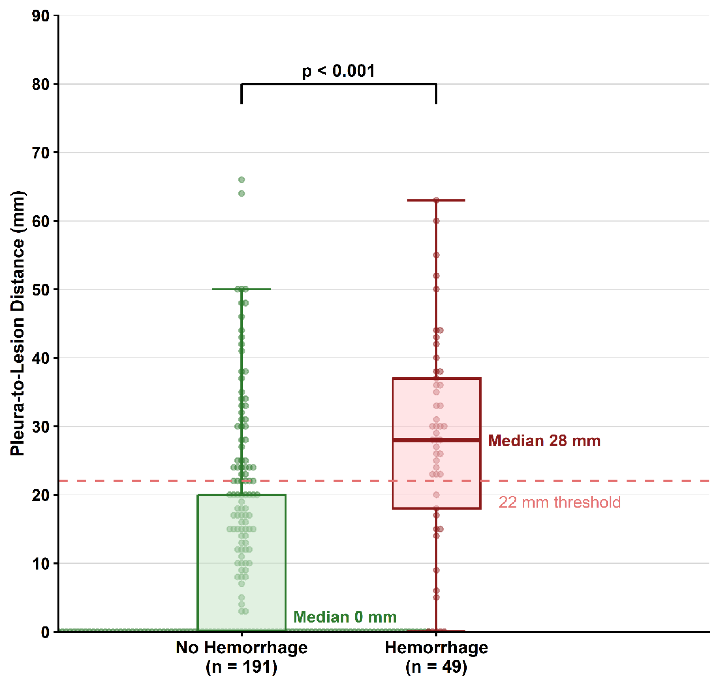
| Pleura-to-lesion distance (mm), median [IQR] | 0 [0–20] | 28 [18–37] | <0.001 |
| Laterality—right lung, n (%) | 106 (55.5%) | 36 (73.5%) | 0.023 |
| RUL apical segment (S1), n (%) | 22 (11.5%) | 15 (30.6%) | 0.003 |
| Lesion morphology, n (%) | |||
| Solid nodule | 164 (85.9%) | 44 (89.8%) | 0.746 |
| Spiculation | 141 (73.8%) | 43 (87.8%) | 0.316 |
| Central necrosis | 33 (21.4%) | 15 (17.9%) | 0.613 |
| Parenchymal background, n (%) | |||
| Emphysema | 92 (48.2%) | 25 (51.0%) | 0.873 |
| Carcinomatous lymphangitis | 40 (24.1%) | 9 (12.3%) | 0.039 |
| Pleural effusion | 44 (22.4%) | 5 (11.4%) | 0.146 |
| Procedural characteristics | |||
| Biopsy approach, n (%) | |||
| Posterior | 95 (49.7%) | 24 (49.0%) | 1.000 |
| Lateral | 52 (27.2%) | 12 (24.5%) | 0.856 |
| Anterior | 44 (23.0%) | 13 (26.5%) | 0.579 |
| Needle–pleural angle (°), median [IQR] | 81 [69–87] | 78 [70–86] | 0.777 |
| Needle–pleural angle < 75°, n (%) | 69 (36.1%) | 22 (44.9%) | 0.260 |
| Needle gauge, n (%) | |||
| 14 gauge | 10 (5.2%) | 2 (4.1%) | 1.000 |
| 16 gauge | 83 (43.5%) | 19 (38.8%) | 0.628 |
| 18 gauge | 98 (51.3%) | 28 (57.1%) | 0.523 |
| Needle length (mm), median [IQR] | 120 [100–120] | 120 [100–160] | <0.001 |
| Fissure traversal, n (%) | 45 (20.5%) | 4 (21.1%) | 1.000 |
| DLP (mGy·cm2), median [IQR] | 802 [494–1259] | 993 [589–1541] | 0.430 |
| Variable | OR | 95% CI | p-Value |
|---|---|---|---|
| Alveolar hemorrhage (n = 240; events = 49; AUC = 0.822; Nagelkerke R2 = 0.319) | |||
| Pleura-to-lesion distance (per 10 mm) | 1.768 | 1.410–2.216 | <0.001 |
| RUL apical segment (S1) | 4.281 | 1.838–9.974 | <0.001 |
| Carcinomatous lymphangitis | 0.359 | 0.142–0.906 | 0.030 |
| Needle length (per 10 mm) | 1.161 | 0.993–1.357 | 0.060 |
| Outcome | n | Events | EPV | R2 | AUC | HL p | Sens. | Spec. | NPV |
|---|---|---|---|---|---|---|---|---|---|
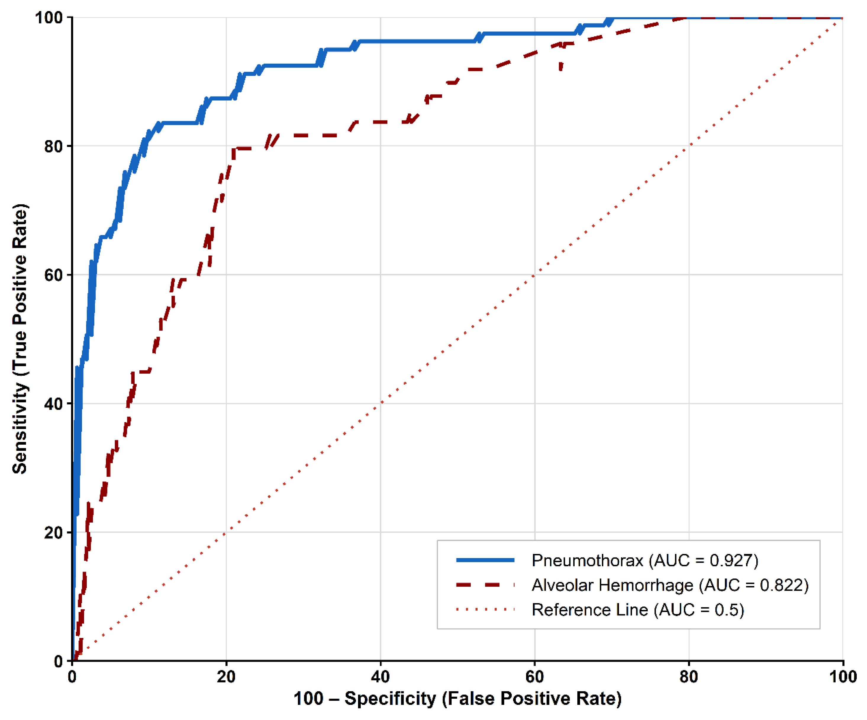
| Alveolar Hemorrhage | 240 | 49 | 12.2 | 0.319 | 0.822 * | 0.470 | 79.6% | 79.1% | 93.8% |
| Pneumothorax | 240 | 79 | 15.6 | 0.656 | 0.927 * | 0.871 | 83.5% | 88.8% | 91.7% |
| Rim Progression † | 76 | 15 | 7.5 | 0.249 | 0.789 | 0.662 | 80.0% | 70.5% | 93.5% |
| Chest Tube Drainage † | 240 | 14 | 7.0 | 0.639 | 0.979 | 0.995 | 100% | 95.1% | 100% |
3.7. Determinants of Hemoptysis
3.8. Hemothorax
4. Discussion
4.1. Main Findings
4.2. Comparison with the Existing Literature
4.2.1. PNX: Incidence and Risk Factors
4.2.2. Alveolar Hemorrhage: Incidence and Risk Factors
4.3. Pathophysiological Interpretation
4.4. Clinical Implications
- Pre-procedural risk stratification and counselling.
- 2.
- Needle angle as a primary modifiable target.
- 3.
- Post-procedural monitoring stratification.
- 4.
- Diagnostic yield and procedural technique.
4.5. Limitations
5. Conclusions
Supplementary Materials
Author Contributions
Funding
Institutional Review Board Statement
Informed Consent Statement
Data Availability Statement
Acknowledgments
Conflicts of Interest
References
- Bray, F.; Laversanne, M.; Sung, H.; Ferlay, J.; Siegel, R.L.; Soerjomataram, I.; Jemal, A. Global Cancer Statistics 2022: GLOBOCAN Estimates of Incidence and Mortality Worldwide for 36 Cancers in 185 Countries. CA Cancer J. Clin. 2024, 74, 229–263. [Google Scholar] [CrossRef]
- Zer, A.; Ahn, M.-J.; Barlesi, F.; Bubendorf, L.; De Ruysscher, D.; Garrido, P.; Gautschi, O.; Hendriks, L.E.; Jänne, P.A.; Kerr, K.M.; et al. Early and Locally Advanced Non-Small-Cell Lung Cancer: ESMO Clinical Practice Guideline for Diagnosis, Treatment and Follow-Up. Ann. Oncol. 2025, 36, 1245–1262. [Google Scholar] [CrossRef]
- Manhire, A.; Charig, M.; Clelland, C.; Gleeson, F.; Miller, R.; Moss, H.; Pointon, K.; Richardson, C.; Sawicka, E. Guidelines for Radiologically Guided Lung Biopsy. Thorax 2003, 58, 920–936. [Google Scholar] [CrossRef]
- Veltri, A.; Bargellini, I.; Giorgi, L.; Almeida, P.A.M.S.; Akhan, O. CIRSE Guidelines on Percutaneous Needle Biopsy (PNB). Cardiovasc. Interv. Radiol. 2017, 40, 1501–1513. [Google Scholar] [CrossRef]
- Balasubramanian, P.; Abia-Trujillo, D.; Barrios-Ruiz, A.; Garza-Salas, A.; Koratala, A.; Chandra, N.C.; Yu Lee-Mateus, A.; Labarca, G.; Fernandez-Bussy, S. Diagnostic Yield and Safety of Diagnostic Techniques for Pulmonary Lesions: Systematic Review, Meta-Analysis and Network Meta-Analysis. Eur. Respir. Rev. 2024, 33, 240046. [Google Scholar] [CrossRef] [PubMed]
- Constantinescu, A.; Stoicescu, E.R.; Iacob, R.; Chira, C.A.; Cocolea, D.M.; Nicola, A.C.; Mladin, R.; Oancea, C.; Manolescu, D. CT-Guided Transthoracic Core-Needle Biopsy of Pulmonary Nodules: Current Practices, Efficacy, and Safety Considerations. J. Clin. Med. 2024, 13, 7330. [Google Scholar] [CrossRef] [PubMed]
- Magnini, A.; Fissi, A.; Cinci, L.; Calistri, L.; Landini, N.; Nardi, C. Diagnostic Accuracy of Imaging-Guided Biopsy of Peripheral Pulmonary Lesions: A Systematic Review. Acta Radiol. 2024, 65, 1222–1237. [Google Scholar] [CrossRef]
- Ho, A.T.N.; Gorthi, R.; Lee, R.; Chawla, M.; Patolia, S. Solitary Lung Nodule: CT-Guided Transthoracic Biopsy vs Transbronchial Biopsy with Endobronchial Ultrasound and Flexible Bronchoscope, a Meta-Analysis of Randomized Controlled Trials. Lung 2023, 201, 85–93. [Google Scholar] [CrossRef] [PubMed]
- Yoon, S.H.; Park, C.M.; Lee, K.H.; Lim, K.Y.; Suh, Y.J.; Im, D.J.; Hur, J.; Han, D.H.; Kang, M.-J.; Choo, J.Y.; et al. Analysis of Complications of Percutaneous Transthoracic Needle Biopsy Using CT-Guidance Modalities In a Multicenter Cohort of 10568 Biopsies. Korean J. Radiol. 2019, 20, 323–331. [Google Scholar] [CrossRef]
- Huo, Y.R.; Chan, M.V.; Habib, A.-R.; Lui, I.; Ridley, L. Pneumothorax Rates in CT-Guided Lung Biopsies: A Comprehensive Systematic Review and Meta-Analysis of Risk Factors. Br. J. Radiol. 2020, 93, 20190866. [Google Scholar] [CrossRef]
- Vachani, A.; Zhou, M.; Ghosh, S.; Zhang, S.; Szapary, P.; Gaurav, D.; Kalsekar, I. Complications After Transthoracic Needle Biopsy of Pulmonary Nodules: A Population-Level Retrospective Cohort Analysis. J. Am. Coll. Radiol. 2022, 19, 1121–1129. [Google Scholar] [CrossRef] [PubMed]
- Heerink, W.J.; De Bock, G.H.; De Jonge, G.J.; Groen, H.J.M.; Vliegenthart, R.; Oudkerk, M. Complication Rates of CT-Guided Transthoracic Lung Biopsy: Meta-Analysis. Eur. Radiol. 2017, 27, 138–148. [Google Scholar] [CrossRef]
- Zhang, H.; Wang, S.; Zhong, F.; Liao, M. Risk Factors for Air Embolism Following Computed Tomography-Guided Percutaneous Transthoracic Needle Biopsy: A Systematic Review and Meta-Analysis. Diagn. Interv. Radiol. 2023, 29, 478–491. [Google Scholar] [CrossRef]
- Baerlocher, M.O.; Nikolic, B.; Sze, D.Y. Adverse Event Classification: Clarification and Validation of the Society of Interventional Radiology Specialty–Specific System. J. Vasc. Interv. Radiol. 2023, 34, 1–3. [Google Scholar] [CrossRef]
- Patel, I.J.; Rahim, S.; Davidson, J.C.; Hanks, S.E.; Tam, A.L.; Walker, T.G.; Wilkins, L.R.; Sarode, R.; Weinberg, I. Society of Interventional Radiology Consensus Guidelines for the Periprocedural Management of Thrombotic and Bleeding Risk in Patients Undergoing Percutaneous Image-Guided Interventions—Part II: Recommendations. J. Vasc. Interv. Radiol. 2019, 30, 1168–1184.e1. [Google Scholar] [CrossRef]
- Hadi, M.; Walker, C.; Desborough, M.; Basile, A.; Tsetis, D.; Hunt, B.; Müller-Hüllsbeck, S.; Rand, T.; Van Delden, O.; Uberoi, R. CIRSE Standards of Practice on Peri-Operative Anticoagulation Management During Interventional Radiology Procedures. Cardiovasc. Interv. Radiol. 2021, 44, 523–536. [Google Scholar] [CrossRef]
- Lin, Y.; Xu, Y.; Lin, J.; Fu, L.; Sun, H.; Huang, Z.; Ooi, B.Y.; Xie, S. Improving CT-Guided Transthoracic Biopsy Diagnostic Yield of Lung Masses Using Intraprocedural CT and Prior PET/CT Fusion Imaging. BMC Pulm. Med. 2022, 22, 311. [Google Scholar] [CrossRef] [PubMed]
- MacMahon, H.; Naidich, D.P.; Goo, J.M.; Lee, K.S.; Leung, A.N.C.; Mayo, J.R.; Mehta, A.C.; Ohno, Y.; Powell, C.A.; Prokop, M.; et al. Guidelines for Management of Incidental Pulmonary Nodules Detected on CT Images: From the Fleischner Society 2017. Radiology 2017, 284, 228–243. [Google Scholar] [CrossRef]
- Ancusa, V.M.; Trusculescu, A.A.; Constantinescu, A.; Burducescu, A.; Fira-Mladinescu, O.; Manolescu, D.L.; Traila, D.; Wellmann, N.; Oancea, C.I. Temporal Trends and Patient Stratification in Lung Cancer: A Comprehensive Clustering Analysis from Timis County, Romania. Cancers 2025, 17, 2305. [Google Scholar] [CrossRef] [PubMed]
- Constantinescu, A.; Căprariu, R.-N.; Stoicescu, E.-R.; Iacob, R.; Mânzatu, M.; Drimus, J.C.; Roșian, A.-S.; Ionescu, A.; Oancea, C.; Manolescu, D. From TNM 8 to TNM 9: Stage Migration and Histology-Specific Patterns in Lung Cancer. Cancers 2025, 17, 3290. [Google Scholar] [CrossRef]
- Peng, B.; Deng, Z.; Wang, Y.; Xu, S.; Luo, D.; Du, Z.; Liu, L.; Hu, Y.; Ren, Y. The Risk of Immediate Pneumothorax after CT-Guided Lung Needle Biopsy: Pleural Tail Sign as a Novel Factor. Quant. Imaging Med. Surg. 2023, 13, 707–719. [Google Scholar] [CrossRef] [PubMed]
- Johansen, K.B.; Valtersson, J.; Laursen, C.B.; Mussmann, B.; Rasmussen, B.; Graumann, O.; Pietersen, P.I. Diagnostic Yield and Complications of CT-Guided Biopsy of Lung Lesions as a Radiological Outpatient Clinic Procedure. Acta Radiol. Open 2025, 14. [Google Scholar] [CrossRef]
- Deng, X.-B.; Xie, L.; Zhu, H.-B.; Liu, Y.-L.; Yang, S.-X.; Zhao, B.; Sun, R.-J.; Li, X.-T.; Chen, M.-L.; Sun, Y.-S. The Nodule-Pleura Relationship Affects Pneumothorax in CT-Guided Percutaneous Transthoracic Needle Biopsy: Avoiding to Cross Pleural Tail Sign May Reduce the Incidence of Pneumothorax. BMC Pulm. Med. 2024, 24, 490. [Google Scholar] [CrossRef]
Disclaimer/Publisher’s Note: The statements, opinions and data contained in all publications are solely those of the individual author(s) and contributor(s) and not of MDPI and/or the editor(s). MDPI and/or the editor(s) disclaim responsibility for any injury to people or property resulting from any ideas, methods, instructions or products referred to in the content. |
© 2026 by the authors. Licensee MDPI, Basel, Switzerland. This article is an open access article distributed under the terms and conditions of the Creative Commons Attribution (CC BY) license.
Share and Cite
Constantinescu, A.; Roșian, A.-S.; Căprariu, R.-N.; Muntean, I.-A.; Ancușa, V.M.; Nicola, A.C.; Oancea, C.; Manolescu, D. Determinants of Pneumothorax and Alveolar Hemorrhage After CT-Guided Lung Biopsy. Diagnostics 2026, 16, 1848. https://doi.org/10.3390/diagnostics16121848
Constantinescu A, Roșian A-S, Căprariu R-N, Muntean I-A, Ancușa VM, Nicola AC, Oancea C, Manolescu D. Determinants of Pneumothorax and Alveolar Hemorrhage After CT-Guided Lung Biopsy. Diagnostics. 2026; 16(12):1848. https://doi.org/10.3390/diagnostics16121848
Chicago/Turabian StyleConstantinescu, Amalia, Alessia-Stephania Roșian, Radu-Nicolae Căprariu, Ionel-Alin Muntean, Versavia Maria Ancușa, Alin Ciprian Nicola, Cristian Oancea, and Diana Manolescu. 2026. "Determinants of Pneumothorax and Alveolar Hemorrhage After CT-Guided Lung Biopsy" Diagnostics 16, no. 12: 1848. https://doi.org/10.3390/diagnostics16121848
APA StyleConstantinescu, A., Roșian, A.-S., Căprariu, R.-N., Muntean, I.-A., Ancușa, V. M., Nicola, A. C., Oancea, C., & Manolescu, D. (2026). Determinants of Pneumothorax and Alveolar Hemorrhage After CT-Guided Lung Biopsy. Diagnostics, 16(12), 1848. https://doi.org/10.3390/diagnostics16121848