


Artificial Intelligence in Dermatopathology: An Update and Review of the Current Literature




Abstract
1. Introduction
1.1. Overview of Dermatopathology and Its Challenges
1.2. Introduction to Artificial Intelligence (AI) and Machine Learning (ML)
1.2.1. Definitions and Fundamental Concepts
1.2.2. Historical Context of AI in Medicine
1.3. Rationale for AI Integration in Dermatopathology
1.3.1. Potential to Enhance Diagnostic Accuracy and Efficiency
1.3.2. Role in Addressing Workforce Challenges
2. Review Methodology
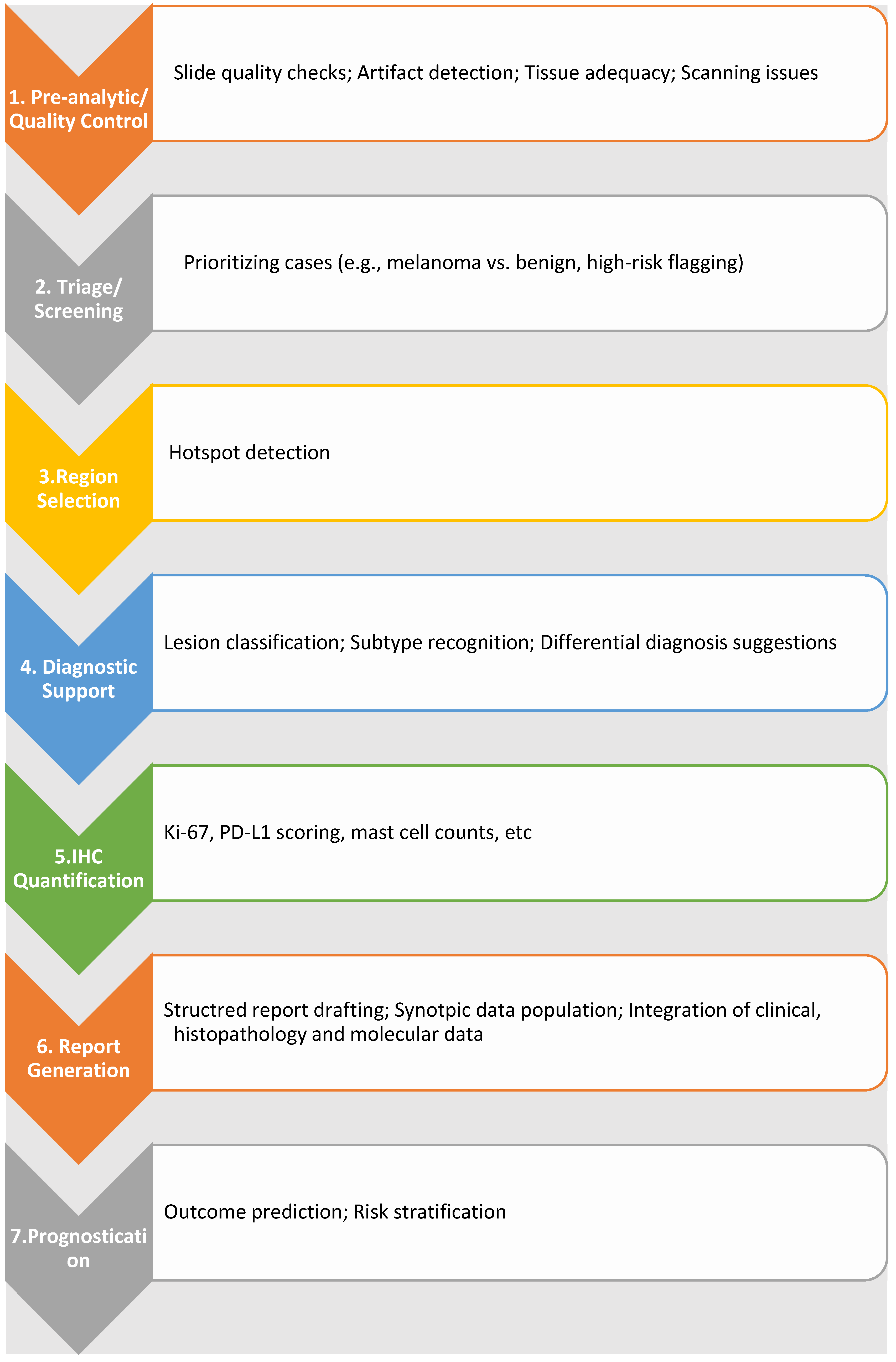
3. AI Applications in Dermatopathology: Current Landscape
3.1. Diagnostic Support for Specific Skin Conditions
3.1.1. Melanocytic Lesions (Nevi vs. Melanoma)
- a.
- Discrimination between benign nevi and malignant melanoma
- b.
- Recognition of specific melanoma subtypes
- c.
- Performance metrics (accuracy, sensitivity, specificity) compared to human experts
- d.
- Examples of successful deep learning models (e.g., CNNs like ResNet50)
3.1.2. Non-Melanoma Skin Cancers (e.g., Basal Cell Carcinoma, Squamous Cell Carcinoma)
- a.
- Detection and classification on whole-slide images (WSIs)
- b.
- Identification of tumor margins
- c.
- Examples of AI-driven tools in Mohs micrographic surgery
3.1.3. Inflammatory Dermatoses
- a.
- Challenges in classification due to overlapping features
- b.
- Emerging applications and limitations
3.1.4. Adnexal Tumors and Other Rare Entities
- a.
- Current research and the need for larger datasets
3.2. Workflow Optimization and Automation
3.2.1. Automated Detection of Mitotic Figures
3.2.2. Identification of Hotspots for Further Review
3.2.3. Quantification of Immunohistochemical Stains
3.2.4. Digital Slide Management and Annotation
3.3. Predictive Analytics and Prognostication
3.3.1. Predicting Disease-Specific Survival in Melanoma
3.3.2. Integration of Histological, Genetic, and Clinical Data (Multimodal AI)
3.4. Educational Tools and Training
3.4.1. AI-Driven Simulators for Skill Refinement
3.4.2. Exposure to Diverse and Challenging Cases

| Application Area | AI Tool | Clinical Use | Dataset Example | Key Findings | References |
|---|---|---|---|---|---|
| Convolutional neural network (CNN): ResNet50, and ensemble CNNs | Discrimination between benign nevi and malignant melanoma | Hematoxylin and eosin (H&E)-stained slides, 595 images (350 nevi, 345 melanomas) | ResNet50 CNNs can match/exceed pathologists; binary classification limits consideration of intermediate lesions | [14] |
| Whole-slide image (WSI) of 50 melanomas and 50 nevi | Ensemble CNN matched or slightly exceeded diagnostic performance | [15] | |||
| Fast random forest (FRF) algorithm | Early screening of nevoid melanoma | 18 histopathological photomicrographs of nevoid melanoma | Enhances screening for rare melanoma subtypes | [16] | |
| Deep learning framework; U-Net | Detection and subtyping of basal cell carcinoma (BCC) | 1400 image patches; multi-center BCC cohorts | Automates detection and subtyping; reduces need for handcrafted features | [22] | |
| Deep learning platform (ArcticAI) | Tumor margin identification during Mohs surgery | 194 BCC patients, with 178 cases used for histological analysis generating 351 WSIs | Improves intraoperative margin assessment; integrates with surgical workflow | [24] | |
| U-shaped deep CNN; cascaded CNN framework | Classification of inflammatory dermatoses | 90 annotated psoriasis skin biopsy images | Effective for segmenting epidermis/dermis; overlapping features and lack of clinical context remain challenges | [29] | |
| Psoriasis: skin biopsy images; superficial perivascular dermatitis: 3954 images, 327 cases | AI can effectively assist in identifying key pathological features and classifying inflammatory skin conditions | [30] | |||
| AI-based mitosis detection algorithms | Mitotic figure detection | 99 digitized melanocytic lesion slides (including 10 nevoid melanomas) | Mitosis detection algorithms may be helpful in particularly challenging cases like nevoid melanoma but offer limited benefit in routine lesion interpretation | [32] |
| Attention-based multiple-instance learning, transformers, saliency maps | Hotspot identification | Reduce review time and improve safety but face challenges such as spurious attention to artifacts, sensitivity–specificity trade-offs in small-lesion detection, and overconfidence on out-of-distribution detection | [12,33,34,35,36] | ||
| DL-based cell detection (ResNet), QuPath | Objective quantification of Ki-67, Melan-A, SOX10, CD3, CD8, PD-L1 | Multicenter dataset of MelanA immunostain and H&E WSIs from 464 melanocytic lesions (melanomas, melanoma in situ, and nevi) [38] | ResNet-based AI models using MelanA immunostain achieved melanoma classification performance comparable to H&E-based models, while combining both stains improved diagnostic accuracy and generalizability [38] | [37,38,39] | |
| 20 full-thickness skin punch biopsies from 10 psoriasis patients, including 10 lesional and 10 peri-lesional unaffected skin [39] | Adobe Photoshop and QuPath showed good agreement for quantifying CD8+ T cells in psoriasis biopsies, with normalization to epidermal length providing more consistent results than area-based normalization [39] | ||||
| WSI platforms, QuPath, AI-assisted annotation | Digital slide management and annotation | [42,43,44,45] | |||
| Deep learning applied to digitized slides | Predicts disease-specific survival and risk stratification in melanoma | [49] | ||
| Integrated histology + genomic + clinical AI | Improved personalized prognostication and treatment guidance | [50] | |||
| Hypertext Atlas of Dermatopathology; SlideTutor; ReportTutor | Skill refinement, diagnostic feedback, report quality improvement | [53,54,55] | ||
| College of American pathologists (CAP) “AI Playground” | Exposure to diverse and complex cases | https://www.pathpresenter.com/cap-announces-immersive-ai-playground-powered-by-pathpresenter/ (accessed on 29 May 2026) |
4. Methodological Considerations and Technical Aspects
4.1. Data Acquisition and Curation
4.1.1. Importance of High-Quality, Annotated Datasets
4.1.2. Whole-Slide Imaging (WSI) and Its Role
4.1.3. Data Augmentation Techniques
4.2. Algorithm Development and Training
4.2.1. Convolutional Neural Networks (CNNs) and Their Architectures
- -
- Basic CNNs: Layers of convolution, pooling, and fully connected layers [59].
- -
- ResNet: Introduced residual connections to enable training of very deep networks without vanishing gradients [60].
- -
- DenseNet: Densely connected layers to improve feature reuse and efficiency [61].
- -
- Inception/GoogLeNet: Multi-scale convolutional filters for extracting features at different resolutions [62].
- -
- Vision transformers (ViTs): More recently used for pathology and leveraging attention mechanisms [63].
4.2.2. Transfer Learning
4.2.3. Explainable AI (XAI) for Interpretability
4.2.4. Large Language Models (LLMs)
4.3. Validation and Performance Evaluation
4.3.1. Internal vs. External Validation
4.3.2. Metrics
4.3.3. Benchmarking Against Human Experts and Real-World Evaluation
4.4. Hardware and Software Infrastructure Requirements
| Domain | Key Components | Key Points/Role in AI Development | Main Challenges | References |
|---|---|---|---|---|
| Data acquisition and curation | Annotated datasets | High-quality, expert-labeled datasets are essential for supervised learning and validation | Time-consuming annotation, inter-observer variability, lack of standardization | [43] |
| Whole-slide imaging (WSI) | Enables digitization of entire histology slides, supporting computational pathology and data sharing | Large file sizes, scanner variability, cross-institution standardization | [41,56] | |
| Data augmentation | Rotation, scaling, color/stain normalization increase dataset diversity and model robustness | Risk of unrealistic synthetic variation, inconsistent augmentation practices | [57] | |
| Algorithm development and training | Convolutional neural networks (CNNs) architectures | Learn hierarchical histologic features (cell → tissue → architecture); include ResNet, DenseNet, Inception | Overfitting, interpretability limitations | [58,59,60,61,62] |
| Transfer learning | Uses pre-trained models to improve performance on small medical datasets | Domain shift between natural and histopathology images | [58] | |
| Explainable AI (XAI) | Saliency maps, multiple instance learning, prototype models improve interpretability | May misidentify artifacts; limited clinical alignment with histopathologic patterns | [65,66] | |
| Large language models (multimodal) | Combine clinical + dermoscopic image interpretation; show promise in screening and triage | Variable performance, low specificity, not reliable standalone tools | [67,68,69,70] | |
| Validation and performance evaluation | Internal vs. external validation | External validation better reflects real-world generalizability | Limited availability of independent datasets | [71] |
| Performance metrics | Accuracy, sensitivity, specificity, F1-score, and receiver operating characteristic area under the curve assess classification performance | Metrics alone may not reflect clinical utility | [72] | |
| Calibration and uncertainty | Ensures predicted probabilities reflect true risk; supports clinical decision-making | Often under-reported in studies | [73,74] | |
| Decision curve analysis | Evaluates net clinical benefit vs. traditional workflows | Requires robust clinical modeling assumptions | [75] | |
| Human benchmarking | Comparison with dermatopathologists establishes expert-level performance | Variability among human experts | ||
| Prospective and silent trials | Evaluate real-world integration without influencing care | Logistically complex, resource-intensive | [76] | |
| Infrastructure requirements | Hardware | Graphic processing unit (GPU) or tensor processing unit (TPU) systems required for WSI processing and deep learning | High cost, scalability limitations | [77] |
| Software tools | TensorFlow, PyTorch, QuPath (https://qupath.github.io) for model development and annotation | Interoperability and workflow integration challenges | [44] |
5. Challenges and Limitations
5.1. Data-Related Challenges
5.2. Algorithmic and Technical Challenges
5.3. Integration and Adoption Challenges
6. Ethical and Legal Considerations
7. Future Directions and Emerging Trends
7.1. Multi-Modal AI and Foundation Models
7.2. Federated Learning and Workflow Integration
8. Conclusions
Author Contributions
Funding
Institutional Review Board Statement
Informed Consent Statement
Data Availability Statement
Conflicts of Interest
References
- Hay, R.J.; Johns, N.E.; Williams, H.C.; Bolliger, I.W.; Dellavalle, R.P.; Margolis, D.J.; Marks, R.; Naldi, L.; Weinstock, M.A.; Wulf, S.K.; et al. The global burden of skin disease in 2010: An analysis of the prevalence and impact of skin conditions. J. Investig. Dermatol. 2014, 134, 1527–1534. [Google Scholar] [CrossRef] [Scilit]
- Cramer, S.F. Interobserver variability in dermatopathology. Arch. Dermatol. 1997, 133, 1033–1036. [Google Scholar] [CrossRef] [Scilit] [PubMed]
- Roky, A.H.; Islam, M.M.; Ahasan, A.M.F.; Mostaq, M.S.; Mahmud, M.Z.; Amin, M.N.; Mahmud, M.A. Overview of skin cancer types and prevalence rates across continents. Cancer Pathog. Ther. 2025, 3, 89–100. [Google Scholar] [CrossRef] [Scilit]
- Walsh, E.; Orsi, N.M. The current troubled state of the global pathology workforce: A concise review. Diagn. Pathol. 2024, 19, 163. [Google Scholar] [CrossRef] [Scilit]
- Dave, M.; Patel, N. Artificial intelligence in healthcare and education. Br. Dent. J. 2023, 234, 761–764. [Google Scholar] [CrossRef] [Scilit]
- Eapen, B.R. Artificial Intelligence in Dermatology: A Practical Introduction to a Paradigm Shift. Indian Dermatol. Online J. 2020, 11, 881–889. [Google Scholar] [CrossRef] [Scilit]
- Sadr, H.; Nazari, M.; Khodaverdian, Z.; Farzan, R.; Yousefzadeh-Chabok, S.; Ashoobi, M.T.; Hemmati, H.; Hendi, A.; Ashraf, A.; Pedram, M.M.; et al. Unveiling the potential of artificial intelligence in revolutionizing disease diagnosis and prediction: A comprehensive review of machine learning and deep learning approaches. Eur. J. Med. Res. 2025, 30, 418. [Google Scholar] [CrossRef] [Scilit]
- Wells, A.; Patel, S.; Lee, J.B.; Motaparthi, K. Artificial intelligence in dermatopathology: Diagnosis, education, and research. J. Cutan. Pathol. 2021, 48, 1061–1068. [Google Scholar] [CrossRef] [Scilit] [PubMed]
- Ramesh, A.N.; Kambhampati, C.; Monson, J.R.; Drew, P.J. Artificial intelligence in medicine. Ann. R. Coll. Surg. Engl. 2004, 86, 334–338. [Google Scholar] [CrossRef] [Scilit] [PubMed]
- Kaul, V.; Enslin, S.; Gross, S.A. History of artificial intelligence in medicine. Gastrointest. Endosc. 2020, 92, 807–812. [Google Scholar] [CrossRef] [Scilit]
- Potter, B.; Ronan, S.G. Computerized dermatopathologic diagnosis. J. Am. Acad. Dermatol. 1987, 17, 119–131. [Google Scholar] [CrossRef] [Scilit] [PubMed]
- Cazzato, G.; Rongioletti, F. Artificial intelligence in dermatopathology: Updates, strengths, and challenges. Clin. Dermatol. 2024, 42, 437–442. [Google Scholar] [CrossRef] [Scilit]
- Polesie, S.; McKee, P.H.; Gardner, J.M.; Gillstedt, M.; Siarov, J.; Neittaanmäki, N.; Paoli, J. Attitudes Toward Artificial Intelligence Within Dermatopathology: An International Online Survey. Front. Med. 2020, 7, 591952. [Google Scholar] [CrossRef] [Scilit] [PubMed]
- Hekler, A.; Utikal, J.S.; Enk, A.H.; Berking, C.; Klode, J.; Schadendorf, D.; Jansen, P.; Franklin, C.; Holland-Letz, T.; Krahl, D.; et al. Pathologist-level classification of histopathological melanoma images with deep neural networks. Eur. J. Cancer 2019, 115, 79–83. [Google Scholar] [CrossRef] [Scilit]
- Brinker, T.J.; Schmitt, M.; Krieghoff-Henning, E.I.; Barnhill, R.; Beltraminelli, H.; Braun, S.A.; Carr, R.; Fernandez-Figueras, M.T.; Ferrara, G.; Fraitag, S.; et al. Diagnostic performance of artificial intelligence for histologic melanoma recognition compared to 18 international expert pathologists. J. Am. Acad. Dermatol. 2022, 86, 640–642. [Google Scholar] [CrossRef] [Scilit]
- Cazzato, G.; Massaro, A.; Colagrande, A.; Trilli, I.; Ingravallo, G.; Casatta, N.; Lupo, C.; Ronchi, A.; Franco, R.; Maiorano, E.; et al. Artificial Intelligence Applied to a First Screening of Naevoid Melanoma: A New Use of Fast Random Forest Algorithm in Dermatopathology. Curr. Oncol. 2023, 30, 6066–6078. [Google Scholar] [CrossRef] [Scilit]
- Hekler, A.; Utikal, J.S.; Enk, A.H.; Solass, W.; Schmitt, M.; Klode, J.; Schadendorf, D.; Sondermann, W.; Franklin, C.; Bestvater, F.; et al. Deep learning outperformed 11 pathologists in the classification of histopathological melanoma images. Eur. J. Cancer 2019, 118, 91–96. [Google Scholar] [CrossRef] [Scilit]
- Elshahawy, M.; Elnemr, A.; Oproescu, M.; Schiopu, A.-G.; Elgarayhi, A.; Elmogy, M.M.; Sallah, M. Early Melanoma Detection Based on a Hybrid YOLOv5 and ResNet Technique. Diagnostics 2023, 13, 2804. [Google Scholar] [CrossRef] [Scilit]
- Duschner, N.; Baguer, D.O.; Schmidt, M.; Griewank, K.G.; Hadaschik, E.; Hetzer, S.; Wiepjes, B.; Le’Clerc Arrastia, J.; Jansen, P.; Maass, P.; et al. Applying an artificial intelligence deep learning approach to routine dermatopathological diagnosis of basal cell carcinoma. J. Dtsch. Dermatol. Ges. 2023, 21, 1329–1337. [Google Scholar] [CrossRef] [Scilit] [PubMed]
- Yin, W.; Zhou, D.; Nie, R. DI-UNet: Dual-branch interactive U-Net for skin cancer image segmentation. J. Cancer Res. Clin. Oncol. 2023, 149, 15511–15524. [Google Scholar] [CrossRef] [Scilit]
- Nogales, A.; Garrido, M.C.; Guitian, A.; Rodriguez-Peralto, J.-L.; Villanueva, C.P.; Díaz-Prieto, D.; García-Tejedor, Á.J. A Hybrid Artificial Intelligence Framework for Melanoma Diagnosis Using Histopathological Images. Technologies 2025, 13, 330. [Google Scholar] [CrossRef] [Scilit]
- Cruz-Roa, A.A.; Arevalo Ovalle, J.E.; Madabhushi, A.; González Osorio, F.A. A deep learning architecture for image representation, visual interpretability and automated basal-cell carcinoma cancer detection. In Proceedings of the International Conference on Medical Image Computing and Computer-Assisted Intervention MICCAI 2013, Nagoya, Japan, 22–26 September 2013; Volume 16, pp. 403–410. [Google Scholar]
- Lan, X.; Guo, G.; Wang, X.; Yan, Q.; Xue, R.; Li, Y.; Zhu, J.; Dong, Z.; Wang, F.; Li, G.; et al. Differentiation and risk stratification of basal cell carcinoma with deep learning on histopathologic images and measuring nuclei and tumor microenvironment features. Skin. Res. Technol. 2024, 30, e13571. [Google Scholar] [CrossRef] [Scilit]
- Levy, J.J.; Davis, M.J.; Chacko, R.S.; Davis, M.J.; Fu, L.J.; Goel, T.; Pamal, A.; Nafi, I.; Angirekula, A.; Suvarna, A.; et al. Intraoperative margin assessment for basal cell carcinoma with deep learning and histologic tumor mapping to surgical site. npj Precis. Oncol. 2024, 8, 2. [Google Scholar] [CrossRef] [Scilit] [PubMed]
- Tighe, D.; Tekeli, K.; Gouk, T.; Smith, J.; Ho, M.; Moody, A.; Walsh, S.; Provost, S.; Freitas, A. Machine learning methods applied to audit of surgical margins after curative surgery for facial (non-melanoma) skin cancer. Br. J. Oral Maxillofac. Surg. 2023, 61, 94–100. [Google Scholar] [CrossRef] [Scilit] [PubMed]
- Bittner, G.C.; Cerci, F.B.; Kubo, E.M.; Tolkachjov, S.N. Mohs micrographic surgery: A review of indications, technique, outcomes, and considerations. An. Bras. Dermatol. 2021, 96, 263–277. [Google Scholar] [CrossRef] [Scilit]
- Davis, M.J.; Srinivasan, G.; Chacko, R.; Chen, S.; Suvarna, A.; Vaickus, L.J.; Torres, V.C.; Hodge, S.; Chen, E.Y.; Preum, S.; et al. A deep learning algorithm to detect cutaneous squamous cell carcinoma on frozen sections in Mohs micrographic surgery: A retrospective assessment. Exp. Dermatol. 2024, 33, e14949. [Google Scholar] [CrossRef] [Scilit] [PubMed]
- Della Mura, M.; Sorino, J.; Colagrande, A.; Daruish, M.; Ingravallo, G.; Massaro, A.; Cazzato, G.; Lupo, C.; Casatta, N.; Ribatti, D.; et al. Artificial Intelligence in the Histopathological Assessment of Non-Neoplastic Skin Disorders: A Narrative Review with Future Perspectives. Med. Sci. 2025, 13, 70. [Google Scholar] [CrossRef] [Scilit]
- Pal, A.; Garain, U.; Chandra, A.; Chatterjee, R.; Senapati, S. Psoriasis skin biopsy image segmentation using Deep Convolutional Neural Network. Comput. Methods Programs Biomed. 2018, 159, 59–69. [Google Scholar] [CrossRef] [Scilit]
- Bao, Y.; Zhang, J.; Zhang, Q.; Chang, J.; Lu, D.; Fu, Y. Artificial Intelligence-Aided Recognition of Pathological Characteristics and Subtype Classification of Superficial Perivascular Dermatitis. Front. Med. 2021, 8, 696305. [Google Scholar] [CrossRef] [Scilit]
- Jansen, P.; Arrastia, J.L.; Baguer, D.O.; Schmidt, M.; Landsberg, J.; Wenzel, J.; Emberger, M.; Schadendorf, D.; Hadaschik, E.; Maass, P.; et al. Deep learning based histological classification of adnex tumors. Eur. J. Cancer 2024, 196, 113431. [Google Scholar] [CrossRef] [Scilit]
- Sturm, B.; Creytens, D.; Smits, J.; Ooms, A.H.A.G.; Eijken, E.; Kurpershoek, E.; Küsters-Vandevelde, H.V.N.; Wauters, C.; Blokx, W.A.M.; van der Laak, J.A.W.M. Computer-Aided Assessment of Melanocytic Lesions by Means of a Mitosis Algorithm. Diagnostics 2022, 12, 436. [Google Scholar] [CrossRef] [Scilit]
- Su, Z.; Rezapour, M.; Sajjad, U.; Niu, S.; Gurcan, M.N.; Niazi, M.K.K. Cross-Attention-Based Saliency Inference for Predicting Cancer Metastasis on Whole Slide Images. IEEE J. Biomed. Health Inform. 2024, 28, 7206–7216. [Google Scholar] [CrossRef] [Scilit]
- Dolezal, J.M.; Srisuwananukorn, A.; Karpeyev, D.; Ramesh, S.; Kochanny, S.; Cody, B.; Mansfield, A.S.; Rakshit, S.; Bansal, R.; Bois, M.C.; et al. Uncertainty-informed deep learning models enable high-confidence predictions for digital histopathology. Nat. Commun. 2022, 13, 6572. [Google Scholar] [CrossRef] [Scilit]
- Cai, L.; Huang, S.; Zhang, Y.; Lu, J.; Zhang, Y. AttriMIL: Revisiting attention-based multiple instance learning for whole-slide pathological image classification from a perspective of instance attributes. Med. Image Anal. 2025, 103, 103631. [Google Scholar] [CrossRef] [Scilit]
- Linmans, J.; Elfwing, S.; van der Laak, J.; Litjens, G. Predictive uncertainty estimation for out-of-distribution detection in digital pathology. Med. Image Anal. 2023, 83, 102655. [Google Scholar] [CrossRef] [Scilit]
- Brogård, M.B.; Steiniche, T.; Lade-Keller, J.; Wandler, A.; Christensen, K.B.; Georgsen, J.B.; Nielsen, P.S. Digital quantification of Ki67 and PRAME in challenging melanocytic lesions—A novel diagnostic tool. Pathol. Res. Pract. 2025, 270, 155953. [Google Scholar] [CrossRef] [Scilit] [PubMed]
- Wies, C.; Schneider, L.; Haggenmüller, S.; Bucher, T.C.; Hobelsberger, S.; Heppt, M.V.; Ferrara, G.; Krieghoff-Henning, E.I.; Brinker, T.J. Evaluating deep learning-based melanoma classification using immunohistochemistry and routine histology: A three center study. PLoS ONE 2024, 19, e0297146. [Google Scholar] [CrossRef] [Scilit]
- Emmanuel, T.; Brent, M.B.; Iversen, L.; Johansen, C. Quantification of Immunohistochemically Stained Cells in Skin Biopsies. Dermatopathology 2022, 9, 82–93. [Google Scholar] [CrossRef] [Scilit] [PubMed]
- Baxi, V.; Lee, G.; Duan, C.; Pandya, D.; Cohen, D.N.; Edwards, R.; Chang, H.; Li, J.; Elliott, H.; Pokkalla, H.; et al. Association of artificial intelligence-powered and manual quantification of programmed death-ligand 1 (PD-L1) expression with outcomes in patients treated with nivolumab ± ipilimumab. Mod. Pathol. 2022, 35, 1529–1539. [Google Scholar] [CrossRef] [Scilit]
- Al-Janabi, S.; Huisman, A.; Vink, A.; Leguit, J.; Offerhaus, G.J.A.; ten Kate, J.F.W.; van Dijk, R.; van Diest, P.J. Whole slide images for primary diagnostics in dermatopathology: A feasibility study. J. Clin. Pathol. 2012, 65, 152–158. [Google Scholar] [CrossRef] [Scilit] [PubMed]
- Weinstein, R.S.; Graham, A.R.; Richter, L.C.; Barker, G.P.; Krupinski, E.A.; Lopez, A.M.; Erps, K.A.; Bhattacharyya, A.K.; Yagi, Y.; Gilbertson, J.R. Overview of telepathology, virtual microscopy, and whole slide imaging: Prospects for the future. Hum. Pathol. 2009, 40, 1057–1069. [Google Scholar] [CrossRef] [Scilit]
- Tschandl, P.; Rinner, C.; Apalla, Z.; Argenziano, G.; Codella, N.; Halpern, A.; Janda, M.; Lallas, A.; Longo, C.; Malvehy, J.; et al. Human-computer collaboration for skin cancer recognition. Nat. Med. 2020, 26, 1229–1234. [Google Scholar] [CrossRef] [Scilit]
- Bankhead, P.; Loughrey, M.B.; Fernández, J.A.; Dombrowski, Y.; McArt, D.G.; Dunne, P.D.; McQuaid, S.; Gray, R.T.; Murray, L.J.; Coleman, H.G.; et al. QuPath: Open source software for digital pathology image analysis. Sci. Rep. 2017, 7, 16878. [Google Scholar] [CrossRef] [Scilit]
- Amgad, M.; Elfandy, H.; Hussein, H.; Atteya, L.A.; Elsebaie, M.A.T.; Abo Elnasr, L.S.; Sakr, R.A.; Salem, H.S.E.; Ismail, A.F.; Saad, A.M.; et al. Structured crowdsourcing enables convolutional segmentation of histology images. Bioinformatics 2019, 35, 3461–3467. [Google Scholar] [CrossRef] [Scilit]
- Liu, Y.; He, Q.; Duan, H.; Shi, H.; Han, A.; He, Y. Using Sparse Patch Annotation for Tumor Segmentation in Histopathological Images. Sensors 2022, 22, 6053. [Google Scholar] [CrossRef] [Scilit] [PubMed]
- Tavolara, T.E.; Su, Z.; Gurcan, M.N.; Niazi, M.K.K. One label is all you need: Interpretable AI-enhanced histopathology for oncology. Semin. Cancer Biol. 2023, 97, 70–85. [Google Scholar] [CrossRef] [Scilit] [PubMed]
- Comes, M.C.; Fucci, L.; Mele, F.; Bove, S.; Cristofaro, C.; De Risi, I.; Fanizzi, A.; Milella, M.; Strippoli, S.; Zito, A. A deep learning model based on whole slide images to predict disease-free survival in cutaneous melanoma patients. Sci. Rep. 2022, 12, 20366. [Google Scholar] [CrossRef] [Scilit] [PubMed]
- Li, X.; Yu, X.; Tian, D.; Liu, Y.; Li, D. Exploring and validating the prognostic value of pathomics signatures and genomics in patients with cutaneous melanoma based on bioinformatics and deep learning. Med. Phys. 2023, 50, 7049–7059. [Google Scholar] [CrossRef] [Scilit]
- Peng, Y.; Chu, Y.; Chen, Z.; Zhou, W.; Wan, S.; Xiao, Y.; Zhang, Y.; Li, J. Combining texture features of whole slide images improves prognostic prediction of recurrence-free survival for cutaneous melanoma patients. World J. Surg. Oncol. 2020, 18, 130. [Google Scholar] [CrossRef] [Scilit]
- Zehra, T.; Parwani, A.; Abdul-Ghafar, J.; Ahmad, Z. A suggested way forward for adoption of AI-Enabled digital pathology in low resource organizations in the developing world. Diagn. Pathol. 2023, 18, 68. [Google Scholar] [CrossRef] [Scilit]
- Shahriari, N.; Grant-Kels, J.; Murphy, M.J. Dermatopathology education in the era of modern technology. J. Cutan. Pathol. 2017, 44, 763–771. [Google Scholar] [CrossRef] [Scilit]
- Feit, J.; Kempf, W.; Jedlicková, H.; Burg, G. Hypertext atlas of dermatopathology with expert system for epithelial tumors of the skin. J. Cutan. Pathol. 2005, 32, 433–437. [Google Scholar] [CrossRef] [Scilit]
- Crowley, R.S.; Medvedeva, O. An intelligent tutoring system for visual classification problem solving. Artif. Intell. Med. 2006, 36, 85–117. [Google Scholar] [CrossRef] [Scilit]
- Crowley, R.S.; Tseytlin, E.; Jukic, D. ReportTutor—An intelligent tutoring system that uses a natural language interface. AMIA Annu. Symp. Proc. 2005, 2005, 171–175. [Google Scholar] [PubMed]
- Mukhopadhyay, S.; Feldman, M.D.; Abels, E.; Ashfaq, R.; Beltaifa, S.; Cacciabeve, N.G.; Cathro, H.P.; Cheng, L.; Cooper, K.; Dickey, G.E.; et al. Whole Slide Imaging Versus Microscopy for Primary Diagnosis in Surgical Pathology: A Multicenter Blinded Randomized Noninferiority Study of 1992 Cases (Pivotal Study). Am. J. Surg. Pathol. 2018, 42, 39–52. [Google Scholar] [CrossRef] [Scilit] [PubMed]
- Tellez, D.; Litjens, G.; Bándi, P.; Bulten, W.; Bokhorst, J.M.; Ciompi, F.; van der Laak, J. Quantifying the effects of data augmentation and stain color normalization in convolutional neural networks for computational pathology. Med. Image Anal. 2019, 58, 101544. [Google Scholar] [CrossRef] [Scilit] [PubMed]
- Khosravi, P.; Kazemi, E.; Imielinski, M.; Elemento, O.; Hajirasouliha, I. Deep Convolutional Neural Networks Enable Discrimination of Heterogeneous Digital Pathology Images. eBioMedicine 2018, 27, 317–328. [Google Scholar] [CrossRef] [Scilit]
- Colliot, O. (Ed.) Machine Learning for Brain Disorders; Humana: New York, NY, USA, 2023. [Google Scholar]
- Xu, W.; Fu, Y.L.; Zhu, D. ResNet and its application to medical image processing: Research progress and challenges. Comput. Methods Programs Biomed. 2023, 240, 107660. [Google Scholar] [CrossRef] [Scilit]
- Zhou, T.; Ye, X.; Lu, H.; Zheng, X.; Qiu, S.; Liu, Y. Dense Convolutional Network and Its Application in Medical Image Analysis. Biomed. Res. Int. 2022, 2022, 2384830. [Google Scholar] [CrossRef] [Scilit]
- Coy, H.; Hsieh, K.; Wu, W.; Nagarajan, M.B.; Young, J.R.; Douek, M.L.; Brown, M.S.; Scalzo, F.; Raman, S.S. Deep learning and radiomics: The utility of Google TensorFlow™ Inception in classifying clear cell renal cell carcinoma and oncocytoma on multiphasic CT. Abdom. Radiol. 2019, 44, 2009–2020. [Google Scholar] [CrossRef] [Scilit]
- Park, S.Y.; Ayana, G.; Wako, B.D.; Jeong, K.C.; Yoon, S.-D.; Choe, S.-w. Vision Transformers for Low-Quality Histopathological Images: A Case Study on Squamous Cell Carcinoma Margin Classification. Diagnostics 2025, 15, 260. [Google Scholar] [CrossRef] [Scilit]
- Bungărdean, R.M.; Şerbănescu, M.S.; Streba, C.T.; Crişan, M. Deep learning with transfer learning in pathology. Case study: Classification of basal cell carcinoma. Rom. J. Morphol. Embryol. 2021, 62, 1017–1028. [Google Scholar] [CrossRef] [Scilit] [PubMed]
- van der Velden, B.H.M.; Kuijf, H.J.; Gilhuijs, K.G.A.; Viergever, M.A. Explainable artificial intelligence (XAI) in deep learning-based medical image analysis. Med. Image Anal. 2022, 79, 102470. [Google Scholar] [CrossRef] [Scilit] [PubMed]
- Plass, M.; Kargl, M.; Kiehl, T.R.; Regitnig, P.; Geißler, C.; Evans, T.; Zerbe, N.; Carvalho, R.; Holzinger, A.; Müller, H. Explainability and causability in digital pathology. J. Pathol. Clin. Res. 2023, 9, 251–260. [Google Scholar] [CrossRef] [Scilit]
- Shah, A.; Wahood, S.; Guermazi, D.; Brem, C.E.; Saliba, E. Skin and Syntax: Large Language Models in Dermatopathology. Dermatopathology 2024, 11, 101–111. [Google Scholar] [CrossRef] [Scilit]
- Boostani, M.; Lallas, A.; Goldust, M.; Nádudvari, N.; Lőrincz, K.; Bánvölgyi, A.; Holló, P.; Wikonkál, N.M.; Paragh, G.; Kiss, N. Diagnostic performance of multimodal large language models in distinguishing melanoma from nevi in clinical and dermoscopic images. JAAD Int. 2025, 23, 58–60. [Google Scholar] [CrossRef] [Scilit]
- Boostani, M.; Pellacani, G.; Goldust, M.; Nádudvari, N.; Rátky, D.; Cantisani, C.; Lőrincz, K.; Bánvölgyi, A.; Wikonkál, N.M.; Paragh, G.; et al. Diagnosing Actinic Keratosis and Squamous Cell Carcinoma With Large Language Models From Clinical Images. Int. J. Dermatol. 2026, 65, 1246–1248. [Google Scholar] [CrossRef] [Scilit]
- Boostani, M.; Zouboulis, C.C.; Pellacani, G.; Navarrete-Dechent, C.; Boussingault, L.; Kiss, T.; Goldfarb, N.; Cantisani, C.; Nádudvari, N.; Bánvölgyi, A.; et al. ChatGPT, Gemini, and Claude in clinical and dermoscopic image analysis of basal cell carcinoma and its common mimickers: A comparative performance analysis. JID Innov. 2026, 6, 100463. [Google Scholar] [CrossRef] [Scilit]
- Lalmalani, R.M.; Lim, C.X.Y.; Oh, C.C. Artificial intelligence in dermatopathology: A systematic review. Clin. Exp. Dermatol. 2025, 50, 251–259. [Google Scholar] [CrossRef] [Scilit] [PubMed]
- Flores, J.; Misra, R.; Shah, B.; Williams, Y.; Haghighat, B.; Miranda, G.; Jani, P.; Frasier, K. Artificial Intelligence and Machine Learning Transforming Dermatopathology with Diagnosis and Predictive Analytics. Dermis 2025, 5, 29. [Google Scholar] [CrossRef] [Scilit]
- Van Calster, B.; McLernon, D.J.; van Smeden, M.; Wynants, L.; Steyerberg, E.W. Calibration: The Achilles heel of predictive analytics. BMC Med. 2019, 17, 230. [Google Scholar] [CrossRef] [Scilit] [PubMed]
- Olsson, H.; Kartasalo, K.; Mulliqi, N.; Capuccini, M.; Ruusuvuori, P.; Samaratunga, H.; Delahunt, B.; Lindskog, C.; Janssen, E.A.M.; Blilie, A. Estimating diagnostic uncertainty in artificial intelligence assisted pathology using conformal prediction. Nat. Commun. 2022, 13, 7761. [Google Scholar] [CrossRef] [Scilit]
- Vickers, A.J.; Elkin, E.B. Decision curve analysis: A novel method for evaluating prediction models. Med. Decis. Mak. 2006, 26, 565–574. [Google Scholar] [CrossRef] [Scilit] [PubMed]
- Kwong, J.C.C.; Erdman, L.; Khondker, A.; Skreta, M.; Goldenberg, A.; McCradden, M.D.; Lorenzo, A.J.; Rickard, M. The silent trial—The bridge between bench-to-bedside clinical AI applications. Front. Digit. Health 2022, 4, 929508. [Google Scholar] [CrossRef] [Scilit] [PubMed]
- Landvater, R.E.; Balis, U. Iris: A Next Generation Digital Pathology Rendering Engine. J. Pathol. Inform. 2025, 16, 100414. [Google Scholar] [CrossRef] [Scilit]
- Jartarkar, S.R. Artificial intelligence: Its role in dermatopathology. Indian J. Dermatol. Venereol. Leprol. 2023, 89, 549–552. [Google Scholar] [CrossRef] [Scilit]
- Tizhoosh, H.R.; Pantanowitz, L. Artificial Intelligence and Digital Pathology: Challenges and Opportunities. J. Pathol. Inform. 2018, 9, 38. [Google Scholar] [CrossRef] [Scilit]
- Cestonaro, C.; Delicati, A.; Marcante, B.; Caenazzo, L.; Tozzo, P. Defining medical liability when artificial intelligence is applied on diagnostic algorithms: A systematic review. Front. Med. 2023, 10, 1305756. [Google Scholar] [CrossRef] [Scilit]
- Gilal, N.U.; Zegour, R.; Al-Thelaya, K.; Özer, E.; Agus, M.; Schneider, J.; Boughorbel, S. PathVLM-Eval: Evaluation of open vision language models in histopathology. J. Pathol. Inform. 2025, 18, 100455. [Google Scholar] [CrossRef] [Scilit]
| Aspect | Advantages | Challenges |
|---|---|---|
| Diagnostic accuracy | AI algorithms can identify subtle histopathologic patterns, improve lesion classification, and reduce diagnostic errors. | Performance may decline when applied to external datasets due to limited generalizability and dataset bias. |
| Efficiency and workflow | AI can rapidly analyze large numbers of slides, assist in triaging cases, and reduce turnaround times in high-volume practices. | Integration into existing laboratory workflows and digital pathology systems can be technically complex and costly. |
| Standardization | AI-assisted interpretation may improve consistency across institutions and reduce inter-observer variability. | Lack of standardized validation protocols and regulatory frameworks may limit widespread implementation. |
| Workforce support | AI may help alleviate dermatopathologist shortages by functioning as a second reader and automating repetitive tasks. | Overreliance on AI could reduce independent diagnostic skills and may raise concerns regarding replacement of human expertise. |
| Educational applications | AI tools can support students and resident education, image annotation, and training through pattern recognition assistance. | Training datasets may contain labeling errors or lack diversity. AI could lead to reduced independent diagnostic reasoning. |
| Accessibility | AI-assisted systems may improve access to expert-level diagnostic support in underserved or remote regions. | Implementation requires substantial digital infrastructure, including slide scanners and storage capacity. |
Disclaimer/Publisher’s Note: The statements, opinions and data contained in all publications are solely those of the individual author(s) and contributor(s) and not of MDPI and/or the editor(s). MDPI and/or the editor(s) disclaim responsibility for any injury to people or property resulting from any ideas, methods, instructions or products referred to in the content. |
© 2026 by the authors. Licensee MDPI, Basel, Switzerland. This article is an open access article distributed under the terms and conditions of the Creative Commons Attribution (CC BY) license.
Share and Cite
Abu-Dayeh, A.; Cazzato, G.; Giubellino, A. Artificial Intelligence in Dermatopathology: An Update and Review of the Current Literature. Diagnostics 2026, 16, 1702. https://doi.org/10.3390/diagnostics16111702
Abu-Dayeh A, Cazzato G, Giubellino A. Artificial Intelligence in Dermatopathology: An Update and Review of the Current Literature. Diagnostics. 2026; 16(11):1702. https://doi.org/10.3390/diagnostics16111702
Chicago/Turabian StyleAbu-Dayeh, Ala’, Gerardo Cazzato, and Alessio Giubellino. 2026. "Artificial Intelligence in Dermatopathology: An Update and Review of the Current Literature" Diagnostics 16, no. 11: 1702. https://doi.org/10.3390/diagnostics16111702
APA StyleAbu-Dayeh, A., Cazzato, G., & Giubellino, A. (2026). Artificial Intelligence in Dermatopathology: An Update and Review of the Current Literature. Diagnostics, 16(11), 1702. https://doi.org/10.3390/diagnostics16111702

