Ultrasound Radiomics in Pediatric Imaging: Current Applications, Challenges, and Future Directions Toward Clinical Implementation

 , , and
Abstract
1. Introduction
2. Overview of Ultrasound Radiomics
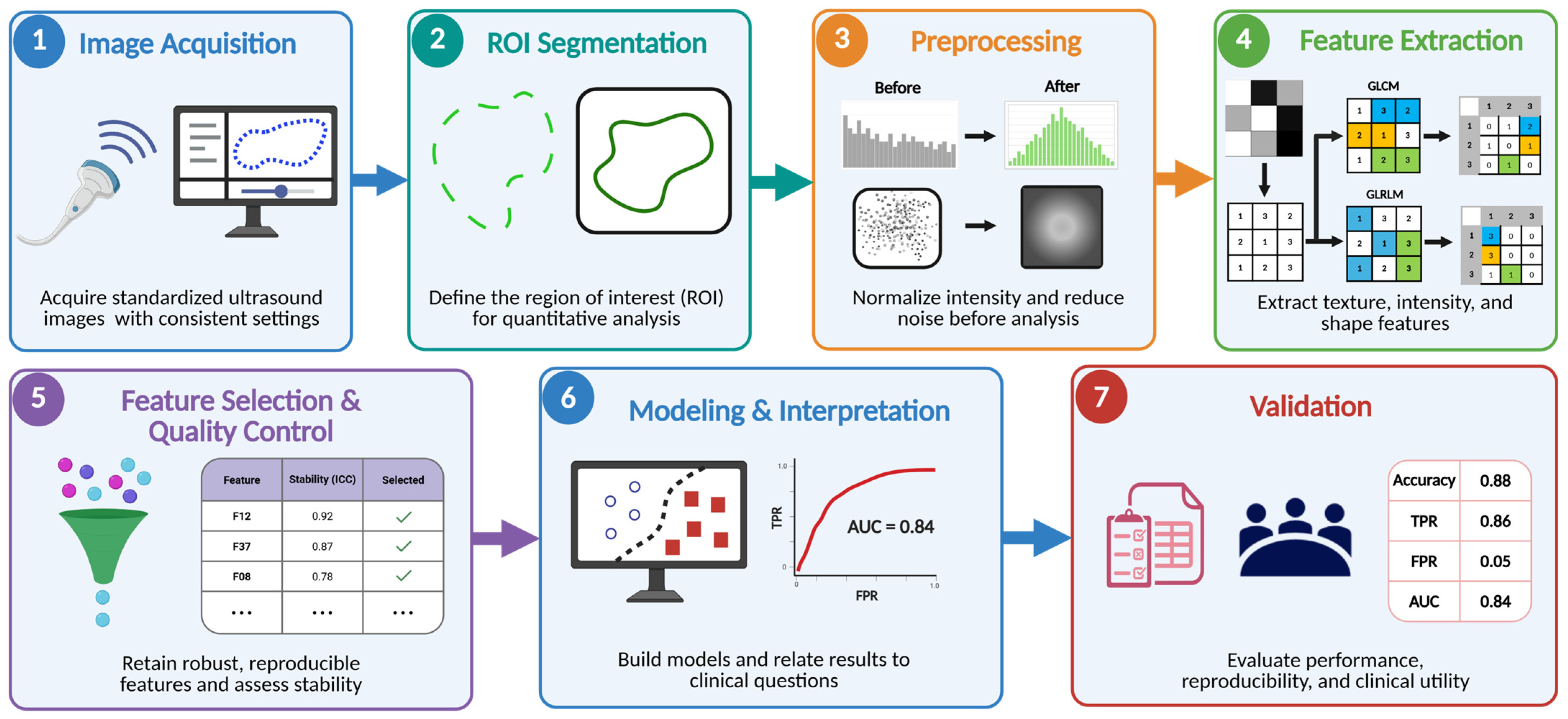
2.1. Radiomics Workflow in Ultrasound
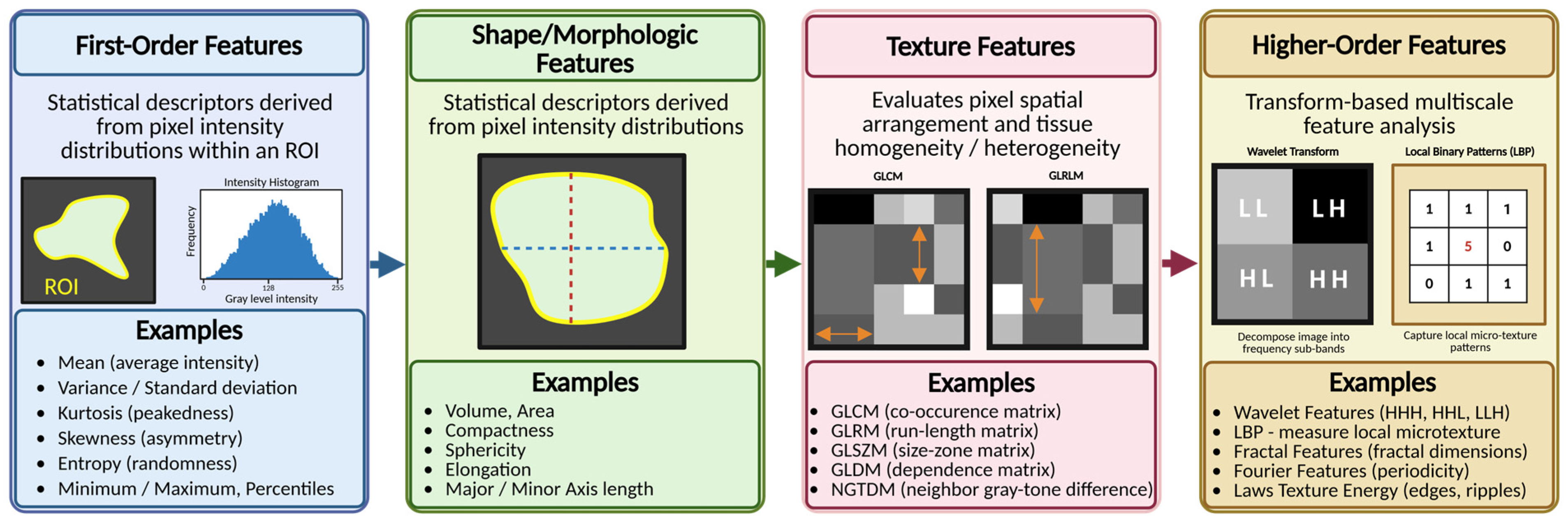
2.2. Radiomic Feature Categories
2.3. Ultrasound-Specific Considerations
3. Technical Challenges Unique to Ultrasound Radiomics
3.1. Acquisition-Related Variability
3.2. Preprocessing and Normalization
3.3. Radiomic Feature Reproducibility and Robustness
3.4. Speckle Noise and Image Artifacts
3.5. Segmentation Variability and Automated Segmentation
3.6. Model Calibration and Clinical Utility
3.7. Small Pediatric Cohorts and Limited Generalizability
3.8. Need for End-to-End Standardization
4. Review Design
4.1. Literature Search Strategy
4.2. Eligibility Criteria
- Studies were included if:
- The cohort included pediatric patients (<18 years);
- They applied grayscale US-based radiomic, texture, or quantitative feature extraction methodologies;
- They evaluated diagnostic, prognostic, predictive, or classification performance;
- They included original research data;
- They were published in English.
- Exclusively included adult (>18 years) subjects;
- Did not apply quantitative feature extraction based on grayscale US image information including other US modalities, e.g., Doppler parameters and advanced ultrasound techniques (e.g., elastography, CEUS) were not included or critically assessed.
- Were not clinical or translational research (review articles, editorials, case series or reports);
- Analyzed non-US imaging modalities (CT or MRI);
- Did not clearly define methodology employed for radiomic analysis (feature extraction or model development);
- Study Selection and Data Extraction
- Eligible studies were reviewed and categorized by primary clinical applications. This included:
- Brain imaging;
- Liver imaging;
- Renal Imaging;
- Oncologic Imaging;
- Emerging areas—including lung and musculoskeletal.
- The following variables were analyzed, when available, to enhance review uniformity:
- Study design, sample size, clinical application, and patient population;
- Ultrasound modality and acquisition parameters;
- Segmentation methodology and radiomic pipeline;
- Radiomic features extracted and models used;
- Study limitations including validation technique or image acquisition methods;
- Level of evidence (per Oxford Center for Evidence-Based Medicine guidelines) [59].
4.3. Evidence Synthesis
5. Current Clinical Applications of Ultrasound Radiomics in Pediatrics
5.1. Brain Imaging
5.1.1. Preterm White Matter Injury and Early Detection
5.1.2. Longitudinal Monitoring and Neurodevelopmental Risk
5.1.3. Expanding Applications Beyond Preterm Injury
5.2. Liver Imaging
5.2.1. Steatosis and Diffuse Liver Disease
5.2.2. Fibrosis and Pediatric-Specific Barriers
5.2.3. Critical Summary
5.3. Renal Imaging
5.3.1. Parenchymal Disease Characterization
5.3.2. Glomerulonephropathy and Histologic Risk Stratification
5.3.3. Hydronephrosis and Longitudinal Monitoring
5.3.4. Critical Summary
5.4. Oncologic Imaging
5.4.1. Tumor Characterization and Prognostic Stratification
5.4.2. Thyroid and Thoracic Applications
5.4.3. Critical Summary
5.5. Emerging Applications
5.5.1. Lung Ultrasound Radiomics
5.5.2. Musculoskeletal and Muscle Applications
5.5.3. Critical Summary

| Sample Size and Subject Age | Primary Disease/Purpose/Clinical Task | Segmentation Method/Radiomics Pipeline | Radiomic Features & Models Used/Validation (Model Algorithm, Logistic Regression, Training/Testing/Division) | Model Used/Performance | Limitations (Validation, Single Center, Sample Size, Study Design) | Evidence Level (Oxford Center for Evidence-Based Medicine) | |
|---|---|---|---|---|---|---|---|
| Brain | |||||||
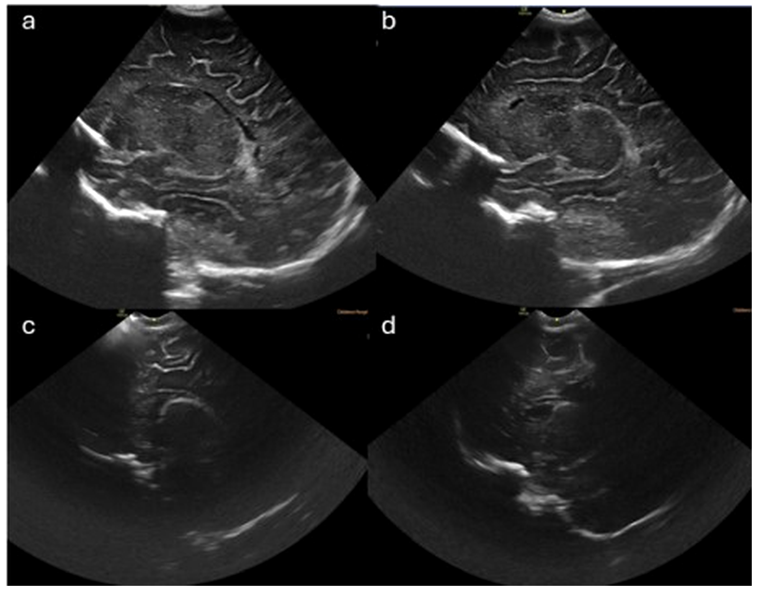
| (Narchi et al. 2013 [60]) | 20 preterm neonates (Mean GA 28.5 ± 1.9 weeks) | White Matter Injury (WMI)-Cystic Periventricular Leukomalacia (PVL) Purpose: Test whether texture analysis of PVE on initial cranial US can differentiate transient echogenicities (which resolve) from those that develop into cystic PVL | Manual ROI segmentation-Software: MaZda v4.5 (B11) Feature extraction–305 features per image | Co-occurrence matrix (COM), run-length matrix (RLM), and the gradient matrix (Gr) Top 10 features selected by Fisher (F)-coefficient | Classifier: Linear discriminant analysis (LDA) → two most discriminant features (MDF1, MDF2) Validation: Leave-one-out cross-validation | - Lack of correlation to PVL risk factors - Lack of long-term follow-up - No standardization of US settings - Small sample size - No multivariate analysis - Single center - Retrospective observational case-control study - Risk of over-fitting (over-conformity) due to small sample size relative to the dimensionality of radiomic feature space | Level 3b |
| (You et al. 2015 [61]) | 33 preterm neonates (Mean GA 28.45 ± 2.7 weeks) | WMI–Diffuse cystic or cavitary periventricular changes Purpose: Quantitatively analyze GLCM texture features on cranial sonography and correlate with WMI severity graded by MRI (normal vs. mild vs. severe) | Manual ROI segmentation Software ImageJ v1.44 and In-house C++ software (Medical Imaging Solution for Segmentation and Texture Analysis) MRI grading by 2 blinded board-certified pediatric radiologists 48 features per image | Angular second moment (ASM), inverse differential moment (IDM), contrast, and entropy Statistical analysis: Kruskal-Wallis test, post-hoc Mann-Whitney U (Bonferroni-corrected α = 0.017) ROC curve analysis for discrimination No ML model; statistical comparison with MRI-based severity grading | Custom GLCM pipeline | - Retrospective study - Single center - Different scanner settings - Interval between US and MRI - Small sample size - No image normalization/postprocessing - Could not discriminate mild WMI from normal - No neurodevelopmental outcome correlation - Risk of over-fitting (over-conformity) due to small sample size relative to the dimensionality of radiomic feature space | Level 3b |
| (Zhu et al. 2023 [62]) | 158 preterm neonates (GA < 37 weeks, age ≤ 7 days) | WMI-Cystic PVL Purpose: Develop an ultrasound radiomics diagnostic system combining traditional radiomics and multi-task deep learning (MTDL-Net) for automated WM segmentation and WMI risk prediction from cranial US images | Manual segmentation by radiologists (≥10 years experience) as gold standard ROIs Automatic segmentation: SDL-Net (U-Net) and MTDL-Net (Mask R-CNN) Software: PyTorch, MATLAB for preprocessing followed by feature extraction–350 features per image | Histogram statistics, GLCM, GLRLM- based features, gray-level size zone matrix (GLSZM) based features, neighboring gray tone difference matrix (NGTDM) based features, and four wavelet-transformed modes Feature selection: Sparse representation-based classification (SRC) → top 52 features | Classifier: SVM-based C-SVC Deep learning: MTDL-Net (Mask R-CNN) for simultaneous segmentation + classification Fusion: deep learning features + manual radiomics features Validation: 70:30 train-test split; 5-fold cross-validation for MTDL-Net (avg AUC 0.843) | Retrospective study Single center Small sample size with class imbalance (32 WMI vs. 126 normal) No external validation Static image-based analysis (not video/cine) PVL not separately categorized Limited to anterior fontanelle coronal plane No neurodevelopmental outcome correlation - Risk of over-fitting (over-conformity) due to small sample size relative to the dimensionality of radiomic feature space | Level 3b |
| (Zhu et al. 2024 [63]) | 267 premature infants (GA 26–36 weeks, mean GA 32.55 ± 2.55 weeks) | White Matter Injury (WMI) Purpose: Develop and evaluate a multiplanar radiomics model from CUS to predict WMI, dynamically monitor WMI recovery at 2 and 4 weeks, and explore correlation with neurodevelopment | Manual ROI segmentation followed by voxel intensity fixed at 25, voxel size resampled 1 × 1 mm, then feature extraction via Pyradiomics 3.0.1 software | Shape, first-order, GLCM, GLRLM, GLSZM, GLDM, NGTDM Feature selection: Spearman correlation + LASSO with 5-fold cross-validation → 58 features | Least absolute shrinkage and selection operator (LASSO) algorithm | Retrospective study Small sample size Limited vendor/probe types (2 machines) No independent external validation Does not classify WMI severity (mild/moderate/severe) Manual ROI segmentation (inefficient) Low sensitivity for microcystic (<1mm) and non-cystic diffuse WMI Single center - Risk of over-fitting (over-conformity) due to small sample size relative to the dimensionality of radiomic feature space | Level 3b |
| (Jung et al. 2019 [64]) | N = 20 very preterm infants (GA 25–33 weeks) | WMI-PVL Purpose: Compare texture parameters of serial CUS images between PVL and normal PVE groups; evaluate early predictive value of texture analysis for PVL within 2–3 weeks of life | Manual ROI segmentation: Software: MaZda v4.5 (Technical University of Lodz) Normalization: histogram remapping within ±3 SD of mean Blinded neuroradiologist (4 years CUS experience) | Features: 308 texture features + variance-to-mean ratio (VMR); first-order histogram (variance, Perc.99%, MaxNorm), co-occurrence matrix, run-length, gradient features Statistical tests: Wilcoxon signed-rank, Mann-Whitney U ROC analysis for discrimination No ML model; statistical comparison approach No train/test split | Key metric: R21 of VMR (ratio of 2nd to 1st CUS values) | Retrospective study design Very small sample size Single center PVL diagnosed by CUS or MRI (not all had MRI)—possible false negatives in normal PVE group PVL severity not stratified Scanner settings not standardized (though same machine/operator used) - Risk of over-fitting (over-conformity) due to small sample size relative to the dimensionality of radiomic feature space | Level 3b |
| (Laccetta et al. 2023 [65]) | N = 46 preterm infants (GA < 32 weeks) | Non-cystic WMI Purpose: Evaluate quantitative CUS echogenicity of periventricular WM (pixel brightness intensity) as a predictor of middle-term neurodevelopment (Bayley-III at 12 mo CA) | Manual segmentation: Software: QLAB13 (Philips, Amsterdam, The Netherlands) Metric: Relative echogenicity (RECP) = mPBI_WMmax/mPBI_CPmax | Feature: Pixel brightness intensity (PBI)— first-order intensity ratio (RECP) Statistical models: Pearson correlation, multivariate linear regression with covariates (GA, arterial pH, PN duration, hospital stay, ROP, hsPDA) No train/test split (single cohort analysis) | No ML classifier; correlational/regression approach | Small sample size Single center Bayley-III at 12 months may be early for cognitive/language assessment Operators aware of clinical history (though blinded to study aims) Acoustic window changes with age No MRI correlation - Risk of over-fitting (over-conformity) due to small sample size relative to the dimensionality of radiomic feature space | Level 2b |
| (Zimina et al. 2026 [66]) | N = 89 full-term newborns (GA > 37 weeks) | Diabetic fetopathy in newborns born to mothers with gestational diabetes mellitus Purpose: Investigate feasibility of radiomic analysis of brain US images to detect brain changes associated with diabetic fetopathy not visible on standard neurosonography | Manual segmentation Software: PyRadiomics (Python 3.9, Visual Studio 2022) 1395 features extracted per image | Feature classes: first-order, shape, GLCM, GLRLM, GLSZM, GLDM, NGTDM Feature selection: Shapiro-Wilk → t-test/Mann-Whitney U.; Spearman correlation filtering (|r| < 0.7) Validation: 80/20 train-test split with stratification; repeated stratified 5-fold cross-validation StandardScaler normalization | Model: Decision tree classifiers (scikit-learn) with automatic hyperparameter tuning | Small sample size Single center Retrospective design No external validation cohort No neurodevelopmental outcome correlation Overfitting concern (Model 4 test AUC 0.85 vs. CV AUC 0.65) Color-to-grayscale conversion may introduce artifacts - Risk of over-fitting (over-conformity) due to small sample size relative to the dimensionality of radiomic feature space | Level 3b |
| (Sultan et al. 2025 [67]) | N = 33 full-term newborns (≥37 weeks GA) | HIV-exposed uninfected (HEU) vs. HIV-unexposed (HU) full-term newborns Purpose: Explore brain ultrasound radiomics (texture analysis) as early neurodevelopmental biomarker comparing by in utero HIV exposure status | Manual ROI segmentation Software: MaZda v4.6 (Technical University of Lodz) Blinded expert (10 years experience) reviewed and excluded artifact-affected images | Features: 12 features including; First-order statistics (heterogeneity), GLCM (entropy, correlation), run-length matrix (RLNonUni, GLevNonU) Validation: ROC analysis with AUC, sensitivity, specificity No formal train/test split described; single cohort analysis | Model: Logistic regression combining 5 features | Small sample size Single site Cross-sectional (no longitudinal neurodevelopmental follow-up) Homogeneous ART exposure (all dolutegravir-based) Images acquired by study nurses (point-of-care US) 14 of 47 studies excluded for image quality No external validation - Risk of over-fitting (over-conformity) due to small sample size relative to the dimensionality of radiomic feature space | Level 2b |
| Liver | |||||||
| (Das et al. 2021 [71]) | 181 pediatric participants in total. Model development dataset contained 132 subjects: 93 normal subjects (484 ROIs) and 39 NAFLD subjects (260 ROIs) used to develop the ML model Age: 6–18 years old | Pediatric Non-alcoholic fatty liver disease (NAFLD) Purpose: Develop a machine learning (ML) based classification model capable of identifying NAFLD from healthy liver tissue using ultrasound texture analysis. | Manual ROI segmentation of 25 × 25 pixel rectangles per image. Texture extraction was performed with ImageJ and MaZda software. | 28 texture features including histogram, co-occurrence matrix (GLCM), run-length matrix, gradient, autoregressive, and Haar wavelet features | Best models: Support Vector Machine, multi-layered perceptron neural net, and extreme gradient boost Testing AUROC 0.95 (95% CI 0.93–0.97). Internal validation AUROC 0.969. External validation AUROC 0.92 (95% CI 0.91–0.94). Outperformed HRI-only (AUROC 0.81), HEAI-only (0.75), and combined HRI + HEAI (0.82). | - Single-center study Relatively small number of NAFLD - Retrospective secondary analysis of a prospective cohort - Manual ROI placement | Level 3b |
| Renal | |||||||
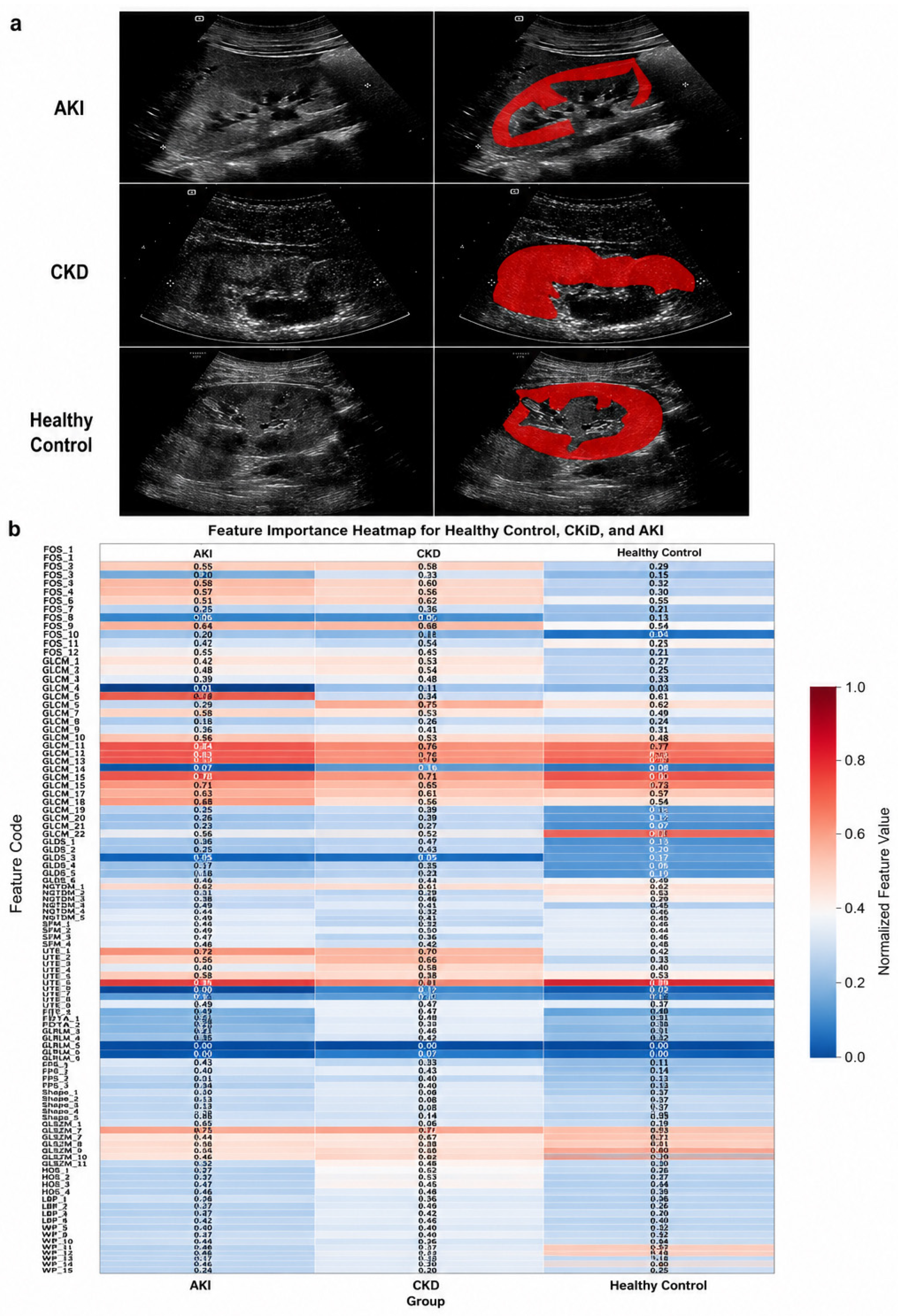
| (De Leon-Benedetti et al. 2025 [25]) | 31 pediatric subjects (60 kidney units): 8 AKI (median age 3.5 years [IQR: 0–11.5]); 14 CKD (median 3.5 years [IQR: 0–6.8]); 9 healthy controls (median 15.5 years [IQR: 12.8–21]) | To determine whether grayscale US radiomics can differentiate AKI vs. CKD vs. healthy kidneys. Purpose: can US radiomics quantify ‘medical renal disease’ beyond subjective echogenicity | Manual ROI segmentation of renal parenchyma on sagittal long-axis B-mode US images by trained researchers with pediatric nephrologist and radiologist review Software: MaZda v4.6 (Technical University of Lodz) Gray level normalization before feature extraction, within each ROI, to rescale pixel intensities to standardized range of 0–255 | Feature extraction (124 total) using PyFeats v1.0.11: GLCM, GLDS, NGTDM, SFM, LTE, FDTA, GLRLM, FPS, GLSZM, HOS, LBP and wavelet packet decomposition Model: principal component analysis–top 10 features selected for modeling. Features used to evaluate 4 ML models: random forest, SVM with radial basis function kernel, logistic regression, and XGBoost Validation: 5-fold cross validation only | Best model: XGBoost, accuracy 0.90, macro F1 0.90 Other models: SVM accuracy 0.90, macro F1 0.88; random forest accuracy 0.88, macro F1 0.89; logistic regression accuracy 0.88, macro F1 0.88 | Retrospective pilot study, very small cohort, single-center, no external validation, age not matched between controls and disease groups; heterogeneous US machines, transduces and settings; manual segmentation Risk of over-fitting (over-conformity) due to small sample size relative to the dimensionality of radiomic feature space | Level 3b |
| (Kou et al. 2024 [74]) | 313 biopsy confirmed pediatric glomerulonephropathy cases: 127 IgA nephropathy, 83 minimal change disease, 103 henoch-schonlein purpura nephritis 469 renal US images used total Age not specified, but pediatric cases | To noninvasively differentiate biopsy-confirmed pediatric GN subtypes (IgAN vs. MCD vs. HSPN) | Manual ROI segmentation of renal parenchymal trasnverse B-mode US images acquired immediately prior to biopsy using LabelMe cropped to standardized 512 × 512 pixels. Then a U-Net segmentation model was trained on manually segmented images. | 1422 features extracted with PyRadiomics: first order features, shape, GLCM, GLSZM, GLRLM, NGTDM and GLDM. Random 8:2 split into training and validation sets. ANOVA, LASSO regression and k-fold cross-validation to determine optimal subset of features. Then RF classification model used to evaluate selected features. No external validation | Final classification model: Random Forest with 37 selected radiomic features Validation AUCs: IgAN (0.94); MCD (0.91); HSPN (0.98) | Single-center study; only 3 subtypes of GN included with no healthy controls; no external validation; age/sex/clinical characteristics not reported; possible data leakage if image-level not patient-level splitting occured Risk of overfitting (over-conformity) due to 1422 initial features (large original feature space) extracted relative to only 313 cases All images acquired on one US system with same probe/settings improves standardization but limits results generalizability | Level 3b |
| (Chen et al. 2024 [75]) | 440 biopsy-confirmed HSPN patients. Grouped (n = 100) ISKDC I-II (no cresecents) vs. (n = 340) ISKDC III-V (crescentic) Training cohort 308, validation 132 (7:3 split). Median training cohort age 9 years (IQR: 6–11), validation cohort median age 8 years (IQR: 5–11) | HSPN classification using renal US radiomics Purpose: to predict crescentic vs. non-crescentic using biopsy proven ISKDC grading through a noninvasive surrogate | Manual ROI segmentation of renal cortex and medulla (excluding collecting system/hilum) using largest long-axis US image of right kidney in 3D slicer. Segmentations performed by a sonographer and reviewed by a second sonographer. No automated segmentation or deep learning pipeline | 105 radiomic features extracted with Pyradiomics: first-order, shape, GLCM, GLSZM, GLRLM, NGTDM and GLDM Feature selection with spearman correlation filtering (if > 0.9, feature removed) then LASSO regression with 5-fold cross-validation: 14 features selected 14 features (3 First order, 4 shape [2D], 7 texture) used to build 3 ML models: logistic regression, k-nearest neighbor, and SVM Internal validation using the n = 132 validation cohort | Best performing model: SVM-Training AUC 0.910, validation AUC 0.870 (95% CI 0.795–0.944). Validation sensitivity 0.706, specificity 0.950, accuracy 0.761, F1 score 0.821 KNN validation AUC 0.810 (95% CI 0.712–0.909) LR validation AUC 0.751 (95% CI 0.625–0.878) | Retrospective, single-center, internal validation only, manual segmentation, feature reproducibility/interobserver ICCs not reported | Level 3b |
| (Sloan et al. 2023 [76]) | 592 US images 90 patients (ages 0–8 years) with clinical diagnosis of hydronephrosis based on Society for Fetal Urology (SFU) grading system: categorized to low-grade (SFU I-II) and high-grade (SFU III-IV). 74 high-grade kidneys (145 images) and 227 low-grade kidneys (447 images) were included | Pediatric hydronephrosis severity classification based on US Purpose: To develop a ML algorithm based on radiomic texture features to differentiate SFU-defined low-grade from high-grade pediatric hydronephrosis | Each kidney manually outlined by urology resident with pediatric urologist expert review (5% required re-contouring) No automated segmentation | 25 chosen radiomic features extracted: 14 GLCM-derived (contrast, entropy, homogeneity, IMC1/2, correlation etc.), grayscale statistics, and morphologic features (circularity, compactness, effective diameter, depth-to-width ratio) 5-fold cross-validation (constant low-grade to high-grade ratio in each fold & patients/kidneys kept within folds to prevent data leakage). Stepwise linear discriminant using Wilks’ lambda as the feature selection criterion during training folds. Linear SVM used as final classifier | SVM performance analyzed by AUC of ROC curve output. Mann-Kendall test used to determine if positive correlation between SVM output and hydronephrosis grade. By kidney unit, AUC 0.86 (95% CI 0.81–0.92), sensitivity 75.7%, specificity 86.3%, accuracy 83.7%, PPV 64.4%, NPV 91.6%, F1-score 69.6%. Significant positive trend between model ouput and increasing SFU grade (p < 0.001) | Retrospective, single-center pilot study; relatively small patient cohort; excluded many structural abnormalities; manual segmentation may introduce observer variability; possible image-selection bias; standardization of image acquisition unclear from text (“main ultrasound system”) Lower risk of over-fitting (over-conformity) as only 25 features extracted, reduced data leakage (patient kept within a validation fold), and cross-validation by kidney (not image) | Level 3b |
| Oncology | |||||||
| (Zhu et al. 2025 [78]) | 73 pediatric patients in total. 25 cases of ganglioneuroma (GN), 12 cases of ganglioneuroblastoma intermixed (GNBi), 2 cases of ganglioneuroblastoma nodular (GNBn), and 34 cases of neuroblastoma (NB). Mean age 45.5 ± 39.8 months (range 0.4–180 months) | Peripheral neuroblastic tumors Purpose: Construct and select a better model for prognostic subsets of pediatric neuroblastic tumors using US. | Images of the largest sections of tumors were chosen for feature extraction. Radiomics feature extraction using Pyradiomics. | 1674 radiomics features were extracted. 324 first-order statistical features and 1350 textural features including GLCM, GLDM, GLRLM, GLSZM and NGTDM. Filters applied include Laplacian of Gaussian (LOG), wavelet with transform, square, square root, logarithm, exponential and gradient. LASSO regression was used for combined models. | Best model: Radiomics model surpassed the combined model at differentiating prognostic subsets of pNTs (AUC 0.918), despite the combined model having the highest AUC (0.941) with p < 0.05 Radiomics models used included RF, KNN, LR, SVM, and xgboost. | - retrospective data - limited sample size - Mono-center cohort study - imbalance between favorable and unfavorable histology groups | Level 3b |
| (Wei et al. 2026 [80]) | 609 patients were included; 422 diagnosed with malignant conditions, and 187 with benign tumors. 487 cases were used for training, and 122 cases were used for validation. Mean age was not specified. | Subpleural pulmonary lesions (SPLs). Purpose: develop a clinical deep learning model (CDLR) for differential diagnosis of benign and malignant SPLs using US. | Images were exported in JPG format and uniformly converted to NIfTI format using SimpleITK library before being imported into Insight Segmentation and Registration Toolkit SNAP (ITK-SNAP) software. Manual segmentation and ROI delineation. Radiomics and deep transfer learning (DTL) features were extracted using Pyradiomics. | 1561 radiomics features were extracted, including include shape, first-order, and texture features. 128 deep transfer learning features were identified. | DTL and DLR models demonstrated superior value to the RAD model. Clinical, RAD and DTL features were integrated through a SVM algorithm model to construct the CDLR model. | - retrospective cohort study - single center study - only grayscale ultrasound was used - use of JPG rather than DICOM images - class imbalance between malignant and benign lesions | Level 3b |
| (Li et al. 2023 [79]) | 164 pediatric patients in total; 103 with pathologically identified ETE, and 61 non-ETE. 115 were used for training, and 49 were used for validation. Mean age was 14.60 ± 3.52 years. | Papillary thyroid carcinoma Purpose: explore extrathyroidal extension (ETE) in children and adolescents. | ROI delineation was performed layer by layer along the edge of the tumor contour using ITK-SNAP software. ETE diagnosis according to AKCC standards. | 1421 image features were extracted using Pyradiomics. Features were divided into 4 categories: shape, first-order statistics, texture, and higher-order statistical features. 217 features with correlation coefficient >0.90. 16 radiomics features were chosen using LASSO. | Best model: radiomics-random forest (RF) model with AUC of 0.999 in the training set, and LightGBM model with AUC of 0.832. Models used included KNN, SVM, RF, and LightGBM. | - retrospective cohort study - single center cohort - small sample size - Imbalance between ETE and non-ETE cases - Did not compare dimensionality reduction algorithms - tumor boundaries were ill-defined in some instances - Only greyscale ultrasound images were used | Level 3b |
| Emerging Areas | |||||||
| (Lin et al. 2024 [81]) | 150 consecutive cases of neonatal lung disease in one NICU: NRDS (n = 60), neonatal pneumonia (n = 30), MAS (n = 30), TTN (n = 30) 8:2 split --> Training cohort (n = 120), median age 263 days (IQR 216–277); validation cohort (n = 30), median age 264 days (IQR 228–277) | In patients with neonatal respiratory distress syndrome (NRDS), investigation of lung ultrasound radiomics Purpose: to develop an operator-independent US radiomics model to improve diagnostic consistency in neonatal lung US for NRDS | All US images acquired on one US system (GE LOGIQ P6) with standardized probe (9–12 MHz linear) and settings Lesion ROIs manually segmented by two senior physicians specializing in neonatal lung US in ITK-SNAP 3.8.0 Two representative images saved for each patient in DICOM format | 107 image features extracted using PyRadiomics including GLCM (n = 24), first-order (n = 18), GLRLM (n = 16), GLSZM (n = 16), GLDM (n = 14), shape (n = 14), NGTDM (n = 5). Multiple features combined if spearman correlation > 0.9, greedy recursive strategy used to filter irrelevant features. Remaining figures underwent LASSO regression with 10-fold cross-validation Final model used 22 non-zero features | ML models, RF, SVM, MLO, KNN, and logistic regression employed with validation by a temporally separate internal cohort Best performing model: Random Forest. Validation cohort AUC 0.951, sensitivity 95,83%, specificity 94.44%, accuracy 95.0%, PPV 92.0%, NPV 97.14%. RF model performance was statistically comparable with experienced physicians (AUC 0.99 vs. 0.98) and significantly better than junior physicians (AUC 0.85) | Single-center retrospective study; relatively small cohort; no external validation; single US system standardized image acquisition; manual ROI segmentation introduces observer variability; potential for model dependence on institution-specific acquisition settings; possible image-level data leakage concerns [2 images per patient’ not fully clarified in methods Risk of over-fitting (over-conformity): small dataset relative to classification complexity; due to single-vendor acquisition—study generalizability limited; possible image-selection bias for model training has potential to make ML model classification easier than in real-world with greater clinical variability | Level 3b |
| (Cai et al. 2026 [82]) | 301 patients were included. 210 were used for training, and 91 were used for testing. Age range was 1–18 years old. | Acute heart failure (AHF) Purpose: develop an integrated machine learning model based on lung US radiomics and clinical data to diganogs AHF in patients with acute dyspnea. | Standardized 6-zone lung US images were used. Images in DICOM format were imported to 3D Slicer software. ROI delineation was manually performed. | 159 features were used in total: 107 radiomics features were extracted using Pyradiomics and combined with 52 clinical features. Features included shape, first-order, and texture features such as GLCM, GLRLM, GLSZM, GLDM and NGTDM. Synthetic Minority Oversampling Technique (SMOTE) was applied to account for the mild class imbalance. | Best model: the integrated RF model achieved optimal performance with an AUC of 0.976 (95% CI: 0.950–0.994), particularly GLRLM texture features. 3 RF models were developed: clinical-only, radiomics-only, and an integrated model. | - retrospective study - single-center cohort study - feature-to-event ratio was 1:0.93, which is below the recommended 10:1, posing a risk of overfitting - experts were blinded to radiomics features, leading to potential artificially inflated performance of the model. - model was not compared to physicians’ diagnostic performance - model used 6-zone protocol rather than the internationally recommended 8- or 12-zone standard protocol. | Level 3b |
| (Mohamed et al. 2026 [83]) | 10 pediatric cases in total. 5 cases with pneumonia findings, and 5 cases with normal findings. Age range was 3–32 months. | Pediatric Pneumonia-related US findings Purpose: evaluate semi-automated, AI-assisted system at identifying clinically relevant lung abnormalities on US. | Automated segmentation algorithm developed by the authors that targets key pathological features such as consolidation, pleural line abnormalities, and B-lines via region growing, selecting pixels with similar greyscale intensity values (±10 gray levels). Minor manual correction was performed. | Quantitative features extracted include tissue thickness, margin irregularity, and variation in echogenicity. Custom-developed software in IDL based on computerized analysis of echogenicity variation, margin irregularity, and structural continuity. | Accuracy in detecting consolidation, B-lines and pleural lines. | - small sample size - validation consists of simple visual confirmation | Level 3b |
| (Wijntjes et al. 2022) [86]) | Predominantly adult but pediatric subset included 994 patients: pediatric subgroup limited to <5 years (n = 50), 5% & 6–18 years (n = 112), 11.3% | In patients with neuromuscular disease (myopathies, neurogenic, NMJ disorders, and non-muscle NMDs), comparison of visual muscle ultrasound assessment (Heckmatt grading) and quantitative muscle ultrasound echogenicity z-score analysis Purpose: to determine the correlation between qualitative and QMUS | No traditional radiomic feature extraction. Manually segmented ROIs representing maximal muscle area with computed echogenicity quantification scores | No ML classifier or advanced radiomic texture pipeline was developed | Not Applicable | Not applicable | |
| (Shklyar et al. (2015) [84]) | 25 boys with Duchenne muscular dystrophy (DMD) and 25 healthy controls Age range: 2–14 years | Duchenne muscular dystrophy Purpose: Compare grayscale level (GSL) and quantitative backscatter analysis (QBA) for differentiating DMD muscle from healthy muscle and examine relationships with age and functional status. | No conventional radiomics pipeline. Quantitative muscle US measurements were obtained from 6 unilateral muscles. GSL derived from standard grayscale US images; QBA derived from raw backscatter signal data requiring specialized processing. | Quantitative US intensity/scattering measures: GSL and QBA. Reliability assessed with intraclass correlation coefficients (ICCs). Associations evaluated with age and functional status (North Star Ambulatory Assessment). No ML classifier, train/test split, or radiomics feature-selection pipeline. | Both GSL and QBA were highly reliable (ICC > = 0.87) and differentiated DMD from controls. Superficial muscle measurements increased with age and worsening functional status, suggesting that anatomic sampling strategy affects diagnostic sensitivity. GSL may be more feasible for routine use because it can be extracted from standard US images. | Small single-center case-control cohort; not a conventional grayscale radiomics study; QBA requires raw signal data and specialized processing; no external validation; no ML model; potential dependence on acquisition settings and muscle depth/sampling location. | Level 3b |
| (Chuang et al. 2025 [87]) | 47 boys with Duchenne muscular dystrophy | Duchenne muscular dystrophy Purpose: Introduce quantitative US-based scatteromics to distinguish early vs. late ambulatory decline in DMD using gastrocnemius ultrasound envelope-statistics parametric imaging. | Not conventional grayscale radiomics. Gastrocnemius ultrasound envelope-statistics parametric maps were generated. Maps were based on Nakagami, homodyned K, and entropy parameters; first-order features were extracted from parametric images. | Scatteromics features derived from Nakagami, homodyned K, and entropy parametric maps. Classifiers: support vector machine (SVM), random forest (RF), and linear discriminant analysis (LDA). Validation approach not fully detailed in the review table source text. | Full scatteromics models achieved high average AUROCs: SVM 0.97, RF 0.98, and LDA 0.83. Findings support ultrasound-derived scattering/texture features as objective biomarkers of pediatric muscle disease severity. | Small cohort; disease-specific DMD population; not a conventional grayscale radiomics workflow; requires quantitative US envelope-statistics processing and parametric map generation; external validation and broader multicenter testing are needed; potential risk of over-fitting relative to sample size and feature/model complexity. | Level 3b |
| (Hao et al. 2025 [88]) | 125 infants total: 59 with developmental dysplasia of the hip (DDH) and 66 healthy controls | Developmental dysplasia of the hip Purpose: Evaluate whether hip US radiomics can identify microstructural femoral head changes in DDH and provide a quantitative adjunct to conventional Graf-based US assessment. | Femoral head ROIs manually segmented on hip US images using 3D Slicer, excluding the ossification center. Retrospective study design. | 92 radiomic features extracted, including first-order, GLCM, gray-level dependence matrix (GLDM), and gray-level size zone matrix (GLSZM) features. Feature selection: maximum relevance minimum redundancy (mRMR) and LASSO regression. Final model: 9 selected radiomic features. | 69 features differed significantly between DDH and controls. Nine-feature radiomics model achieved validation AUC 0.91. Suggests US radiomics may capture subtle femoral head tissue alterations in DDH. | Retrospective study; modest sample size; likely single-center cohort; manual segmentation; no multicenter external validation described; model requires prospective validation and assessment of reproducibility across scanners/operators; possible risk of over-fitting given initial feature number relative to cohort size. | Level 3b |
6. Future Directions Toward Clinical Translation and Real-World Implementation
6.1. Standardization as the Foundation for Clinical Use
6.2. Multicenter and Multi-Vendor Validation
6.3. Integration into the Ultrasound Workflow
6.4. Clinically Actionable Decision Support
6.5. Application-Specific Implementation Pathways
6.6. AI-Enabled Ultrasound Radiomics Platforms
6.7. Regulatory Approval, Reporting, and Clinical Adoption
Closing Perspective
7. Conclusions
Author Contributions
Funding
Institutional Review Board Statement
Informed Consent Statement
Data Availability Statement
Conflicts of Interest
References
- Hwang, M.; Piskunowicz, M.; Darge, K. Advanced ultrasound techniques for pediatric imaging. Pediatrics 2019, 143, e20182609. [Google Scholar] [CrossRef] [Scilit]
- Thukral, B.B. Problems and preferences in pediatric imaging. Indian. J. Radiol. Imaging 2015, 25, 359–364. [Google Scholar] [CrossRef] [Scilit] [PubMed]
- World Health Organization. Communicating Radiation Risks in Paediatric Imaging: Information to Support Health Care Discussions About Benefit and Risk; World Health Organization: Geneva, Switzerland, 2016. [Google Scholar]
- U.S. Food and Drug Administration. Ultrasound Imaging; U.S. Food and Drug Administration: Silver Spring, MD, USA, 2024.
- de Borja, C.; Watkins, R.; Woolridge, T. Common ultrasound applications for pediatric musculoskeletal conditions. Curr. Rev. Musculoskelet. Med. 2022, 15, 447–455. [Google Scholar] [CrossRef] [Scilit]
- Frush, D.P.; Callahan, M.J.; Coley, B.D.; Nadel, H.R.; Paul Guillerman, R. Comparison of the different imaging modalities used to image pediatric oncology patients: A COG diagnostic imaging committee/SPR oncology committee white paper. Pediatr. Blood Cancer 2023, 70, e30298. [Google Scholar] [CrossRef] [Scilit]
- Gamboa, H.E.; Molle-Rios, Z.; Anupindi, S.A. Underutilization of Bowel Ultrasound in North America in Children with Inflammatory Bowel Disease. Dig. Dis. 2020, 38, 390–397. [Google Scholar] [CrossRef] [Scilit] [PubMed]
- Kuban, K.; Adler, I.; Allred, E.N.; Batton, D.; Bezinque, S.; Betz, B.W.; Cavenagh, E.; Durfee, S.; Ecklund, K.; Feinstein, K.; et al. Observer variability assessing US scans of the preterm brain: The ELGAN study. Pediatr. Radiol. 2007, 37, 1201–1208. [Google Scholar] [CrossRef] [Scilit]
- Moran, C.M.; Thomson, A.J.W. Preclinical ultrasound imaging—A review of techniques and imaging applications. Front. Phys. 2020, 8, 124. [Google Scholar] [CrossRef] [Scilit]
- Rix, A.; Lederle, W.; Theek, B.; Lammers, T.; Moonen, C.; Schmitz, G.; Kiessling, F. Advanced ultrasound technologies for diagnosis and therapy. J. Nucl. Med. 2018, 59, 740–746. [Google Scholar] [CrossRef] [Scilit]
- Yu, L.; Che, M.; Wu, X.; Luo, H. Research on ultrasound-based radiomics: A bibliometric analysis. Quant. Imaging Med. Surg. 2024, 14, 4520–4539. [Google Scholar] [CrossRef] [Scilit] [PubMed]
- Cloutier, G.; Destrempes, F.; Yu, F.; Tang, A. Quantitative ultrasound imaging of soft biological tissues: A primer for radiologists and medical physicists. Insights Imaging 2021, 12, 127. [Google Scholar] [CrossRef] [Scilit]
- Sun, F.-Y.; Meng, D.-L.; Liu, L.; Cao, X.-Q.; Fu, L.; Meng, L.; Cui, X.-W.; Pan, X.-F. Ultrasound-based radiomics for the evaluation of breast cancer. Front. Oncol. 2025, 15, 1710405. [Google Scholar] [CrossRef] [Scilit] [PubMed]
- Raunig, D.L.; McShane, L.M.; Pennello, G.; Gatsonis, C.; Carson, P.L.; Voyvodic, J.T.; Wahl, R.L.; Kurland, B.F.; Schwarz, A.J.; Gönen, M.; et al. Quantitative imaging biomarkers: A review of statistical methods for technical performance assessment. Stat. Methods Med. Res. 2015, 24, 27–67. [Google Scholar] [CrossRef] [Scilit]
- Avanzo, M.; Wei, L.; Stancanello, J.; Vallières, M.; Rao, A.; Morin, O.; Mattonen, S.A.; El Naqa, I. Machine and deep learning methods for radiomics. Med. Phys. 2020, 47, e185–202. [Google Scholar] [CrossRef] [Scilit]
- Christensen, A.M.; Rosado-Mendez, I.M.; Hall, T.J. A Systematized Review of Quantitative Ultrasound Based on First-Order Speckle Statistics. IEEE Trans. Ultrason. Ferroelectr. Freq. Control 2024, 71, 872–886. [Google Scholar] [CrossRef] [Scilit]
- Oelze, M.L.; Mamou, J. Review of quantitative ultrasound: Envelope statistics and backscatter coefficient imaging and contributions to diagnostic ultrasound. IEEE Trans. Ultrason. Ferroelectr. Freq. Control 2016, 63, 336–351. [Google Scholar] [CrossRef] [Scilit]
- Theek, B.; Opacic, T.; Magnuska, Z.; Lammers, T.; Kiessling, F. Radiomic analysis of contrast-enhanced ultrasound data. Sci. Rep. 2018, 8, 11359. [Google Scholar] [CrossRef] [Scilit] [PubMed]
- van Timmeren, J.E.; Cester, D.; Tanadini-Lang, S.; Alkadhi, H.; Baessler, B. Radiomics in medical imaging-“how-to” guide and critical reflection. Insights Imaging 2020, 11, 91. [Google Scholar] [CrossRef] [Scilit] [PubMed]
- McCague, C.; Ramlee, S.; Reinius, M.; Selby, I.; Hulse, D.; Piyatissa, P.; Bura, V.; Crispin-Ortuzar, M.; Sala, E.; Woitek, R. Introduction to radiomics for a clinical audience. Clin. Radiol. 2023, 78, 83–98. [Google Scholar] [CrossRef] [Scilit]
- Varghese, B.A.; Cen, S.Y.; Hwang, D.H.; Duddalwar, V.A. Texture analysis of imaging: What radiologists need to know. AJR Am. J. Roentgenol. 2019, 212, 520–528. [Google Scholar] [CrossRef] [Scilit]
- Zwanenburg, A.; Vallières, M.; Abdalah, M.A.; Aerts, H.J.W.L.; Andrearczyk, V.; Apte, A.; Ashrafinia, S.; Bakas, S.; Beukinga, R.J.; Boellaard, R.; et al. The Image Biomarker Standardization Initiative: Standardized Quantitative Radiomics for High-Throughput Image-based Phenotyping. Radiology 2020, 295, 328–338. [Google Scholar] [CrossRef] [Scilit]
- van Griethuysen, J.J.M.; Fedorov, A.; Parmar, C.; Hosny, A.; Aucoin, N.; Narayan, V.; Beets-Tan, R.G.H.; Fillion-Robin, J.-C.; Pieper, S.; Aerts, H.J.W.L. Computational radiomics system to decode the radiographic phenotype. Cancer Res. 2017, 77, e104–e107. [Google Scholar] [CrossRef] [Scilit] [PubMed]
- Avery, E.; Sanelli, P.C.; Aboian, M.; Payabvash, S. Radiomics: A primer on processing workflow and analysis. Semin. Ultrasound CT MRI 2022, 43, 142–146. [Google Scholar] [CrossRef] [Scilit] [PubMed]
- De Leon-Benedetti, L.; Sultan, L.R.; Otero, H.J.; Morales-Tisnés, T.; Sims, J.; Fitzpatrick, K.; Fitzgerald, J.C.; Furth, S.; Laskin, B.L.; Viteri, B. Quantifying “medical renal disease”: A pediatric pilot study using ultrasound radiomics for differentiating acute kidney injury and chronic kidney disease. Diagnostics 2025, 15, 2112. [Google Scholar] [CrossRef] [Scilit]
- Al-Hasani, M.; Sultan, L.R.; Sagreiya, H.; Cary, T.W.; Karmacharya, M.B.; Sehgal, C.M. Ultrasound Radiomics for the Detection of Early-Stage Liver Fibrosis. Diagnostics 2022, 12, 2737. [Google Scholar] [CrossRef] [Scilit] [PubMed]
- Whybra, P.; Zwanenburg, A.; Andrearczyk, V.; Schaer, R.; Apte, A.P.; Ayotte, A.; Baheti, B.; Bakas, S.; Bettinelli, A.; Boellaard, R.; et al. The image biomarker standardization initiative: Standardized convolutional filters for reproducible radiomics and enhanced clinical insights. Radiology 2024, 310, e231319. [Google Scholar] [CrossRef] [Scilit]
- Zhang, W.; Guo, Y.; Jin, Q. Radiomics and its feature selection: A review. Symmetry 2023, 15, 1834. [Google Scholar] [CrossRef] [Scilit]
- Fournier, L.; Costaridou, L.; Bidaut, L.; Michoux, N.; Lecouvet, F.E.; de Geus-Oei, L.-F.; Boellaard, R.; Oprea-Lager, D.E.; Obuchowski, N.A.; Caroli, A.; et al. Incorporating radiomics into clinical trials: Expert consensus endorsed by the European Society of Radiology on considerations for data-driven compared to biologically driven quantitative biomarkers. Eur. Radiol. 2021, 31, 6001–6012. [Google Scholar] [CrossRef] [Scilit]
- Lambin, P.; Rios-Velazquez, E.; Leijenaar, R.; Carvalho, S.; van Stiphout, R.G.P.M.; Granton, P.; Zegers, C.M.L.; Gillies, R.; Boellard, R.; Dekker, A.; et al. Radiomics: Extracting more information from medical images using advanced feature analysis. Eur. J. Cancer 2012, 48, 441–446. [Google Scholar] [CrossRef] [Scilit]
- Szczypiński, P.M.; Strzelecki, M.; Materka, A.; Klepaczko, A. MaZda—A software package for image texture analysis. Comput. Methods Programs Biomed. 2009, 94, 66–76. [Google Scholar] [CrossRef] [Scilit] [PubMed]
- Haralick, R.M.; Shanmugam, K.; Dinstein, I. Textural Features for Image Classification. IEEE Trans. Syst. Man. Cybern. 1973, 3, 610–621. [Google Scholar] [CrossRef] [Scilit]
- Galloway, M.M. Texture analysis using gray level run lengths. Comput. Graph. Image Process. 1975, 4, 172–179. [Google Scholar] [CrossRef] [Scilit]
- Ojala, T.; Pietikainen, M.; Maenpaa, T. Multiresolution gray-scale and rotation invariant texture classification with local binary patterns. IEEE Trans. Pattern Anal. Mach. Intell. 2002, 24, 971–987. [Google Scholar] [CrossRef] [Scilit]
- Jia, Y.; Yang, J.; Zhu, Y.; Nie, F.; Wu, H.; Duan, Y.; Chen, K. Ultrasound-based radiomics: Current status, challenges and future opportunities. Med. Ultrason. 2022, 24, 451–460. [Google Scholar] [CrossRef] [Scilit]
- Traverso, A.; Wee, L.; Dekker, A.; Gillies, R. Repeatability and reproducibility of radiomic features: A systematic review. Int. J. Radiat. Oncol. Biol. Phys. 2018, 102, 1143–1158. [Google Scholar] [CrossRef] [Scilit] [PubMed]
- Cheng, M.-Q.; Chen, L.-D.; Hu, H.-T.; Zhang, J.-C.; Ruan, S.-M.; Huang, H.; Kuang, M.; Lu, M.-D.; Li, W.; Wang, W. Reproducibility of radiomics features from ultrasound images: Influence of image acquisition and processing. Eur. Radiol. 2022, 32, 5843–5851. [Google Scholar] [CrossRef] [Scilit]
- Kim, Y.H. Artificial intelligence in medical ultrasonography: Driving on an unpaved road. Ultrasonography 2021, 40, 313–317. [Google Scholar] [CrossRef] [Scilit]
- Bettinelli, A.; Marturano, F.; Avanzo, M.; Loi, E.; Menghi, E.; Mezzenga, E.; Pirrone, G.; Sarnelli, A.; Strigari, L.; Strolin, S.; et al. A Novel Benchmarking Approach to Assess the Agreement among Radiomic Tools. Radiology 2022, 303, 533–541. [Google Scholar] [CrossRef] [Scilit]
- Triantafyllou, M.; Vassalou, E.E.; Goulianou, A.M.; Tosounidis, T.H.; Marias, K.; Karantanas, A.H.; Klontzas, M.E. The Effect of Ultrasound Image Pre-Processing on Radiomics Feature Quality: A Study on Shoulder Ultrasound. J. Digit. Imaging Inform. Med. 2025, 38, 4184–4195. [Google Scholar] [CrossRef] [Scilit] [PubMed]
- Xue, C.; Yuan, J.; Lo, G.G.; Chang, A.T.Y.; Poon, D.M.C.; Wong, O.L.; Zhou, Y.; Chu, W.C.W. Radiomics feature reliability assessed by intraclass correlation coefficient: A systematic review. Quant. Imaging Med. Surg. 2021, 11, 4431–4460. [Google Scholar] [CrossRef] [Scilit]
- Zwanenburg, A.; Leger, S.; Agolli, L.; Pilz, K.; Troost, E.G.C.; Richter, C.; Löck, S. Assessing robustness of radiomic features by image perturbation. Sci. Rep. 2019, 9, 614. [Google Scholar] [CrossRef] [Scilit]
- Van Timmeren, J.E.; Leijenaar, R.T.H.; van Elmpt, W.; Wang, J.; Zhang, Z.; Dekker, A.; Lambin, P. Test-Retest Data for Radiomics Feature Stability Analysis: Generalizable or Study-Specific? Tomography 2016, 2, 361–365. [Google Scholar] [CrossRef] [Scilit] [PubMed]
- Michailovich, O.V.; Tannenbaum, A. Despeckling of medical ultrasound images. IEEE Trans. Ultrason. Ferroelectr. Freq. Control 2006, 53, 64–78. [Google Scholar] [CrossRef] [Scilit]
- Bottenus, N.; LeFevre, M.; Cleve, J.; Crowley, A.L.; Trahey, G. Resolution and speckle reduction in cardiac imaging. IEEE Trans. Ultrason. Ferroelectr. Freq. Control 2021, 68, 1131–1143. [Google Scholar] [CrossRef] [Scilit] [PubMed]
- Baad, M.; Lu, Z.F.; Reiser, I.; Paushter, D. Clinical significance of US artifacts. Radiographics 2017, 37, 1408–1423. [Google Scholar] [CrossRef] [Scilit]
- Jin, J.; Zhu, H.; Zhang, J.; Ai, Y.; Zhang, J.; Teng, Y.; Xie, C.; Jin, X. Multiple U-Net-Based Automatic Segmentations and Radiomics Feature Stability on Ultrasound Images for Patients with Ovarian Cancer. Front. Oncol. 2020, 10, 614201. [Google Scholar] [CrossRef] [Scilit]
- Yang, Y.; Chen, F.; Liang, H.; Bai, Y.; Wang, Z.; Zhao, L.; Ma, S.; Niu, Q.; Li, F.; Xie, T.; et al. CNN-based automatic segmentations and radiomics feature reliability on contrast-enhanced ultrasound images for renal tumors. Front. Oncol. 2023, 13, 1166988. [Google Scholar] [CrossRef] [Scilit] [PubMed]
- Rainio, O.; Roshan, E.; Hosseini, S.M.; Rehman, R.; Okenwa, J.; Klén, R. Deep learning for medical ultrasound image segmentation: A systematic review of the current research. J. Digit. Imaging Inform. Med. 2026; epub ahead of print. [CrossRef] [Scilit]
- Ronneberger, O.; Fischer, P.; Brox, T. U-Net: Convolutional networks for biomedical image segmentation. In Medical Image Computing and Computer-Assisted Intervention (MICCAI); Navab, N., Hornegger, J., Wells, W.M., Frangi, A.F., Eds.; Springer International Publishing: Cham, Switzerland, 2015; pp. 234–241. [Google Scholar]
- Steyerberg, E.W.; Vickers, A.J.; Cook, N.R.; Gerds, T.; Gonen, M.; Obuchowski, N.; Pencina, M.J.; Kattan, M.W. Assessing the performance of prediction models: A framework for traditional and novel measures. Epidemiology 2010, 21, 128–138. [Google Scholar] [CrossRef] [Scilit]
- Moons, K.G.M.; Altman, D.G.; Reitsma, J.B.; Ioannidis, J.P.A.; Macaskill, P.; Steyerberg, E.W.; Vickers, A.J.; Ransohoff, D.F.; Collins, G.S. Transparent Reporting of a multivariable prediction model for Individual Prognosis or Diagnosis (TRIPOD): Explanation and elaboration. Ann. Intern. Med. 2015, 162, W1–W73. [Google Scholar] [CrossRef] [Scilit]
- Vickers, A.J.; Elkin, E.B. Decision curve analysis: A novel method for evaluating prediction models. Med. Decis. Mak. 2006, 26, 565–574. [Google Scholar] [CrossRef] [Scilit] [PubMed]
- Assel, M.; Sjoberg, D.D.; Vickers, A.J. The Brier score does not evaluate the clinical utility of diagnostic tests or prediction models. Diagn. Progn. Res. 2017, 1, 19. [Google Scholar] [CrossRef] [Scilit]
- Moro, F.; Ciancia, M.; Sciuto, M.; Baldassari, G.; Tran, H.E.; Carcagnì, A.; Fagotti, A.; Testa, A.C. Performance of radiomics analysis in ultrasound imaging for differentiating benign from malignant adnexal masses: A systematic review and meta-analysis. Acta Obstet. Gynecol. Scand. 2025, 104, 1433–1442. [Google Scholar] [CrossRef] [Scilit]
- Demircioğlu, A. Reproducibility and interpretability in radiomics: A critical assessment. Diagn. Interv. Radiol. 2025, 31, 321–328. [Google Scholar] [CrossRef] [Scilit]
- Mongan, J.; Moy, L.; Kahn, C.E. Checklist for artificial intelligence in medical imaging (CLAIM): A guide for authors and reviewers. Radiol. Artif. Intell. 2020, 2, e200029. [Google Scholar] [CrossRef] [Scilit]
- Kocak, B.; Baessler, B.; Bakas, S.; Cuocolo, R.; Fedorov, A.; Maier-Hein, L.; Mercaldo, N.; Müller, H.; Orlhac, F.; dos Santos, D.P.; et al. CheckList for EvaluAtion of Radiomics research (CLEAR): A step-by-step reporting guideline for authors and reviewers endorsed by ESR and EuSoMII. Insights Imaging 2023, 14, 75. [Google Scholar] [CrossRef] [Scilit]
- OCEBM Levels of Evidence—Centre for Evidence-Based Medicine (CEBM), University of Oxford. Available online: https://www.cebm.ox.ac.uk/resources/levels-of-evidence/ocebm-levels-of-evidence (accessed on 16 May 2026).
- Narchi, H.; Mahmoud-Ghoneim, D.; Skinner, A.; Cogings, P. Texture analysis of periventricular echogenicity on neonatal cranial ultrasound predicts periventricular leukomalacia. J. Neonatal Perinat. Med. 2013, 6, 117–124. [Google Scholar] [CrossRef] [Scilit]
- You, S.K.; Choi, Y.H.; Park, S.J.; Cheon, J.-E.; Kim, I.-O.; Kim, W.-S.; Lee, M.S.; Cho, H.-H. Quantitative sonographic texture analysis in preterm neonates with white matter injury: Correlation of texture features with white matter injury severity. J. Ultrasound Med. 2015, 34, 1931–1940. [Google Scholar] [CrossRef] [Scilit]
- Zhu, J.; Yao, S.; Yao, Z.; Yu, J.; Qian, Z.; Chen, P. White matter injury detection based on preterm infant cranial ultrasound images. Front. Pediatr. 2023, 11, 1144952. [Google Scholar] [CrossRef] [Scilit]
- Zhu, T.; Zhang, S.; Jiang, W.; Chai, D.; Mao, J.; Wei, Y.; Xiong, J. A multiplanar radiomics model based on cranial ultrasound to predict the white matter injury in premature infants and an analysis of its correlation with neurodevelopment. J. Ultrasound Med. 2024, 43, 899–911. [Google Scholar] [CrossRef] [Scilit]
- Jung, H.N.; Suh, S.-I.; Park, A.; Kim, G.-H.; Ryoo, I. Early prediction of periventricular leukomalacia using quantitative texture analysis of serial cranial ultrasound scans in very preterm infants. Ultrasound Med. Biol. 2019, 45, 2658–2665. [Google Scholar] [CrossRef] [Scilit]
- Laccetta, G.; Di Chiara, M.; De Nardo, M.C.; Tagliabracci, M.; Travaglia, E.; De Santis, B.; Spiriti, C.; Dito, L.; Regoli, D.; Caravale, B.; et al. Quantitative ultrasonographic examination of cerebral white matter by pixel brightness intensity as marker of middle-term neurodevelopment: A prospective observational study. Sci. Rep. 2023, 13, 16816. [Google Scholar] [CrossRef] [Scilit]
- Zimina, N.D.; Pleshkov, M.O.; Voshchenko, A.O.; Fomina, S.V.; Samoilova, Y.G.; Kudlai, D.A.; Mitselya, E.V.; Tolmachev, I.V. Radiomics in assessing neurosonographic changes in newborns with diabetic fetopathy: Analysis of ultrasound images. Sib. J. Clin. Exp. Med. 2026, 40, 9. [Google Scholar] [CrossRef] [Scilit]
- Sultan, L.R.; Ramirez-Suarez, K.I.; Schenkel, S.R.; Schaeubinger, M.M.; Cerron-Vela, C.; Kgole, S.W.; Masasa, G.; Phale, B.; Makhema, J.; Mokane, T.; et al. Brain ultrasound radiomics identify textural differences in basal ganglia and white matter between full term newborns HIV-exposed uninfected and HIV-unexposed in Botswana. Early Hum. Dev. 2025, 210, 106368. [Google Scholar] [CrossRef] [Scilit]
- Lee, E.J.; Choi, M.; Ahn, S.B.; Yoo, J.-J.; Kang, S.H.; Cho, Y.; Song, D.S.; Koh, H.; Jun, D.W.; Lee, H.W. Prevalence of nonalcoholic fatty liver disease in pediatrics and adolescents: A systematic review and meta-analysis. World J. Pediatr. 2024, 20, 569–580. [Google Scholar] [CrossRef] [Scilit]
- Vos, M.B.; Abrams, S.H.; Barlow, S.E.; Caprio, S.; Daniels, S.R.; Kohli, R.; Mouzaki, M.; Sathya, P.; Schwimmer, J.B.; Sundaram, S.S.; et al. NASPGHAN Clinical Practice Guideline for the Diagnosis and Treatment of Nonalcoholic Fatty Liver Disease in Children: Recommendations from the Expert Committee on NAFLD (ECON) and the North American Society of Pediatric Gastroenterology, Hepatology and Nutrition (NASPGHAN). J. Pediatr. Gastroenterol. Nutr. 2017, 64, 319–334. [Google Scholar]
- Yoon, H.; Kim, J.; Lim, H.J.; Lee, M.-J. Quantitative liver imaging in children. Investig. Radiol. 2025, 60, 60–71. [Google Scholar] [CrossRef] [Scilit]
- Das, A.; Connell, M.; Khetarpal, S. Digital image analysis of ultrasound images using machine learning to diagnose pediatric nonalcoholic fatty liver disease. Clin. Imaging 2021, 77, 62–68. [Google Scholar] [CrossRef] [Scilit]
- Zhou, Z.; Gao, A.; Zhang, Q.; Wu, W.; Wu, S.; Tsui, P.-H. Ultrasound backscatter envelope statistics parametric imaging for liver fibrosis characterization: A review. Ultrason. Imaging 2020, 42, 92–109. [Google Scholar] [CrossRef] [Scilit]
- Wang, X.-M.; Zhang, X.-J. Role of radiomics in staging liver fibrosis: A meta-analysis. BMC Med. Imaging 2024, 24, 87. [Google Scholar] [CrossRef] [Scilit]
- Kou, J.; Li, Z.; You, Y.; Wang, R.; Chen, J.; Tang, Y. Automatic identification and classification of pediatric glomerulonephritis on ultrasound images based on deep learning and radiomics. J. Big Data 2024, 11, 165. [Google Scholar] [CrossRef] [Scilit]
- Chen, J.; Wen, Z.; Yang, X.; Jia, J.; Zhang, X.; Pian, L.; Zhao, P. Ultrasound-Based Radiomics for the Classification of Henoch-Schönlein Purpura Nephritis in Children. Ultrason. Imaging 2024, 46, 110–120. [Google Scholar] [CrossRef] [Scilit]
- Sloan, M.; Li, H.; Lescay, H.A.; Judge, C.; Lan, L.; Hajiyev, P.; Giger, M.L.; Gundeti, M.S. Pilot study of machine learning in the task of distinguishing high and low-grade pediatric hydronephrosis on ultrasound. Investig. Clin. Urol. 2023, 64, 588–596. [Google Scholar] [CrossRef] [Scilit]
- SPR 2024. Pediatr. Radiol. 2024, 54, 1–167. [CrossRef] [Scilit]
- Zhu, H.; Ye, J.; Luo, H.; Ni, S.; Pan, Y.; Zhao, Z.; Yang, Y. Ultrasonic-Based Radiomics Signature with Machine Learning for Differentiating Prognostic Subsets of Pediatric Peripheral Neuroblastic Tumors: A Retrospective Study. Ultrasound Med. Biol. 2025, 51, 852–859. [Google Scholar] [CrossRef] [Scilit]
- Li, J.; Xia, F.; Wang, X.; Jin, Y.; Yan, J.; Wei, X.; Zhao, Q. Multiclassifier radiomics analysis of ultrasound for prediction of extrathyroidal extension in papillary thyroid carcinoma in children. Int. J. Med. Sci. 2023, 20, 278–286. [Google Scholar] [CrossRef] [Scilit] [PubMed]
- Wei, L.; Zeng, J.; Feng, Y.; Liao, X.; Yang, H. Ultrasound-based deep learning radiomics for the differential diagnosis of benign and malignant subpleural pulmonary lesions. Front. Oncol. 2026, 16, 1786674. [Google Scholar] [CrossRef] [Scilit]
- Lin, W.; Ruan, J.; Liu, Z.; Liu, C.; Wang, J.; Chen, L.; Zhang, W.; Lyu, G. Exploring the diagnostic value of ultrasound radiomics for neonatal respiratory distress syndrome. BMC Pediatr. 2024, 24, 215. [Google Scholar] [CrossRef] [Scilit]
- Cai, J.; Tong, N.; Hang, C.; Qi, X.; Su, L.; Guo, S. A Lung Ultrasound Radiomics-Based Machine Learning Model for Diagnosing Acute Heart Failure in the Emergency Department. Diagnostics 2026, 16, 598. [Google Scholar] [CrossRef] [Scilit]
- Mohamed, M.K.; Sultan, L.R.; Venkatakkrishna, S.S.B.; Cary, T.W.; Workman, L.; Otero, H.; Zar, H.J.; Sehgal, C.M.; Andronikou, S. Automated detection of pediatric pneumonia via clinically driven AI analysis of lung ultrasound. Ultrasound Q. 2026, 42, e00740. [Google Scholar] [CrossRef] [Scilit] [PubMed]
- Shklyar, I.; Geisbush, T.R.; Mijialovic, A.S.; Pasternak, A.; Darras, B.T.; Wu, J.S.; Rutkove, S.B.; Zaidman, C.M. Quantitative muscle ultrasound in Duchenne muscular dystrophy: A comparison of techniques. Muscle Nerve 2015, 51, 207–213. [Google Scholar] [CrossRef] [Scilit]
- Pillen, S.; Arts, I.M.P.; Zwarts, M.J. Muscle ultrasound in neuromuscular disorders. Muscle Nerve 2008, 37, 679–693. [Google Scholar] [CrossRef] [Scilit] [PubMed]
- Wijntjes, J.; van der Hoeven, J.; Saris, C.G.J.; Doorduin, J.; van Alfen, N. Visual versus quantitative analysis of muscle ultrasound in neuromuscular disease. Muscle Nerve 2022, 66, 253–261. [Google Scholar] [CrossRef] [Scilit] [PubMed]
- Chuang, Y.-W.; Lin, C.-W.; Weng, W.-C.; Tsui, P.-H. Ultrasound scatteromics: A multimodal QUS-based solution for detecting ambulatory function deterioration in Duchenne muscular dystrophy. Ultrasonics 2025, 154, 107679. [Google Scholar] [CrossRef] [Scilit] [PubMed]
- Hao, J.; Wang, X.; Pan, Z.; Zuo, M.; Jia, Y.; Lv, Y.; Wang, Y.; Liu, X. Ultrasound radiomics for identifying microstructural changes in the femoral head with developmental dysplasia of the hip. Pediatr. Radiol. 2025, 55, 2745–2751. [Google Scholar] [CrossRef] [Scilit]


Disclaimer/Publisher’s Note: The statements, opinions and data contained in all publications are solely those of the individual author(s) and contributor(s) and not of MDPI and/or the editor(s). MDPI and/or the editor(s) disclaim responsibility for any injury to people or property resulting from any ideas, methods, instructions or products referred to in the content. |
© 2026 by the authors. Licensee MDPI, Basel, Switzerland. This article is an open access article distributed under the terms and conditions of the Creative Commons Attribution (CC BY) license.
Share and Cite
Mezher, M.; Elgamal, M.; Schoeman, S.; Al-Hasani, M.; Otero, H.J.; Sultan, L.R. Ultrasound Radiomics in Pediatric Imaging: Current Applications, Challenges, and Future Directions Toward Clinical Implementation. Diagnostics 2026, 16, 1669. https://doi.org/10.3390/diagnostics16111669
Mezher M, Elgamal M, Schoeman S, Al-Hasani M, Otero HJ, Sultan LR. Ultrasound Radiomics in Pediatric Imaging: Current Applications, Challenges, and Future Directions Toward Clinical Implementation. Diagnostics. 2026; 16(11):1669. https://doi.org/10.3390/diagnostics16111669
Chicago/Turabian StyleMezher, Maria, Mohannad Elgamal, Sean Schoeman, Maryam Al-Hasani, Hansel J. Otero, and Laith R. Sultan. 2026. "Ultrasound Radiomics in Pediatric Imaging: Current Applications, Challenges, and Future Directions Toward Clinical Implementation" Diagnostics 16, no. 11: 1669. https://doi.org/10.3390/diagnostics16111669
APA StyleMezher, M., Elgamal, M., Schoeman, S., Al-Hasani, M., Otero, H. J., & Sultan, L. R. (2026). Ultrasound Radiomics in Pediatric Imaging: Current Applications, Challenges, and Future Directions Toward Clinical Implementation. Diagnostics, 16(11), 1669. https://doi.org/10.3390/diagnostics16111669