Article

Automated Cephalometric Points Marking System

1
Poznan University of Medical Sciences, Department of Orthodontics and Craniofacial Anomalies, Fredry 10, 60-812 Poznan, Poland
2
Institute of Computing Science, Poznan University of Technology, Piotrowo 2, 60-965 Poznan, Poland
*
Author to whom correspondence should be addressed.
These authors contributed equally to this work.
Diagnostics 2026, 16(11), 1638; https://doi.org/10.3390/diagnostics16111638

Submission received: 1 December 2025 / Revised: 30 April 2026 / Accepted: 15 May 2026 / Published: 27 May 2026
(This article belongs to the Section Machine Learning and Artificial Intelligence in Diagnostics)

Abstract

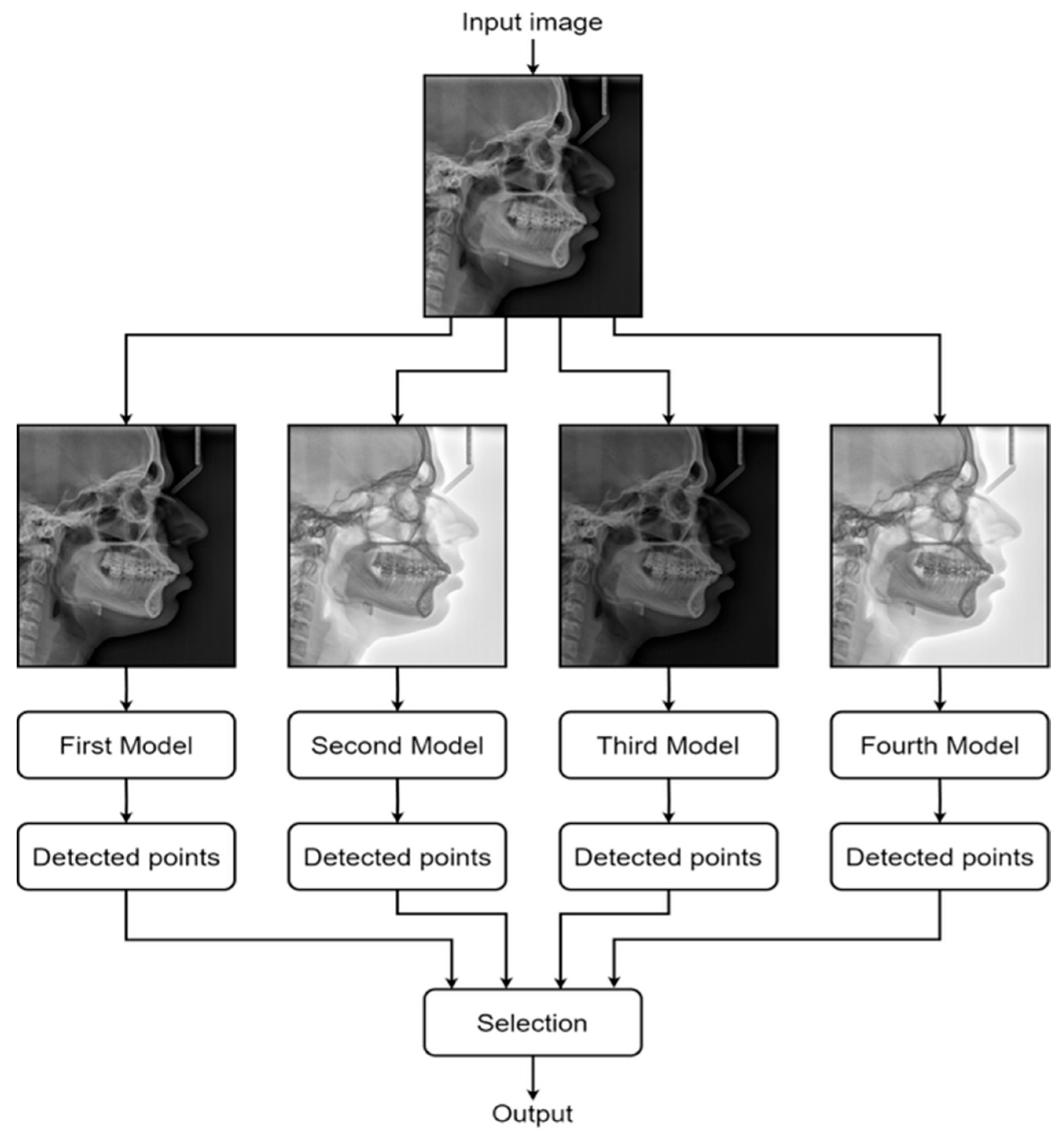
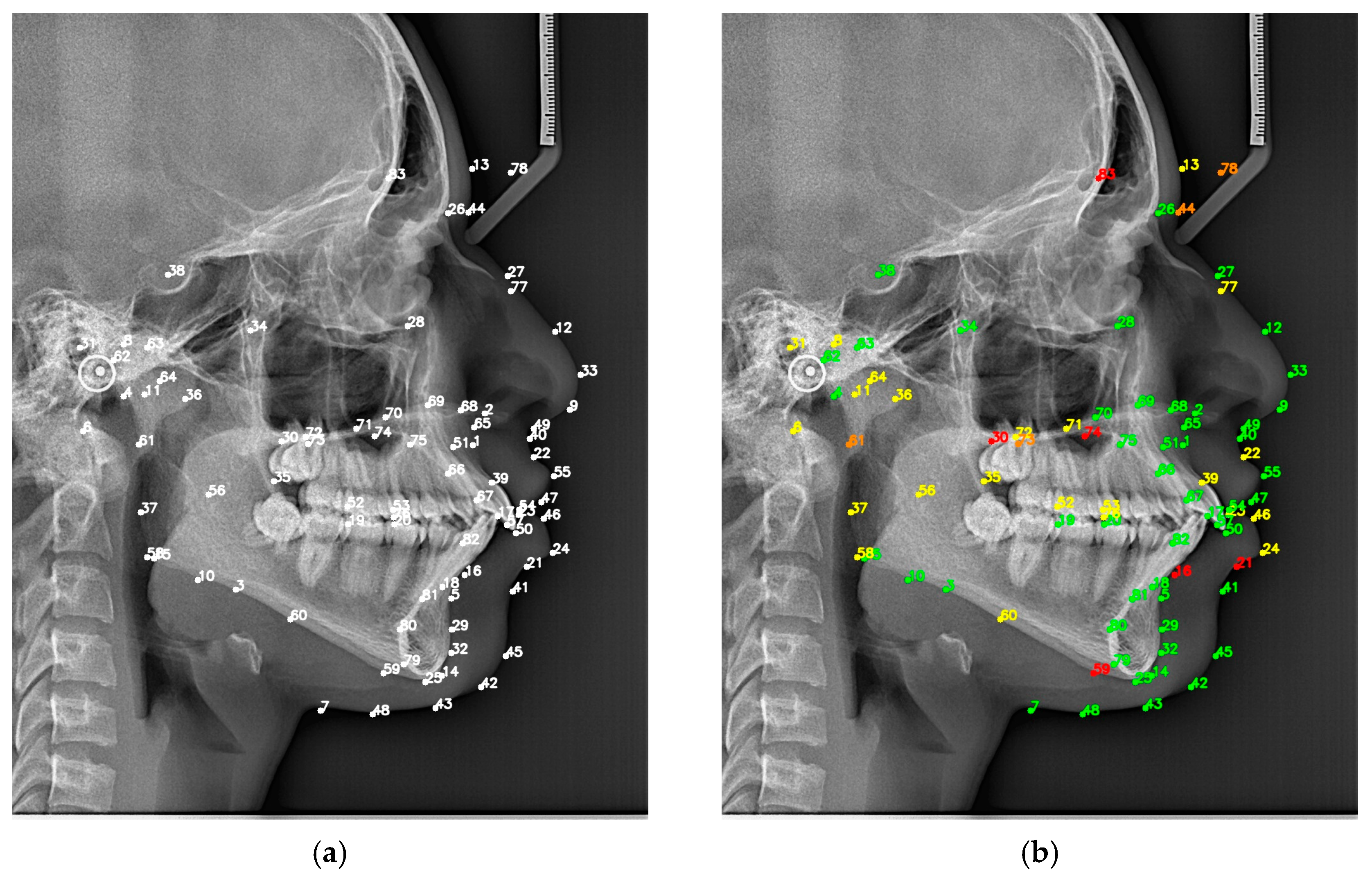
Background/Objectives: Modern artificial intelligence methods are increasingly used in medical and dental image analysis to support diagnosis and treatment planning. In orthodontics, automatic detection of cephalometric landmarks from X-ray images remains a challenging and clinically relevant task. Methods: This study proposes a multi-model approach for cephalometric landmark detection based on the ALD algorithm and three derived models trained with extended image augmentation techniques. The applied augmentations, including contrast and negative transformations, improved the detection of specific anatomical landmarks. The final detection strategy integrates outputs from all four models, selecting the most accurate prediction for each landmark based on historical performance results. Results: The proposed method was evaluated on real datasets. It achieved a mean radial error (MRE) of 2.12 mm compared to 2.26 mm for the baseline model, and a successful detection rate (SDR) of 72.22% within a 2.5 mm threshold compared to 68.87% for the baseline model. Conclusions: The results demonstrate that the ensemble-based approach improves landmark detection accuracy and has the potential to support clinical orthodontic workflows.
Keywords: medical decisions; algorithms; AI; decision support system; cephalometric analysis; deep learning; image processing medical decisions; algorithms; AI; decision support system; cephalometric analysis; deep learning; image processing