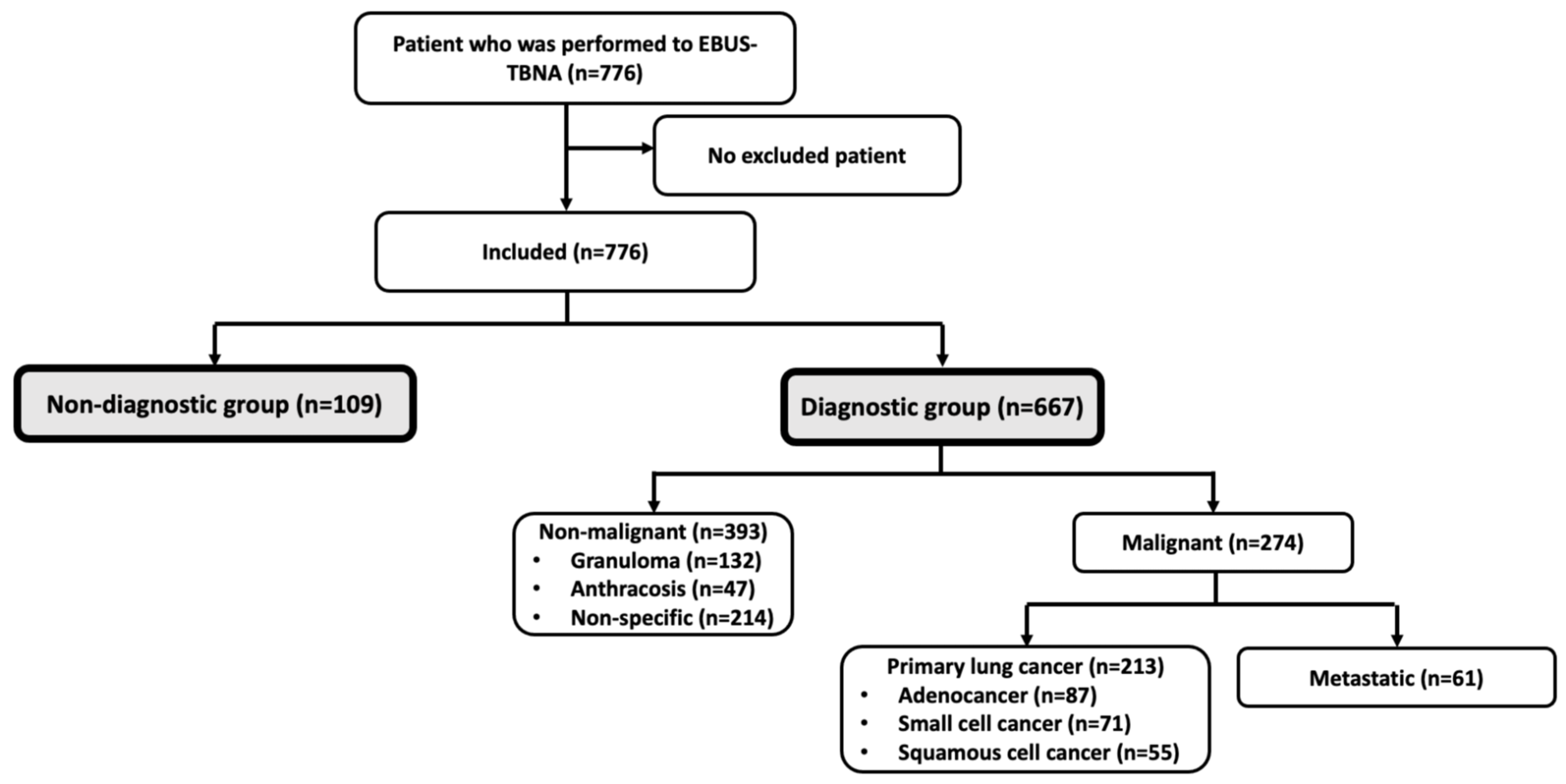
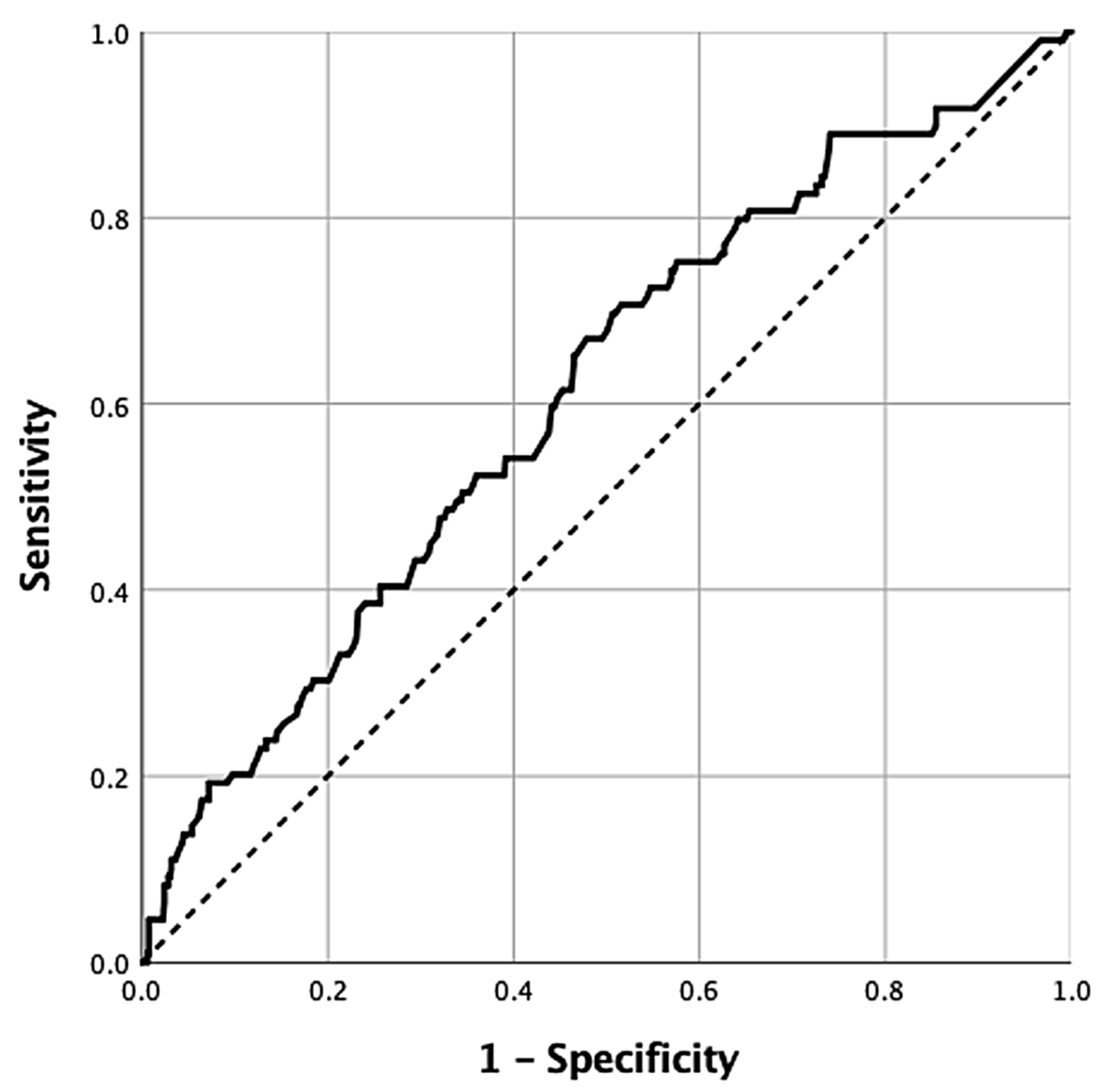
Associated Factors for Non-Diagnostic Cytopathology in the Endobronchial Ultrasound-Transbronchial Needle Aspiration: A Retrospective Cohort Study

 ,
,
Abstract
1. Introduction
2. Materials and Methods
2.1. Patients
2.2. Variables
2.3. Endobronchial Ultrasound-Transbronchial Needle Aspiration Procedure
2.4. Statistical Analysis
3. Results
3.1. Patient Characteristics
3.2. Factors Associated with Non-Diagnostic Cytology Results in EBUS-TBNA Patients
3.3. Performance of Lymph Node Size as a Diagnostic Factor
4. Discussion
4.1. Strengths of the Study
4.2. Limitations
5. Conclusions
Author Contributions
Funding
Institutional Review Board Statement
Informed Consent Statement
Data Availability Statement
Acknowledgments
Conflicts of Interest
Abbreviations
| aOR | Adjusted Odds Ratios |
| AUROC | The Area Under the ROC curve |
| CI | Confidence Intervals |
| CT | Computed Tomography |
| COPD | Chronic Obstructive Pulmonary Disease |
| EBUS-TBNA | Endobronchial Ultrasound-guided Transbronchial Needle Aspiration |
| ICU | Intensive Care Unit |
| IQR | Interquartile Range |
| ROC | Receiver Operating Characteristic |
| NPV | Negative Predictive Value |
| PPV | Positive Predictive Value |
| ROSE | Rapid On-Site Evaluation |
| SUVmax | Maximum Standardized Uptake Value |
| 18F-FDG PET-CT | 18F-Fluorodeoxyglucose Positron Emission Tomography-Computed Tomography |
References
- Fantin, A.; Castaldo, N.; Tirone, C.; Sartori, G.; Crisafulli, E.; Patrucco, F.; Vetrugno, L.; Patruno, V. Endobronchial ultrasound: A pictorial essay. Acta Biomed. 2023, 94, e2023113. [Google Scholar] [CrossRef] [PubMed]
- Sandoh, K.; Ishida, M.; Okano, K.; Ito, H.; Tsuta, K. Usefulness and problems of cytological examination by endobronchial ultrasound-guided transbronchial needle aspiration in the diagnosis of mediastinal lymphadenopathy: A retrospective single-centre study. Mol. Clin. Oncol. 2021, 15, 138. [Google Scholar] [CrossRef] [PubMed]
- Erer, O.F.; Erol, S.; Anar, C.; Aydogdu, Z.; Ozkan, S.A. Diagnostic yield of EBUS-TBNA for lymphoma and review of the literature. Endosc. Ultrasound 2017, 6, 317–322. [Google Scholar] [CrossRef] [PubMed]
- Herth, F.; Becker, H.D.; Ernst, A. Conventional vs. endobronchial ultrasound-guided transbronchial needle aspiration: A randomized trial. Chest 2004, 125, 322–325. [Google Scholar] [CrossRef] [PubMed]
- Bonifazi, M.; Tramacere, I.; Zuccatosta, L.; Mei, F.; Sediari, M.; Paonessa, M.C.; Gasparini, S. Conventional versus Ultrasound-Guided Transbronchial Needle Aspiration for the Diagnosis of Hilar/Mediastinal Lymph Adenopathies: A Randomized Controlled Trial. Respiration 2017, 94, 216–223. [Google Scholar] [CrossRef] [PubMed]
- Sun, X.; Zhang, T.; Shao, C.; Wang, P.; Chen, M.; Zhao, J.; Wang, M. All-shot Aspirations in Endobronchial Ultrasound-Guided Transbronchial Needle Aspiration (EBUS-TBNA) Are Associated with Malignancy. Technol. Cancer Res. Treat. 2023, 22, 15330338231169881. [Google Scholar] [CrossRef] [PubMed]
- Lee, H.S.; Lee, G.K.; Lee, H.S.; Kim, M.S.; Lee, J.M.; Kim, H.Y.; Nam, B.H.; Zo, J.I.; Hwangbo, B. Real-time endobronchial ultrasound-guided transbronchial needle aspiration in mediastinal staging of non-small cell lung cancer: How many aspirations per target lymph node station? Chest 2008, 134, 368–374. [Google Scholar] [CrossRef] [PubMed]
- Diacon, A.H.; Schuurmans, M.M.; Theron, J.; Brundyn, K.; Louw, M.; Wright, C.A.; Bolliger, C.T. Transbronchial needle aspirates: How many passes per target site? Eur. Respir. J. 2007, 29, 112–116. [Google Scholar] [CrossRef] [PubMed]
- About Us. Available online: https://ankarasehir.saglik.gov.tr/EN-845880/about-us.html (accessed on 8 December 2024).
- Sarinc Ulasli, S.; Koksal, D.; Karcioglu, O.; Babaoglu, E.; Onder, S.; Emri, S. Diagnostic utility of conventional transbronchial needle aspiration in older patients. Eur. Geriatr. Med. 2018, 9, 263–267. [Google Scholar] [CrossRef] [PubMed]
- Kobayashi, F.; Saraya, T.; Akizawa, T.; Abe, T.; Takagi, R.; Ieki, E.; Ishikawa, N.; Kurokawa, N.; Aso, J.; Nunokawa, H.; et al. Impact of Cough Severity on the Diagnostic Yield of Endobronchial Ultrasonography Transbronchial Biopsy with Guide Sheath: A Retrospective Observational Study. J. Clin. Med. 2024, 13, 347. [Google Scholar] [CrossRef] [PubMed]
- Kim, B.K.; Choi, H.; Kim, C.Y. Factors associated with increased diagnostic yield of endobronchial ultrasound-guided transbronchial needle aspiration: An observational single center study. J. Thorac. Dis. 2024, 16, 439–449. [Google Scholar] [CrossRef] [PubMed]
- Luthra, K.; Singh, J. Evaluation of 18FDG PET-CT-positive mediastinal-hilar lymph nodes in extrathoracic malignancies by EBUS-TBNA; correlation of SUVmax, and short-axis diameter with the final diagnosis. Indian J. Cancer 2021, 60, 106–113. [Google Scholar] [CrossRef] [PubMed]
- Wang, L.; Wu, W.; Hu, Y.; Teng, J.; Zhong, R.; Han, B.; Sun, J. Sonographic Features of Endobronchial Ultrasonography Predict Intrathoracic Lymph Node Metastasis in Lung Cancer Patients. Ann. Thorac. Surg. 2015, 100, 1203–1209. [Google Scholar] [CrossRef] [PubMed]
- Jensen, A.R.; Mainz, J.; Overgaard, J. Impact of delay on diagnosis and treatment of primary lung cancer. Acta Oncol. 2002, 41, 147–152. [Google Scholar] [CrossRef] [PubMed]
- Oki, M.; Saka, H.; Kitagawa, C.; Kogure, Y.; Murata, N.; Adachi, T.; Ando, M. Rapid on-site cytologic evaluation during endobronchial ultrasound-guided transbronchial needle aspiration for diagnosing lung cancer: A randomized study. Respiration 2013, 85, 486–492. [Google Scholar] [CrossRef] [PubMed]
- Nakajima, T.; Yasufuku, K.; Saegusa, F.; Fujiwara, T.; Sakairi, Y.; Hiroshima, K.; Nakatani, Y.; Yoshino, I. Rapid on-site cytologic evaluation during endobronchial ultrasound-guided transbronchial needle aspiration for nodal staging in patients with lung cancer. Ann. Thorac. Surg. 2013, 95, 1695–1699. [Google Scholar] [CrossRef] [PubMed]



| n | % | ||
|---|---|---|---|
| Male gender | 502 | 64.7 | |
| BAL | 663 | 81.6 | |
| BAL cytology | |||
| Malignant | 73 | 11.5 | |
| No malignant | 560 | 88.5 | |
| Forceps biopsy | 147 | 18.9 | |
| Malignant | 76 | 53.1 | |
| No malignant | 67 | 46.9 | |
| Non-diagnostic | 4 | - | |
| Brush biopsy | 85 | 11.0 | |
| Malignant | 47 | 55.3 | |
| No malignant | 38 | 44.7 | |
| Transthoracic needle aspiration biopsy | 47 | 6.1 | |
| Malignant | 31 | 66.0 | |
| No malignant | 16 | 34.7 | |
| EBUS-TBNA stations | |||
| 2R | 6 | 0.8 | |
| 4R | 192 | 24.7 | |
| 10R | 81 | 10.4 | |
| 11R | 94 | 12.1 | |
| 7 | 566 | 72.9 | |
| 4L | 26 | 3.4 | |
| 10L | 79 | 10.2 | |
| 11L | 66 | 8.5 | |
| 2R | 6 | 0.8 | |
| n | Mean ± Std. Dev. | Median (IQR) | ||
|---|---|---|---|---|
| Age (years) | 776 | 61.5 ± 12.6 | 63.0 (15.0) | |
| Maximum size of lymph node (mm) with EBUS | 776 | 20.7 ± 8.4 | 19.5 (13.8) | |
| 4R | 641 | 13.8 ± 8.3 | 11.3 (10.5) | |
| 10R | 518 | 13.6 ± 7.9 | 11.5 (10.7) | |
| 11R | 393 | 13.3 ± 7.4 | 12.0 (9.3) | |
| 7 | 723 | 18.4 ± 10.6 | 16.7 (13.6) | |
| 4L | 395 | 9.2 ± 5.9 | 7.5 (5.9) | |
| 10L | 363 | 11.8 ± 7.3 | 9.4 (8.7) | |
| 11L | 300 | 11.6 ± 6.3 | 9.8 (6.4) | |
| EBUS-TBNA sample count | 774 | 8.1 ± 13.1 | 7.0 (4.0) | |
| 4R | 192 | 5.2 ± 1.6 | 5.0 (2.0) | |
| 10R | 81 | 5.4 ± 1.5 | 5.0 (2.0) | |
| 11R | 94 | 5.4 ± 1.7 | 5.0 (2.0) | |
| 7 | 564 | 5.9 ± 1.4 | 6.0 (2.0) | |
| 4L | 26 | 5.1 ± 1.4 | 5.0 (2.0) | |
| 10L | 79 | 5.3 ± 1.6 | 5.0 (2.0) | |
| 11L | 66 | 5.7 ± 1.4 | 6.0 (2.0) | |
| Total biopsied station with EBUS-TBNA | 776 | 1.4 ± 0.6 | 1.0 (1.0) | |
| Total adjacent biopsy technique | 776 | 1.2 ± 0.8 | 1.0 (1.0) | |
| 18 F-PET-CT FDG SUV-max | 494 | 13.5 ± 50.1 | 9.5 (9.8) | |
| Total (n = 776) | Non-Diagnostic (n = 109) | Diagnostic (n = 667) | ||||
| n (%) | n (%) | n (%) | OR (95% CI) | p-Value | ||
| Male gender | 502 (64.7) | 76 (69.7) | 426 (63.9) | 1.30 (0.84–2.02) | 0.236 | |
| BAL | 633 (81.6) | 89 (81.7) | 544 (81.6) | 1.01 (0.60–1.70) | 0.982 | |
| Forceps biopsy | 147 (18.9) | 16 (14.7) | 131 (19.6) | 0.70 (0.40–1.24) | 0.220 | |
| Brush biopsy | 85 (11.0) | 13 (11.9) | 72 (10.8) | 1.12 (0.60–2.10) | 0.726 | |
| Transthoracic needle asp. | 47 (6.1) | 8 (7.3) | 39 (5.8) | 1.28 (0.58–2.81) | 0.545 | |
| EBUS-TBNA stations | ||||||
| 4R | 192 (24.7) | 25 (22.9) | 167 (25.0) | 0.90 (0.55–1.44) | 0.637 | |
| 10R | 81 (10.4) | 11 (10.1) | 70 (10.5) | 0.95 (0.49–1.87) | 0.898 | |
| 11R | 94 (12.1) | 16 (14.7) | 78 (11.7) | 1.30 (0.73–2.32) | 0.376 | |
| 7 | 566 (72.9) | 83 (76.1) | 483 (72.4) | 1.22 (0.76–1.95) | 0.416 | |
| 4L | 26 (3.4) | 3 (2.8) | 23 (3.4) | 0.80 (0.23–2.69) | >0.999 | |
| 10L | 79 (10.2) | 8 (7.3) | 71 (10.6) | 0.67 (0.31–1.42) | 0.290 | |
| 11L | 66 (8.5) | 6 (5.5) | 60 (9.0) | 0.59 (0.25–1.40) | 0.226 | |
| 2R | 6 (0.8) | 1 (0.9) | 5 (0.7) | 1.23 (0.14–10.64) | 0.598 | |
| Non-Diagnostic | Diagnostic | |||||
| n | Median (IQR) | n | Median (IQR) | p-Value | ||
| Age (years) | 109 | 65.0 (11.5) | 667 | 63.0 (16.0) | 0.018 | |
| EBUS-TBNA stations | ||||||
| 4R mm | 25 | 19.9 (16.3) | 167 | 17.0 (12.9) | 0.354 | |
| 4R bx count | 25 | 4.0 (1.5) | 167 | 5.0 (2.0) | 0.017 | |
| 10R mm | 11 | 11.0 (4.3) | 70 | 19.4 (14.7) | 0.003 | |
| 10R bx count | 11 | 4.0 (3.0) | 70 | 6.0 (1.3) | 0.041 | |
| 11R mm | 16 | 13.5 (8.6) | 78 | 15.0 (10.7) | 0.034 | |
| 11R bx count | 16 | 5.0 (1.8) | 78 | 5.5 (3.0) | 0.249 | |
| 7 bx mm | 83 | 15.3 (10.0) | 483 | 19.4 (13.2) | 0.001 | |
| 7 bx count | 82 | 6.0 (1.0) | 482 | 6.0 (2.0) | 0.273 | |
| 4L mm | 3 | 15.1 (-) | 23 | 15.0 (17.0) | 0.880 | |
| 4L bx count | 3 | 5.0 (-) | 23 | 5.0 (2.0) | 0.762 | |
| 10L mm | 8 | 15.6 (7.7) | 71 | 18.1 (12.6) | 0.338 | |
| 10L bx count | 8 | 5.5 (2.8) | 71 | 5.0 (3.0) | 0.679 | |
| 11L mm | 6 | 15.3 (11.3) | 60 | 12.0 (9.3) | 0.330 | |
| 11L bx count | 6 | 5.0 (2.3) | 60 | 6.0 (2.0) | 0.330 | |
| 2R mm | 1 | - | 5 | 11.7 (7.5) | 0.667 | |
| 2R bx count | 1 | - | 6 | 4.0 (2.8) | 0.571 | |
| Total bx count | 109 | 7.0 (3.0) | 666 | 8.0 (4.0) | 0.022 | |
| Max size of biopsied LN, mm | 109 | 16.3 (11.2) | 667 | 19.9 (13.9) | <0.001 | |
| Total biopsied station with EBUS-TBNA | 109 | 1.0 (1.0) | 667 | 1.0 (1.0) | 0.526 | |
| Total adjacent biopsy technique | 109 | 1.0 (0.0) | 667 | 1.0 (1.0) | 0.734 | |
| 18 F-PET-CT FDG SUV-max | 64 | 7.7 (9.8) | 430 | 9.8 (9.7) | 0.165 | |
| Univariate | Multivariate * | |||
|---|---|---|---|---|
| OR (95% CI) | p-Value | adjOR (95% CI) | p-Value | |
| Age, year | 1.02 (1.00–1.04) | 0.029 | 1.02 (1.00–1.03) | 0.086 |
| Male gender | 1.30 (0.84–2.02) | 0.237 | 1.27 (0.81–1.99) | 0.290 |
| Total bx count | 0.92 (0.86–0.99) | 0.023 | 0.94 (0.87–1.01) | 0.077 |
| Max biopsied LAP, mm | 0.95 (0.93–0.98) | <0.001 | 0.96 (0.93–0.98) | 0.002 |
Disclaimer/Publisher’s Note: The statements, opinions and data contained in all publications are solely those of the individual author(s) and contributor(s) and not of MDPI and/or the editor(s). MDPI and/or the editor(s) disclaim responsibility for any injury to people or property resulting from any ideas, methods, instructions or products referred to in the content. |
© 2026 by the authors. Licensee MDPI, Basel, Switzerland. This article is an open access article distributed under the terms and conditions of the Creative Commons Attribution (CC BY) license.
Share and Cite
Ozden Sertcelik, U.; Parlak, E.S.; Hezer, H.; Ceylan, E.G.; Sertcelik, A.; Karalezli, A. Associated Factors for Non-Diagnostic Cytopathology in the Endobronchial Ultrasound-Transbronchial Needle Aspiration: A Retrospective Cohort Study. Diagnostics 2026, 16, 1509. https://doi.org/10.3390/diagnostics16101509
Ozden Sertcelik U, Parlak ES, Hezer H, Ceylan EG, Sertcelik A, Karalezli A. Associated Factors for Non-Diagnostic Cytopathology in the Endobronchial Ultrasound-Transbronchial Needle Aspiration: A Retrospective Cohort Study. Diagnostics. 2026; 16(10):1509. https://doi.org/10.3390/diagnostics16101509
Chicago/Turabian StyleOzden Sertcelik, Umran, Ebru Sengul Parlak, Habibe Hezer, Eren Goktug Ceylan, Ahmet Sertcelik, and Ayşegul Karalezli. 2026. "Associated Factors for Non-Diagnostic Cytopathology in the Endobronchial Ultrasound-Transbronchial Needle Aspiration: A Retrospective Cohort Study" Diagnostics 16, no. 10: 1509. https://doi.org/10.3390/diagnostics16101509
APA StyleOzden Sertcelik, U., Parlak, E. S., Hezer, H., Ceylan, E. G., Sertcelik, A., & Karalezli, A. (2026). Associated Factors for Non-Diagnostic Cytopathology in the Endobronchial Ultrasound-Transbronchial Needle Aspiration: A Retrospective Cohort Study. Diagnostics, 16(10), 1509. https://doi.org/10.3390/diagnostics16101509