Article

Serum P-Cresyl Sulfate Levels Correlate with Peripheral Arterial Disease in Hypertensive Patients

1
Division of Nephrology, Department of Internal Medicine, Yuan’s General Hospital, Kaohsiung 80249, Taiwan
2
Division of Nephrology, Department of Internal Medicine, Dalin Tzu Chi Hospital, Buddhist Tzu Chi Medical Foundation, Chiayi 62247, Taiwan
3
School of Medicine, Tzu Chi University, Hualien 97004, Taiwan
4
Division of Nephrology, Hualien Tzu Chi Hospital, Buddhist Tzu Chi Medical Foundation, Hualien 97004, Taiwan
5
Graduate Institute of Clinical Pharmacy, School of Medicine, Tzu Chi University, Hualien 97004, Taiwan
6
School of Pharmacy, Tzu Chi University, Hualien 97004, Taiwan
7
Division of Cardiology, Hualien Tzu Chi Hospital, Buddhist Tzu Chi Medical Foundation, Hualien 97004, Taiwan
*
Author to whom correspondence should be addressed.
These authors contributed equally to this work.
Diagnostics 2025, 15(9), 1097; https://doi.org/10.3390/diagnostics15091097
Submission received: 19 March 2025 / Revised: 11 April 2025 / Accepted: 23 April 2025 / Published: 25 April 2025

Abstract

:
Background/Objectives: p-Cresyl sulfate (PCS) is implicated in inflammation, oxidative stress and vascular dysfunction. Hypertension is a major risk factor for peripheral arterial disease (PAD), which is linked to increased mortality in patients with hypertension. This study aimed to evaluate the association between serum PCS levels and PAD in hypertension cases. Methods: We analyzed fasting blood samples and clinical data from 105 patients with hypertension in a cardiovascular outpatient clinic. Serum PCS levels were quantified using high-performance liquid chromatography–mass spectrometry. Ankle–brachial index (ABI) was measured using an automated oscillometric device; ABI < 0.9 indicated PAD. Results: A total of 24 patients (22.9%) had PAD. The PAD group had a higher prevalence of diabetes mellitus (p = 0.026), elevated serum C-reactive protein (CRP) levels (p < 0.001) and increased PCS levels (p = 0.002) than the normal ABI group. Multivariate logistic regression showed that PCS (odds ratio [OR]: 1.154, 95% confidence interval [CI]: 1.013–1.315, p = 0.031) and CRP (per 0.1 mg/dL increase, OR: 1.649, 95% CI: 1.138–2.389, p = 0.008) were independently associated with PAD. According to Spearman’s correlation analysis, log-transformed PCS (log-PCS) levels negatively correlated with left or right ABI (p = 0.001 and p = 0.004, respectively) and estimated glomerular filtration rate (p = 0.001) but positively correlated with log-CRP (p = 0.024). Conclusions: Elevated serum PCS and CRP levels are significantly associated with PAD in patients with hypertension, suggesting the potential role of PCS in PAD pathogenesis.

1. Introduction

Peripheral arterial disease (PAD) is characterized by peripheral artery narrowing with subsequent reduction in blood flow to distal extremities, caused mainly by advanced atherosclerosis [1]. More importantly, PAD has been viewed as a risk equivalent of atherosclerotic cardiovascular disease (ASCVD) and a significant risk factor for predicting cardiovascular mortality [2]. Hypertension is a well-established major risk factor for ASCVD, as well as PAD, but its importance in these diseases cannot be stressed enough [3,4,5,6,7,8]. Nonetheless, a worldwide pooled analysis revealed the stable global age-standardized prevalence of hypertension [9]. In 2015, the prevalence of PAD was 5.6% [10]. Owing to the significant burden of PAD and its associated risk factors, an approach that can better detect PAD development in hypertension cases is needed.
p-Cresyl sulfate (PCS) has been extensively studied as a protein-bound uremic toxin derived from the intake of aromatic amino acids, mainly tyrosine and phenylalanine, that are subsequently processed by intestinal bacteria into phenolic end products [11]. Regarding toxicity on the human cardiovascular system, elevated PCS levels have been linked to various cardiovascular diseases [12,13], including peripheral arterial stiffness [14], coronary artery disease (CAD) [15,16,17], arrhythmia [18] and left ventricular dysfunction [19]. Increased PCS levels were reported to be associated with a low ankle–brachial index (ABI) and vascular access failure events in patients undergoing hemodialysis [20]. However, the relationship between PCS and PAD in patients with hypertension remains unclear, particularly in outpatients with a range of renal statuses. By investigating whether PCS is associated with PAD in this population regardless of the renal status, our study aimed to explore whether PCS could be another biomarker that can be integrated into current risk stratification models and enhance early detection strategies for PAD to improve cardiovascular outcomes in patients with hypertension.

2. Materials and Methods

2.1. Patients

We recruited 105 patients with hypertension from the cardiovascular outpatient department of a medical center in Hualien, Taiwan, between July 2020 and December 2020. The exclusion criteria were as follows: recent acute coronary syndrome within 3 months, acute decompensated heart failure, acute stroke within 3 months, malignancy, limb amputation, acute infection during blood sampling, ABI > 1.3, or patient refusal to provide informed consent for the study. This study obtained approval from the Research Ethics Committee, Hualien Tzu Chi Hospital, Buddhist Tzu Chi Medical Foundation (IRB108-219-A). All participants provided their written informed consent to participate in this study. After resting for at least 10 min, our trained staff measured the participants’ morning blood pressure (BP) using standard mercury sphygmomanometers with appropriate cuff sizes. Systolic BP (SBP) and diastolic BP (DBP) values were measured thrice at a 5 min interval and were averaged for the analysis. In this study, hypertension was defined as SBP ≥ 140 mm Hg and/or DBP ≥ 90 mmHg or intake of antihypertensive drugs in the past 2 weeks according to the Eighth Joint National Committee (JNC 8) guidelines. Moreover, diabetes mellitus (DM) was defined as fasting plasma glucose levels ≥ 126 mg/dL or intake of hypoglycemic agents.

2.2. Anthropometric Analysis

We measured all patients’ anthropometric variables in the morning after overnight fasting. Body weight and height measurements were recorded to the nearest 0.5 kg and 0.5 cm, respectively. Additionally, the body mass index was calculated as weight (kg) divided by the square of height (m2).

2.3. Biochemical Investigations

Immediately after collection, fasting blood samples (approximately 5 mL) were centrifuged at 3000× g for 10 min. The serum levels of blood urea nitrogen, creatinine, fasting glucose, total cholesterol (TCH), triglycerides (TG), high-density lipoprotein cholesterol (HDL-C), low-density lipoprotein cholesterol (LDL-C), total calcium, phosphorus and C-reactive protein (CRP) were checked using an autoanalyzer (Siemens Advia 1800; Siemens Healthcare GmbH, Henkestr, Erlangen, Germany) [21]. To measure the serum levels of human intact parathyroid hormone (iPTH), we used a commercially available enzyme-linked immunosorbent assay (Abcam, Cambridge, MA, USA) [21]. For calculating the estimated glomerular filtration rate (eGFR), we applied the chronic kidney disease epidemiology collaboration equation.

2.4. Determination of Serum PCS Levels by High-Performance Liquid Chromatography–Mass Spectrometry

A Waters e2695 high-performance liquid chromatography system integrated with a mass spectrometer (ACQUITY QDa, Waters Corporation, Milford, MA, USA) was used in measuring serum PCS levels in this study [14]. As for the analytical column, we used the Phenomenex Luna® C18 (2) (5 µ, 250 × 4.60 mm, 100 Å) with the following operational parameters: column temperature, 40 °C; flow rate, 0.8 mL/min; an injection volume, 30 µL. In the mobile phase, a binary gradient was implemented: the initial composition (95% [A] water with 0.1% formic acid/5% [B] methanol with 0.1% formic acid) remained constant for 1 min; subsequently, solvent B was incrementally increased to 70% over a span of 12 min and held constant for an additional 2 min. For column re-equilibration, solvent B was diminished to 50% within 1 min and sustained at this level for 2 min.
The liquid chromatography–mass spectrometry (LC-MS) gradient conditions were modified, given that the pretreated samples were concurrently analyzed. The specifications of the instrument were as follows: desolvation temperature, 600 °C; capillary voltage, 0.8 kV; sample cone voltage, 15.0 V. The mass spectrometer functioned in full scan mode, covering a range of 50–450 m/z for positive ionization and 100–350 m/z for negative ionization. We employed a single-ion recording mode to monitor the specific masses of each compound (PCS: 187.0 m/z). Data acquisition and processing were conducted using the Empower® 3.0 software (Waters Corporation, Milford, MA, USA). The retention time observed for PCs was approximately 16.56 min. In quantifying endogenous compounds, we measured and compared the peak areas against the calibration curve derived from the standard solutions. All the determination coefficients of linearity (r2) exceeded 0.995. The LC-MS single-ion recording mode was utilized for analyzing individual ions.

2.5. ABI Measurements

BP was measured using the oscillometric technique on both the upper and lower extremities, with each measurement taken thrice while the participant was in a supine position, generating data from the brachial, dorsalis pedis and posterior tibial arteries (VaSera VS-1000, Fukuda Denshi, Tokyo, Japan) [22]. ABI was computed as the ratio of the highest recorded SBP in the dorsalis pedis or posterior tibial artery at either the right or left ankle to that in the brachial artery of either upper limb. Continuous electrocardiographic monitoring was performed for over 15 min. Patients exhibiting a reduced ABI of 0.9 in either the left or right extremity were considered to have PAD, as previously mentioned [22].

2.6. Strategies to Avoid Bias

Several strategies were applied to reduce the risk of bias in this study. Selection bias was minimized by strict inclusion and exclusion criteria to make the study population reflect hypertensive patients in general. Recruitment was performed in a systematic manner without over-representation of certain subgroups by consecutively enrolling all eligible participants. We used validated methods and protocols to standardize data collection procedures to minimize potential information bias. Serum PCS concentrations were determined by standard laboratory methods and ABI values were assessed by trained staff members using Doppler ultrasound according to prevailing clinical protocols. Third, to minimize potential bias in how results are considered, we performed a thorough review of the literature in order to contextualize our findings with respect to related evidence while also considering the potential for publication bias.

2.7. Statistical Analysis

We performed a power analysis to determine whether our sample size was sufficient to detect statistically significant correlations between serum PCS levels and PAD, based on previously published findings of vascular markers and PAD [23,24]. At an α of 0.05, and 80% statistical power to detect a medium correlation coefficient (r = 0.30), which was of clinical significance in our context, the required sample size for this current study was calculated [25]. As a minimum sample size to provide sufficient power to detect any such correlation our calculations indicated at least 85 participants would be required. Our study population of 105 hypertensive patients was greater than this threshold and provided adequate statistical power. Post hoc power analysis confirmed that our sample size provided 86% power to detect the correlation observed with log-PCS and left ABI (r = −0.321, p = 0.001) and 78% power for the correlation with right ABI (r = −0.281, p = 0.004). These outcomes confirm our findings to be sufficiently reliable and suggest that the number of enrolled patients was sufficient for adequate statistical analysis.
The normality of data distribution was assessed using the Kolmogorov–Smirnov test. Continuous variables that are normally distributed are presented as means ± standard deviation, whereas those that are not normally distributed are presented as medians with interquartile ranges; comparisons between patients were conducted using the two-tailed Student’s independent t-test and the Mann–Whitney U test, respectively. Categorical variables are expressed as counts with corresponding percentages and they were analyzed using the χ2 test. Variables demonstrating a significant association with PAD were subsequently examined through multivariable logistic regression analysis. We also utilized the receiver operating characteristic (ROC) curve to calculate the area under the curve (AUC) and to ascertain the serum levels of PCS and CRP as predictors of PAD in individuals with hypertension. In light of the nonnormally distributed variables, we applied a base 10 logarithmic transformation to facilitate normality. The correlation between log-PCS left ABI, right ABI values and clinical variables was evaluated using Spearman’s rank correlation coefficient. All statistical data were analyzed using the SPSS software for Windows (version 19.0; SPSS, Chicago, IL, USA). A p-value of 0.05 was considered statistically significant.

3. Results

Table 1 delineates the baseline characteristics of the cohort comprising 105 individuals with hypertension, stratified into normal (n = 81) and low (n = 24) ABI categories. Within this cohort, 71 patients (67.6%) were male, and the prevalence rates of DM and CAD were 31.4% (n = 33) and 66.7% (n = 70), respectively. The prevalence of DM was significantly elevated in the low ABI group compared with that in the normal ABI group (p = 0.026). Additionally, the low ABI group had markedly higher levels of CRP and PCS than the normal ABI group (p < 0.001 and p = 0.002, respectively). Conversely, SBP and DBP, sex distribution, CAD presence, or antihypertensive medication use showed no statistically significant differences between the two groups.
A multivariate logistic regression analysis was conducted after the adjustment for significant variables (p < 0.2) correlated with PAD. These variables included DM, BMI, TCH, eGFR, CRP and PCS levels. Among patients with hypertension, the serum levels of CRP (each 0.1 mg/dL increment, odds ratio [OR]: 1.649, 95% confidence interval [CI]: 1.138–2.389, p = 0.008) and PCS (OR: 1.154, 95% CI: 1.013–1.315, p = 0.031) were identified as independent predictors for PAD (Table 2). Furthermore, the ROC curve for PAD prediction indicated that the AUC values for PCS and CRP were 0.714 (95% CI, 0.618–0.798; p = 0.002) and 0.739 (95% CI, 0.645–0.820; p < 0.001), respectively (Figure 1).
Table 3 shows the association between serum log-PCS and various clinical variables through Spearman’s correlation analysis. The left and right ABI values revealed a negative correlation with log-PCS (r = −0.321; p = 0.001 and r = −0.281, p = 0.004, respectively). Furthermore, serum log-PCS positively correlated with log-CRP (r = 0.220, p = 0.024) but negatively correlated with estimated eGFR (r = −0.325, p = 0.001). Likewise, the left and right ABI values negatively correlated with log-CRP (r = −0.382 and r = −0.446, p < 0.001, respectively). Notably, the right ABI values positively correlated with eGFR (r = 0.199, p = 0.042).

4. Discussion

This study demonstrated that among the recruited patients with hypertension visiting our cardiovascular outpatient department, significantly higher prevalence rates of DM and higher levels of PCS and CRP were noted in those diagnosed with PAD measured by ABI. In the subsequent multivariable logistic regression analysis, higher serum PCS and CRP levels independently correlated with PAD presence. Furthermore, Spearman’s correlation analysis revealed that the log-PCS levels negatively correlated with the left or right ABI values and eGFR and positively correlated with the log-CRP.
Results such as higher DM prevalence and CRP levels in patients with hypertension with PAD, as well as the independent correlation of CRP levels with PAD, align well with current evidence. First, DM has been well recognized as one of the risk factors of ASCVD, including PAD [2,26,27]. As for the role of inflammation in PAD pathogenesis, CRP has been found to be a biomarker that can not only predict the presence but also the severity of PAD [26,28,29,30]. However, some authors argued that although patients with PAD exhibit elevated CRP levels, such increase may be more of a reflection of an underlying systemic inflammation resulting from various traditional and nontraditional ASCVD risk factors rather than causally linked to PAD development [31]. Therefore, some experts suggested combining CRP with other biomarkers to predict and risk-stratify PAD more accurately [32].
Considering the positive correlation between PCS levels and PAD presence, the underlying mechanism could be the involvement of PCS in atherosclerosis and vascular calcification [33,34]. Various in vitro and in vivo studies demonstrated that PCS induces reactive oxygen species (ROS) [35], activates the renin–angiotensin–aldosterone system [11], stimulates and damages endothelial cells [13,35], causes vascular smooth muscles to migrate and proliferate [33] and interacts with chemotactic proteins and complement interleukin-1 receptors leading to inflammation [36]. Thus, PCS may have the capacity to initiate and accelerate the process of atherosclerosis, which constitutes a significant risk factor for PAD. Additionally, PCS reportedly promotes osteogenesis in arterial smooth muscle cells; osteogenesis can contribute to vascular calcification and could explain the positive correlation between PCS levels and arterial stiffness observed in patients with chronic kidney disease (CKD) [14,37]. Overlapping risk factors characterize arterial stiffness and PAD and exhibit a complex interrelationship; in fact, some experts regarded arterial stiffness as a precursory lesion of PAD. Regarding the identified negative association between PCS and eGFR, research indicates that diminished eGFR results in impaired tubular secretion, which represents a principal pathway for the elimination of protein-bound uremic toxins such as PCS [11,38,39]. Moreover, metabolic acidosis, altered protein assimilation, gut microbiota changes and DM, which are commonly found in patients with CKD, could contribute to increased PCS levels [11,17,40]. Lastly, the right ABI positively correlated with eGFR. The clinical significance and potential implications of this result require further well-designed studies to specifically identify the optimal cutoff value of ABI for risk-stratifying PAD presence in patients with hypertension.
This study has several limitations that need to be addressed. First, the study is cross-sectional in design; thus, we cannot establish a causal relationship between elevated PCS levels and PAD. Second, while CRP levels are recognized as a marker of inflammation and PAD severity, their elevation may signify various underlying atherosclerotic risk factors instead of serving as a direct contributor to PAD pathogenesis. Third, the present study was limited solely to hypertensive patients who were asymptomatic for carotid artery disease and PAD. As a result, data regarding claudication and carotid disease were not obtained, and thus, symptomatic PAD or carotid artery involvement could not be evaluated. Asymptomatic PAD is a prevalent condition that frequently goes undiagnosed even though it is associated with considerable cardiovascular risk. We did not include symptomatic patients, which may limit the generalizability of our findings to other PAD presentations in the wider population. Additional studies should include both symptomatic and asymptomatic patients, as we indicate an indirect relationship between vascular markers and the presence of PAD. Another issue to be considered is that using ABI as the primary diagnostic tool for PAD may underestimate disease prevalence in hypertensive patients with medial artery calcification, a common feature in CKD and diabetes. Further studies must include adjunctive techniques like toe–brachial index or imaging modalities to better establish the diagnosis of PAD in this high-risk population. Finally, our participants were recruited from a single center; hence, they may not represent the broader hypertensive population. Considering these limitations, further studies with larger and more diverse populations with hypertension are required to clarify the potential mechanics of PCS, inflammation and PAD pathogenesis.

5. Conclusions

This study demonstrates that PCS may serve as a biomarker of evolving PAD in hypertensive patients. In a multivariate logistic regression analysis, elevated serum levels of PCS and CRP are independent predictors of PAD in hypertensive patients. Meanwhile, PCS positively correlates with CRP and PAD presence in this patient population and inversely correlates with eGFR. PCS showed an area under the curve (AUC) of 0.714, establishing it as a suitable diagnostic for PAD in hypertensive patients. Our findings show the possibility of PCS as a promising biomarker for early identification and risk stratification of PAD in a clinical setting, thereby potentially enabling timely intervention and subsequent appropriate follow-up management to improve patient outcomes.

Author Contributions

Conceptualization, B.-G.H. and J.-H.W.; methodology, C.-H.L. and J.-H.W.; investigation, J.-H.W.; formal analysis, J.-P.T. and B.-G.H.; data curation, B.-G.H., C.-H.L. and J.-H.W.; writing—original draft preparation, Y.-B.C. and B.-G.H.; writing—review and editing, J.-P.T., B.-G.H. and J.-H.W.; supervision, J.-H.W.; funding acquisition, B.-G.H. and J.-H.W. All authors have read and agreed to the published version of the manuscript.

Funding

This study was funded by a grant from Hualien Tzu Chi Hospital, Buddhist Tzu Chi Medical Foundation, Hualien, Taiwan, grant number TCRD-112-043 and TCMF-A 106-01-08.

Institutional Review Board Statement

The research conducted herein has been reviewed and approved by the Research Ethics Committee, Hualien Tzu Chi Hospital, adhering to the ethical standards for research involving human subjects. (IRB108-219-A and approval on 1 January 2020).

Informed Consent Statement

Informed consent was procured from all study participants.

Data Availability Statement

Upon request, the corresponding author can provide the data utilized in this study.

Conflicts of Interest

The authors declare no conflicts of interest.

References

  1. Abraham, A.T.; Mojaddedi, S.; Loseke, I.H.; Bray, C. Hypertension in patients with peripheral artery disease: An updated literature review. Cureus 2024, 16, e62246. [Google Scholar] [CrossRef] [PubMed]
  2. Gornik, H.L.; Aronow, H.D.; Goodney, P.P.; Arya, S.; Brewster, L.P.; Byrd, L.; Chandra, V.; Drachman, D.E.; Eaves, J.M.; Ehrman, J.K.; et al. 2024 ACC/AHA/AACVPR/APMA/ABC/SCAI/SVM/SVN/SVS/SIR/VESS guideline for the management of lower extremity peripheral artery disease: A report of the American College of Cardiology/American Heart Association joint committee on clinical practice guidelines. Circulation 2024, 149, e1313–e1410. [Google Scholar] [PubMed]
  3. Kharawala, A.; Nagraj, S.; Pargaonkar, S.; Seo, J.; Kokkinidis, D.G.; Altin, S.E. Hypertension management in peripheral artery disease: A mini review. Curr. Hypertens. Rev. 2024, 20, 1–9. [Google Scholar] [CrossRef]
  4. Poredoš, P.; Mikhailidis, D.P.; Paraskevas, K.I.; Blinc, A.; Antignani, P.L.; Stanek, A.; Mansilha, A.; Cevc, M. Management of arterial hypertension in patients with peripheral arterial disease. Int. Angiol. 2024, 43, 541–547. [Google Scholar] [CrossRef]
  5. Kjeldsen, S.E. Hypertension and cardiovascular risk: General aspects. Pharmacol. Res. 2018, 129, 95–99. [Google Scholar] [CrossRef] [PubMed]
  6. He, J.; Gu, D.; Chen, J.; Wu, X.; Kelly, T.N.; Huang, J.F.; Chen, J.C.; Chen, C.S.; Bazzano, L.A.; Reynolds, K.; et al. Premature deaths attributable to blood pressure in China: A prospective cohort study. Lancet 2009, 374, 1765–1772. [Google Scholar] [CrossRef]
  7. Luo, D.; Cheng, Y.; Zhang, H.; Ba, M.; Chen, P.; Li, H.; Chen, K.; Sha, W.; Zhang, C.; Chen, H. Association between high blood pressure and long term cardiovascular events in young adults: Systematic review and meta-analysis. BMJ 2020, 370, m3222. [Google Scholar] [CrossRef]
  8. Lewington, S.; Clarke, R.; Qizilbash, N.; Peto, R.; Collins, R.; Prospective Studies Collaboration. Age-specific relevance of usual blood pressure to vascular mortality: A meta-analysis of individual data for one million adults in 61 prospective studies. Lancet 2002, 360, 1903–1913. [Google Scholar]
  9. Zhou, B.; Carrillo-Larco, R.M.; Danaei, G.; Riley, L.M.; Paciorek, C.J.; Stevens, G.A.; Gregg, E.W.; Bennett, J.E.; Solomon, B.; Singleton, R.K.; et al. Worldwide trends in hypertension prevalence and progress in treatment and control from 1990 to 2019: A pooled analysis of 1201 population-representative studies with 104 million participants. Lancet 2021, 398, 957–980. [Google Scholar]
  10. Song, P.; Rudan, D.; Zhu, Y.; Fowkes, F.J.I.; Rahimi, K.; Fowkes, F.G.R.; Rudan, I. Global, regional, and national prevalence and risk factors for peripheral artery disease in 2015: An updated systematic review and analysis. Lancet Glob. Health 2019, 7, e1020–e1030. [Google Scholar] [CrossRef]
  11. Gryp, T.; Vanholder, R.; Vaneechoutte, M.; Glorieux, G. P-cresyl sulfate. Toxins 2017, 9, 52. [Google Scholar] [CrossRef]
  12. Shafi, T.; Meyer, T.W.; Hostetter, T.H.; Melamed, M.L.; Parekh, R.S.; Hwang, S.; Banerjee, T.; Coresh, J.; Powe, N.R. Free levels of selected organic solutes and cardiovascular morbidity and mortality in hemodialysis patients: Results from the retained organic solutes and clinical outcomes (ROSCO) investigators. PLoS ONE 2015, 10, e0126048. [Google Scholar] [CrossRef] [PubMed]
  13. Glorieux, G.; Vanholder, R.; Van Biesen, W.; Pletinck, A.; Schepers, E.; Neirynck, N.; Speeckaert, M.; De Bacquer, D.; Verbeke, F. Free p-cresyl sulfate shows the highest association with cardiovascular outcome in chronic kidney disease. Nephrol. Dial. Transplant. 2021, 36, 998–1005. [Google Scholar] [CrossRef] [PubMed]
  14. Chang, Y.C.; Lin, Y.L.; Lai, Y.H.; Wang, C.H.; Hsu, B.G. Serum p-cresyl sulfate level is an independent marker of peripheral arterial stiffness as assessed using brachial-ankle pulse wave velocity in patients with non-dialysis chronic kidney disease stage 3 to 5. Toxins 2022, 14, 287. [Google Scholar] [CrossRef]
  15. Hsu, H.J.; Yen, C.H.; Wu, I.W.; Hsu, K.H.; Chen, C.K.; Sun, C.Y.; Chou, C.C.; Chen, C.Y.; Tsai, C.J.; Wu, M.S.; et al. The association of uremic toxins and inflammation in hemodialysis patients. PLoS ONE 2014, 9, e102691. [Google Scholar] [CrossRef]
  16. Wang, C.P.; Lu, L.F.; Yu, T.H.; Hung, W.C.; Chiu, C.A.; Chung, F.M.; Yeh, L.R.; Chen, H.J.; Lee, Y.J.; Houng, J.Y. Serum levels of total p-cresylsulphate are associated with angiographic coronary atherosclerosis severity in stable angina patients with early stage of renal failure. Atherosclerosis 2010, 211, 579–583. [Google Scholar] [CrossRef]
  17. Chiu, C.A.; Lu, L.F.; Yu, T.H.; Hung, W.C.; Chung, F.M.; Tsai, I.T.; Yang, C.Y.; Hsu, C.C.; Lu, Y.C.; Wang, C.P.; et al. Increased levels of total p-cresylsulphate and indoxyl sulphate are associated with coronary artery disease in patients with diabetic nephropathy. Rev. Diabet. Stud. 2010, 7, 275–284. [Google Scholar] [CrossRef]
  18. Tang, W.H.; Wang, C.P.; Yu, T.H.; Hung, W.C.; Chung, F.M.; Lu, Y.C.; Hsu, C.C.; Lu, L.F.; Huang, L.L.; Lee, Y.J.; et al. Serum total p-cresylsulfate level is associated with abnormal QTc interval in stable angina patients with early stage of renal failure. Clin. Chim. Acta 2014, 437, 25–30. [Google Scholar] [CrossRef] [PubMed]
  19. Lu, L.F.; Tang, W.H.; Hsu, C.C.; Tsai, I.T.; Hung, W.C.; Yu, T.H.; Wu, C.C.; Chung, F.M.; Lu, Y.C.; Lee, Y.J.; et al. Associations among chronic kidney disease, high total p-cresylsulfate and left ventricular systolic dysfunction. Clin. Chim. Acta 2016, 457, 63–68. [Google Scholar] [CrossRef]
  20. Lin, C.J.; Pan, C.F.; Liu, H.L.; Chuang, C.K.; Jayakumar, T.; Wang, T.J.; Chen, H.H.; Wu, C.J. The role of protein-bound uremic toxins on peripheral artery disease and vascular access failure in patients on hemodialysis. Atherosclerosis 2012, 225, 173–179. [Google Scholar] [CrossRef]
  21. Chen, Y.L.; Huang, P.Y.; Tsai, J.P.; Wang, J.H.; Hsu, B.G. Serum osteoprotegerin levels and the vascular reactivity index in patients with hypertension. Medicina 2023, 59, 1794. [Google Scholar] [CrossRef] [PubMed]
  22. Chiu, L.T.; Lin, L.; Lin, H.J.; Lai, Y.H.; Hsu, B.G. Positive correlation of serum indoxyl sulfate level with peripheral arterial disease in hemodialysis patients. Vascular 2022, 30, 928–933. [Google Scholar] [CrossRef]
  23. Signorelli, S.S.; Fiore, V.; Malaponte, G. Inflammation and Peripheral Arterial Disease: The Value of Circulating Biomarkers (Review). Int. J. Mol. Med. 2014, 33, 777–783. [Google Scholar] [CrossRef] [PubMed]
  24. Celebi, S.; Berkten, B.; Amasyali, B. The Association between Thrombotic and Inflammatory Biomarkers and Lower-Extremity Peripheral Artery Disease. Int. Wound J. 2020, 17, 1346–1355. [Google Scholar] [CrossRef] [PubMed]
  25. Cohen, J. Statistical Power Analysis for the Behavioral Sciences, 2nd ed.; Lawrence Erlbaum Associates: Hillsdale, NJ, USA, 1988. [Google Scholar]
  26. Fowkes, F.G.; Rudan, D.; Rudan, I.; Aboyans, V.; Denenberg, J.O.; McDermott, M.M.; Norman, P.E.; Sampson, U.K.; Williams, L.J.; Mensah, G.A.; et al. Comparison of global estimates of prevalence and risk factors for peripheral artery disease in 2000 and 2010: A systematic review and analysis. Lancet 2013, 382, 1329–1340. [Google Scholar] [CrossRef]
  27. Joosten, M.M.; Pai, J.K.; Bertoia, M.L.; Rimm, E.B.; Spiegelman, D.; Mittleman, M.A.; Mukamal, K.J. Associations between conventional cardiovascular risk factors and risk of peripheral artery disease in men. JAMA 2012, 308, 1660–1667. [Google Scholar] [CrossRef]
  28. Ridker, P.M.; Stampfer, M.J.; Rifai, N. Novel risk factors for systemic atherosclerosis: A comparison of C-reactive protein, fibrinogen, homocysteine, lipoprotein(a), and standard cholesterol screening as predictors of peripheral arterial disease. JAMA 2001, 285, 2481–2485. [Google Scholar] [CrossRef]
  29. De Haro, J.; Acin, F.; Medina, F.J.; Lopez-Quintana, A.; March, J.R. Relationship between the plasma concentration of C-reactive protein and severity of peripheral arterial disease. Clin. Med. Cardiol. 2008, 3, 1–7. [Google Scholar] [CrossRef]
  30. Kim, B.G.; Seo, J.; Kim, G.S.; Jin, M.N.; Lee, H.Y.; Byun, Y.S.; Kim, B.O. Elevated C-reactive protein/albumin ratio is associated with lesion complexity, multilevel involvement, and adverse outcomes in patients with peripheral artery disease. Angiology 2022, 73, 843–851. [Google Scholar] [CrossRef]
  31. Sharma, P.; Klarin, D.; Voight, B.F.; Tsao, P.S.; Levin, M.G.; Damrauer, S.M. Evaluation of plasma biomarkers for causal association with peripheral artery disease. Arterioscler. Thromb. Vasc. Biol. 2024, 44, 1114–1123. [Google Scholar] [CrossRef]
  32. Saenz-Pipaon, G.; Martinez-Aguilar, E.; Orbe, J.; González Miqueo, A.; Fernandez-Alonso, L.; Paramo, J.A.; Roncal, C. The role of circulating biomarkers in peripheral arterial disease. Int. J. Mol. Sci. 2021, 22, 3601. [Google Scholar] [CrossRef] [PubMed]
  33. Han, H.; Chen, Y.; Zhu, Z.; Su, X.; Ni, J.; Du, R.; Zhang, R.; Jin, W. p-Cresyl sulfate promotes the formation of atherosclerotic lesions and induces plaque instability by targeting vascular smooth muscle cells. Front. Med. 2016, 10, 320–329. [Google Scholar] [CrossRef] [PubMed]
  34. Opdebeeck, B.; Maudsley, S.; Azmi, A.; De Maré, A.; De Leger, W.; Meijers, B.; Verhulst, A.; Evenepoel, P.; D’Haese, P.C.; Neven, E. Indoxyl sulfate and p-cresyl sulfate promote vascular calcification and associate with glucose intolerance. J. Am. Soc. Nephrol. 2019, 30, 751–766. [Google Scholar] [CrossRef] [PubMed]
  35. Watanabe, H.; Miyamoto, Y.; Enoki, Y.; Ishima, Y.; Kadowaki, D.; Kotani, S.; Nakajima, M.; Tanaka, M.; Matsushita, K.; Mori, Y.; et al. p-Cresyl sulfate, a uremic toxin, causes vascular endothelial and smooth muscle cell damages by inducing oxidative stress. Pharmacol. Res. Perspect. 2015, 3, e00092. [Google Scholar] [CrossRef]
  36. Wu, P.H.; Lin, Y.T.; Chiu, Y.W.; Baldanzi, G.; Huang, J.C.; Liang, S.S.; Lee, S.C.; Chen, S.C.; Hsu, Y.L.; Kuo, M.C.; et al. The relationship of indoxyl sulfate and p-cresyl sulfate with target cardiovascular proteins in hemodialysis patients. Sci. Rep. 2021, 11, 3786. [Google Scholar] [CrossRef]
  37. Chang, J.F.; Hsieh, C.Y.; Liou, J.C.; Liu, S.H.; Hung, C.F.; Lu, K.C.; Lin, C.C.; Wu, C.C.; Ka, S.M.; Wen, L.L.; et al. Scavenging intracellular ros attenuates p-cresyl sulfate-triggered osteogenesis through MAPK signaling pathway and NF-κB activation in human arterial smooth muscle cells. Toxins 2020, 12, 472. [Google Scholar] [CrossRef]
  38. Di Iorio, B.R.; Rocchetti, M.T.; De Angelis, M.; Cosola, C.; Marzocco, S.; Di Micco, L.; di Bari, I.; Accetturo, M.; Vacca, M.; Gobbetti, M.; et al. Nutritional therapy modulates intestinal microbiota and reduces serum levels of total and free indoxyl sulfate and p-cresyl sulfate in chronic kidney disease (Medika study). J. Clin. Med. 2019, 8, 1424. [Google Scholar] [CrossRef]
  39. Meijers, B.K.I.; Evenepoel, P. The gut-kidney axis: Indoxyl sulfate, p-cresyl sulfate and CKD progression. Nephrol. Dial. Transplant. 2011, 26, 759–761. [Google Scholar] [CrossRef]
  40. Poesen, R.; Evenepoel, P.; de Loor, H.; Kuypers, D.; Augustijns, P.; Meijers, B. Metabolism, protein binding, and renal clearance of microbiota-derived p-cresol in patients with CKD. Clin. J. Am. Soc. Nephrol. 2016, 11, 1136–1144. [Google Scholar] [CrossRef]
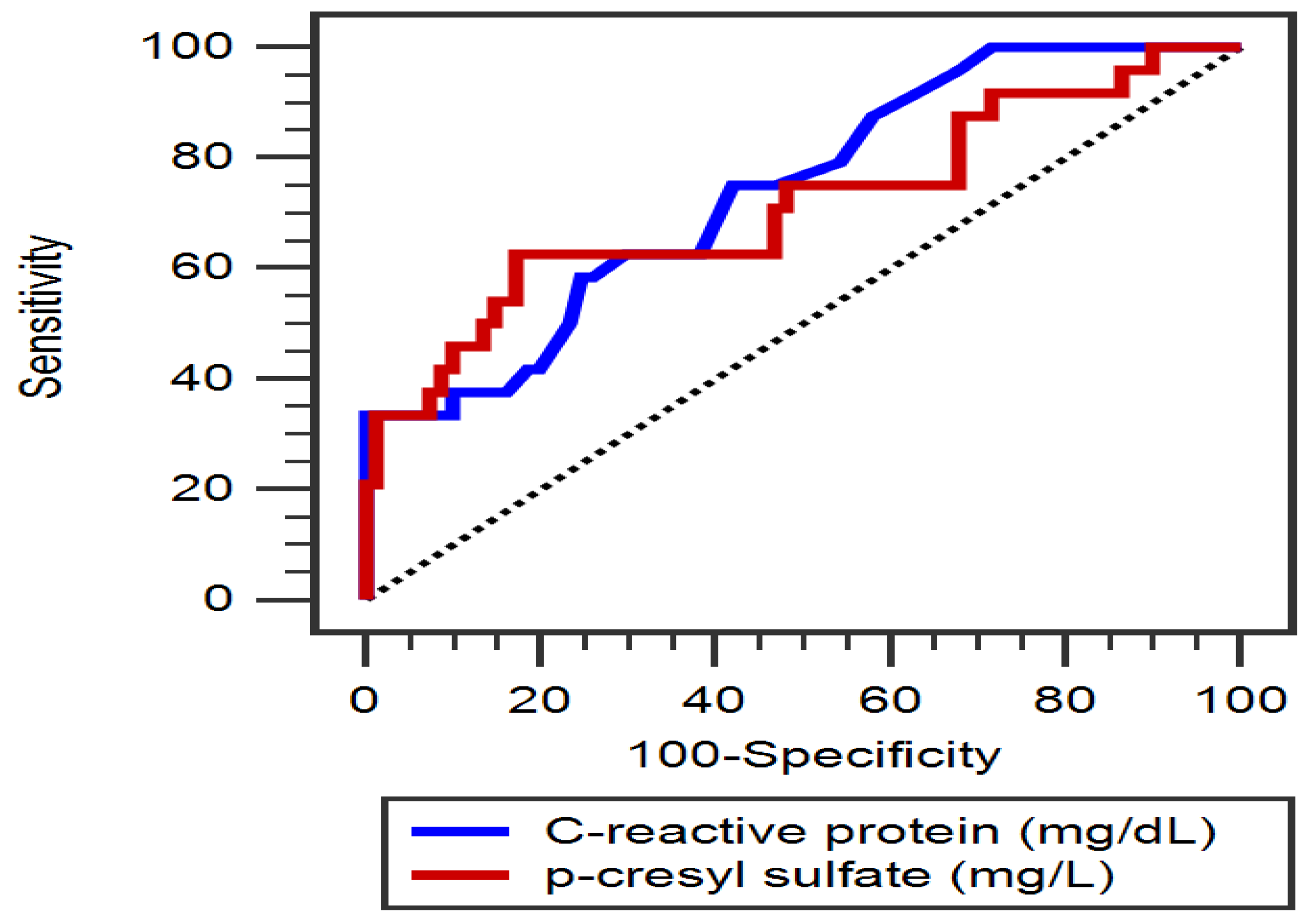
Figure 1. The area under the receiver operating characteristic curve indicates the diagnostic power of serum total p-cresyl sulfate level and C-reactive protein level for predicting peripheral artery disease among 105 hypertensive patients.
Diagnostics 15 01097 g001
Table 1. The clinical variables of the 105 hypertensive patients in the normal or low ankle brachial index group.
CharacteristicsAll Patients
(n = 105)
Control Group
(n = 81)
Low ABI Group
(n = 24)
p Value
Age (years)65.07 ± 10.4164.51 ± 9.8066.96 ± 12.290.313
Height (cm)161.01 ± 8.63161.41 ± 8.18159.67 ± 10.080.386
Body weight (kg)69.53 ± 12.8168.93 ± 12.2371.58 ± 14.710.375
Body mass index (kg/m2)26.73 ± 3.8226.36 ± 3.6027.96 ± 4.350.071
Left ankle-brachial index1.09 (1.01–1.13)1.11 (1.07–1.16)0.89 (0.83–0.94)<0.001 *
Right ankle-brachial index1.07 (1.00–1.13)1.09 (1.05–1.14)0.87 (0.83–0.93)<0.001 *
Systolic blood pressure (mmHg)131.95 ± 16.70132.02 ±17.13131.71 ± 15.520.936
Diastolic blood pressure (mmHg)74.50 ± 10.1774.60 ± 9.8574.13 ± 11.400.840
Total cholesterol (mg/dL)170.94 ± 41.04167.83 ± 40.10181.46 ± 43.310.154
Triglyceride (mg/dL)128.00 (91.00–172.50)119.00 (88.50–179.50)141.50 (122.50–164.25)0.262
HDL-C (mg/dL)44.52 ± 12.5944.32 ± 12.5145.21 ± 13.090.763
LDL-C (mg/dL)99.88 ± 31.8398.04 ± 32.70106.08 ± 28.470.279
Fasting glucose (mg/dL)107.00 (95.00–133.50)106.00 (94.50–131.50)114.00 (97.00–163.50)0.312
Blood urea nitrogen (mg/dL)17.02 ± 5.0716.79 ± 4.1117.79 ± 7.520.398
Creatinine (mg/dL)1.13 ± 0.301.10 ± 0.271.23 ± 0.390.054
eGFR (mL/min)66.89 ± 18.9468.69 ± 17.7560.80 ± 21.840.073
Total calcium (mg/dL)9.14 ± 0.379.14 ± 0.399.13 ± 0.300.919
Phosphorus (mg/dL)3.49 ± 0.523.50 ± 0.523.48 ± 0.510.860
iPTH (pg/mL)48.10 (33.75–65.25)47.30 (33.35–62.25)51.50 (34.68–75.08)0.296
CRP (mg/dL)0.20 (0.15–0.27)0.19 (0.14–0.25)0.26 (0.20–0.81)<0.001 *
Total p-cresyl sulfate (mg/L)6.91 (5.02–10.55)6.59 (4.53–8.32)11.02 (5.70–17.93) 0.002 *
Male, n (%)71 (67.6)56 (69.1)15 (62.5)0.542
Diabetes mellitus, n (%)33 (31.4)21 (25.9)12 (50.0) 0.026 *
Coronary artery disease, n (%)70 (66.7)52 (64.2)18 (75.0)0.324
Hyperlipidemia86 (81.9)68 (84)18 (75)0.319
Stable congestive heart failure16 (15.2)11 (13.6)5 (20.8)0.388
Stroke5 (4.8)3 (3.7)2 (8.3)0.352
ACE inhibitor use, n (%)34 (32.5)28 (34.6)6 (25.0)0.379
ARB use, n (%)49 (46.7)37 (45.7)12 (50.0)0.709
β-blocker use, n (%)57 (54.3)42 (51.90)15 (62.5)0.358
CCB use, n (%)40 (38.1)33 (40.7)7 (29.2)0.305
Statin use, n (%)60 (57.1)46 (56.8)14 (58.3)0.893
Fibrate use, n (%)21 (20.0)14 (17.3)7 (29.2)0.201
Aspirin, n (%)63 (60.0)48 (59.3)15 (62.5)0.776
Clopidogrel, n (%)30 (28.6)25 (30.9)5 (20.8)0.339
Values for continuous variables are shown as mean ± standard deviation after analysis by Student’s t-test; variables not normally distributed are shown as median and interquartile range after analysis by the Mann–Whitney U test; values are presented as number (%) and analysis after analysis by the chi-square test. ABI, ankle-brachial index; HDL-C, high-density lipoprotein cholesterol; LDL-C, low-density lipoprotein cholesterol; eGFR, estimated glomerular filtration rate; iPTH, intact parathyroid hormone; CRP, C-reactive protein; ACE, angiotensin-converting enzyme; ARB, angiotensin-receptor blocker; CCB, calcium-channel blocker. * p < 0.05 was considered statistically significant.
Table 2. Multivariable logistic regression analysis of the factors correlated to peripheral artery disease among the 105 hypertensive patients.
VariablesOdds Ratio95% Confidence Intervalp Value
Total p-cresyl sulfate, 1 mg/L1.1541.013–1.3150.031 *
C-reactive protein, 0.1 mg/dL1.6491.138–2.3890.008 *
Diabetes mellitus, present2.0950.609–7.1990.240
Body mass index, 1 kg/m21.0980.936–1.2880.249
eGFR, 1 mL/min0.9950.960–1.0300.766
Total cholesterol, 1 mg/dL1.0040.991–1.0170.547
eGFR, estimated glomerular filtration rate. Data was analyzed using the multivariate logistic regression analysis (adopted factors: diabetes mellitus, body mass index, estimated glomerular filtration rate, total cholesterol, C-reactive protein and total p-cresyl sulfate). * p < 0.05 was considered statistically significant.
Table 3. Spearman correlation coefficients between left ABI, right ABI, log-endocan and clinical variables in 164 patients with chronic kidney disease.
VariablesLeft Log-ABIRight Log-ABILog-PCS (mg/L)
Spearman Coefficient of Correlationp ValueSpearman Coefficient of Correlationp ValueSpearman Coefficient of Correlationp Value
Age (years)–0.1120.255–0.1870.0560.0310.752
Body mass index (kg/m2)–0.1390.157–0.0570.5610.0490.621
Left ABI0.690<0.001 *–0.321 0.001 *
Right ABI0.690<0.001 *–0.281 0.004 *
Log-PCS (mg/L)–0.321 0.001 *–0.281 0.004 *
SBP (mmHg)0.0420.669–0.0360.7180.0110.910
DBP (mmHg)0.0600.5450.0400.685–0.0090.926
Total cholesterol (mg/dL)–0.1790.068–0.0560.5740.1040.292
Log-Triglyceride (mg/dL)–0.0350.7230.0240.8110.0930.347
HDL-C (mg/dL)–0.0920.349–0.1200.224–0.0790.421
LDL-C (mg/dL)–0.1250.2030.0060.9480.1000.308
Log-Glucose (mg/dL)0.0040.972–0.0420.673–0.0050.961
eGFR (mL/min)0.1490.1290.199 0.042 *–0.325 0.001 *
Total calcium (mg/dL)–0.0050.964–0.0410.6750.1390.159
Phosphorus (mg/dL)0.0010.992–0.0220.8240.1560.111
Log-iPTH (pg/mL)–0.1530.119–0.0510.605–0.0610.576
Log-CRP (mg/L)–0.382<0.001 *–0.446<0.001 *0.220 0.024 *
Age (years)–0.1120.255–0.1870.0560.0310.752
Data of PCS, triglyceride, glucose, iPTH and CRP levels showed skewed distribution and, therefore, were log-transformed before analysis. Analysis of data was performed using the Spearman correlation analysis. ABI, ankle-brachial index; PCS, p-cresyl sulfate; SBP, systolic blood pressure; DBP, diastolic blood pressure; HDL-C, high-density lipoprotein cholesterol; LDL-C, low-density lipoprotein cholesterol; eGFR, estimated glomerular filtration rate; iPTH, intact parathyroid hormone; CRP, C-reactive protein. * p < 0.05 was considered statistically significant (two-tailed).
Disclaimer/Publisher’s Note: The statements, opinions and data contained in all publications are solely those of the individual author(s) and contributor(s) and not of MDPI and/or the editor(s). MDPI and/or the editor(s) disclaim responsibility for any injury to people or property resulting from any ideas, methods, instructions or products referred to in the content.