Article

Clinical and Electrophysiological Predictors of Isthmus Dependency in Atrial Flutter

Department of Cardiology, Ulm University Heart Center, Albert-Einstein-Allee 23, 89081 Ulm, Germany
*
Author to whom correspondence should be addressed.
Diagnostics 2025, 15(9), 1095; https://doi.org/10.3390/diagnostics15091095
Submission received: 10 February 2025 / Revised: 13 April 2025 / Accepted: 25 April 2025 / Published: 25 April 2025
(This article belongs to the Section Clinical Diagnosis and Prognosis)

Abstract

Background: Atrial flutter (AFL) is a macro-reentrant tachycardia classified as cavotricuspid isthmus (CTI)-dependent or non-CTI-dependent based on its reliance on the CTI for conduction. CTI dependence can present as type I ECG (sawtooth flutter waves in inferior leads and positive P-waves in V1) or type II ECG (absence of these characteristics). This study aimed to identify clinical and electrophysiological parameters to improve CTI dependence prediction in AFL. Methods: Patients at the Ulm University Heart Center between 2010 and 2019 with AFL undergoing electrophysiological studies and ablation were enrolled. Clinical and electrophysiological parameters such as age, gender, prior comorbidities, interventions, and medication use were analyzed. Results: The study included 383 patients, with 70% presenting with type I ECG AFL. CTI dependence was observed in 242 (90.3%) type I ECG patients and 52 (45.2%) type II ECG patients. CTI-dependent AFL patients were younger and had fewer comorbidities. Predictors for CTI dependence in type I ECG included male gender (p = 0.006), absence of beta-blocker use (p = 0.031), no prior atrial fibrillation (p = 0.035), and no prior pulmonary vein isolation (p < 0.001). In type II ECG, predictors for CTI dependence included younger age (p = 0.016), male gender (p = 0.007), absence of arterial hypertension (p = 0.036), and longer atrial cycle length (p < 0.001). Conclusions: Identifying clinical and electrophysiological parameters enhances the ability to predict CTI dependence in AFL, offering valuable insights for tailored diagnostic and therapeutic approaches. Coupling these parameters with ECG findings holds promise for refining prediction accuracy and optimizing patient care.

1. Introduction

Atrial flutter (AFL) is a common supraventricular arrhythmia characterized by a macro-reentrant circuit in the atria, with an incidence of 88 per 100,000 person-years [1]. According to its pathophysiologic mechanism, AFL can be classified into cavotricuspid isthmus (CTI)-dependent AFL, commonly referred to as typical AFL, and non-CTI-dependent AFL, known as atypical AFL [2]. Among these, typical AFL is the most prevalent and clinically relevant form. This arrhythmia is defined by a reentrant circuit that depends on the CTI, an anatomical region in the right atrium bordered by the tricuspid valve and the inferior vena cava (IVC). The CTI plays a pivotal role in the pathophysiology of typical AFL due to its slower conduction velocity compared to adjacent atrial tissue, which facilitates the formation and maintenance of a stable reentrant circuit with a cycle length (CL) between 200 and 260 ms [3,4,5,6,7]. Typical, counterclockwise (CCW) AFL is identified on electrocardiography (ECG) by its characteristic pattern of negative sawtooth flutter waves in the inferior leads (II, III, and aVF) and positive flutter waves in lead V1, referred to as the type I ECG pattern. In contrast, atypical AFL is not CTI-dependent and presents as a heterogeneous group of macro-reentrant arrhythmias, characterized more commonly by their inability to meet the criteria for a type I ECG and instead exhibiting a type II ECG pattern [3,8]. The reentrant circuit often involves other anatomical structures other than the CTI, such as atrial scars caused by prior surgeries, catheter ablations, or degenerative processes. Atypical AFL generally presents with less distinct ECG features, making its diagnosis more complex [3,6,9].
Clinically, AFL is associated with a variety of symptoms, including palpitations, dyspnea, fatigue, and dizziness, which can significantly impair a patient’s quality of life. AFL also carries a risk of serious complications, including new onset of atrial fibrillation (AF) (50%), stroke, thromboembolism, and tachycardia-induced cardiomyopathy [10,11,12,13,14]. While the diagnosis of AFL often begins with a 12-lead ECG, definitive confirmation of the arrhythmia mechanism and CTI dependency typically requires invasive electrophysiological studies (EPSs) [3]. As CTI dependence can be reliably confirmed through EPSs, non-CTI-dependent mechanisms remain more diverse, often involving macro-reentrant circuits around left atrial scars (post-ablation or surgical), the mitral isthmus, or septal regions, and require advanced mapping techniques for precise characterization [10,15].
Catheter ablation, specifically CTI ablation for typical AFL, is the preferred treatment due to its high success rates and a recurrence risk of less than 10% [16,17,18]. In patients with atypical, non-CTI-dependent AFL, the variability in circuit location and mechanism increases procedural complexity and reduces success rates, necessitating advanced mapping techniques and individualized approaches for accurate identification and effective treatment [16]. However, identifying predictors of CTI dependency in AFL is critical to improving diagnostic efficiency, guiding treatment decisions, and optimizing patient outcomes. This study aims to analyze clinical, electrophysiological, and ECG parameters associated with CTI-dependent AFL. By identifying reliable clinical predictors, this research seeks to enhance diagnostic accuracy, streamline invasive procedures, and improve the overall management of patients with AFL.

2. Materials and Methods

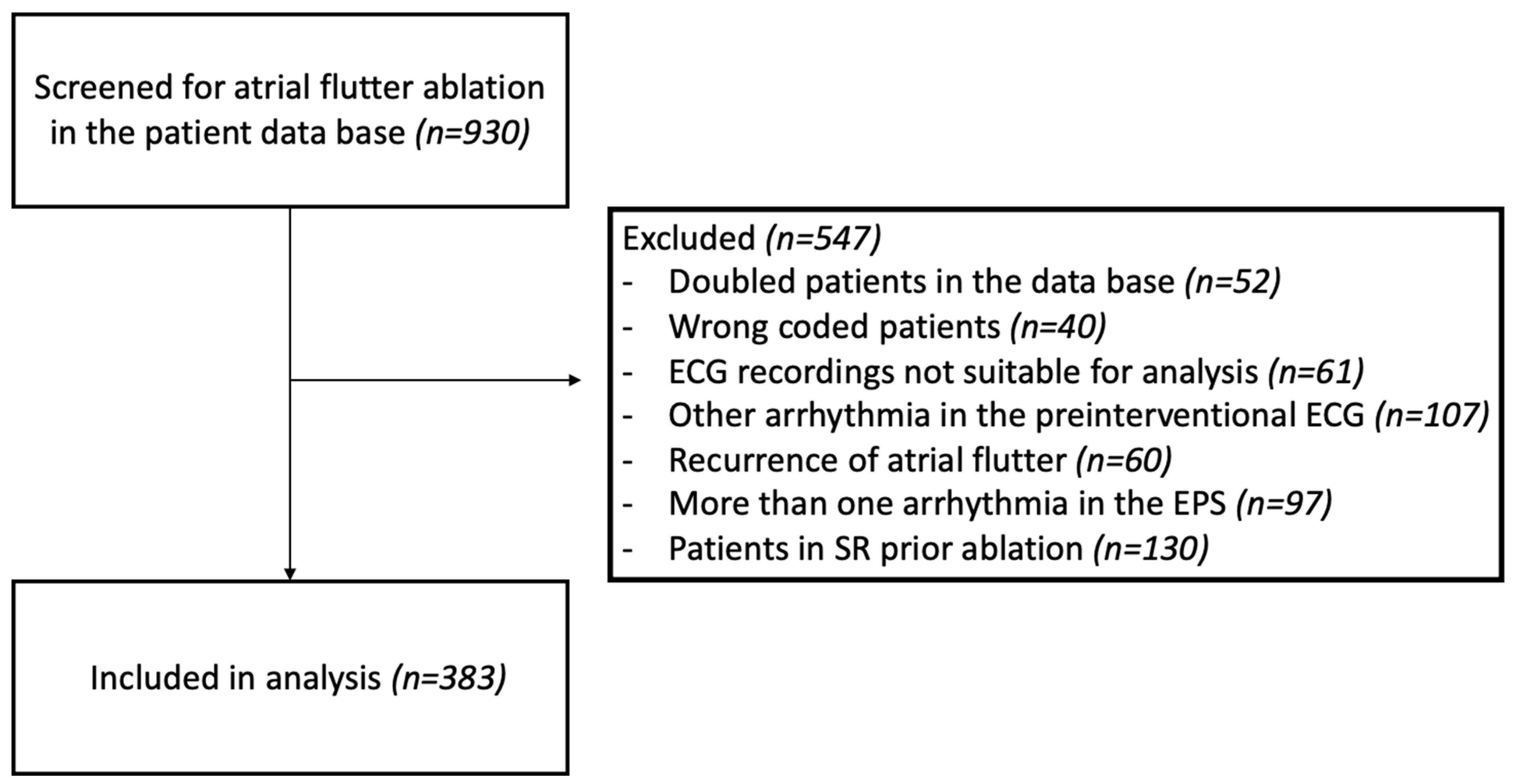
2.1. Study Population

For this retrospective cohort study, we screened 930 consecutive patients who underwent an EPS at Ulm University heart center between January 2010 and December 2019, including cases encoded as typical (ICD-10: I48.3) or atypical atrial flutter (ICD-10: I48.4) in the clinical data system. Additional cases were identified through a review of catheterization lab intervention plans. Informed consent was obtained from each patient. In total, 547 patients of the screened population were not eligible for enrolment for the study due to one or more of the following reasons: multiple listing or wrong coding of cases in the clinical data management system, ECG tracings not suitable for analyzing atrial waves, recurrence of AFL, clinical arrhythmia either not inducible or unstable or with changeable patterns or converted in sinus rhythm and a different mechanism of tachyarrhythmia detected at EPS (Figure 1).
A total of 383 patients presented with stable AFL ECG patterns and were eligible for further analysis. All patients underwent EPSs, and CTI-dependent AFL was confirmed by entrainment mapping and termination of AFL under CTI ablation. Patients’ characteristics, medical history, and ECGs were obtained from medical records prior to EPS and ablation. The study protocol adheres to the ethical guidelines outlined in the Declaration of Helsinki and was approved by the local ethics committee (ethics approval Nr. 324/16).

2.2. EPS and Ablation
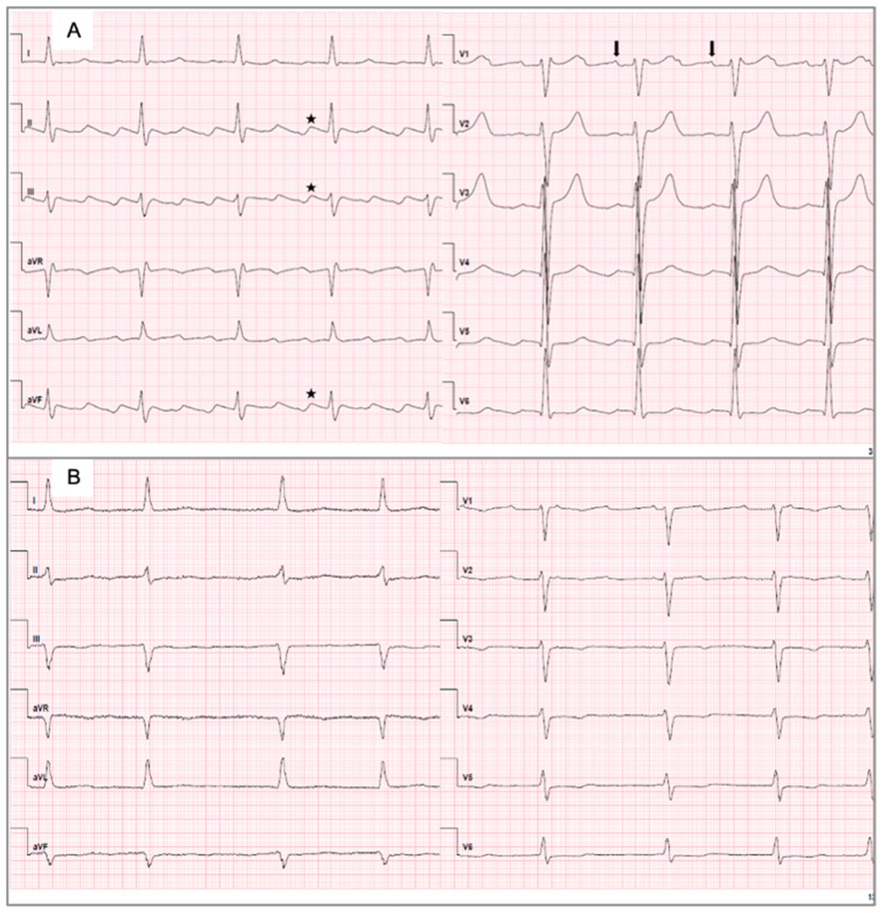
To categorize atrial arrhythmia in CTI-dependent and non-CTI-dependent AFL, all patients underwent EPSs. Patients with suspected CTI-dependent flutter received a conventional EPS to confirm the macro-reentrant tachycardia in the right atrium. Access to the right atrium was obtained via the right femoral vein, and a steerable decapolar diagnostic catheter (Inquiry, Abbott, formerly St. Jude Medical, St. Paul, MN, USA) was placed in the coronary sinus (CS) or positioned at the tricuspid annulus. To determine the post-pacing interval (PPI), entrainment was performed from the two proximal and two distal electrodes of the diagnostic catheter with a CL slightly shorter than the baseline AFL CL. A return CL equal to or within 20 ms of AFL CL at the CS ostium, the lower free wall of the right atrium (low right atrium‚ LRA), and on the CTI and a concealed entrainment at these sites are considered as CTI-dependent or typical AFL [4].
The direction of typical AFL was determined by the activation pattern of the decapolar catheter at the tricuspid annulus and classified as clockwise (CW) or CCW. After confirming CTI-dependent AFL, a deflectable-tip ablation catheter (Blazer II XP™, 8 mm, Boston Scientific, Marlborough, MA, USA; Thermocool® Celcius, Thermocool® Smarttouch SF, Biosense Webster, Irvine, CA, USA; Therapy™ Cool Flex™, St. Jude Medical, St. Paul, MN, USA; AlCath LT Gold FullCircle, Biotronik, Berlin, Germany) was placed on the CTI and radiofrequency energy was applied at 6 o’clock of the tricuspid annulus in left anterior oblique (LAO) 40° view. The ablation line was established between the tricuspid annulus and the IVC orifice. Termination of AFL confirmed ablation within the macro-reentrant circuit. Directly after completion of CTI ablation and after a waiting time of up to 30 min, a bidirectional block was confirmed by determination of the activation pattern and conduction time from the CS ostium to the LRA and vice versa. If EPS detected non-CTI-dependent AFL, atrial arrhythmia was terminated via electrical direct current cardioversion and ablation was rescheduled using an electroanatomical mapping and ablation system.

2.3. ECG Analysis

A 12-lead ECG of every patient was recorded prior to the EPS and ablation. All ECGs were independently reviewed by at least two experienced electrophysiologists from a high-volume tertiary electrophysiology center, each with substantial expertise in arrhythmia interpretation. In cases of disagreement, a third independent electrophysiologist was consulted to reach consensus. This rigorous multi-review approach aimed to ensure high diagnostic accuracy and reproducibility in distinguishing between type I and type II ECG patterns. ECGs were considered as type I ECGs (Figure 2A) if the characteristic ECG sawtooth pattern was present in leads II and III, and/or aVF with slow downsloping and a negative segment, followed by a shorter, sharply positive deflection leading to the next downsloping segment, and a positive P-wave in V1. On the contrary, either an inverse sawtooth pattern, with a slow upsloping positive segment followed by a shorter negative deflection, no or no clear sawtooth pattern in the inferior leads, negative or biphasic P-waves or low voltages without clear, identifiable P-waves in V1 were considered as type II ECGs (Figure 2B).

2.4. Statistical Analysis

Statistical analyses were performed using SPSS® software (SPSS, V29, Chicago, IL, USA). Categorical variables are described as absolute and relative frequencies, and continuous variables are expressed as mean ± standard deviation (SD). Univariate analyses were performed using a binominal logistic regression model, and predictors that were independently associated with an unusual ECG pattern were analyzed in a multivariate logistic regression model. A p-value of <0.05 in the univariate analysis qualified to enter the multivariate model. Groups were compared using a two-sided t-test or a Mann–Whitney test, as appropriate. For subgroup analyses, p-values reflected overall group differences, and post hoc testing was conducted to identify specific between-group differences. A two-tailed p-value of <0.05 was considered statistically significant. Receiver operator characteristic (ROC) analyses were performed to determine the area under the curve (AUC) and the sensitivity/specificity of the predictors. Analysis of variance (ANOVA) tests were conducted to assess differences among multiple groups for continuous variables, while chi-square tests were used for categorical variables to evaluate associations across multiple groups. If ANOVA showed significant differences between groups, additional post hoc tests were performed to determine which specific groups differed from each other.

3. Results

3.1. Baseline Characteristics

We included 383 patients diagnosed with AFL due to characteristic ECG patterns. The majority of patients were male (300/383 patients, 78.3%), and the mean age of patients was 69.3 ± 11.2 years. On hospital admission, 268 patients (70%) presented a type I ECG and 115 patients (30%) showed a type II ECG. The majority of patients were diagnosed with CTI-dependent atrial flutter (294/383 patients, 77%) (Table 1).
There was a high prevalence of cardiovascular comorbidities, like arterial hypertension (275/383 patients, 72%), diabetes mellitus (89/383 patients, 23%), coronary heart disease (149/383 patients, 39%), and prior AF (145/383 patients, 38%). A total of 185 patients (48.3%) had heart failure (HF), of whom 70 patients (18.3%) were diagnosed with HF with reduced ejection fraction (HFrEF), 61 patients (15.9%) with HF with preserved ejection fraction (HFpEF), and 67 patients (17.5%) with HF with mildly reduced ejection fraction (HFmrEF). The mean left ventricular ejection fraction (LVEF) was 50 ± 15%. A history of prior right or left atrial ablation was present in 3.4% (13/383 patients) of these patients, 9.1% (35/383 patients) of the cohort had prior pulmonary vein isolation, and 16.4% (63/383 patients) of patients had prior heart surgery. Detailed information on patients’ baseline characteristics is provided in Table 2.

3.1.1. Baseline Characteristics of Patients with Type I ECGs and Type II ECGs

In total, 70% (268 patients) of the cohort exhibited type I ECG recordings, while 30% (115 patients) presented with a type II ECG pattern. Patients with type II ECGs were older (73.0 ± 11.1 vs. 67.8 ± 10.9; p < 0.001) and more frequently female (30.4% vs. 17.9%; p = 0.006). Regarding cardiovascular risk factors, diabetes mellitus was more prevalent in patients with type II ECGs (33.0% vs. 19.1%; p = 0.003). Furthermore, patients with type II ECGs had a higher CHA2DS2-VASc score (3.8 ± 1.7 vs. 2.7 ± 1.6; p < 0.001), a greater incidence of stroke history (15.7% vs. 6.7%; p = 0.006), and borderline significantly more often a diagnosis of AF compared to patients with type I ECGs (45.2% vs. 34.7%; p = 0.052). In 90.3% of cases, patients with type I ECGs exhibited CTI-dependent AFL, while patients with type II ECGs showed CTI-dependent atrial flutter in almost half of the cases (45.2%). Detailed information on patients’ characteristics compared between type I and type II ECGs is provided in Table 3.

3.1.2. Baseline Characteristics of Patients with CTI-Dependent and Non-CTI-Dependent AFL

Among the cohort, 76.7% of the patients had CTI-dependent AFL, while 23.2% of the patients had non-CTI-dependent AFL. Patients with non-CTI-dependent AFL were older (74.1 ± 10.1 vs. 67.9 ± 11.1; p < 0.001) and more frequently female (40.4% vs. 16.0%, p < 0.001). Almost three-quarters (83.1%) of patients with non-CTI-dependent AFL were diagnosed with arterial hypertension, whereas patients with CTI-dependent AFL were significantly less likely to have arterial hypertension (68.4%). Patients with non-CTI-dependent AFL had a significantly higher CHA2DS2-VASc score compared to patients with CTI-dependent AFL (3.9 ± 1.7 vs. 2.8 ± 1.7; p < 0.001), were more frequently diagnosed with AF (50.6% vs. 34.0%; p = 0.005), had a higher prevalence of prior pulmonary vein isolation (20.2% vs. 5.8%, p < 0.001), were more frequently prescribed beta blockers (87.7% vs. 72.9%; p = 0.006), and had a higher incidence of stroke history (14.6% vs. 7.8%; p = 0.055). Among patients with CTI-dependent AFL, 82.3% showed a type I ECG, while 29.2% of patients with non-CTI-dependent AFL also exhibited a type I ECG. Detailed information on patient characteristics for those with CTI-dependent and non-CTI-dependent AFL is provided in Table 4.

3.1.3. Baseline Characteristics of Subgroups Based on CTI Dependence and ECG Records

Based on the cross-tabulation (Table 1) mentioned earlier, four distinct subgroups can be identified: patients with CTI-dependent AFL and type I ECGs, patients with non-CTI-dependent AFL and type I ECGs, patients with CTI-dependent AFL and type II ECGs, and patients with non-CTI-dependent AFL and type II ECGs. Analysis of the patient characteristics of these groups revealed significant differences in age, sex, arterial hypertension, diabetes mellitus, beta-blocker intake, prior AF, prior pulmonary vein isolation, and prior heart surgery. Details are provided in Table 5.

3.2. Characteristics of the ECG and Electrophysiological Parameters Between Subgroups

The analysis of ECG and EPS characteristics between the subgroups during ongoing tachycardia demonstrated no statistically significant differences in terms of heart rate (p = 0.461) or QRS width (p = 0.315). The atrial CL in the group with CTI-dependent AFL and type II ECG was significantly longer (250.6 ± 44.1 ms) compared to the other groups (based on ECG characteristics: p = 0.023; EPS characteristics: p = 0.001). Furthermore, a statistically significant deviation was observed in the number of patients with an atrial CL over 245 ms (p ≤ 0.001) (Table 6).

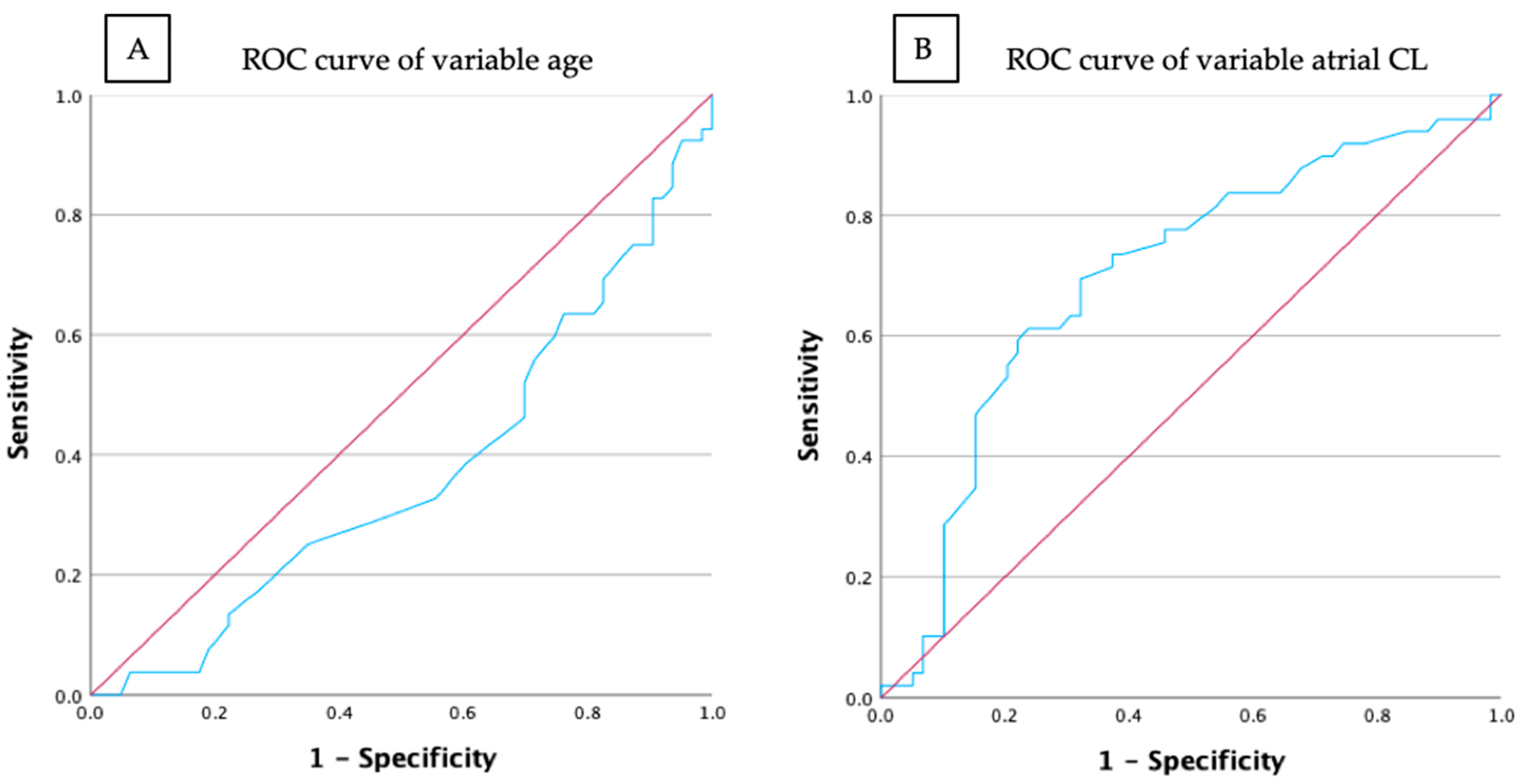
3.3. Prediction Models for CTI-Dependent AFL and Type I ECG

Binary logistic regression revealed that female gender significantly decreases the likelihood of CTI-dependent AFL (p = 0.006). Furthermore, beta-blocker use (odds ratio 0.109, p = 0.014), a history of AF (odds ratio 0.415, p = 0.035), and previous pulmonary vein isolation (odds ratio 0.099, p < 0.001) significantly reduced the likelihood of CTI-dependent AFL (Table 7).
Multivariate analysis with the independent variable including patients with a history of AF, male gender increased the likelihood of CTI-dependent AFL by 201.6% (odds ratio 3.016, p = 0.019), and beta-blocker use reduced it by 87.8% (odds ratio 0.122, p = 0.043) (Table 8). Prior AF and prior PVI are interrelated and mutually influence each other, leading to potential multicollinearity. Therefore, only one variable was included to maintain statistical validity.

3.4. Prediction Models for CTI-Dependent AFL and Type II ECG

In the binary logistic regression analysis of patients with type II ECGs, arterial hypertension (odds ratio 0.375, p = 0.036), a higher CHA2DS2VASc score (odds ratio 0.758, p = 0.017), and increasing age were associated with a reduced likelihood of CTI-dependent AFL. In contrast, male gender (odds ratio 3.356, p = 0.007), prior cardiac surgery (odds ratio 3.056, p = 0.021), and longer atrial CL were associated with an increased likelihood of CTI-dependent AFL. Specifically, patients with an atrial CL > 245 ms were nearly three times more likely to develop CTI-dependent AFL and type II ECG compared to those with shorter atrial CL (Table 9).
The ROC analysis identified 75 years as the optimal threshold for differentiating CTI-dependent from CTI-independent AFL in patients with type II ECGs. However, the low AUC of 0.363 indicates limited discriminatory power of the parameter “Age > 75 years” for predicting CTI dependency. The ROC curve and the calculated AUC of 0.703 for the atrial CL measured during the EPS demonstrated superior discriminatory ability compared to age (Figure 3).
In the multivariate analysis, which included the significant variables from the binary logistic regression, male gender was associated with a 163.1% increase in the likelihood of CTI-dependent AFL (p = 0.045). Additionally, an atrial CL over 245 ms increased the likelihood of CTI-dependent AFL by 146.5% (p = 0.035). The remaining variables did not demonstrate statistical significance (Table 10).

4. Discussion

AFL is one of the most common cardiac arrhythmias, often occurring in combination with other arrhythmias such as AF [19,20]. Although it is characterized by a distinct electrophysiological substrate, it is frequently misdiagnosed by physicians [21,22]. Moreover, AFL is associated with elevated mortality rates, primarily due to its correlation with an increased risk of developing AF (50%), stroke, and other heart diseases [22,23,24,25]. Direct current cardioversion and pharmacological therapy are recommended for acute management of AFL, particularly in patients presenting with hemodynamic instability or symptomatic arrhythmia. While these modalities are effective for acute rhythm/rate control, catheter ablation is the most effective intervention for the long-term management of symptomatic and recurrent AFL episodes (class I recommendation) [16]. Consequently, in specialized centers, it is the most commonly performed procedure following pulmonary vein isolation [26,27,28]. However, there are various diagnostic modalities, making the differentiation between CTI-dependent and non-CTI-dependent AFL crucial for determining the optimal ablation approach. The aim of this study is to conduct a comprehensive analysis of the AFL cohort, encompassing patients with type I and type II ECGs as well as those with CTI-dependent and non-CTI-dependent AFL. Furthermore, the study aimed to identify clinical parameters with prognostic value for CTI dependence in AFL and to evaluate their predictive accuracy and diagnostic reliability.
In this study, patients with type I ECGs represented a younger and healthier cohort compared to those with type II ECGs, who had a higher prevalence of diabetes mellitus and stroke, as well as higher CHA2DS2-VASc scores. Similarly, CTI-dependent AFL was more prevalent in younger patients with fewer comorbidities. Compared to the study cohort of Barbato et al., where patients with prior valve interventions were excluded, non-CTI-dependent AFL in our study was significantly associated with older age [29]. The predictive relevance of age may result from an increased prevalence of cardiovascular diseases in older individuals, leading to atrial remodeling and favoring non-CTI-dependent conduction patterns [30]. Additional factors related to cardiac aging, such as oxidative stress, fibrosis, and slowed atrial conduction, may contribute to the formation of macro-reentrant tachycardias and atypical AFL pathways [31,32,33,34]. Although EPS remains the definitive method to confirm the mechanism of AFL, the clinical and electrophysiological predictors identified in our study are not intended as a substitute but rather as supportive tools to enhance diagnostic efficiency and patient stratification prior to invasive evaluation.
The current study demonstrated that additional clinical parameters also provide valuable predictive insights into the CTI dependence of AFL. In patients with type I ECGs, prior pulmonary vein isolation and AF significantly reduced the likelihood of CTI-dependent AFL, likely due to atrial remodeling induced by these conditions and their treatment [34,35,36]. Additionally, as previously reported by Gu et al., CTI-dependent AFL may present atypically on ECGs following ablation procedures in the left atrium, a finding that aligns with the results of this study [11]. Moreover, the use of beta blockers increased the probability of non-CTI-dependent AFL in patients with type I ECGs, possibly due to their negative bathmotropic effects on atrial conduction and their interaction with rate control in the context of AF management [37,38]. Male gender was strongly associated with CTI dependence in patients with both type I and type II ECGs, consistent with epidemiological data demonstrating a higher prevalence of AFL in men [1,16]. In contrast, women in this study tended to be older and more likely to present with non-CTI-dependent AFL, possibly due to secondary structural atrial remodeling linked to advanced age and cardiovascular comorbidities [30,34,39].
Longer atrial CL was identified as another significant predictive parameter for CTI dependence in patients with type II ECGs. Previous studies have shown a positive correlation between age and CL in CTI-dependent AFL, suggesting that age-related slowing of atrial conduction contributes to this association [32,40]. In the current study, ROC analysis of EPS-measured atrial CL demonstrated moderate discriminatory accuracy in differentiating CTI-dependent from CTI-independent AFL. However, the atrial CL was not a significant predictor of CTI dependence in patients with type I ECGs. Due to the retrospective nature of our study, only atrial CLs were systematically available for both CTI-dependent and non-CTI-dependent AFL. More detailed electrophysiological data, especially for the non-CTI-dependent group, were not uniformly recorded and thus could not be included.
The subsequent data analysis revealed that prior cardiac surgery increased the likelihood of CTI-dependent AFL in patients with type II ECGs, aligning with studies identifying CTI-dependent AFL as the most common atrial tachycardia following open-heart surgery. Postoperatively, larger atrial scars were associated with non-CTI-dependent AFL [41].
The final predictive factor analyzed in this study was arterial hypertension, which was associated with a reduced likelihood of CTI-dependent AFL in patients with type II ECGs. The increasing prevalence of hypertension with advancing age contributes to atrial remodeling processes, including the formation of low-voltage areas, thereby indicating a potential interaction between these two predictors [42].
The analysis presented in this study highlights the intricate interplay between clinical and electrophysiological parameters in predicting CTI dependence in AFL. A deeper understanding of these interactions has the potential to refine diagnostic criteria and guide the development of personalized therapeutic strategies. Key mechanistic factors, such as atrial scar burden or atrial size, were not systematically available in our retrospective cohort and should be incorporated in future studies. However, further investigation is required to validate these findings in larger cohorts, and improve the predictive accuracy for distinguishing CTI-dependent from non-CTI-dependent AFL.

Limitations

As a retrospective, single-center analysis, this study may have limited generalizability. Although basic EPS parameters were included, detailed electrophysiological mapping data, especially for non-CTI-dependent AFL, were not uniformly available. Due to the observational design, causal relationships cannot be established.

5. Conclusions

In this study, the vast majority of patients with type I ECGs and approximately half of those with type II ECGs demonstrated CTI-dependent AFL. Nonetheless, well-defined clinical and electrophysiological predictors are crucial for improving diagnostic precision and optimizing therapeutic strategies. Our analysis identified male gender, absence of beta-blocker use, and no prior AF/PVI as the most reliable predictors of CTI dependence in patients with type I ECG AFL. For type II ECG AFL, significant predictors of CTI dependence included younger age, male gender, absence of arterial hypertension, lower CHA2DS2-VASc scores, prior cardiac surgery, and longer atrial CL. Coupling these parameters with ECG findings holds promise for refining prediction accuracy and optimizing patient care. Further studies are required to validate these findings, investigate additional predictive factors, and develop comprehensive scoring systems for more accurate risk stratification and individualized treatment approaches.

Author Contributions

Conceptualization: L.K. and K.W.-E.; methodology: L.K. and K.W.-E.; software: L.K., K.W.-E. and S.R.; validation of ECG: S.R., A.S., D.A. and K.W.-E.; formal analysis: L.K., S.R., F.D. and K.W.-E.; investigation: L.K., S.R. and K.W.-E.; resources: K.W.-E. and W.R.; data curation: L.K., K.W.-E., D.A., Y.T., S.R. and A.S.; writing—original draft preparation, L.K. and K.W.-E.; writing—review and editing: Y.T., F.D., C.B., K.W.-E. and W.R.; visualization: L.K. and K.W.-E.; supervision: K.W.-E. and W.R.; project administration: L.K. and K.W.-E. All authors have read and agreed to the published version of the manuscript.

Funding

This research received no external funding.

Institutional Review Board Statement

The study protocol adheres to the ethical guidelines outlined in the Declaration of Helsinki and was approved by the local ethics committee (ethics approval Nr. 324/16, 12 October 2016).

Informed Consent Statement

Informed consent was obtained from all subjects involved in the study.

Data Availability Statement

The data presented in this study are available on request from the corresponding author. The data are not publicly available due to data privacy laws.

Acknowledgments

K.W.-E. was supported by a Hertha-Nathorff fellowship and the clinician scientist program of Ulm University.

Conflicts of Interest

The authors declare no conflicts of interest.

Abbreviations

The following abbreviations are used in this manuscript:
AAsAntiarrhythmics
AATAntiarrhythmic therapy
AFAtrial fibrillation
AFLAtrial flutter
ANOVAAnalysis of variance
AUCArea under the curve
BMIBody mass index
BpmBeats per minute
CCWCounterclockwise
CLCycle length
COPDChronic obstructive pulmonary disease
CSCoronary sinus
CTICavotricuspid isthmus
CWClockwise
DfDegrees of freedom
ECGElectrocardiogram
EPSElectrophysiological study
ICDInternational classification of diseases
IVCInferior vena cava
LALeft atrial
LAOLeft anterior oblique
LRALow right atrium
N/ANot applicable
OACOral anticoagulation
OROdds ratio
OSASObstructive sleep apnea syndrome
PMPacemaker
PPIPost-pacing interval
PVIPulmonary vein isolation
RARight atrial
ROCReceiver operator characteristics
SDStandard deviation
Sig.Significance

References

  1. Granada, J.; Uribe, W.; Chyou, P.-H.; Maassen, K.; Vierkant, R.; Smith, P.N.; Hayes, J.; Eaker, E.; Vidaillet, H. Incidence and Predictors of Atrial Flutter in the General Population. J. Am. Coll. Cardiol. 2000, 36, 2242–2246. [Google Scholar] [CrossRef]
  2. Ebner, B.; Grant, J.; Vincent, L.; Maning, J.; Olorunfemi, O.; Olarte, N.; Colombo, R.; Lambrakos, L.; Mendoza, I. Comparison of In-Hospital Outcomes of Patients Undergoing Catheter Ablation for Typical versus Atypical Atrial Flutter. J. Interv. Card. Electrophysiol. 2022, 63, 295–302. [Google Scholar] [CrossRef]
  3. Bun, S.-S.; Latcu, D.G.; Marchlinski, F.; Saoudi, N. Atrial Flutter: More than Just One of a Kind. Eur. Heart J. 2015, 36, 2356–2363. [Google Scholar] [CrossRef]
  4. Bun, S.-S.; Lațcu, D.G.; Wedn, A.M.; Squara, F.; Scarlatti, D.; Theodore, G.; Al Amoura, A.; Benaïch, F.A.; Hasni, K.; Saoudi, N.; et al. Cavotricuspid Isthmus Is Constantly a Zone of Slow Conduction: Data from Ultra-High-Resolution Mapping. Pacing Clin. Electrophysiol. PACE 2020, 43, 189–193. [Google Scholar] [CrossRef] [PubMed]
  5. Olgin, J.E.; Kalman, J.M.; Fitzpatrick, A.P.; Lesh, M.D. Role of Right Atrial Endocardial Structures as Barriers to Conduction during Human Type I Atrial Flutter. Activation and Entrainment Mapping Guided by Intracardiac Echocardiography. Circulation 1995, 92, 1839–1848. [Google Scholar] [CrossRef] [PubMed]
  6. Friedman, P.A.; Luria, D.; Fenton, A.M.; Munger, T.M.; Jahangir, A.; Shen, W.K.; Rea, R.F.; Stanton, M.S.; Hammill, S.C.; Packer, D.L. Global Right Atrial Mapping of Human Atrial Flutter: The Presence of Posteromedial (Sinus Venosa Region) Functional Block and Double Potentials: A Study in Biplane Fluoroscopy and Intracardiac Echocardiography. Circulation 2000, 101, 1568–1577. [Google Scholar] [CrossRef]
  7. Doiny, D.; Merino, J.L. Atrial Flutter: Common and Main Atypical Forms. Available online: https://www.escardio.org/Journals/E-Journal-of-Cardiology-Practice/Volume-11/Atrial-flutter-common-and-main-atypical-forms (accessed on 9 May 2024).
  8. Puech, P.; Latour, H.; Grolleau, R. Flutter and his limits. Arch. Mal. Coeur Vaiss. 1970, 63, 116–144. [Google Scholar] [PubMed]
  9. Saoudi, N.; Cosío, F.; Waldo, A.; Chen, S.A.; Iesaka, Y.; Lesh, M.; Saksena, S.; Salerno, J.; Schoels, W.; Working Group of Arrhythmias of the European of Cardiology and the North American Society of Pacing and Electrophysiology. A Classification of Atrial Flutter and Regular Atrial Tachycardia According to Electrophysiological Mechanisms and Anatomical Bases; a Statement from a Joint Expert Group from The Working Group of Arrhythmias of the European Society of Cardiology and the North American Society of Pacing and Electrophysiology. Eur. Heart J. 2001, 22, 1162–1182. [Google Scholar] [CrossRef]
  10. Cosio, F.G. Atrial Flutter, Typical and Atypical: A Review. Arrhythm. Electrophysiol. Rev. 2017, 6, 55–62. [Google Scholar] [CrossRef]
  11. Gu, Z.; Jiao, J.; Shen, Y.; Ding, X.; Zhu, C.; Li, M.; Chen, H.; Ju, W.; Gu, K.; Yang, G.; et al. A Simple Score to Predict New-Onset Atrial Fibrillation After Ablation of Typical Atrial Flutter. Can. J. Cardiol. 2024. [CrossRef]
  12. Giehm-Reese, M.; Kronborg, M.B.; Lukac, P.; Kristiansen, S.B.; Jensen, H.K.; Gerdes, C.; Kristensen, J.; Nielsen, J.M.; Nielsen, J.C. Recurrent Atrial Arrhythmia in a Randomised Controlled Trial Comparing Contact Force-Guided and Contact Force-Blinded Ablation for Typical Atrial Flutter. J. Interv. Card. Electrophysiol. 2022, 63, 699–707. [Google Scholar] [CrossRef]
  13. Li, J.-H.; Xie, H.-Y.; Chen, Y.-Q.; Cao, Z.-J.; Tang, Q.-H.; Guo, X.-G.; Sun, Q.; Ma, J. Risk of New-Onset Atrial Fibrillation Post-Cavotricuspid Isthmus Ablation in Typical Atrial Flutter Without History of Atrial Fibrillation. Front. Physiol. 2021, 12, 763478. [Google Scholar] [CrossRef] [PubMed]
  14. Maskoun, W.; Pino, M.I.; Ayoub, K.; Llanos, O.L.; Almomani, A.; Nairooz, R.; Hakeem, A.; Miller, J. Incidence of Atrial Fibrillation After Atrial Flutter Ablation. JACC Clin. Electrophysiol. 2016, 2, 682–690. [Google Scholar] [CrossRef] [PubMed]
  15. Glover, B.M.; Chen, J.; Hong, K.L.; Boveda, S.; Baranchuk, A.; Haugaa, K.H.; Dorian, P.; Potpara, T.S.; Crystal, E.; Mitchell, B.; et al. Catheter ablation for atrial flutter: A survey by the European Heart Rhythm Association and Canadian Heart Rhythm Society. EP Eur. 2016, 18, 1880–1885. [Google Scholar] [CrossRef] [PubMed]
  16. Brugada, J.; Katritsis, D.G.; Arbelo, E.; Arribas, F.; Bax, J.J.; Blomström-Lundqvist, C.; Calkins, H.; Corrado, D.; Deftereos, S.G.; Diller, G.-P.; et al. 2019 ESC Guidelines for the Management of Patients with Supraventricular tachycardiaThe Task Force for the Management of Patients with Supraventricular Tachycardia of the European Society of Cardiology (ESC): Developed in Collaboration with the Association for European Paediatric and Congenital Cardiology (AEPC). Eur. Heart J. 2020, 41, 655–720. [Google Scholar] [CrossRef]
  17. Schwartzman, D.; Callans, D.J.; Gottlieb, C.D.; Dillon, S.M.; Movsowitz, C.; Marchlinski, F.E. Conduction Block in the Inferior Vena Caval-Tricuspid Valve Isthmus: Association with Outcome of Radiofrequency Ablation of Type I Atrial Flutter. J. Am. Coll. Cardiol. 1996, 28, 1519–1531. [Google Scholar] [CrossRef]
  18. Natale, A.; Newby, K.H.; Pisanó, E.; Leonelli, F.; Fanelli, R.; Potenza, D.; Beheiry, S.; Tomassoni, G. Prospective Randomized Comparison of Antiarrhythmic Therapy versus First-Line Radiofrequency Ablation in Patients with Atrial Flutter. J. Am. Coll. Cardiol. 2000, 35, 1898–1904. [Google Scholar] [CrossRef]
  19. Lloyd-Jones, D.M.; Wang, T.J.; Leip, E.P.; Larson, M.G.; Levy, D.; Vasan, R.S.; D’Agostino, R.B.; Massaro, J.M.; Beiser, A.; Wolf, P.A.; et al. Lifetime Risk for Development of Atrial Fibrillation. Circulation 2004, 110, 1042–1046. [Google Scholar] [CrossRef]
  20. Van Gelder, I.C.; Rienstra, M.; Bunting, K.V.; Casado-Arroyo, R.; Caso, V.; Crijns, H.J.G.M.; De Potter, T.J.R.; Dwight, J.; Guasti, L.; Hanke, T.; et al. 2024 ESC Guidelines for the Management of Atrial Fibrillation Developed in Collaboration with the European Association for Cardio-Thoracic Surgery (EACTS): Developed by the Task Force for the Management of Atrial Fibrillation of the European Society of Cardiology (ESC), with the Special Contribution of the European Heart Rhythm Association (EHRA) of the ESC. Endorsed by the European Stroke Organisation (ESO). Eur. Heart J. 2024, 45, 3314–3414. [Google Scholar] [CrossRef]
  21. Knight, B.P.; Michaud, G.F.; Strickberger, S.A.; Morady, F. Electrocardiographic Differentiation of Atrial Flutter from Atrial Fibrillation by Physicians. J. Electrocardiol. 1999, 32, 315–319. [Google Scholar] [CrossRef]
  22. Rahman, F.; Wang, N.; Yin, X.; Ellinor, P.T.; Lubitz, S.A.; LeLorier, P.A.; McManus, D.D.; Sullivan, L.M.; Seshadri, S.; Vasan, R.S.; et al. Atrial Flutter: Clinical Risk Factors and Adverse Outcomes in the Framingham Heart Study. Heart Rhythm 2016, 13, 233–240. [Google Scholar] [CrossRef]
  23. Katov, L.; Teumer, Y.; Schlarb, A.; Reiländer, S.; Aktolga, D.; Diofano, F.; Bothner, C.; Rottbauer, W.; Weinmann-Emhardt, K. Comparative Efficacy of Cavotricuspid Isthmus Ablation in Sinus Rhythm Versus Typical Atrial Flutter. Hearts 2024, 5, 482–490. [Google Scholar] [CrossRef]
  24. Jilek, C.; Gleirscher, L.; Strzelczyk, E.; Sepela, D.; Tiemann, K.; Lewalter, T. Rechtsatriales isthmusabhängiges Vorhofflattern. Herzschrittmachertherapie Elektrophysiol. 2023, 34, 291–297. [Google Scholar] [CrossRef] [PubMed]
  25. Chinitz, J.S.; Gerstenfeld, E.P.; Marchlinski, F.E.; Callans, D.J. Atrial Fibrillation Is Common after Ablation of Isolated Atrial Flutter during Long-Term Follow-Up. Heart Rhythm 2007, 4, 1029–1033. [Google Scholar] [CrossRef]
  26. García-Fernández, F.J.; Ibáñez Criado, J.L.; Quesada Dorador, A.; Collaborators of the Spanish Catheter Ablation Registry; REGISTRY COLLABORATORS. Spanish Catheter Ablation Registry. 17th Official Report of the Spanish Society of Cardiology Working Group on Electrophysiology and Arrhythmias (2017). Rev. Esp. Cardiol. 2018, 71, 941–951. [Google Scholar] [CrossRef] [PubMed]
  27. Hosseini, S.M.; Rozen, G.; Saleh, A.; Vaid, J.; Biton, Y.; Moazzami, K.; Heist, E.K.; Mansour, M.C.; Kaadan, M.I.; Vangel, M.; et al. Catheter Ablation for Cardiac Arrhythmias: Utilization and In-Hospital Complications, 2000 to 2013. JACC Clin. Electrophysiol. 2017, 3, 1240–1248. [Google Scholar] [CrossRef]
  28. Holmqvist, F.; Kesek, M.; Englund, A.; Blomström-Lundqvist, C.; Karlsson, L.O.; Kennebäck, G.; Poçi, D.; Samo-Ayou, R.; Sigurjónsdóttir, R.; Ringborn, M.; et al. A Decade of Catheter Ablation of Cardiac Arrhythmias in Sweden: Ablation Practices and Outcomes. Eur. Heart J. 2019, 40, 820–830. [Google Scholar] [CrossRef]
  29. Barbato, G.; Carinci, V.; Tomasi, C.; Frassineti, V.; Margheri, M.; Di Pasquale, G. Is Electrocardiography a Reliable Tool for Identifying Patients with Isthmus-Dependent Atrial Flutter? EP Eur. 2009, 11, 1071–1076. [Google Scholar] [CrossRef]
  30. Tsao, C.W.; Aday, A.W.; Almarzooq, Z.I.; Anderson, C.A.M.; Arora, P.; Avery, C.L.; Baker-Smith, C.M.; Beaton, A.Z.; Boehme, A.K.; Buxton, A.E.; et al. Heart Disease and Stroke Statistics-2023 Update: A Report From the American Heart Association. Circulation 2023, 147, e93–e621. [Google Scholar] [CrossRef]
  31. Chiao, Y.A.; Rabinovitch, P.S. The Aging Heart. Cold Spring Harb. Perspect. Med. 2015, 5, a025148. [Google Scholar] [CrossRef]
  32. Havránek, S.; Simek, J.; Sťovíček, P.; Wichterle, D. Distribution of Mean Cycle Length in Cavo-Tricuspid Isthmus Dependent Atrial Flutter. Physiol. Res. 2012, 61, 43–51. [Google Scholar] [CrossRef] [PubMed]
  33. Lin, Y.-K.; Chen, Y.-A.; Lee, T.-I.; Chen, Y.-C.; Chen, S.-A.; Chen, Y.-J. Aging Modulates the Substrate and Triggers Remodeling in Atrial Fibrillation. Circ. J. 2018, 82, 1237–1244. [Google Scholar] [CrossRef] [PubMed]
  34. Shen, M.J.; Arora, R.; Jalife, J. Atrial Myopathy. JACC Basic Transl. Sci. 2019, 4, 640–654. [Google Scholar] [CrossRef] [PubMed]
  35. Lim, M.W.; Morton, M.; Fernando, R.; Elbracht-Leong, S.; Better, N.; Segan, L.; William, J.; Crowley, R.; Morton, J.B.; Sparks, P.B.; et al. Impact of Posterior Wall Isolation During AF Ablation on the Incidence of Left Atrial Flutter. JACC Clin. Electrophysiol. 2024, 10, 1620–1630. [Google Scholar] [CrossRef]
  36. Pott, A.; Teumer, Y.; Weinmann, K.; Baumhardt, M.; Schweizer, C.; Buckert, D.; Bothner, C.; Rottbauer, W.; Dahme, T. Substrate-based ablation of atypical atrial flutter in patients with atrial cardiomyopathy. Int. J. Cardiol. Heart Vasc. 2022, 40, 101018. [Google Scholar] [CrossRef]
  37. Grandi, E.; Ripplinger, C.M. Antiarrhythmic Mechanisms of Beta Blocker Therapy. Pharmacol. Res. 2019, 146, 104274. [Google Scholar] [CrossRef]
  38. Van Gelder, I.C.; Rienstra, M.; Crijns, H.J.G.M.; Olshansky, B. Rate Control in Atrial Fibrillation. Lancet 2016, 388, 818–828. [Google Scholar] [CrossRef]
  39. Dong, X.-J.; Wang, B.-B.; Hou, F.-F.; Jiao, Y.; Li, H.-W.; Lv, S.-P.; Li, F.-H. Global Burden of Atrial Fibrillation/Atrial Flutter and Its Attributable Risk Factors from 1990 to 2019. EP Eur. 2023, 25, 793–803. [Google Scholar] [CrossRef]
  40. Hoffmayer, K.S.; Yang, Y.; Joseph, S.; McCabe, J.M.; Bhave, P.; Hsu, J.; Ng, R.K.; Lee, B.K.; Badhwar, N.; Lee, R.J.; et al. Predictors of Unusual ECG Characteristics in Cavotricuspid Isthmus-Dependent Atrial Flutter Ablation. Pacing Clin. Electrophysiology 2011, 34, 1251–1257. [Google Scholar] [CrossRef]
  41. Pap, R.; Kohári, M.; Makai, A.; Bencsik, G.; Traykov, V.B.; Gallardo, R.; Klausz, G.; Zsuzsanna, K.; Forster, T.; Sághy, L. Surgical Technique and the Mechanism of Atrial Tachycardia Late after Open Heart Surgery. J. Interv. Card. Electrophysiol. 2012, 35, 127–135. [Google Scholar] [CrossRef]
  42. Neuhauser, H.K.; Adler, C.; Rosario, A.S.; Diederichs, C.; Ellert, U. Hypertension Prevalence, Awareness, Treatment and Control in Germany 1998 and 2008-11. J. Hum. Hypertens. 2015, 29, 247–253. [Google Scholar] [CrossRef] [PubMed]
Figure 1. Consort diagram illustrating patient selection. Of 930 patients screened for AFL ablation, 547 were excluded due to duplicates, incorrect coding, unsuitable ECG recordings, other arrhythmias on pre-interventional ECG, recurrence of atrial flutter, multiple arrhythmias during EPSs, and sinus rhythm prior to ablation. A total of 383 patients were included in the final analysis. AFL, atrial flutter; EPS, electrophysiological study.
Diagnostics 15 01095 g001
Figure 2. Depiction of a type I ECG, also referred to as typical AFL, and of a type II ECG, also referred to as atypical AFL. (A) Type I ECG shows the characteristic sawtooth pattern in II, III, aVF (black stars) and positive P-waves in V1 (black arrows). (B) Type II ECG is a flutter ECG that does not fulfill the criteria of a type I ECG. AFL, atrial flutter; ECG, electrocardiogram.
Diagnostics 15 01095 g002
Figure 3. ROC curves for the predictive variables age and atrial cycle length. (A) ROC curve of the variable age (blue) and the reference line (red). (B) ROC curve of the cycle length measured during the EPS (blue) and the reference line (red). The x-axis represents 1-specificity (false positive rate), and the y-axis represents sensitivity (true positive rate). CL, cycle length; EPS, electrophysiological study; ROC, receiver operating characteristic.
Diagnostics 15 01095 g003
Table 1. Cross-table of patients with CTI-dependent and non-CTI-dependent AFL categorized by type I and type II ECG findings.
Type I ECGType II ECGTotal Participants
CTI-dependent AFL242 (63.1)52 (13.6)294 (76.7)
Non-CTI-dependent AFL26 (6.8)63 (16.4)89 (23.2)
Total participants268 (70.0)115 (30.0)
A total of 268 patients showed type I ECGs and 115 patients showed type II ECGs, with AFL in 294 cases being CTI-dependent and 89 being non-CTI-dependent. AFL, atrial flutter; CTI, cavotricuspid isthmus; ECG, electrocardiogram.
Table 2. Baseline characteristics of the included patients with atrial flutter.
Baseline CharacteristicsAll Patients (n = 383)
Age [years], mean ± SD69.3 ± 11.2
Male, n (%)300 (78.3)
BMI [kg/m2], mean ± SD28.1 ± 5.7
Coronary heart disease, n (%)149 (39.2)
Arterial hypertension, n (%)275 (71.8)
Hyperlipoproteinemia, n (%)201 (52.5)
Diabetes mellitus, n (%)89 (23.3)
Chronic obstructive pulmonary disease, n (%)45 (11.7)
Obstructive sleep apnea syndrome, n (%)22 (5.7)
Pulmonary hypertension, n (%)27 (7.0)
Stroke, n (%)36 (9.4)
Oral anticoagulation prior to ablation, n (%)214 (58.0)
CHA2DS2-VASc score, mean ± SD3.0 ± 1.7
Antiarrhythmic drugs
-
Antiarrhythmic drugs class III, n (%)
-
Beta blockers, n (%)
-
Digitalis glycosides, n (%)

31 (8.4)
281 (76.2)
24 (6.5)
Prior atrial fibrillation, n (%)145 (37.9)
Prior PVI, n (%)35 (9.1)
Prior LA/RA ablation (without PVI), n (%)13 (3.4)
Prior heart surgery, n (%)63 (16.4)
BMI, body mass index; LA, left atrial; PVI, pulmonary vein isolation; RA, right atrial; SD, standard deviation.
Table 3. Baseline characteristics of the patients with type I and type II ECGs.
Type I ECGType II ECGp-Value
Total, n (%)268 (70.0)115 (30.0)N/A
Age [years], mean ± SD67.8 ± 10.973.0 ± 11.1<0.001
Male, n (%)220 (82.1)80 (69.6)0.006
BMI [kg/m2], mean ± SD28.1 ± 5.728.1 ± 5.80.920
Coronary heart disease, n (%)97 (36.5)52 (45.6)0.094
Arterial hypertension, n (%)185 (69.0)90 (78.3)0.066
Hyperlipoproteinemia, n (%)136 (50.7)65 (56.5)0.300
Diabetes mellitus, n (%)51 (19.1)38 (33.0)0.003
Chronic obstructive pulmonary disease, n (%)33 (12.3)12 (10.4)0.601
Obstructive sleep apnea syndrome, n (%)15 (5.6)7 (6.1)0.850
Pulmonary hypertension, n (%)17 (6.3)10 (8.7)0.410
Stroke, n (%)18 (6.7)18 (15.7)0.006
Oral anticoagulation prior to ablation, n (%)148 (56.5)66 (61.7)0.359
CHA2DS2-VASc score, mean ± SD2.7 ± 1.63.8 ± 1.7<0.001
Antiarrhythmic drugs
-
Antiarrhythmic drugs class III, n (%)
-
Beta blockers, n (%)
-
Digitalis glycosides, n (%)

25 (9.5)
193 (73.7)
17 (6.5)

6 (5.6)
88 (82.2)
7 (6.5)

0.216
0.079
0.985
Prior atrial fibrillation, n (%)93 (34.7)52 (45.2)0.052
Prior PVI, n (%)21 (7.8)14 (12.2)0.177
Prior heart surgery, n (%)39 (14.6)24 (20.9)0.126
CTI-dependent atrial flutter, n (%)242 (90.3)52 (45.2)<0.001
BMI, body mass index; CTI, cavotricuspid isthmus; ECG, electrocardiogram; N/A, not applicable; PVI, pulmonary vein isolation; SD, standard deviation.
Table 4. Baseline characteristics of the patients with CTI-dependent and non-CTI-dependent AFL.
CTINon-CTIp-Value
Total, n (%)294 (76.7)89 (23.2)n/A
Age [years], mean ± SD67.9 ± 11.174.1 ± 10.1<0.001
Male, n (%)247 (84.0)53 (59.6)<0.001
BMI [kg/m2], mean ± SD28.0 ± 5.328.5 ± 7.10.544
Coronary heart disease, n (%)107 (36.6)42 (47.7)0.478
Arterial hypertension, n (%)201 (68.4)74 (83.1)0.007
Hyperlipoproteinemia, n (%)147 (50.0)54 (60.7)0.077
Diabetes mellitus, n (%)65 (22.2)24 (27.0)0.350
Chronic obstructive pulmonary disease, n (%)38 (12.9)7 (7.9)0.194
Obstructive sleep apnea syndrome, n (%)18 (6.1)4 (4.5)0.563
Pulmonary hypertension, n (%)21 (7.1)6 (6.7)0.897
Stroke, n (%)23 (7.8)13 (14.6)0.055
Oral anticoagulation prior to ablation, n (%)161 (55.9)53 (65.4)0.125
CHA2DS2-VASc score, mean ± SD2.8 ± 1.73.9 ± 1.7<0.001
Antiarrhythmic drugs
-
Antiarrhythmic drugs class III, n (%)
-
Beta blockers, n (%)
-
Digitalis glycosides, n (%)

25 (8.7)
210 (72.9)
18 (6.3)

6 (7.4)
71 (87.7)
6 (7.4)

0.751
0.006
0.709
Prior atrial fibrillation, n (%)100 (34.0)45 (50.6)0.005
Prior PVI, n (%)17 (5.8)18 (20.2)<0.001
Prior heart surgery, n (%)49 (16.7)14 (15.7)0.835
ECG, type I, n (%)242 (82.3)26 (29.2)<0.001
AFL, atrial flutter; BMI, body mass index; CTI, cavotricuspid isthmus; ECG, electrocardiogram; N/A, not applicable; PVI, pulmonary vein isolation; SD, standard deviation.
Table 5. Baseline characteristics of the subgroups based on CTI dependence and ECG records.
CTI +
Type I ECG
Non-CTI +
Type I ECG
CTI +
Type II ECG
Non-CTI +
Type II ECG
p-Value
Total, n (%)242 (63.1)26 (6.8)52 (13.6)63 (16.4)N/A
Age [years], mean ± SD67.4 ± 10.7 *,***71.1 ± 11.9 *70.1 ± 12.6 #75.4 ± 9.0 ***,#0.001
Male, n (%)204 (84.3) **,***16 (61.5) **43 (82.7) *37 (58.7) ***,*0.001
BMI [kg/m2], mean ± SD28.0 ± 5.429.6 ± 7.928.2 ± 4.428.0 ± 6.80.600
Coronary heart disease, n (%)85 (35.1)14 (53.8)24 (46.2)28 (41.9)0.117
Arterial hypertension, n (%)165 (68.2) *20 (76.9)36 (69.2)54 (85.7) *0.044
Hyperlipoproteinemia, n (%)119 (49.2)17 (65.4)28 (53.8)37 (58.7)0.259
Diabetes mellitus, n (%)49 (20.2) *,**2 (7.7) #16 (30.8) *22 (34.9) **,#0.013
COPD, n (%)31 (12.8)2 (7.7)7 (13.5)5 (7.9)0.638
OSAS, n (%)13 (5.4)2 (7.7)5 (9.62 (3.2)0.505
Pulmonary hypertension, n (%)15 (6.2)2 (7.7)6 (11.5)4 (6.3)0.617
Stroke, n (%)16 (6.7) *2 (7.7)7 (13.5)11 (17.5) *0.047
OAC prior ablation, n (%)132 (55.5)16 (61.5)29 (55.8)37 (64.9)0.458
CHA2DS2-VASc score, mean ± SD2.6 ± 1.6 *,***,###3.3 ± 1.5 *3.4 ± 1.8 ***,#4.2 ± 1.6 #,###0.001
Antiarrhythmic drugs
-
AA class III, n (%)
-
Beta blockers, n (%)
-
Digitalis glycosides, n (%)

22 (9.2)
170 (70.2) *,#
16 (6.6)

3 (12.5)
23 (95.8) *
1 (4.2)

3 (5.8)
40 (76.9)
2 (3.8)

3 (5.3)
48 (84.2) #
5 (8.8)

0.591
0.013
0.734
Prior atrial fibrillation, n (%)79 (32.6) *,#14 (53.8) *21 (40.4)31 (49.2) #0.029
Prior PVI, n (%)12 (5.0) *,*** 9 (34.6) ***5 (9.6)9 (14.3) *0.001
Prior heart surgery, n (%)33 (13.6) ***6 (23.1)16 (30.8) ***8 (12.7)0.005
p-values on the right side of the table indicate overall comparisons between the four defined subgroups using ANOVA or chi-square tests, as appropriate. For pairwise between-group comparisons, additional symbols are used to indicate levels of statistical significance: * and # for p < 0.05, ** for p < 0.01, and *** and ### for p < 0.001. AAs, antiarrhythmics; COPD, chronic obstructive pulmonary disease; ECG, electrocardiogram; N/A, not applicable; OAC, oral anticoagulation; OSAS, obstructive sleep apnea syndrome; PVI, pulmonary vein isolation; SD, standard deviation.
Table 6. Comparison of electrophysiological and ECG characteristics between the subgroups.
TotalCTI +
Type I ECG
Non-CTI +
Type I ECG
CTI +
Type II ECG
Non-CTI +
Type II ECG
p-Value
ECG characteristics
Heart rate [bpm],
mean ± SD
98.2 ± 31.396.6 ± 32.598.7 ± 33.398.8 ± 32.7103.6 ± 29.40.461
QRS [ms],
mean ± SD
113.0 ± 32.2112.4 ± 32.5110.5 ± 34.8120.4 ± 32.4110.4 ± 29.20.315
Atrial CL [ms],
mean ± SD
234.3 ± 39.8232.6 ± 36.0224.6 ± 31.3 *,#250.6 ± 44.1 *231.1 ± 49.9 #0.023
Atrial CL > 245 ms,
n (%)
121 (31.7)69 (28.5) ***6 (23.1) **28 (53.8) ***,**18 (29.0)<0.001
Ventricular PM stimulation, n (%)23 (6.0)13 (5.4)2 (7.7)6 (11.5)2 (3.2)0.286
EPS characteristics
Atrial CL [ms],
mean ± SD
244.8 ± 33.7241.6 ± 32.2 **238.0 ± 22.4 *264.7 ± 36.4 **,*243.1 ± 35.60.001
Atrial CL > 245 ms,
n (%)
163 (45.2)97 (40.1) **,*9 (39.1)35 (71.4) **,*22 (37.3)0.001
p-values on the right side of the table indicate overall comparisons between the four defined subgroups using ANOVA or chi-square tests, as appropriate. For pairwise between-group comparisons, additional symbols are used to indicate levels of statistical significance: * and # for p < 0.05, ** for p < 0.01, and *** for p < 0.001. Bpm, beats per minute; CL, cycle length; CTI, cavotricuspid isthmus; ECG, electrocardiogram; EPS, electrophysiological study; PM, pacemaker; SD, standard deviation.
Table 7. Binary logistic regression analysis of the type I ECG group to evaluate predictors of CTI-dependent AFL.
Odds Ratio95% Confidence Intervalp-Value
Age [years]0.9650.924–1.0070.100
Age > 75 years0.6000.259–1.3890.233
Male3.3561.416–7.9370.006
Body mass index [kg/m2]0.9600.906–1.0190.180
Arterial hypertension0.6430.248–1.6650.363
Hyperlipoproteinemia0.5120.220–1.1940.121
Diabetes mellitus3.0620.700–13.4020.137
Coronary heart disease0.4640.205–1.0480.065
CHA2DS2VASc score [points]0.7930.621–1.0130.063
AAT with beta blockers0.1090.014–0.8210.031
Prior atrial fibrillation0.4150.184–0.9400.035
Prior PVI0.0990.036–0.266<0.001
Prior heart surgery0.5260.197–1.4070.201
Atrial CL [ms]1.0040.990–1.0180.591
Atrial CL > 245 ms1.1350.472–2.7280.778
AAT, antiarrhythmic therapy; PVI, pulmonary vein isolation.
Table 8. Coefficient table for multivariate analysis of CTI-dependent atrial flutter and type I ECG with the independent variable “Prior atrial fibrillation”.
Regression CoefficientStandard ErrorWalddfSig.Odds Ratio95% Confidence Interval
Male1.1040.4705.50510.0193.0161.199–7.583
AAT with beta blockers−2.1021.0374.10910.0430.1220.016–0.933
Prior PVI−0.7540.4442.88010.0900.4700.197–1.124
AAT, antiarrhythmic therapy; df, degrees of freedom; PVI, pulmonary vein isolation; Sig., significance.
Table 9. Binary logistic regression analysis of the type II ECG group to evaluate predictors of CTI-dependent AFL.
Odds Ratio95% Confidence Intervalp-Value
Age [years]0.9540.918–0.9910.016
Age > 75 years0.3700.172–0.7960.011
Male3.3561.399–8.0650.007
Body mass index [kg/m2]1.0030.937–1.0740.926
Arterial hypertension0.3750.150–0.9400.036
Hyperlipoproteinemia0.8200.391–1.7200.599
Diabetes mellitus0.8280.378–1.8150.638
Coronary heart disease1.0050.481–2.0990.990
CHA2DS2VASc score [points]0.7580.604–0.9510.017
AAT with beta blockers0.4330.080–2.3390.331
Prior atrial fibrillation0.6990.333–1.4690.345
Prior PVI0.6380.200–2.0380.449
Prior heart surgery3.0561.185–7.8770.021
Atrial CL [ms]1.0171.005–1.0290.004
Atrial CL > 245 ms4.2051.863–9.489<0.001
AAT, antiarrhythmic therapy; CL, cycle length; PVI, pulmonary vein isolation.
Table 10. Coefficient table for multivariate analysis of CTI-dependent atrial flutter and type II ECG.
Regression CoefficientStandard ErrorWalddfSig.Odds Ratio95% Confidence Interval
Age [years]−0.0320.0222.07310.1500.9690.927–1.012
Male0.9670.4824.03010.0452.6311.023–6.765
Arterial hypertension−0.5240.5360.95510.3280.5920.207–1.693
Prior heart surgery0.5470.5311.06210.3031.7280.610–4.895
Atrial CL > 245 ms0.9020.4284.45110.0352.4651.066–5.701
CL, cycle length; df, degrees of freedom; Sig., significance.
Disclaimer/Publisher’s Note: The statements, opinions and data contained in all publications are solely those of the individual author(s) and contributor(s) and not of MDPI and/or the editor(s). MDPI and/or the editor(s) disclaim responsibility for any injury to people or property resulting from any ideas, methods, instructions or products referred to in the content.

