
Fractal Dimension Analysis of Mandibular Trabecular Architecture in Gingival Recession During Orthodontic Retention: A Cross-Sectional Study


Abstract
1. Introduction
2. Materials and Methods
2.1. Study Design, Ethics, and Consent
2.2. Sample, Groups, and Criteria
- Completed non-extraction fixed orthodontic treatment (with incisor crowding/spacing ≤ 3 mm) and in at least the first year of the retention period;
- No missing permanent teeth in the mandible except for the third molars;
- No orthodontic relapse and deformed fixed lingual retention appliance;
- No history of periodontitis;
- Presence of gingival recession ≥ 1 mm in at least one tooth (premolar or incisor-canine) in the relevant mandibular ROIs;
- No gingival recession in the contralateral region of the gingival recession site;
- No history of systemic bone disease or trauma.
2.3. Periodontal Examination
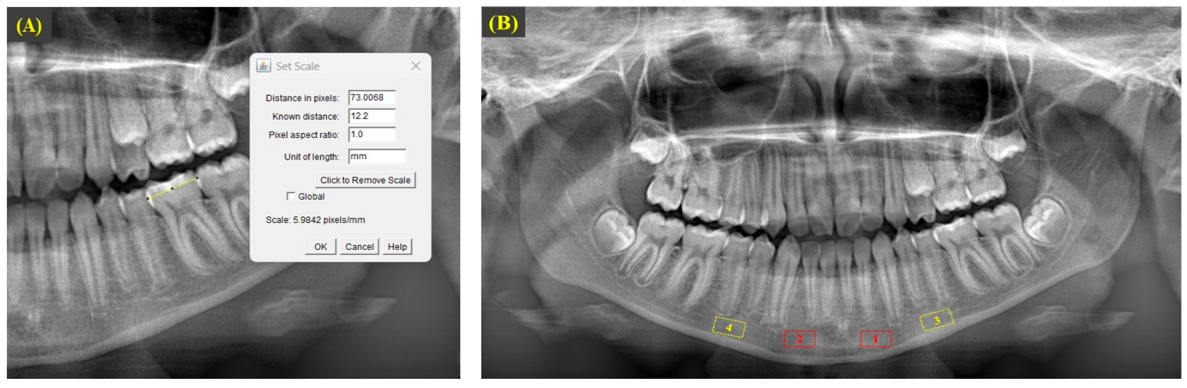
2.4. Radiograph Acquisition and Cephalometric Measurements
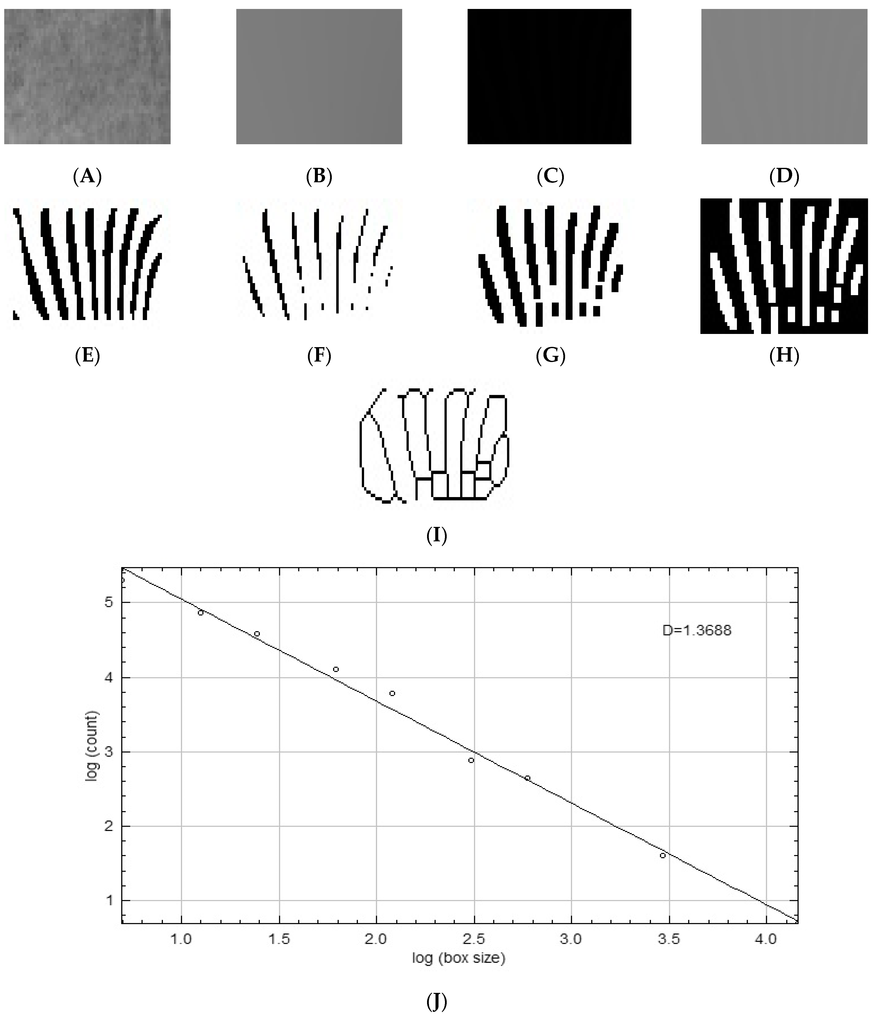
2.5. Fractal Dimension Analysis
2.6. Statistical Analysis
3. Results
4. Discussion
5. Conclusions
Author Contributions
Funding
Institutional Review Board Statement
Informed Consent Statement
Data Availability Statement
Conflicts of Interest
References
- Lindhe, J.; Allen, E.; Maynard, G. Consensus report. Mucogingival therapy. Ann. Periodontol. 1996, 1, 702–706. [Google Scholar]
- Kasaj, A. Etiology and Prevalence of Gingival Recession. In Gingival Recession Management: A Clinical Manual; Kasaj, A., Ed.; Springer International Publishing: Cham, Switzerland, 2018; pp. 19–31. [Google Scholar]
- Çolak, R.; Önder, B.A. Evaluation of the Prevalence and Etiologic Factors of Gingival Recession in Western Black Sea Region. Aydin Dent. J. 2024, 10, 237–245. [Google Scholar] [CrossRef]
- Jati, A.S.; Furquim, L.Z.; Consolaro, A. Gingival recession: Its causes and types, and the importance of orthodontic treatment. Dent. Press J. Orthod. 2016, 21, 18–29. [Google Scholar] [CrossRef] [PubMed]
- Chatzopoulou, D.; Johal, A. Management of gingival recession in the orthodontic patient. Semin. Orthod. 2015, 21, 15–26. [Google Scholar] [CrossRef]
- Vanarsdall, R.; Secchi, A. Periodontal/orthodontic interrelationships. In Orthodontics: Current Principle and Techniques, 2nd ed.; Mosby: St. Louis, MO, USA, 1994; p. 719. [Google Scholar]
- Joss-Vassalli, I.; Grebenstein, C.; Topouzelis, N.; Sculean, A.; Katsaros, C. Orthodontic therapy and gingival recession: A systematic review. Orthod. Craniofac. Res. 2010, 13, 127–141. [Google Scholar] [CrossRef] [PubMed]
- Slutzkey, S.; Levin, L. Gingival recession in young adults: Occurrence, severity, and relationship to past orthodontic treatment and oral piercing. Am. J. Orthod. Dentofacial. Orthop. 2008, 134, 652–656. [Google Scholar] [CrossRef]
- Bollen, A.-M.; Cunha-Cruz, J.; Bakko, D.W.; Huang, G.J.; Hujoel, P.P. The effects of orthodontic therapy on periodontal health: A systematic review of controlled evidence. J. Am. Dent. Assoc. 2008, 139, 413–422. [Google Scholar] [CrossRef]
- Dias, A.T.; Lopes, J.F.; Fernandes, J.C.H.; Fernandes, G.V.O. The Treatment of Gingival Recessions in the Lower Anterior Region Associated with the Use/Absence of Lingual-Fixed Orthodontics Retainers: Three Case Reports Using the Laterally Closed Tunnel Technique and Parallel Incision Methods. J. Dent. 2025, 13, 93. [Google Scholar] [CrossRef]
- Steiner, G.G.; Pearson, J.; Ainamo, J. Changes of the marginal periodontium as a result of labial tooth movement in monkeys. J. Periodontol. 1981, 52, 314–320. [Google Scholar] [CrossRef]
- Wennström, J.L.; Lindhe, J.; Sinclair, F.; Thilander, B. Some periodontal tissue reactions to orthodontic tooth movement in monkeys. J. Clin. Periodontol. 1987, 14, 121–129. [Google Scholar] [CrossRef]
- Farsoun, C.F.; Castro, F.; Farsoun, A.; Farsoun, J.; Fernandes, J.C.H.; Fernandes, G.V.d.O. Gingival recession in canines orthodontically aligned: A narrative review. Rev. Flum. Odontol. (Online) 2023, 3, 100–121. [Google Scholar]
- Mishra, S.; Kumar, M.; Mishra, L.; Panda, S.; Panda, S.; Lewkowicz, N.; Lapinska, B. Estimation of Cancellous Changes Using Fractal Analysis in Patients with Periodontitis. Biomedicines 2023, 11, 2547. [Google Scholar] [CrossRef]
- Aktuna Belgin, C.; Serindere, G. Evaluation of trabecular bone changes in patients with periodontitis using fractal analysis: A periapical radiography study. J. Periodontol. 2020, 91, 933–937. [Google Scholar] [CrossRef]
- Sener, E.; Cinarcik, S.; Baksi, B.G. Use of fractal analysis for the discrimination of trabecular changes between individuals with healthy gingiva or moderate periodontitis. J. Periodontol. 2015, 86, 1364–1369. [Google Scholar] [CrossRef]
- Soltani, P.; Sami, S.; Yaghini, J.; Golkar, E.; Riccitiello, F.; Spagnuolo, G. Application of Fractal Analysis in Detecting Trabecular Bone Changes in Periapical Radiograph of Patients with Periodontitis. Int. J. Dent. 2021, 2021, 3221448. [Google Scholar] [CrossRef] [PubMed]
- Southard, T.E.; Southard, K.A.; Krizan, K.E.; Hillis, S.L.; Haller, J.W.; Keller, J.; Vannier, M.W. Mandibular bone density and fractal dimension in rabbits with induced osteoporosis. Oral Surg. Oral Med. Oral Pathol. Oral Radiol. Endod. 2000, 89, 244–249. [Google Scholar] [CrossRef] [PubMed]
- Prouteau, S.; Ducher, G.; Nanyan, P.; Lemineur, G.; Benhamou, L.; Courteix, D. Fractal analysis of bone texture: A screening tool for stress fracture risk? Eur. J. Clin. Invest. 2004, 34, 137–142. [Google Scholar] [CrossRef]
- Cicek, O.; Arslan, D. Effect of Fixed and Removable Functional Therapy on Mandibular Anterior Bone Structures: A Fractal Analysis Study. Diagnostics 2024, 14, 1713. [Google Scholar] [CrossRef]
- Köse, E.; Ay Ünüvar, Y.; Uzun, M. Assessment of the relationship between fractal analysis of mandibular bone and orthodontic treatment duration: A retrospective study. J. Orofac. Orthop. 2022, 83, 102–110. [Google Scholar] [CrossRef]
- Shrout, M.; Hildebolt, C.; Potter, B. The effect of varying the region of interest on calculations of fractal index. Dentomaxillofac Radiol. 1997, 26, 295–298. [Google Scholar] [CrossRef]
- Lynch, J.; Hawkes, D.; Buckland-Wright, J. A robust and accurate method for calculating the fractal signature of texture in macroradiographs of osteoarthritic knees. Med. Inform. 1991, 16, 241–251. [Google Scholar] [CrossRef] [PubMed]
- Lynch, J.; Hawkes, D.; Buckland-Wright, J. Analysis of texture in macroradiographs of osteoarthritic knees, using the fractal signature. Phys. Med. Biol. 1991, 36, 709. [Google Scholar] [CrossRef]
- Buckland-Wright, J.; Lynch, J.; Rymer, J.; Fogelman, I. Fractal signature analysis of macroradiographs measures trabecular organization in lumbar vertebrae of postmenopausal women. Calcif. Tissue Int. 1994, 54, 106–112. [Google Scholar] [CrossRef] [PubMed]
- Yu, Y.-Y.; Chen, H.; Lin, C.-H.; Chen, C.-M.; Oviir, T.; Chen, S.-K.; Hollender, L. Fractal dimension analysis of periapical reactive bone in response to root canal treatment. Oral Surg. Oral Med. Oral Pathol. Oral Radiol. Endod. 2009, 107, 283–288. [Google Scholar] [CrossRef]
- Fazzalari, N.; Parkinson, I. Fractal properties of cancellous bone of the iliac crest in vertebral crush fracture. Bone 1998, 23, 53–57. [Google Scholar] [CrossRef]
- Bolat Gümüş, E.; Yavuz, E.; Tufekci, C. Effects of functional orthopedic treatment on mandibular trabecular bone in class II patients using fractal analysis. J. Orofac. Orthop. 2023, 84, 155–164. [Google Scholar] [CrossRef] [PubMed]
- Eid, H.A.; Assiri, H.A.M.; Kandyala, R.; Togoo, R.A.; Turakhia, V.S. Gingival enlargement in different age groups during fixed Orthodontic treatment. J. Int. Oral Health. JIOH 2014, 6, 1–4. [Google Scholar]
- Kumar, V.; Singh, P.; Arora, V.K.; Kaur, S.; Sarin, S.; Singh, H. Assessment of effect of fixed orthodontic treatment on gingival health: An observational study. J. Pharm. Bioallied Sci. 2021, 13, S425–S428. [Google Scholar] [CrossRef]
- Theodorelosa, P.; Ferrillob, M.; Pandisc, N.; Kloukosd, D.; Fleminge, P.S.; Katsarosf, C. A Cross-Sectional Evaluation of the Association between Orthodontic Treatment, Retention Modality and the Prevalence of Gingival Recession. Oral Health Prev. Dent. 2024, 22, 647–654. [Google Scholar] [CrossRef]
- Levin, L.; Samorodnitzky-Naveh, G.R.; Machtei, E.E. The association of orthodontic treatment and fixed retainers with gingival health. J. Periodontol. 2008, 79, 2087–2092. [Google Scholar] [CrossRef]
- Renkema, A.M.; Fudalej, P.S.; Renkema, A.A.; Abbas, F.; Bronkhorst, E.; Katsaros, C. Gingival labial recessions in orthodontically treated and untreated individuals: A case–control study. J. Clin. Periodontol. 2013, 40, 631–637. [Google Scholar] [CrossRef] [PubMed]
- Katsaros, C.; Livas, C.; Renkema, A.-M. Unexpected Complications of Bonded Mandibular Lingual Retainers. Inf. Orthod. Kieferor. 2013, 45, 18–21. [Google Scholar] [CrossRef] [PubMed]
- Pazera, P.; Fudalej, P.; Katsaros, C. Severe complication of a bonded mandibular lingual retainer. Am. J. Orthod. Dentofac. Orthop. 2012, 142, 406–409. [Google Scholar] [CrossRef]
- Gebistorf, M.; Mijuskovic, M.; Pandis, N.; Fudalej, P.S.; Katsaros, C. Gingival recession in orthodontic patients 10 to 15 years posttreatment: A retrospective cohort study. Am. J. Orthod. Dentofac. Orthop. 2018, 153, 645–655. [Google Scholar] [CrossRef]
- Juloski, J.; Glisic, B.; Vandevska-Radunovic, V. Long-term influence of fixed lingual retainers on the development of gingival recession: A retrospective, longitudinal cohort study. Angle Orthod. 2017, 87, 658–664. [Google Scholar] [CrossRef] [PubMed]
- Petsos, H.; Usherenko, R.; Dahmer, I.; Eickholz, P.; Kopp, S.; Sayahpour, B. Influence of fixed orthodontic steel retainers on gingival health and recessions of mandibular anterior teeth in an intact periodontium-a randomized, clinical controlled trial. BMC Oral Health 2024, 24, 236. [Google Scholar] [CrossRef]
- Rody, W., Jr.; Elmaraghy, S.; McNeight, A.; Chamberlain, C.; Antal, D.; Dolce, C.; Wheeler, T.; McGorray, S.; Shaddox, L. Effects of different orthodontic retention protocols on the periodontal health of mandibular incisors. Orthod. Craniofac. Res. 2016, 19, 198–208. [Google Scholar] [CrossRef]
- Silness, J.; Löe, H. Periodontal disease in pregnancy: Iii. Response to local treatment. Acta Odontol. Scand. 1966, 24, 747–759. [Google Scholar] [CrossRef]
- Löe, H.; Silness, J. Periodontal disease in pregnancy I. Prevalence and severity. Acta Odontol. Scand. 1963, 21, 533–551. [Google Scholar] [CrossRef]
- White, S.C.; Rudolph, D.J. Alterations of the trabecular pattern of the jaws in patients with osteoporosis. Oral Surg. Oral Med. Oral Pathol. Oral Radiol. Endod. 1999, 88, 628–635. [Google Scholar] [CrossRef]
- Özden, S.; Cicek, O. Assessment of the Mandibular Osseous Architecture in Cleft Lip and Palate Using Fractal Dimension Analysis: A Pilot Study. J. Clin. Med. 2024, 13, 7334. [Google Scholar] [CrossRef]
- Çiçek, O.; Yılmaz, H.; Melayim, H. Evaluation of changes in alveolar bone remodeling after orthodontic treatment using fractal analysis: A retrospective study. Int. Dent. Res. 2024, 14, 19–26. [Google Scholar] [CrossRef]
- Fleming, P.S.; Andrews, J. The role of orthodontics in the prevention and management of gingival recession. Br. Dent. J. 2024, 237, 341–347. [Google Scholar] [CrossRef]
- Bayrak, E.A.; Kirci, P. Fractal analysis usage areas in healthcare. In Proceedings of the IEEE International Conference on System Analysis & Intelligent Computing, Kyiv, Ukraine, 5–9 October 2020; pp. 377–406. [Google Scholar]
- Kato, C.N.; Barra, S.G.; Tavares, N.P.; Amaral, T.M.; Brasileiro, C.B.; Mesquita, R.A.; Abreu, L.G. Use of fractal analysis in dental images: A systematic review. Dentomaxillofac. Radiol. 2020, 49, 20180457. [Google Scholar] [CrossRef] [PubMed]
- Updike, S.X.; Nowzari, H. Fractal analysis of dental radiographs to detect periodontitis-induced trabecular changes. J. Periodontal Res. 2008, 43, 658–664. [Google Scholar] [CrossRef]
- Dosdogru, E.B.; Ziyaettin, M.; Ertürk, A.F.; Ertürk, A.F. Investigation of the Effect of Periodontitis on Trabecular Bone Structure by Fractal Analysis. Cureus 2025, 17, e77833. [Google Scholar] [CrossRef] [PubMed]
- Eser, S.; Sarıbaş, E. Anatomical assessment of the trabecular structure of the alveolar bone in periodontal disease by fractal analysis method. Folia Morphol. 2024, 83, 157–167. [Google Scholar] [CrossRef]
- Gómez-García, F.J.; Lopez-Jornet, P.; Fernández-Martínez, M.; Guerrero-Sanchez, Y. The bone density studied through the fractal dimension in patients with periodontal disease. Fractals 2020, 28, 2040031. [Google Scholar] [CrossRef]
- Uğur Aydın, Z.; Ocak, M.; Bayrak, S.; Göller Bulut, D.; Orhan, K. The effect of type 2 diabetes mellitus on changes in the fractal dimension of periapical lesion in teeth after root canal treatment: A fractal analysis study. Int. Endod. J. 2021, 54, 181–189. [Google Scholar] [CrossRef]
- Amer, M.E.; Heo, M.-S.; Brooks, S.L.; Benavides, E. Anatomical variations of trabecular bone structure in intraoral radiographs using fractal and particles count analyses. Imaging Sci. Dent. 2012, 42, 5–12. [Google Scholar] [CrossRef]
- Rothe, L.E.; Bollen, A.-M.; Little, R.M.; Herring, S.W.; Chaison, J.B.; Chen, C.S.-K.; Hollender, L.G. Trabecular and cortical bone as risk factors for orthodontic relapse. Am. J. Orthod. Dentofac. Orthop. 2006, 130, 476–484. [Google Scholar] [CrossRef] [PubMed]
- Hennessy, J.; Garvey, T.; Al-Awadhi, E.A. A randomized clinical trial comparing mandibular incisor proclination produced by fixed labial appliances and clear aligners. Angle Orthod. 2016, 86, 706–712. [Google Scholar] [CrossRef] [PubMed]
- McLaughlin, R.P.; Bennett, J.C.; Trevisi, H. Systemized Orthodontic Treatment Mechanics, 1st ed.; Mosby Ltd.: St. Louis, MO, USA, 2001. [Google Scholar]
- Melsen, B.; Allais, D. Factors of importance for the development of dehiscences during labial movement of mandibular incisors: A retrospective study of adult orthodontic patients. Am. J. Orthod. Dentofac. Orthop. 2005, 127, 552–561. [Google Scholar] [CrossRef]
- Djeu, G.; Hayes, C.; Zawaideh, S. Correlation between mandibular central incisor proclination and gingival recession during fixed appliance therapy. Angle Orthod. 2002, 72, 238–245. [Google Scholar] [PubMed]
- Pandis, N.; Vlahopoulos, K.; Madianos, P.; Eliades, T. Long-term periodontal status of patients with mandibular lingual fixed retention. Eur. J. Orthod. 2007, 29, 471–476. [Google Scholar] [CrossRef]
- Thomson, W. Orthodontic treatment outcomes in the long term: Findings from a longitudinal study of New Zealanders. The Angle Orthod. 2002, 72, 449–455. [Google Scholar]
- Fragkioudakis, I.; Tassou, D.; Sideri, M.; Vouros, I. Prevalance and clinical characteristics of gingival recession in Greek young adults: A cross-sectional study. Clin. Exp. Dent. Res. 2021, 7, 672–678. [Google Scholar] [CrossRef]

| Parameter | Score | Definition |
|---|---|---|
| For GI | 0 | Healthy gingiva |
| 1 | Mild infection with color change and edema without bleeding on probing | |
| 2 | Moderate infection with color change and edema, along with bleeding on probing | |
| 3 | Severe infection with marked edema and hyperemic gingiva with tendency to spontaneously bleed | |
| For PI | 0 | No plaque |
| 1 | Presence of film-like plaque, not visible by direct observation but detectable by periodontal probing, located at free gingival margin and on relevant tooth surface | |
| 2 | Presence of visible soft deposits at gingival margin and on tooth surface | |
| 3 | Presence of excessive soft deposits at gingival margin and on tooth surface | |
| For gingival recession | Distance measured from cemento-enamel junction to gingival margin on exposed root surface that exceeds 0.5 mm |
| Group 1 | Group 2 | Group 3 | Group 4 | p | ||
|---|---|---|---|---|---|---|
| Age (year) | Mean ±SD | 22.53 ± 2.82 | 22.4 ± 2.89 | 21.46 ± 2.32 | 21.26 ± 2.28 | 0.545 K |
| Gender | Female, n (%) | 10 (67) | 9 (60) | 9 (60) | 7 (47) | 0.728 χ2 |
| Male, n (%) | 5 (33) | 6 (40) | 6 (40) | 8 (53) | ||
| Retention duration (year) | Mean ± SD | 1.98 ± 1.36 | 1.88 ± 1.25 | 1.91 ± 0.9 | 2.06 ± 1.07 | 0.755 K |
| PI | Mean ± SD | 1.93 ± 0.79 | 1.93 ± 0.88 | 2.46 ± 0.99 | 2.2 ± 0.94 | 0.204 K |
| GI | Mean ± SD | 1.33 ± 0.81 | 1.26 ± 0.7 | 1.46 ± 0.51 | 1.4 ± 0.5 | 0.641 K |
| T0 | T1 | T2 | p | T1-T0 | T2-T1 | T2-T0 | |
|---|---|---|---|---|---|---|---|
| Mean ± SD (Median) | Mean ± SD (Median) | Mean ± SD (Median) | p | p | p | ||
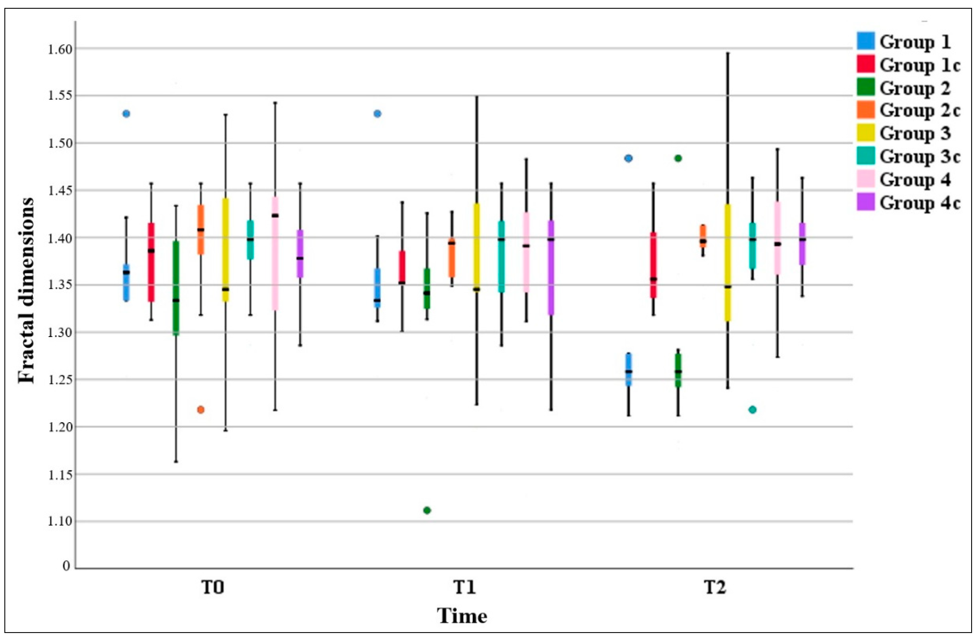
| Group 1 | 1.3680 ± 0.059 (1.3631) | 1.3517 ± 0.060 (1.3336) | 1.2834 ± 0.084 (1.2583) | 0.001 *,F | 0.079 W | 0.011 *,W | 0.005 *,W |
| Group 1c | 1.3812 ± 0.052 (1.386) | 1.3626 ± 0.041 (1.352) | 1.3752 ± 0.47 (1.356) | 0.262 A | 0.15 A | 1.000 A | 0.852 A |
| Group 2 | 1.3546 ± 0.11 (1.3336) | 1.3303 ± 0.075 (1.3412) | 1.2703 ± 0.073 (1.2583) | 0.003 *,F | 0.326 W | 0.027 *,W | 0.041 *,W |
| Group 2c | 1.3959 ± 0.062 (1.408) | 1.3836 ± 0.039 (1.394) | 1.4013 ± 0.035 (1.396) | 0.119 F | 0.147 W | 0.125 W | 0.878 W |
| Group 3 | 1.3792 ± 0.104 (1.3452) | 1.3760 ± 0.095 (1.3412) | 1.3787 ± 0.105 (1.3482) | 0.991 A | 0.874 A | 0.918 A | 1.000 A |
| Group 3c | 1.3919 ± 0.051 (1.398) | 1.3772 ± 0.065 (1.396) | 1.3852 ± 0.064 (1.397) | 0.220 A | 0.211 A | 1.000 A | 0.786 A |
| Group 4 | 1.3809 ± 0.098 (1.4232) | 1.3789 ± 0.87 (1.3912) | 1.3841 ± 0.67 (1.3932) | 0.627 F | 0.733 W | 0.496 W | 0.955 W |
| Group 4c | 1.3819 ± 0.054 (1.378) | 1.3732 ± 0.07 (1.398) | 1.3892 ± 0.054 (1.388) | 0.413 A | 0.568 A | 1.000 A | 0.097 A |
| T0 | p | T1 | p | T2 | p | |
|---|---|---|---|---|---|---|
| Mean ± SD (Median) | Mean ± SD (Median) | Mean ± SD (Median) | ||||
| Group 1 | 1.3680 ± 0.059 (1.3631) | 0.524 t | 1.3517 ± 0.060 (1.3336) | 0.285 M | 1.2834 ± 0.084 (1.2583) | 0.001 *,t |
| Group 1c | 1.3812 ± 0.052 (1.386) | 1.3626 ± 0.041 (1.352) | 1.3752 ± 0.047 (1.356) | |||
| Group 2 | 1.3546 ± 0.11 (1.3336) | 0.218 t | 1.3303 ± 0.075 (1.3412) | 0.01 *,M | 1.2703 ± 0.073 (1.2583) | 0.001 *,t |
| Group 2c | 1.3959 ± 0.062 (1.408) | 1.3836 ± 0.039 (1.394) | 1.4013 ± 0.035 (1.396) | |||
| Group 3 | 1.3792 ± 0.104 (1.3452) | 0.676 t | 1.3760 ± 0.095 (1.3412) | 0.969 t | 1.3787 ± 0.105 (1.3482) | 0.841 t |
| Group 3c | 1.3919 ± 0.051 (1.398) | 1.3772 ± 0.065 (1.396) | 1.3852 ± 0.064 (1.397) | |||
| Group 4 | 1.3809 ± 0.098 (1.4232) | 0.973 t | 1.3789 ± 0.87 (1.3912) | 0.539 M | 1.3841 ± 0.67 (1.3932) | 0.821 t |
| Group 4c | 1.3819 ± 0.054 (1.378) | 1.3732 ± 0.07 (1.398) | 1.3892 ± 0.054 (1.388) |
| T0 | p | T1 | p | T2 | p | |
|---|---|---|---|---|---|---|
| Mean ± SD (Median) | Mean ± SD (Median) | Mean ± SD (Median) | ||||
| Group 1 | 1.3680 ± 0.059 (1.3631) | 0.449 M | 1.3517 ± 0.060 (1.3336) | 0.983 M | 1.2834 ± 0.084 (1.2583) | 0.786 M |
| Group 2 | 1.3546 ± 0.11 (1.3336) | 1.3303 ± 0.075 (1.3412) | 1.2703 ± 0.073 (1.2583) | |||
| Group 3 | 1.3792 ± 0.104 (1.3452) | 0.964 t | 1.3760 ± 0.095 (1.3412) | 0.933 t | 1.3787 ± 0.105 (1.3482) | 0.869 t |
| Group 4 | 1.3809 ± 0.098 (1.4232) | 1.3789 ± 0.87 (1.3912) | 1.3841 ± 0.67 (1.3932) |
| FD Differences | PI | GI | ||
|---|---|---|---|---|
| FD differences | Mean ± SD | −0.002 ± 0.12 | 2.13 ± 0.91 | 1.43 ± 0.69 |
| Pearson coefficient | 1 | 0.269 | 0.273 | |
| p | 0.037 * | 0.035 * | ||
| IMPA differences | Mean ± SD | −0.06 ± 0.12 | 1.93 ± 0.82 | 1.3 ± 0.74 |
| Spearman rho | −0.106 | 0.161 | 0.148 | |
| p | 0.578 | 0.397 | 0.436 |
Disclaimer/Publisher’s Note: The statements, opinions and data contained in all publications are solely those of the individual author(s) and contributor(s) and not of MDPI and/or the editor(s). MDPI and/or the editor(s) disclaim responsibility for any injury to people or property resulting from any ideas, methods, instructions or products referred to in the content. |
© 2025 by the authors. Licensee MDPI, Basel, Switzerland. This article is an open access article distributed under the terms and conditions of the Creative Commons Attribution (CC BY) license (https://creativecommons.org/licenses/by/4.0/).
Share and Cite
Küçükoğlu Çolak, M.; Çolak, R.; Cicek, O. Fractal Dimension Analysis of Mandibular Trabecular Architecture in Gingival Recession During Orthodontic Retention: A Cross-Sectional Study. Diagnostics 2025, 15, 1013. https://doi.org/10.3390/diagnostics15081013
Küçükoğlu Çolak M, Çolak R, Cicek O. Fractal Dimension Analysis of Mandibular Trabecular Architecture in Gingival Recession During Orthodontic Retention: A Cross-Sectional Study. Diagnostics. 2025; 15(8):1013. https://doi.org/10.3390/diagnostics15081013
Chicago/Turabian StyleKüçükoğlu Çolak, Merve, Resul Çolak, and Orhan Cicek. 2025. "Fractal Dimension Analysis of Mandibular Trabecular Architecture in Gingival Recession During Orthodontic Retention: A Cross-Sectional Study" Diagnostics 15, no. 8: 1013. https://doi.org/10.3390/diagnostics15081013
APA StyleKüçükoğlu Çolak, M., Çolak, R., & Cicek, O. (2025). Fractal Dimension Analysis of Mandibular Trabecular Architecture in Gingival Recession During Orthodontic Retention: A Cross-Sectional Study. Diagnostics, 15(8), 1013. https://doi.org/10.3390/diagnostics15081013