
Novel Usefulness of M2BPGi for Predicting Severity and Clinical Outcomes in Hospitalized COVID-19 Patients
 ,
,  , , , ,
Abstract
1. Introduction
2. Materials and Methods
2.1. Study Population
2.2. M2BPGi Assay
2.3. Statistical Analysis
3. Results
4. Discussion
Supplementary Materials
Author Contributions
Funding
Institutional Review Board Statement
Informed Consent Statement
Data Availability Statement
Conflicts of Interest
References
- Davido, B.; Mégarbane, B.; Loubet, P. COVID-19 surge during summer 2024: The phantom menace? Clin. Microbiol. Infect. 2024, 30, 1492–1493. [Google Scholar] [CrossRef] [PubMed]
- World Health Organization. COVID-19 Epidemiological Update, Edition 170, 13 August 2024. Available online: https://iris.who.int/handle/10665/378513 (accessed on 22 August 2024).
- World Health Organization. Clinical Management of COVID-19. Available online: https://www.who.int/publications/i/item/WHO-2019-nCoV-clinical-2023.2 (accessed on 22 August 2024).
- Walsh, T.J.; Bright, R.A.; Ahuja, A.; McCarthy, M.W.; Marfuggi, R.A.; Simpson, S.Q. Meeting the challenges of sepsis in severe Coronavirus Disease 2019: A call to arms. Open Forum. Infect. Dis. 2023, 10, ofac645. [Google Scholar] [CrossRef]
- Heubner, L.; Hattenhauer, S.; Güldner, A.; Petrick, P.L.; Rößler, M.; Schmitt, J.; Schneider, R.; Held, H.C.; Mehrholz, J.; Bodechtel, U.; et al. Characteristics and outcomes of sepsis patients with and without COVID-19. J. Infect. Public Health 2022, 15, 670–676. [Google Scholar] [CrossRef]
- Kang, S.J.; Jung, S.I. Age-related morbidity and mortality among patients with COVID-19. Infect. Chemother. 2020, 52, 154–164. [Google Scholar] [CrossRef] [PubMed]
- Stralin, K.; Wahlstrom, E.; Walther, S.; Bennet-Bark, A.M.; Heurgren, M.; Linden, T.; Holm, J.; Hanberger, H. Mortality trends among hospitalized COVID-19 patients in Sweden: A nationwide observational cohort study. Lancet. Reg. Health Eur. 2021, 4, 100054. [Google Scholar] [CrossRef] [PubMed]
- Park, M.; Hur, M.; Kim, H.; Lee, C.H.; Lee, J.H.; Nam, M. Usefulness of KL-6 for predicting clinical outcomes in hospitalized COVID-19 patients. Medicina 2022, 58, 1317. [Google Scholar] [CrossRef] [PubMed]
- Park, M.; Hur, M.; Kim, H.; Lee, C.H.; Lee, J.H.; Kim, H.W.; Nam, M.; Lee, S. Soluble ST2 as a useful biomarker for predicting clinical outcomes in hospitalized COVID-19 patients. Diagnostics 2023, 13, 259. [Google Scholar] [CrossRef] [PubMed]
- Scott, L.J.; Tavare, A.; Hill, E.M.; Jordan, L.; Juniper, M.; Srivastava, S.; Redfern, E.; Little, H.; Pullyblank, A. Prognostic value of National Early Warning Scores (NEWS2) and component physiology in hospitalized patients with COVID-19: A multicentre study. Emerg. Med. J. 2022, 39, 589–594. [Google Scholar] [CrossRef]
- Angeli, F.; Reboldi, G.; Verdecchia, P. Ageing, ACE2 deficiency and bad outcome in COVID-19. Clin. Chem. Lab. Med. 2020, 59, 1607–1609. [Google Scholar] [CrossRef]
- Henry, B.M.; de Oliveira, M.H.S.; Benoit, S.; Plebani, M.; Lippi, G. Hematologic, biochemical and immune biomarker abnormalities associated with severe illness and mortality in coronavirus disease 2019 (COVID-19): A meta-analysis. Clin. Chem. Lab. Med. 2020, 58, 1021–1028. [Google Scholar] [CrossRef]
- Bivona, G.; Agnello, L.; Ciaccio, M. Biomarkers for prognosis and treatment response in COVID-19 patients. Ann. Lab. Med. 2021, 41, 540–548. [Google Scholar] [CrossRef] [PubMed]
- Shirabe, K.; Bekki, Y.; Gantumur, D.; Araki, K.; Ishii, N.; Kuno, A.; Narimatsu, H.; Mizokami, M. Mac-2 binding protein glycan isomer (M2BPGi) is a new serum biomarker for assessing liver fibrosis: More than a biomarker of liver fibrosis. J. Gastroenterol. 2018, 53, 819–826. [Google Scholar] [PubMed]
- Moon, H.W.; Park, M.; Hur, M.; Kim, H.; Choe, W.H.; Yun, Y.M. Usefulness of enhanced liver fibrosis, glycosylation isomer of Mac-2 binding protein, galectin-3, and soluble suppression of tumorigenicity 2 for assessing liver fibrosis in chronic liver diseases. Ann. Lab. Med. 2018, 38, 331–337. [Google Scholar] [CrossRef]
- Tamaki, N.; Kurosaki, M.; Loomba, R.; Izumi, N. Clinical utility of Mac-2 binding protein glycosylation isomer in chronic liver diseases. Ann. Lab. Med. 2021, 41, 16–24. [Google Scholar]
- Jang, S.Y.; Tak, W.Y.; Park, S.Y.; Kweon, Y.O.; Lee, Y.R.; Kim, G.; Hur, K.; Han, M.H.; Lee, W.K. Diagnostic efficacy of serum Mac-2 binding protein glycosylation isomer and other markers for liver fibrosis in non-alcoholic fatty liver diseases. Ann. Lab. Med. 2021, 41, 302–309. [Google Scholar] [CrossRef] [PubMed]
- Hur, M.; Park, M.; Moon, H.W.; Choe, W.H.; Lee, C.H. Comparison of non-invasive clinical algorithms for liver fibrosis in patients with chronic hepatitis B to reduce the need for liver biopsy: Application of enhanced liver fibrosis and Mac-2 binding protein glycosylation isomer. Ann. Lab. Med. 2022, 42, 249–257. [Google Scholar]
- Dufour, J.F.; Marjot, T.; Becchetti, C.; Tilg, H. COVID-19 and liver disease. Gut 2022, 71, 2350–2362. [Google Scholar]
- Montori, M.; Baroni, G.S.; Santori, P.; Di Giampaolo, C.; Ponziani, F.; Abenavoli, L.; Scarpellini, E. Liver damage and COVID-19: At least a “two-hit” story in systematic review. Curr. Issues Mol. Biol. 2023, 45, 3035–3047. [Google Scholar] [CrossRef] [PubMed]
- Zhang, B.; Zhou, X.; Qiu, Y.; Song, Y.; Feng, F.; Feng, J.; Song, Q.; Jia, Q.; Wang, J. Clinical characteristics of 82 cases of death from COVID-19. PLoS ONE 2020, 15, e0235458. [Google Scholar]
- Park, M.; Hur, M.; Kim, H.; Lee, C.H.; Lee, J.H.; Kim, H.W.; Nam, M. Prognostic utility of procalcitonin, presepsin, and the VACO index for predicting 30-day mortality in hospitalized COVID-19 patients. Ann. Lab. Med. 2022, 42, 406–414. [Google Scholar]
- Singer, M.; Deutschman, C.S.; Seymour, C.W.; Shankar-Hari, M.; Annane, D.; Bauer, M.; Bellomo, R.; Bernard, G.R.; Chiche, J.D.; Coopersmith, C.M.; et al. The Third international consensus definitions for sepsis and septic shock (Sepsis-3). JAMA 2016, 315, 801–810. [Google Scholar]
- Royal College of Physicians, UK. National EarlyWarning Score (NEWS) 2. 2017. Available online: https://www.rcplondon.ac.uk/projects/outputs/national-early-warning-score-news-2 (accessed on 8 August 2024).
- Berzigotti, A.; Tsochatzis, E.; Boursier, J.; Castera, L.; Cazzagon, N.; Friedrich-Rust, M.; Petta, S.; Thiele, M.; European Association for the Study of the Liver. EASL clinical practice guidelines on non-invasive tests for evaluation of liver disease severity and prognosis—2021 update. J. Hepatol. 2021, 75, 659–689. [Google Scholar]
- Šimundić, A.M. Measures of diagnostic accuracy: Basic definitions. EJIFCC 2009, 19, 203–211. [Google Scholar] [PubMed]
- Mukaka, M.M. Statistics corner: A guide to appropriate use of correlation coefficient in medical research. Malawi Med. J. 2012, 24, 69–71. [Google Scholar] [PubMed]
- Nagashima, K.; Noma, H.; Sato, Y.; Gosho, M. Sample size calculations for single-arm survival studies using transformations of the Kaplan–Meier estimator. Pharm. Stat. 2020, 20, 499–511. [Google Scholar] [PubMed]
- Xu, R.; Shaw, P.A.; Mehrotra, D.V. Hazard ratio estimation in small samples. Stat. Biopharm. Res. 2018, 10, 139–149. [Google Scholar]
- Nah, E.H.; Cho, S.; Kim, S.; Chu, J.; Kwon, E.; Cho, H.I. Prevalence of liver fibrosis and associated risk factors in the Korean general population: A retrospective cross-sectional study. BMJ Open 2021, 11, e046529. [Google Scholar]
- Wu, P.S.; Hsieh, Y.C.; Lee, K.C.; Huang, Y.H.; Hou, M.C.; Lin, H.C. Mac-2 binding protein glycosylation isomer is a potential biomarker to predict portal hypertension and bacterial infection in cirrhotic patients. PLoS ONE 2021, 16, e0258589. [Google Scholar]
- Kolesova, O.; Vanaga, I.; Laivacuma, S.; Derovs, A.; Kolesovs, A.; Radzina, M.; Platkajis, A.; Eglite, J.; Hagina, E.; Arutjunana, S.; et al. Intriguing findings of liver fibrosis following COVID-19. BMC Gastroenterol. 2021, 21, 370. [Google Scholar]
- Taneva, G.; Dimitrov, D.; Velikova, T. Liver dysfunction as a cytokine storm manifestation and prognostic factor for severe COVID-19. World J. Hepatol. 2021, 13, 2005–2012. [Google Scholar]
- Nazerian, Y.; Ghasemi, M.; Yassaghi, Y.; Nazerian, A.; Hashemi, S.M. Role of SARS-CoV-2-induced cytokine storm in multi-organ failure: Molecular pathways and potential therapeutic options. Int. Immunopharmacol. 2022, 113, 109428. [Google Scholar] [CrossRef] [PubMed]
- Rani, R.; Gandhi, C.R. Stellate cell in hepatic inflammation and acute injury. J. Cell. Physiol. 2023, 238, 1226–1236. [Google Scholar] [CrossRef] [PubMed]
- Morio, K.; Imamura, M.; Daijo, K.; Teraoka, Y.; Honda, F.; Nakamura, Y.; Kobayashi, T.; Nakahara, T.; Nagaoki, Y.; Kawaoka, T.; et al. Wisteria floribunda agglutinin positive Mac-2-binding protein level increases in patients with acute liver injury. J. Gastroenterol. 2017, 52, 1252–1257. [Google Scholar] [CrossRef] [PubMed]
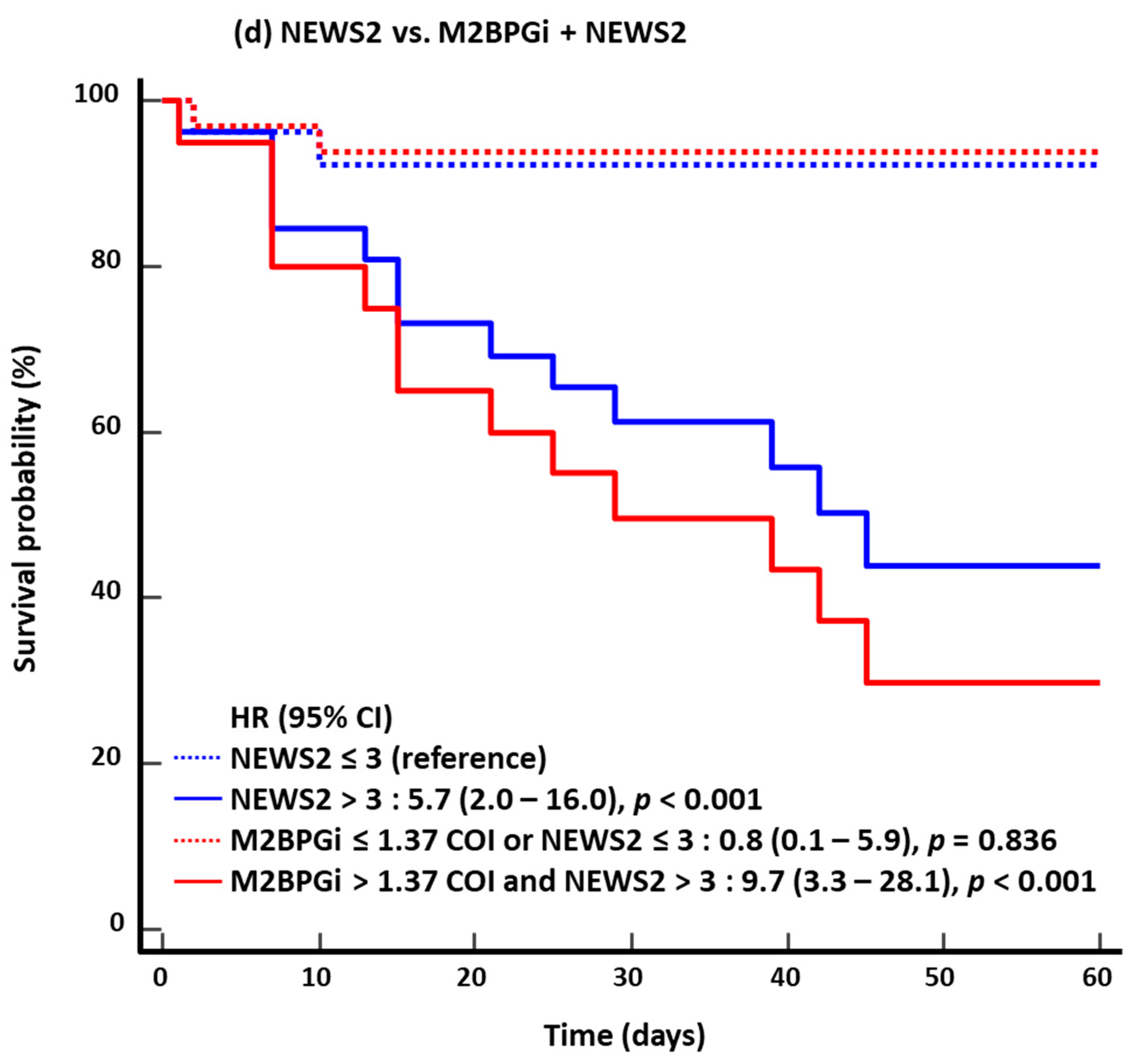

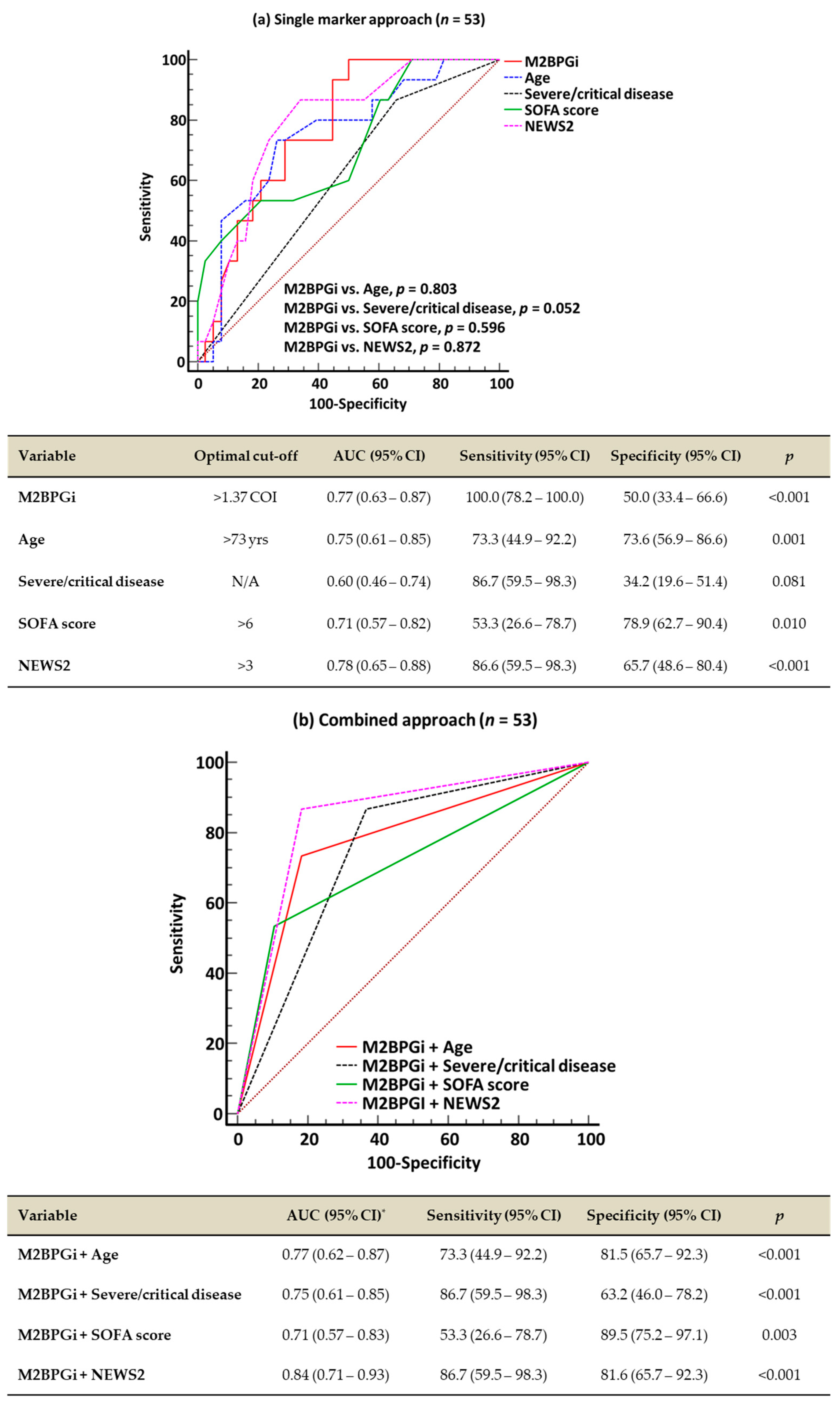
| Variable | All Patients (n = 53) | Mild/Moderate (n = 15) * | Severe/Critical (n = 38) * | p † |
|---|---|---|---|---|
| Age (years) | 72.0 (62.8–79.0) | 67.0 (57.2–75.7) | 72.0 (64.0–79.0) | 0.098 |
| ≥65, n (%) | 36 (67.9) | 8 (53.3) | 28 (73.7) | 0.156 |
| ≥70, n (%) | 30 (56.6) | 6 (40.0) | 24 (63.2) | 0.129 |
| ≥75, n (%) | 21 (39.6) | 4 (26.7) | 17 (44.7) | 0.350 |
| Male, n (%) | 33 (62.3) | 9 (60.0) | 24 (63.2) | 0.832 |
| Body mass index (kg/m2) | 24.1 (22.3–25.8) | 22.9 (21.0) | 24.3 (22.4–26.1) | 0.068 |
| Comorbidities ‡, n (%) | ||||
| n = 0 | 10 (18.9) | 3 (20.0) | 7 (18.4) | 0.486 |
| n = 1 | 12 (22.6) | 5 (33.3) | 7 (18.4) | |
| n = 2 | 14 (26.4) | 2 (13.4) | 12 (31.6) | |
| n ≥ 3 | 17 (32.1) | 5 (33.3) | 12 (31.6) | |
| Hypertension | 27 (50.9) | 7 (46.7) | 20 (52.6) | 0.698 |
| Diabetes mellitus | 19 (35.8) | 3 (20.0) | 16 (42.1) | 0.204 |
| Malignancy | 9 (17.0) | 4 (26.7) | 5 (13.2) | 0.252 |
| Dyslipidemia | 9 (17.0) | 3 (20.0) | 6 (15.8) | 0.700 |
| Chronic neurologic conditions | 8 (15.4) | 3 (20.0) | 5 (13.2) | 0.672 |
| Chronic heart disease | 6 (11.3) | 3 (20.0) | 3 (7.9) | 0.334 |
| Chronic respiratory disease | 5 (9.4) | 2 (13.4) | 3 (7.9) | 0.614 |
| Dementia | 5 (9.4) | 1 (6.7) | 4 (10.5) | 1.000 |
| Chronic kidney disease | 3 (5.7) | 0 (0.0) | 3 (7.9) | 0.549 |
| Connective tissue disease | 3 (5.7) | 0 (0.0) | 3 (7.9) | 0.549 |
| Peripheral artery disease | 2 (3.8) | 1 (6.7) | 1 (2.6) | 0.789 |
| Symptoms, n (%) | ||||
| Respiratory symptoms § | 35 (66.0) | 10 (66.7) | 25 (65.8) | 0.952 |
| Fever | 34 (64.2) | 8 (53.3) | 26 (68.4) | 0.306 |
| General weakness and/or fatigue | 23 (43.4) | 8 (53.3) | 15 (39.5) | 0.363 |
| Gastrointestinal symptoms ‖ | 9 (17.0) | 3 (20.0) | 6 (15.8) | 0.700 |
| Neurological symptoms ¶ | 6 (11.3) | 1 (6.7) | 5 (13.2) | 0.662 |
| Symptom duration (day) | 4.0 (1.0–7.5) | 7.0 (5.0–16.0) | 1.5 (0.0–7.0) | <0.001 |
| COVID-19 dx to admission, n (%) | ||||
| COVID-19 dx + ≤ 48 h | 30 (56.6) | 6 (40.0) | 24 (63.2) | 0.090 |
| COVID-19 dx + 3 to 11 days | 12 (22.6) | 3 (20.0) | 9 (23.7) | |
| COVID-19 dx after admission ** | 11 (20.8) | 6 (40.0) | 5 (13.2) | |
| COVID-19 dx to enrollment (day) | 3.0 (0.0–11.0) | 3.0 (0.2–37.5) | 3.0 (0.0–9.0) | 0.189 |
| Hospital stays (day) | 29.0 (20.8–49.0) | 45.0 (17.3–58.8) | 27.5 (21.0–42.0) | 0.458 |
| Vital signs | ||||
| Systolic BP (mm Hg) | 120.0 (110.0–141.3) | 120.0 (110.0–138.0) | 127.5 (110.0–147.0) | 0.352 |
| Diastolic BP (mm Hg) | 72.0 (70.0–80.0) | 70.0 (68.5–78.5) | 74.5 (70.0–80.0) | 0.523 |
| Pulse rate (beats/min) | 82.0 (74.0–95.5) | 81.0 (71.5–89.0) | 83.0 (77.0–98.0) | 0.195 |
| Respiration rate (breaths/min) | 20.0 (20.0–23.0) | 20.0 (19.0–20.0) | 20.5 (20.0–24.8) | 0.022 |
| Body temperature (°C) | 37.1 (36.8–37.7) | 36.8 (36.6–37.1) | 37.3 (36.9–37.8) | 0.033 |
| Oxygen saturation (%) | 96.0 (94.4–97.0) | 97.0 (96.0–97.0) | 95.5 (93.6–97.0) | 0.148 |
| Laboratory data | ||||
| White blood cells (×109/L) | 6.1 (4.8–8.9) | 4.8 (4.3–5.9) | 6.8 (5.2–9.8) | 0.029 |
| Neutrophils (×109/L) | 4.3 (3.1–6.7) | 3.2 (2.5–3.6) | 5.2 (3.7–7.7) | 0.004 |
| Lymphocytes (×109/L) | 1.1 (0.6–1.5) | 1.5 (1.2–1.8) | 0.8 (0.6–1.2) | <0.001 |
| Aspartate aminotransferas (U/L) | 30.0 (23.0–43.5) | 30.0 (23.7–41.7) | 30.0 (23.0–43.0) | 0.751 |
| Alanine aminotransferase (U/L) | 24.0 (14.8–37.3) | 23.0 (11.5–37.7) | 24.5 (15.0–35.0) | 0.607 |
| Alkaline phosphatase (U/L) | 72.0 (63.0–86.0) | 70.0 (63.0–74.0) | 73.0 (63.0–87.0) | 0.326 |
| γ-glutamyl transferase (U/L) | 28.0 (20.2–48.0) | 24.0 (18.0–29.5) | 30.5 (23.0–54.5) | 0.062 |
| Lactate dehydrogenase (U/L) | 531.0 (420.2–750.0) | 418.0 (338.0–591.0) | 577.0 (449.7–827.3) | 0.017 |
| Total bilirubin (umol/L) | 12.5 (9.5–18.2) | 15.1 (9.7–17.6) | 11.9 (9.4–25.8) | 0.843 |
| Direct bilirubin (umol/L) | 3.5 (2.2–6.7) | 2.9 (1.5–4.7) | 4.0 (2.4–11.3) | 0.113 |
| Creatinine (umol/L) | 76.9 (57.0–95.4) | 60.1 (55.0–89.9) | 80.0 (63.6–110.5) | 0.141 |
| Lactate (mmol/L) | 1.8 (1.2–2.3) | 1.6 (1.0–2.0) | 1.8 (1.3–2.5) | 0.194 |
| C-reactive protein (mg/L) | 39.1 (4.7–132.3) | 4.0 (0.4–38.9) | 63.8 (19.6–151.5) | 0.003 |
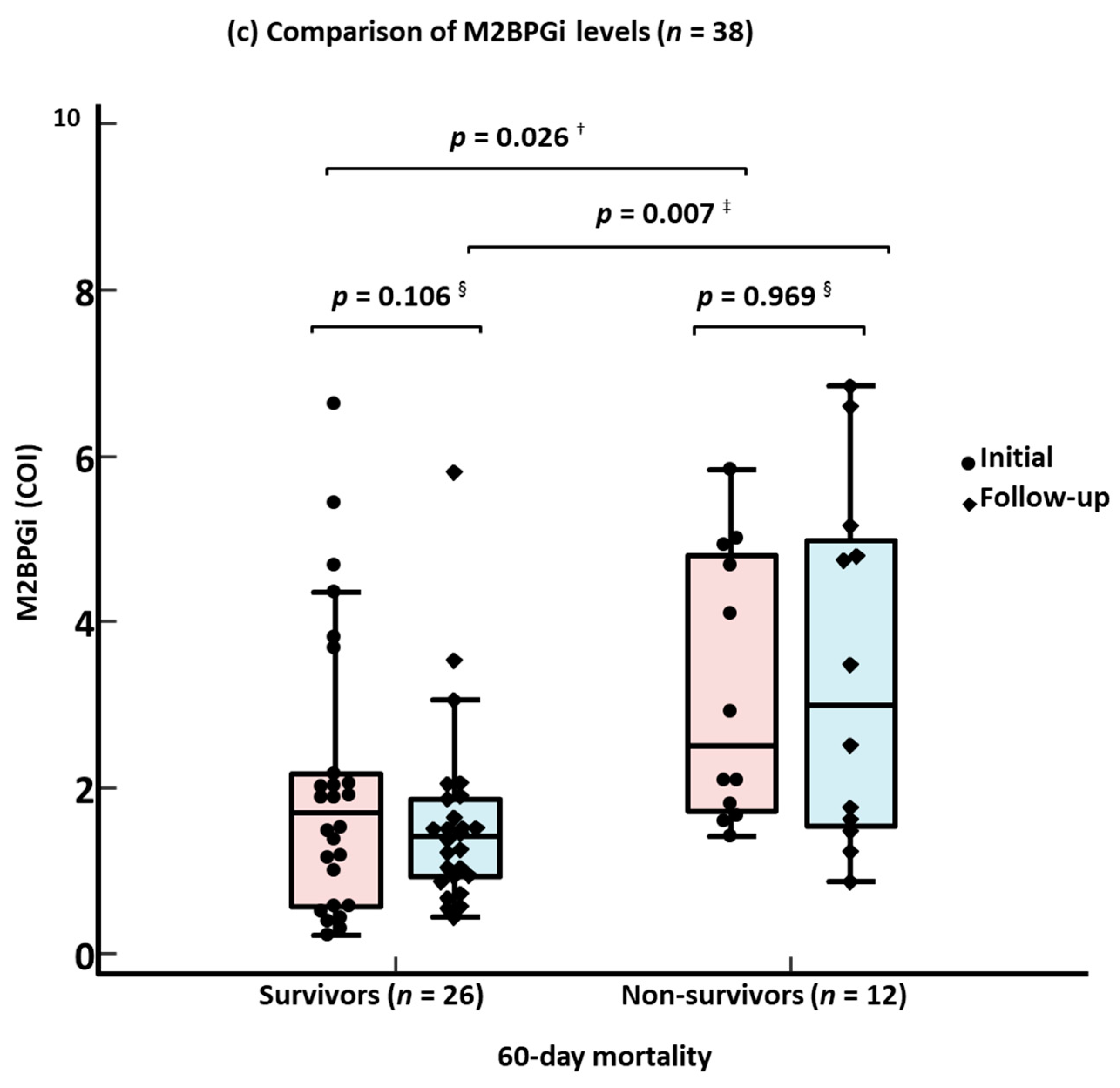
| M2BPGi (COI) | 1.9 (1.0–3.7) | 0.8 (0.4–2.6) | 2.0 (1.4–3.8) | 0.045 |
| >1.37 ††, n (%) | 34 (64.2) | 6 (40.0) | 28 (73.7) | 0.022 |
| Liver fibrosis score | ||||
| FIB-4 | 2.2 (1.2–4.7) | 1.9 (1.5–2.4) | 2.6 (1.2–4.3) | 0.323 |
| ≥1.3 ‡‡ | 39 (73.6) | 12 (80.0) | 27 (71.1) | 0.509 |
| Severity assessment | ||||
| SOFA score | 4.0 (1.0–7.0) | 0.0 (0.0–1.0) | 5.0 (3.0–7.0) | <0.001 |
| NEWS2 | 3.0 (2.0–6.0) | 2.0 (0.0–3.0) | 5.0 (3.0–8.0) | <0.001 |
| Sepsis/septic shock | 31 (58.5)/6 (11.3) | 0 (0.0)/0 (0.0) | 31 (81.6)/6 (15.8) | NA |
| Treatment, n (%) | ||||
| Supplemental oxygen therapy | 23 (43.4) | 2 (13.3) | 21 (55.3) | 0.006 |
| Antibiotics | 34 (64.2) | 10 (66.7) | 24 (63.2) | 0.812 |
| Azithromycin | 29 (54.7) | 8 (53.3) | 21 (55.3) | 0.899 |
| 3rd-generation cephalosporins | 20 (37.7) | 6 (40.0) | 14 (36.8) | 0.832 |
| Piperacillin/tazobactam | 2 (3.8) | 0 (0.0) | 2 (5.3) | 1.000 |
| Fluroquinolone | 2 (3.8) | 0 (0.0) | 2 (5.3) | 1.000 |
| Lopinavir/ritonavir | 26 (49.1) | 10 (66.7) | 16 (42.1) | 0.110 |
| Hydroxychloroquine | 22 (41.5) | 7 (46.7) | 15 (39.5) | 0.635 |
| Clinical outcomes, n (%) | ||||
| ICU admission | 14 (26.4) | 1 (6.7) | 13 (34.2) | 0.080 |
| Ventilator use | 12 (22.6) | 0 (0.0) | 12 (31.6) | 0.012 |
| ECMO use | 7 (13.2) | 0 (0.0) | 7 (18.4) | 0.171 |
| 30-day mortality §§ | 12 (22.6) | 2 (13.3) | 10 (26.3) | 0.471 |
| 60-day mortality §§ | 15 (28.3) | 2 (13.3) | 13 (34.2) | 0.182 |
| Variable | 30-Day Mortality | 60-Day Mortality | ||||
|---|---|---|---|---|---|---|
| Survivors (n = 41) | Non-Survivors (n = 12) | p | Survivors (n = 38) | Non-Survivors (n = 15) | p | |
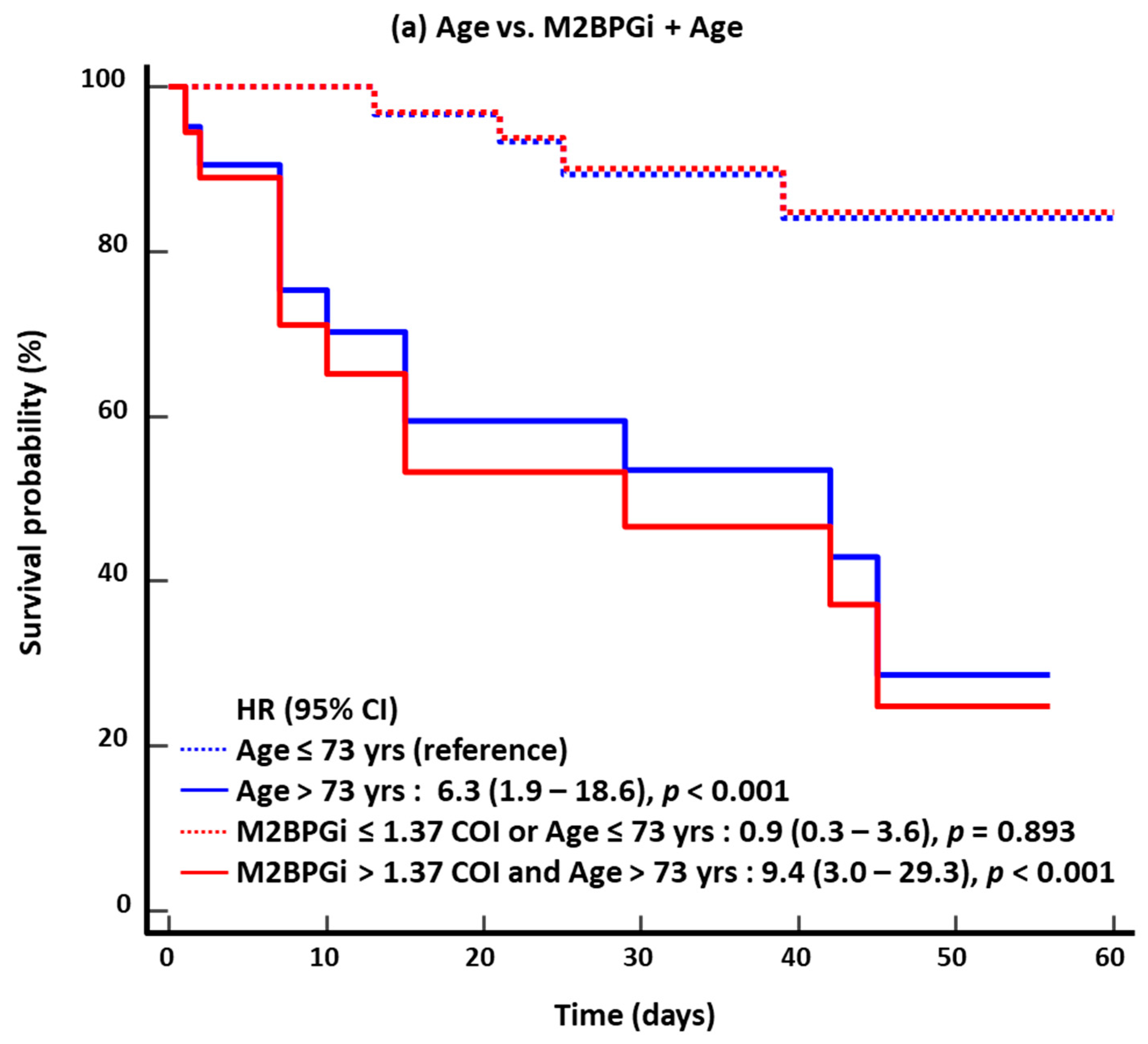
| Age (years) | 69.0 (61.7–77.0) | 78.5 (73.5–83.0) | 0.016 | 68.5 (61.0–75.0) | 79.0 (72.7–83.5) | 0.004 |
| ≥65, n (%) | 26 (63.4) | 10 (83.3) | 0.296 | 23 (60.5) | 13 (86.7) | 0.102 |
| ≥70, n (%) | 20 (48.8) | 10 (83.3) | 0.047 | 18 (47.4) | 12 (80.0) | 0.036 |
| ≥75, n (%) | 12 (29.3) | 9 (75.0) | 0.007 | 10 (26.3) | 11 (73.3) | 0.001 |
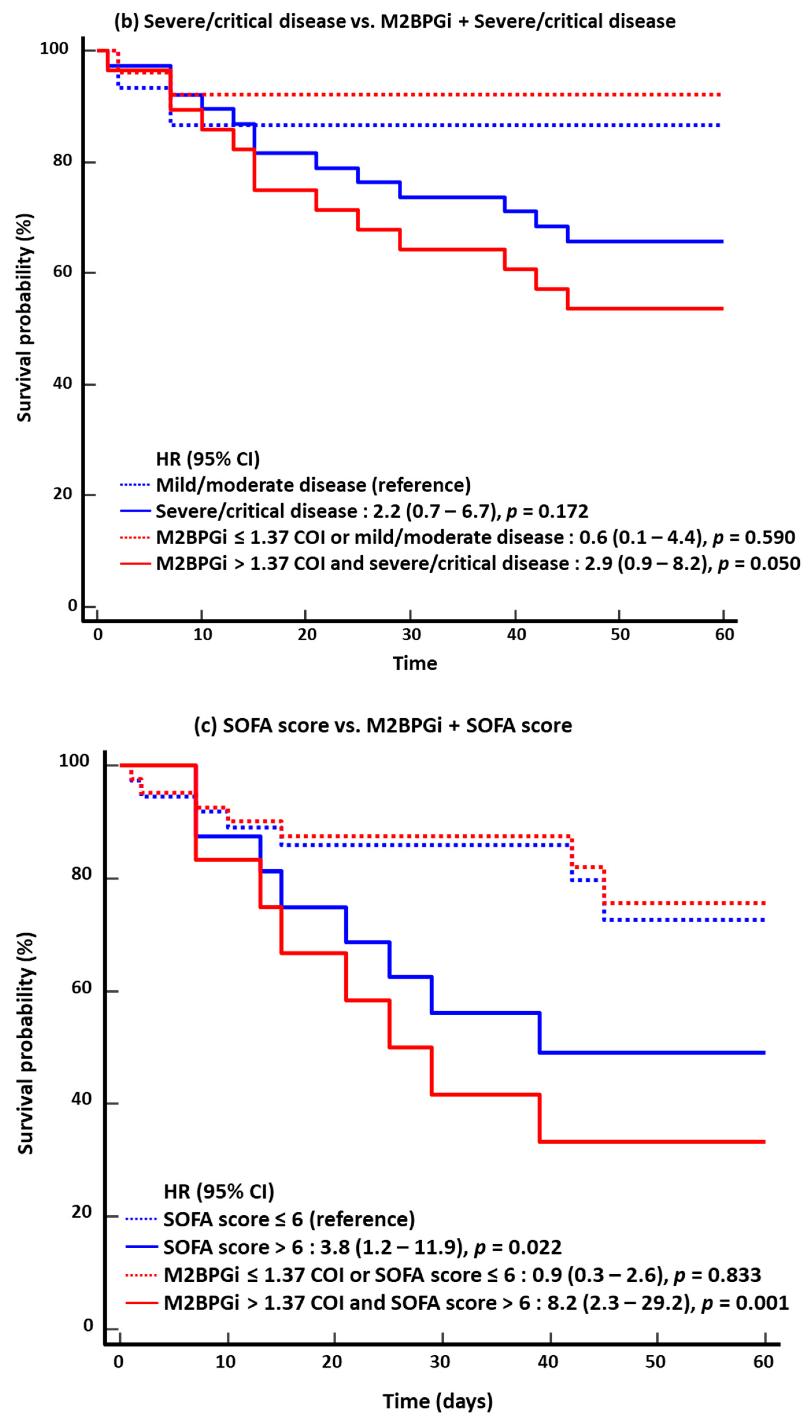
| Severe/critical disease, n (%) | 28 (68.3) | 10 (83.3) | 0.471 | 25 (65.8) | 13 (86.7) | 0.182 |
| SOFA score | 4.0 (0.0–5.2) | 7.5 (3.0–9.5) | 0.018 | 3.5 (0.0–5.0) | 7.0 (3.0–9.0) | 0.016 |
| NEWS2 | 3.0 (1.0–5.3) | 5.5 (4.0–9.0) | 0.010 | 3.0 (1.0–4.0) | 6.0 (4.2–9.0) | 0.001 |
| M2BPGi (COI) | 1.5 (0.5–2.4) | 2.7 (1.8–4.7) | 0.011 | 1.4 (0.5–2.2) | 2.9 (1.8–4.8) | 0.002 |
| >1.37, n (%) | 22 (53.7) | 12 (100.0) | 0.002 | 19 (50.0) | 15 (100.0) | <0.001 |
| Variable | Univariate | Multivariate | ||
|---|---|---|---|---|
| HR (95% CI) | p | HR (95% CI) | p | |
| 30-day mortality | ||||
| Age | 1.06 (1.01–1.12) | 0.031 | 1.01 (0.93–1.10) | 0.753 |
| Male | 0.59 (0.19–1.83) | 0.364 | ||
| Comorbidities (n) | 1.71 (1.04–2.82) | 0.033 | 1.69 (0.89–3.23) | 0.111 |
| Disease severity | 1.47 (0.69–3.12) | 0.319 | ||
| SOFA score | 1.24 (1.03–1.48) | 0.018 | 1.17 (0.63–1.47) | 0.190 |
| NEWS2 | 1.16 (1.03–1.31) | 0.013 | 1.09 (0.92–1.28) | 0.317 |
| M2BPGi | 1.27 (1.01–1.62) | 0.048 | 1.44 (1.05–1.98) | 0.025 |
| 60-day mortality | ||||
| Age | 1.07 (1.02–1.12) | 0.010 | 1.03 (0.96–1.11) | 0.370 |
| Male | 0.65 (0.24–1.80) | 0.412 | ||
| Comorbidities (n) | 1.55 (1.01–2.39) | 0.048 | 1.46 (0.86–2.49) | 0.159 |
| Disease severity | 1.71 (0.81–3.59) | 0.159 | ||
| SOFA score | 1.23 (1.05–1.45) | 0.012 | 1.12 (0.90–1.38) | 0.300 |
| NEWS2 | 1.18 (1.06–1.32) | 0.002 | 1.15 (0.98–1.34) | 0.070 |
| M2BPGi | 1.30 (1.06–1.60) | 0.012 | 1.45 (1.09–1.92) | 0.010 |
Disclaimer/Publisher’s Note: The statements, opinions and data contained in all publications are solely those of the individual author(s) and contributor(s) and not of MDPI and/or the editor(s). MDPI and/or the editor(s) disclaim responsibility for any injury to people or property resulting from any ideas, methods, instructions or products referred to in the content. |
© 2025 by the authors. Licensee MDPI, Basel, Switzerland. This article is an open access article distributed under the terms and conditions of the Creative Commons Attribution (CC BY) license (https://creativecommons.org/licenses/by/4.0/).
Share and Cite
Park, M.; Hur, M.; Kim, H.; Lee, C.H.; Lee, J.H.; Kim, H.W.; Nam, M.; Lee, S. Novel Usefulness of M2BPGi for Predicting Severity and Clinical Outcomes in Hospitalized COVID-19 Patients. Diagnostics 2025, 15, 937. https://doi.org/10.3390/diagnostics15070937
Park M, Hur M, Kim H, Lee CH, Lee JH, Kim HW, Nam M, Lee S. Novel Usefulness of M2BPGi for Predicting Severity and Clinical Outcomes in Hospitalized COVID-19 Patients. Diagnostics. 2025; 15(7):937. https://doi.org/10.3390/diagnostics15070937
Chicago/Turabian StylePark, Mikyoung, Mina Hur, Hanah Kim, Chae Hoon Lee, Jong Ho Lee, Hyung Woo Kim, Minjeong Nam, and Seungho Lee. 2025. "Novel Usefulness of M2BPGi for Predicting Severity and Clinical Outcomes in Hospitalized COVID-19 Patients" Diagnostics 15, no. 7: 937. https://doi.org/10.3390/diagnostics15070937
APA StylePark, M., Hur, M., Kim, H., Lee, C. H., Lee, J. H., Kim, H. W., Nam, M., & Lee, S. (2025). Novel Usefulness of M2BPGi for Predicting Severity and Clinical Outcomes in Hospitalized COVID-19 Patients. Diagnostics, 15(7), 937. https://doi.org/10.3390/diagnostics15070937