Article

Validation of Ultrasound for Quantification of Knee Meniscal Tissue: A Cadaveric Study

by
Jacobo Rodríguez-Sanz
1,2,†,
Miguel Malo-Urriés
3,*,
Sergio Borrella-Andrés
3,
Isabel Albarova-Corral
3 and
Carlos López-de-Celis
1,2,4,†
1
Faculty of Medicine and Health Science, Universitat Internacional de Catalunya, 08195 Barcelona, Spain
2
ACTIUM Functional Anatomy Group, 08195 Barcelona, Spain
3
PhysiUZerapy Health Sciences Research Group, Health Sciences Faculty, Department of Physiatry and Nursing, University of Zaragoza, 50009 Zaragoza, Spain
4
Fundació Institut Universitari per a la Recerca a l’Atenció Primària de Salut Jordi Gol i Gurina, 08035 Barcelona, Spain
*
Author to whom correspondence should be addressed.
These authors contributed equally to this work.
Diagnostics 2025, 15(3), 389; https://doi.org/10.3390/diagnostics15030389

Submission received: 17 December 2024 / Revised: 1 February 2025 / Accepted: 3 February 2025 / Published: 6 February 2025
(This article belongs to the Special Issue New Advances in Forensic Radiology and Imaging)

Abstract

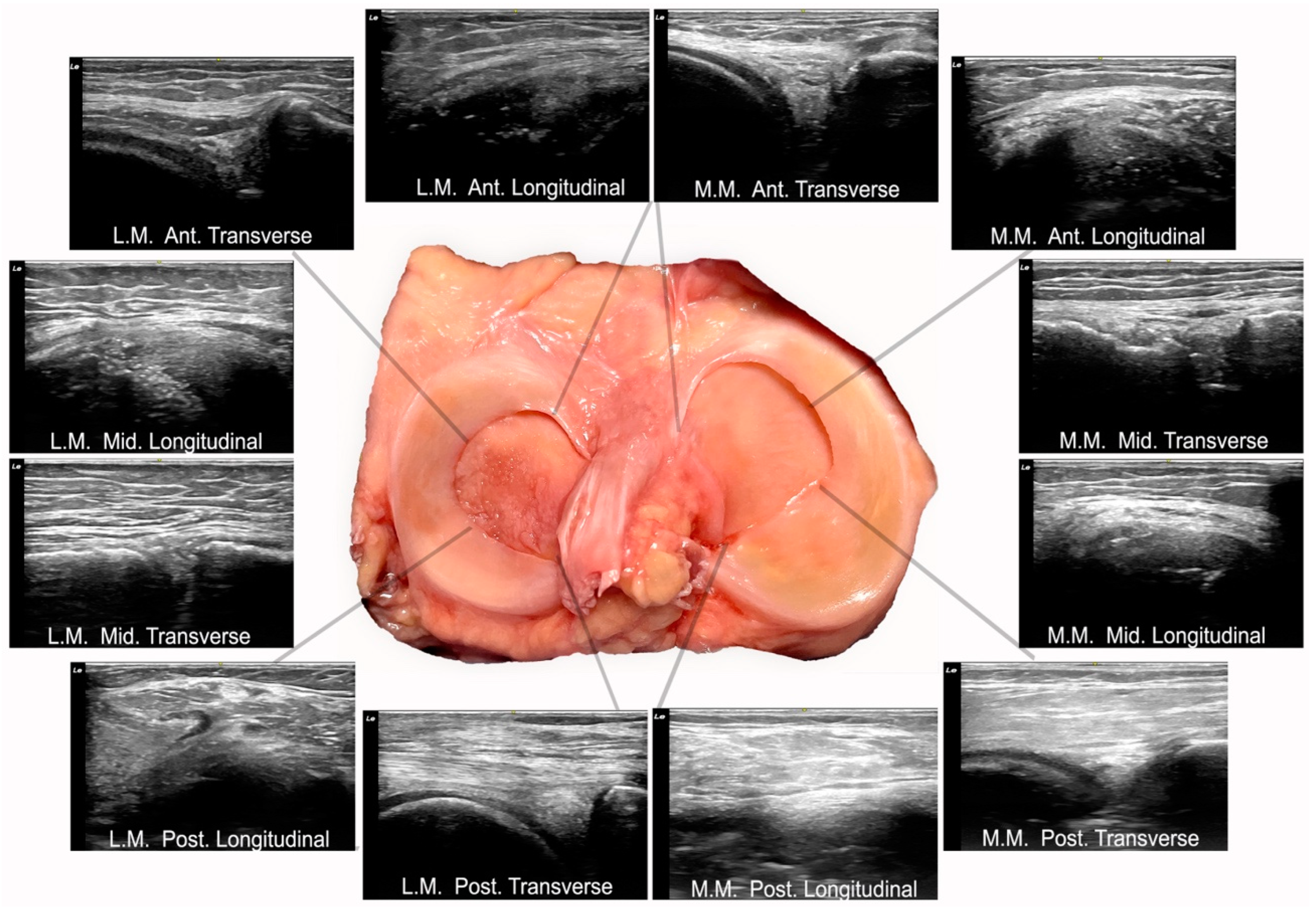
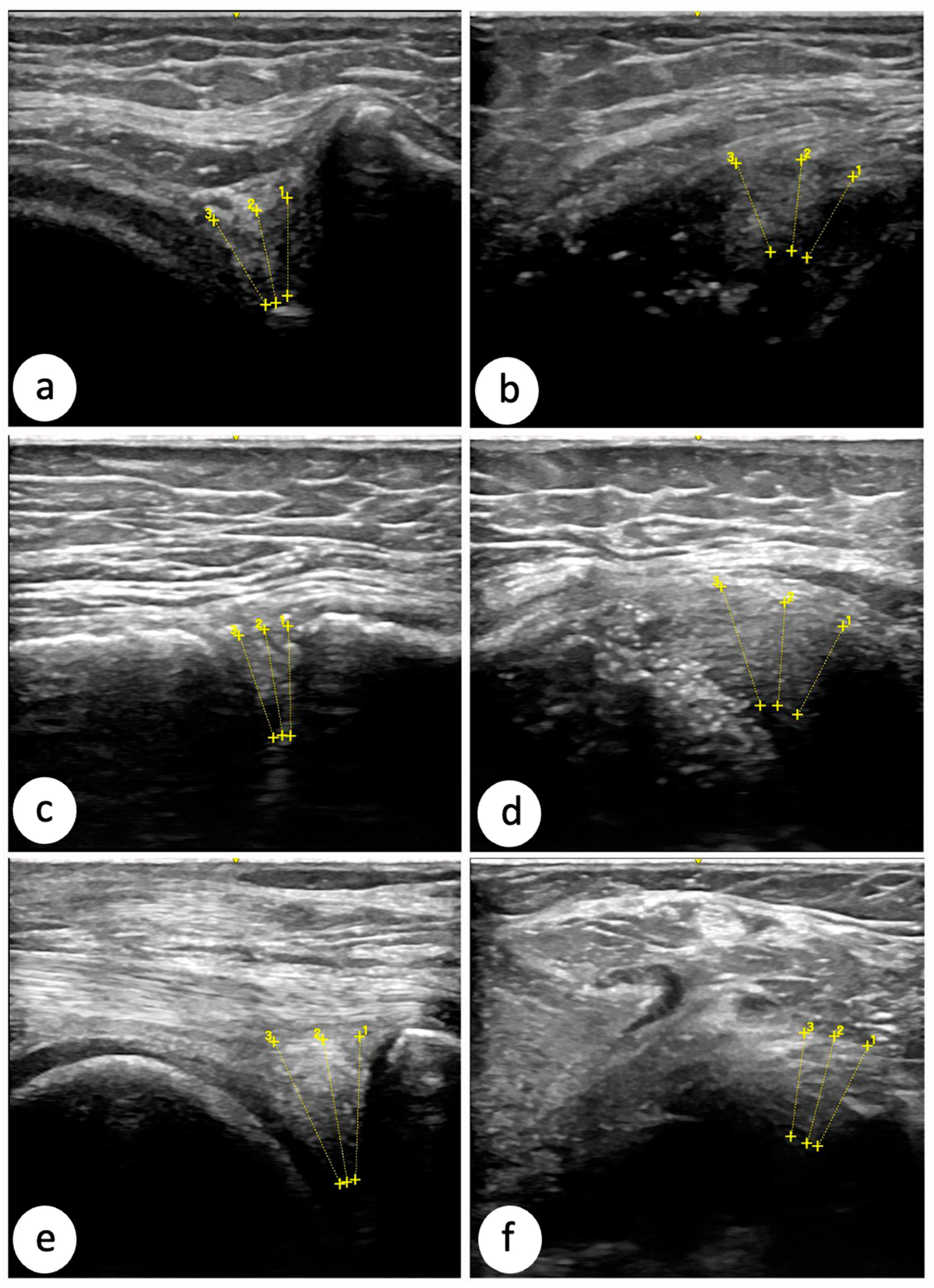
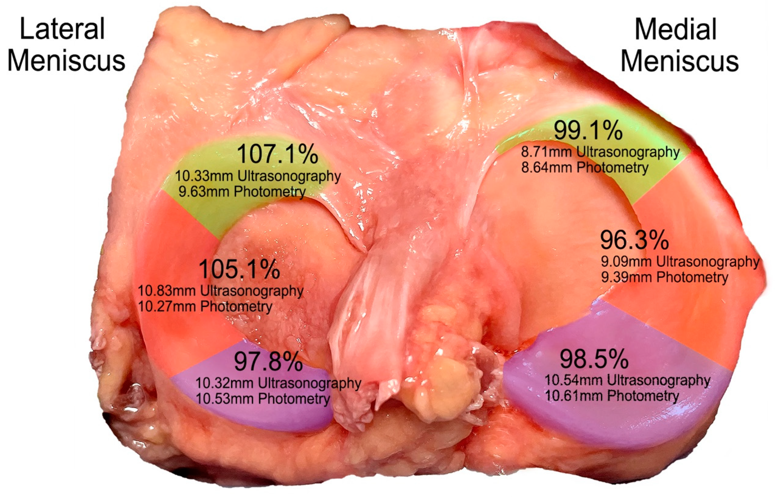
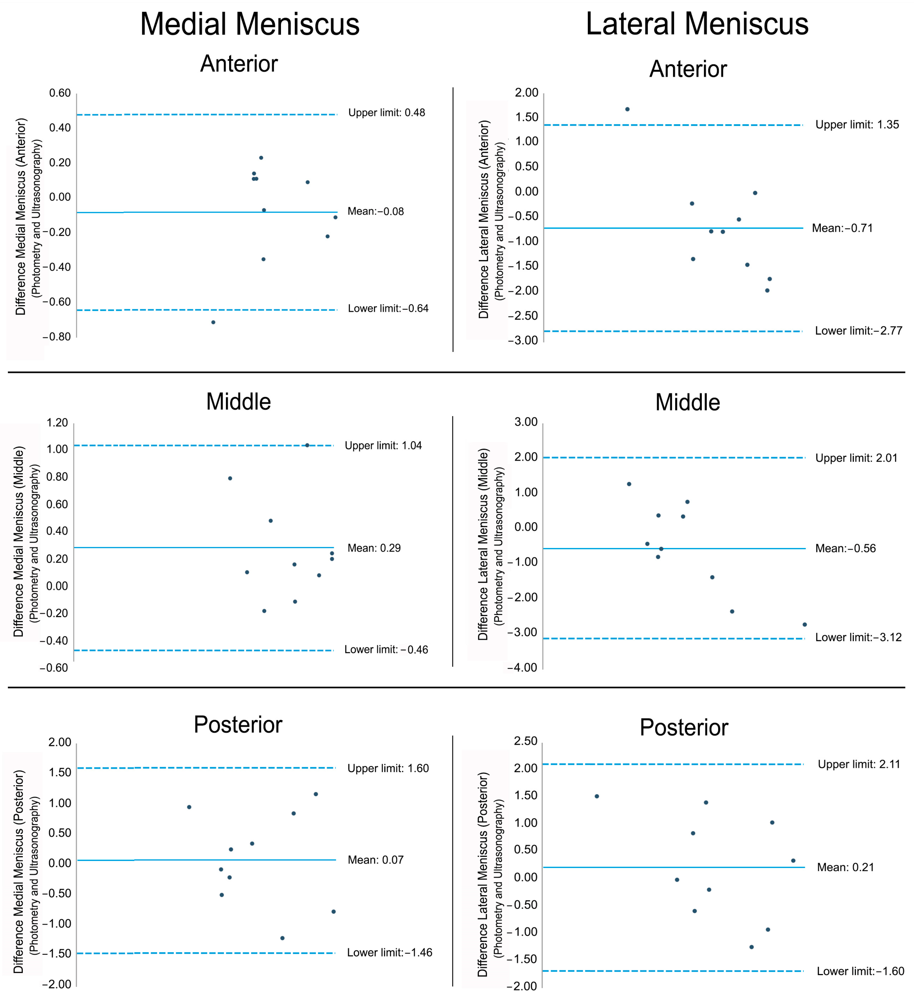
Background: While MRI is the gold standard for meniscal assessment, its cost and accessibility limitations have led to growing interest in ultrasound, though its validity for quantifying meniscal tissue remains unclear. To validate the use of ultrasound in quantifying meniscal tissue across the anterior, middle, and posterior regions of both menisci (medial and lateral) in longitudinal and transverse planes by comparison with cadaveric dissection. Methods: A cross-sectional study was conducted on ten cryopreserved anatomical donors, obtaining a total of 120 ultrasound scans from the different meniscal regions. Following ultrasound imaging, cadaveric dissection was performed to facilitate photometric measurements, thereby enabling validation of the ultrasound findings. The intra-examiner reliability of the ultrasound measurements was also assessed. Results: The intra-examiner reliability of ultrasound measurements ranged from moderate to excellent. A strong and statistically significant positive correlation was observed between ultrasound and photometric measurements across all meniscal regions (r > 0.821; p < 0.05). In the medial meniscus, ultrasound visualized 99.1% of the anterior region (8.71 mm with ultrasound; 8.64 mm with photometry), 96.3% of the middle region (9.09 mm with ultrasound; 9.39 mm with photometry), and 98.5% of the posterior region (10.54 mm with ultrasound; 10.61 mm with photometry). In the lateral meniscus, ultrasound visualized 107.1% of the anterior region, 105.1% of the middle region, and 97.8% of the posterior region. The observed excess in tissue visualization in some regions likely reflects the inclusion of adjacent connective tissue, indistinguishable from meniscal tissue on ultrasound. Conclusions: Ultrasound is a valid and reliable modality for visualizing most meniscal tissue across regions, with a measurement discrepancy under 0.7 mm compared to anatomical dissection. However, caution is advised as adjacent connective tissue may sometimes be misidentified as meniscal tissue during evaluations.
Keywords: ultrasound; meniscus; cadaver; validation; knee ultrasound; meniscus; cadaver; validation; knee