Abstract
Background: Myxoid glomus tumors (mGTs) are an uncommon histologic pattern of glomus tumors, characterized by prominent myxoid stromal changes that may mimic a wide range of soft tissue neoplasms. Recent reports of unexpected CD34 expression in some cases have further complicated their differential diagnosis. Objectives: This study aimed to characterize the histopathological, immunohistochemical, and clinical features of cutaneous mGTs, with particular emphasis on CD34 expression. Methods: We analyzed 8 histologically confirmed cases of cutaneous mGTs underwent to a comprehensive evaluation of morphological features and immunophenotypic profile, with available clinical data. The immunohistochemical panel included smooth muscle actin (SMA), CD34, and S100. Mast cell density was assessed by tryptase in 3 cases. As controls, 8 glomus tumors without myxoid features were also examined for CD34 expression. Results: The cohort consisted of 8 patients (2 males, 6 females; age range 23–71 years). All tumors were located on the distal phalanges of the digits and showed extensive myxoid stromal changes. Immunohistochemistry demonstrated SMA positivity and CD34 expression in all mGTs. In contrast, none of the control GTs without myxoid stroma expressed CD34. Mast cells were consistently identified in the tested cases, predominantly within the myxoid matrix, suggesting a possible role in stromal remodeling. Conclusions: mGTs represent a rare but distinct histological pattern within the glomus tumor spectrum; frequent CD34 expression and mast cell infiltration appear to be characteristic features, although their biological significance remains uncertain. Recognition of these findings is essential to avoid misdiagnosis with other CD34-positive perivascular neoplasms or myxoid soft tissue sarcomas.
1. Introduction
Glomus tumors (GTs) are rare soft tissue neoplasms (<2%), composed of modified smooth muscle cells from the glomus body, a thermoregulatory arteriovenous structure []. They are most common in young adults and typically benign, though rare malignant forms exist []. Sporadic GTs often harbor NOTCH gene rearrangements, with some showing BRAF or KRAS mutations. Rare familial cases, typically presenting with multiple lesions in an autosomal dominant hereditary pattern, are associated with loss-of-function mutations in the GLMN gene (chromosome 1p22.1). These mutations impair the function of glomulin in vascular smooth muscle cells, leading to abnormal vascular development and tumor formation [,]. GTs arise mostly in the skin and superficial soft tissues of distal extremities—especially subungual, hand, wrist, and foot—but have been reported in sites like the stomach, genitourinary tract, mediastinum, bone, and lung [,,]. Cutaneous lesions are typically <1 cm, reddish-blue, and painful, often triggered by cold or touch. Deep or visceral GTs usually present as nonspecific masses [,].
Histologically, the vast majority of GTs are benign and consist of small, uniform glomus cells with round to oval monomorphic nuclei, lightly eosinophilic cytoplasm, and well-defined borders. The solid (conventional) type is the most common, accounting for approximately 75% of cases, and is characterized by nests of glomus cells surrounding capillaries. Other histological patterns may occasionally be observed, including oncocytic, hyalinized or epithelioid forms, as well as symplastic GT, which is defined by the presence of scattered, large, hyperchromatic nuclei in the absence of mitotic activity; these nuclear changes should be interpreted as degenerative (“ancient”) alterations without prognostic significance [,]. Rarely, intermediate or malignant lesions are encountered, defined by the variable presence of deep location, large size, nuclear atypia, increased mitotic activity, or atypical mitoses. In this regard, Folpe et al. [] proposed that a GT should be classified as malignant when one of the following criteria is met: deep location, size ≥ 2 cm, atypical mitoses, or marked nuclear atypia with increased mitotic activity (≥5/50 HPF). GTs of uncertain malignant potential are defined by deep location or large size (≥2 cm), or superficial location with increased mitotic activity (≥5/50 HPF). Immunohistochemically, GTs are consistently positive for vimentin (100%), SMA (99%), MSA (95%), and calponin (80%), with collagen IV and laminin expression in ~91% of cases and h-caldesmon in ~87% []. In contrast, markers such as S100, CD31, cytokeratin, and EMA are negative. CD34 is usually negative, although focal to diffuse positivity may be observed in some cases.
Among the different GT patterns described in the literature, myxoid GTs represent a variant uncommonly reported, although it is not clear if it represents an entity or only a spectrum of the same lesion. These lesions are considered benign and their morphological significance and the underlying mechanisms responsible for their myxoid features remain poorly understood. Furthermore, mGTs can mimic a range of benign and malignant soft tissue neoplasms with prominent myxoid stroma. Recent literature has also highlighted the unexpected expression of CD34 in some of these myxoid cases, a feature not typical of conventional GTs. This observation has led to hypotheses regarding a transitional phenotype between smooth muscle and endothelium as the possible cell of origin [,], and underscores the need for careful morphologic and immunohistochemical evaluation. Herein, we present a case series of 8 mGTs and analyze their clinicopathologic features in the context of previously reported literature.
2. Materials and Methods
This retrospective case–control study includes eight cases of cutaneous mGTs diagnosed between 2019 and 2025 at the University of Bari “Aldo Moro”, Bari, Italy, and the University of Minnesota, Minneapolis, USA, matched with other randomly selected eight cases of typical GT to compare CD34 immuno-expression. The study was conducted in accordance with the principles of the Declaration of Helsinki, and ethical approval was waived by the Institutional Review Board (IRB) due to the use of anonymized archival data and specimens (approval code 22007, approval date 5 September 2025).
All cases included in this study were histologically confirmed cutaneous GTs with available formalin-fixed paraffin-embedded (FFPE) tissue and immunohistochemical analysis was performed. All specimens were obtained from surgical excision, which also represented the definitive treatment in all patients. Cases with incomplete clinical data or poor tissue preservation were excluded. No comorbidities specifically related to glomus tumors were identified. Patient age, sex, and tumor site were retrieved from medical records and are summarized in Table 1.
Table 1.
Clinicopathological and immunohistochemical features of mGTs.
Cases were identified and retrieved through pathology databases using the terms ‘glomus tumor’ and/or ‘myxoid’, and all extracted cases were re-evaluated by two dermatopathologists (G.C. and A.G.) according to the inclusion criteria, consisting of: (1) extensive myxoid stromal changes defined as the sum of ≥30% of myxoid phenomena in the stroma, or in form of multiple scattered foci always ≥30%; (2) availability of immunohistochemical staining; (3) presence of clinical data. The original immunohistochemical panel included CD34 (QBEND/10, Dako-Agilent, Glostrup, Denmark, 1:100), S-100 (Polyclonal, Dako, 1:500), Desmin (DE-R-11, Leica, Nussloch, Germany, ready to use), and SMA (ASM-1/1A4, Thermo Fisher, Waltham, MA, USA, 1:500).
Three cases of mGTs were also tested for tryptase (Leica Biosystem, clone 10D11) to identify the presence of mast cells expressed as the mean number of positive cells/10 HPF (400×).
Finally, a comprehensive literature review was also performed using PubMed, Scopus, and Web of Science databases with the keyword “myxoid glomus tumor”. All articles describing superficial/cutaneous cases were selected for discussion, in order to contextualize our findings.
3. Results
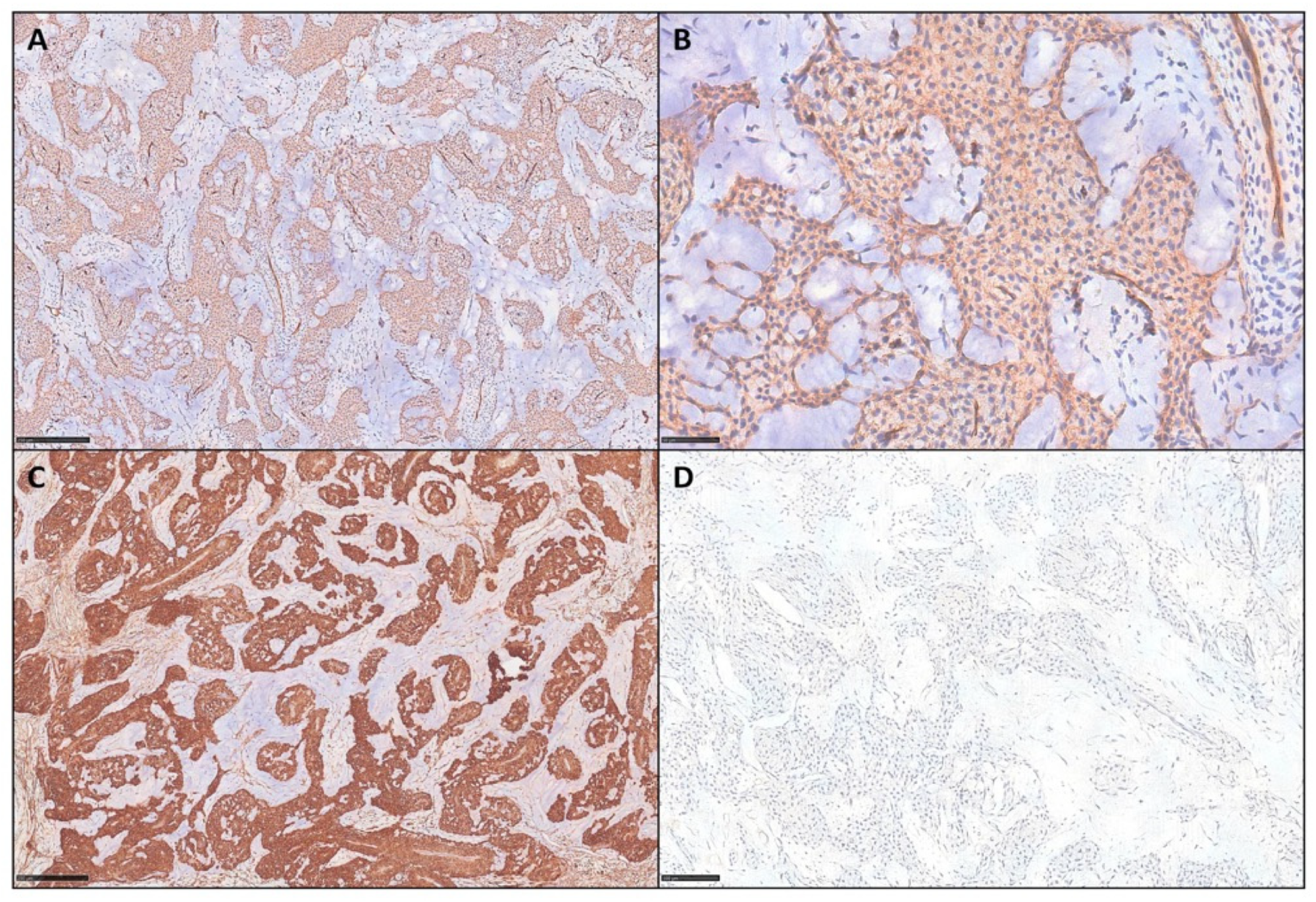
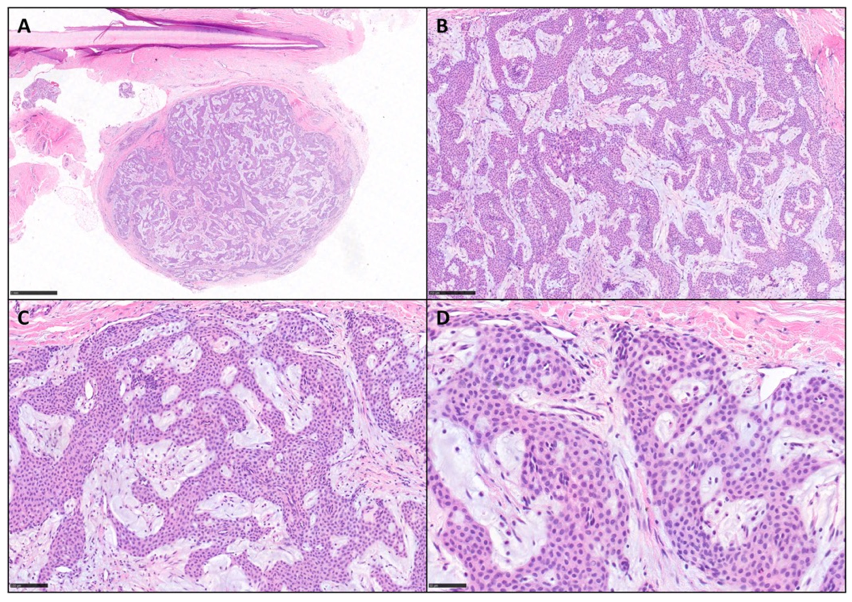
A total of 8 patients (2 males, 6 females; age range: 23–71 years) met the inclusion criteria (Table 1). All tumors were located on the distal phalanges of the digits, near the ungual plate. Histologically, they were characterized by nests and sheets of uniform, round glomus cells with centrally located nuclei and eosinophilic cytoplasm, embedded within an abundant myxoid stroma. A florid, delicate vascular network was also present. According to the criteria proposed by Folpe et al. [], all cases in our series were consistent with benign lesions, lacking any features of malignancy: all tumors were superficially located, measured between 1 and 1.5 cm in diameter, showed 0–1 mitoses per 50 high-power fields (HPF), and exhibited no nuclear atypia or necrosis. Immunohistochemistry showed SMA and CD34 positivity in all cases, with CD34 staining ranging from focal to diffuse. None of the cases expressed S100 and Desmin. In contrast, all 8 cases of GT without myxoid component were totally negative for CD34.
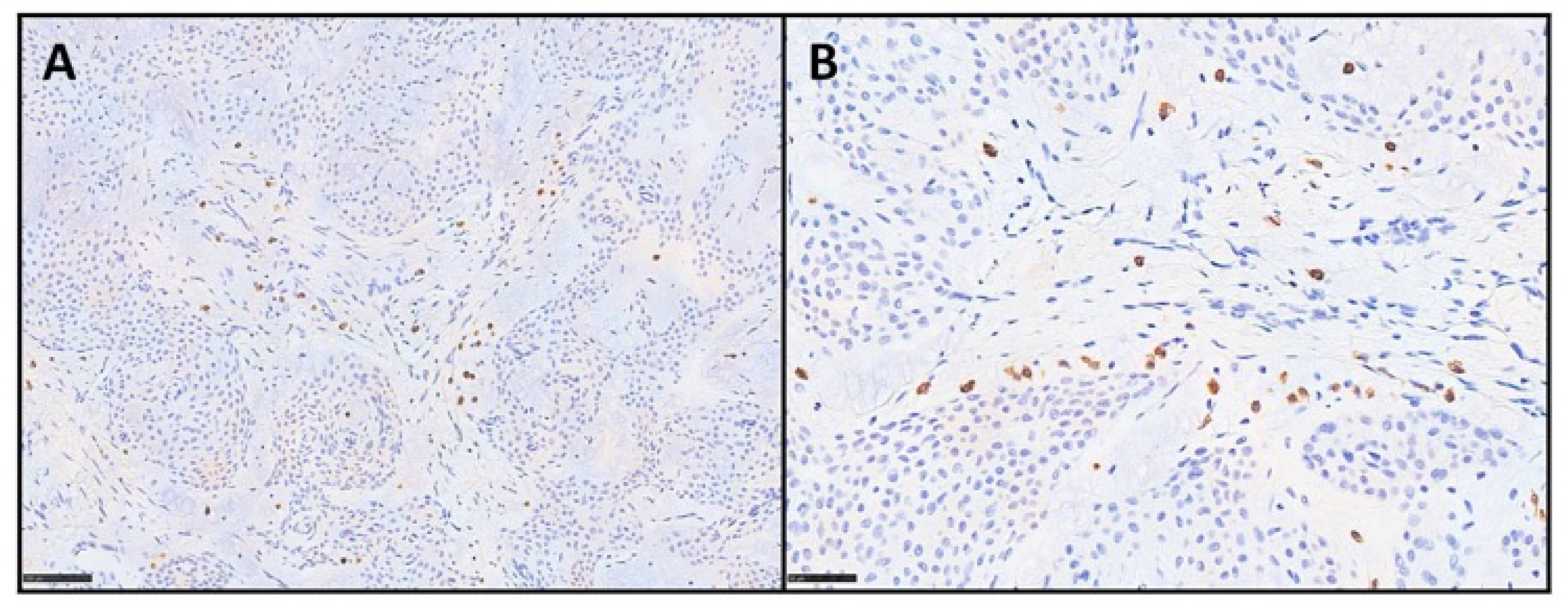
Figure 1 and Figure 2 show some examples of mGTs with histological and immunohistochemical insights.
Figure 1.
Rarely, some glomus tumors harbor abundant myxoid changes. (A) Photomicrograph showing an example of peri-ungueal mGT (H&E, scale bar 1 mm). (B,C) Tumor is made up of nests and cords of glomus cells embedded in fibrous and myxoid stroma (H&E, scale bar 250 and 100 μm). (D) Higher magnification of small, uniform, round glomus cells with centrally located nuclei, amphophilic to lightly eosinophilic cytoplasm, and sharply defined borders immersed in a myxoid stroma (H&E, scale bar 50 μm).
Figure 2.
(A,B) Tumor cells, in these two representative cases of mGTs from our series, show diffuse cytoplasmic expression of CD34 with moderate intensity (IHC for CD34, scale bar 250 μm and 50 μm). (C) All cases displayed strong and diffuse positivity for SMA (IHC for SMA, scale bar 250 μm), (D) as well as negativity for Desmin (IHC for Desmin, scale bar 100 μm).
Moreover, tryptase showed the presence of mast cells in all three tested cases. In particular, mast cells were found within the myxoid matrix, especially at the periphery of the lesions, with counts ranging from 12 to 20 positive cells per HPF (mean count: 16) in the hotspot areas. Figure 3 shows the twoases immunostained with anti-tryptase antibody.
Figure 3.
(A,B) Mast cells are highlighted by tryptase immunostaining in the tested cases. Positive cells are mainly distributed within the myxoid stroma, as single cells or small loose clusters (IHC for tryptase, scale bar 100 and 50 μm).
4. Discussion
GTs, first described by Masson in 1924 [], are rare, mostly benign perivascular neoplasms arising from modified smooth muscle cells of the glomus body [,]. Although their smooth muscle origin is widely accepted [,,,], as confirmed by SMA diffuse positivity, the histogenesis of GTs remains incompletely understood [,,,,,,,,,,]. However, occasional CD34 expression in GTs—typically considered a progenitor and endothelial marker—has been reported [], although its functional significance remains unclear. In this regard, some authors have proposed a transitional phenotype between smooth muscle and endothelial cells, based on the co-expression of myoid markers (SMA) and vascular antigens (CD31, CD34) in glomus cells located within sinusoidal vessel walls [,,,]. Preliminary studies have demonstrated CD34 positivity in both benign and malignant GTs, particularly within the endothelial components [,,,,,,,,], but its biological role as well as diagnostic value are still debated.
Among the histologic variants described in the literature, the myxoid subtype has emerged as a peculiar form (Table 2). While generally regarded as benign, the morphological significance and underlying mechanisms contributing to the myxoid stromal features are not fully elucidated. Furthermore, the absence of clear classificatory criteria may have led the authors not to describe this feature, also leading us not to include all cases of GTs with myxoid features published in the literature.
Table 2.
Summary of published cases of mGTs.
Tsuneyoshi and Enjoji [] identified a myxoid subtype in 23 of 63 GTs, characterized by myxoid stroma and cords of epithelioid tumor cells arranged around vascular spaces and the frequent presence of mast cells within the stroma. They also proposed that GTs may represent a mesodermal developmental anomaly rather than a true neoplasm, based on their ultrastructural features and slow-growing clinical course. In another mGT case, Pabuççuoğlu and Lebe [] observed CD34 and MMP-9 expression, suggesting that MMP-9, produced by both tumor cells and mast cells, may contribute to the development of the myxoid stroma through extracellular matrix remodeling. Notably, the presence of mast cells in GTs has only previously been documented in another non-cutaneous deep-seated case, also showing myxoid changes, in relation to pain generation: indeed, the presence of mast cells may play a significant algogenic role by releasing histamine and substance P [].
Mentzel et al. [] further demonstrated the coexpression of SMA and CD34 in 6 mGTs, underlining that this immunophenotype could complicate the differential diagnosis with other CD34-positive tumors, such as myopericytoma or solitary fibrous tumor (SFT), particularly in the presence of myxoid stroma.
Our series included eight cutaneous GTs with prominent myxoid stromal changes (≥30%), all demonstrating CD34 expression in addition to the classical immunophenotype. CD34 expression has previously been reported in mGTs, although it is not regarded as a consistent immunophenotypic feature [,,]. As summarized in Table 2, several mGTs have been described in the literature, but CD34 immunostaining was not consistently performed by all authors. With this background, our intent was to demonstrate—through a relatively larger case series—that CD34 expression can represent a recurrent finding in mGTs. Therefore, our results provide additional evidence supporting the presence of CD34 immunoreactivity in this rare variant.
Histologically, mGTs are characterized by small, uniform round cells arranged in nests or sheets around capillary-sized vessels, embedded in a pale, myxoid stroma. This morphology may mimic several benign and malignant soft tissue tumors, such as superficial acral fibromyxoma, myopericytoma, angioleiomyoma, myxoid SFT, low-grade fibromyxoid sarcoma, and myxoid liposarcoma (Table 3). The presence of prominent myxoid matrix, coupled with CD34 immunopositivity, could increase the risk of misdiagnosis, both in cutaneous and deep-seated lesions, where the clinical suspicion for GTs is lower.
Table 3.
Immunohistochemical profile of mGTs and their main mimickers.
Superficial acral fibromyxoma, often found in acral sites like mGTs, is CD34 and CD99 positive, lacking the perivascular round cell morphology typical of GTs []. Angioleiomyomas with myxoid stromal changes can also mimic mGTs; however, they are composed of interlacing bundles of smooth muscle cells and demonstrate diffuse immunoreactivity for SMA and calponin, with strong and consistent positivity for h-caldesmon []. Desmin staining is typically variable, ranging from focal expression to complete absence in a subset of cases []. These tumors are characteristically negative for melanocytic markers such as HMB45 and Melan A []. Myopericytoma also shares a perivascular growth pattern but is predominantly characterized by concentric arrangements of spindle cells. By immunohistochemistry, it expresses SMA and h-caldesmon, while desmin and/or CD34 are only focally positive []. Myxoid solitary fibrous tumors, rarely cutaneous in location, have been described as CD34 and STAT6 positive, with a patternless architecture that lacks muscle marker expression []. Low-grade fibromyxoid sarcoma, composed of deceptively bland spindle cells within a fibrous to myxoid background, shows positivity for MUC4, a highly sensitive and specific marker []. Myxoid liposarcoma may mimic mGTs due to its delicate capillary vasculature and pale stroma, but it is distinguished by the presence of lipoblasts and characteristic FUS-DDIT3 translocation [].
Taken together, our findings support the notion that mGTs represent anhistologic pattern within the GT spectrum, characterized by unusual morphologic (myxoid matrix) and immunohistochemial (CD34-positivity) features, that may lead to diagnostic confusion. Whether they also represent a true biological distinct entity, as suggested by CD34 expression and mast cell colonization, remains an open question. Recognizing the benign behavior of these lesions is essential to prevent overtreatment and inappropriate classification. Awareness of this rare variant, as well as careful morphologic evaluation in conjunction with a targeted immunohistochemical panel, remains the cornerstone of accurate diagnosis.
5. Conclusions
In conclusion, our series highlights the importance of recognizing mGTs as a rare but distinct histological variant of glomus tumors, characterized by frequent CD34 expression on immunohistochemistry. These findings expand the morphologic and immunophenotypic spectrum of GTs and underline the need for the cautious interpretation of CD34 positivity in the setting of mesenchymal tumors with myxoid stroma. Accurate diagnosis relies on integrating histologic features, immunoprofiles, and clinical context, in order to avoid misclassification with more aggressive neoplasms and ensure appropriate patient management.
Future studies on mGTs should aim to clarify whether this variant represents a distinct biological entity or merely a morphologic spectrum within the glomus tumor family. Larger multicenter cohorts, supported by molecular profiling, could help elucidate the pathogenetic mechanisms underlying the myxoid stroma and the unexpected CD34 expression. In particular, the potential role of mast cells in extracellular matrix remodeling and in symptom generation deserves further investigation. Comparative genomic studies between conventional GTs and mGTs may also uncover genetic or epigenetic differences that could explain their peculiar phenotype.
Author Contributions
Conceptualization, G.C. and A.G.; methodology, G.C., C.R. and F.F. (Francesco Fortarezza); investigation, J.S., M.D.M. and F.F. (Francesco Fanelli); writing—original draft preparation, J.S. and M.D.M.; writing—review and editing, G.C., A.G. and G.I.; supervision, G.I. and C.R.; figures and tables, A.C. and F.F. (Francesco Fortarezza). All authors have read and agreed to the published version of the manuscript.
Funding
This research received no external funding.
Institutional Review Board Statement
The study was conducted in accordance with the principles of the Declaration of Helsinki and ethical approval was waived by the Institutional Review Board (IRB) of University of Minnesota due to the use of anonymized archival data and specimens (approval code 22007, approval date 5 September 2025).
Informed Consent Statement
Informed consent was obtained from all subjects involved in the study.
Data Availability Statement
The original contributions presented in this study are included in the article. Further inquiries can be directed to the corresponding author.
Conflicts of Interest
The authors declare no conflicts of interest.
References
- Gombos, Z.; Zhang, P.J. Glomus tumor. Arch. Pathol. Lab. Med. 2008, 132, 1448–1452. [Google Scholar] [CrossRef] [PubMed]
- Brenn, T.; Goodlad, J.; Mentzel, T. AFIP Series 5 Fasc. N. 7-Non Melanocytic Tumors of the Skin; American Registry of Pathology: Arlington, VA, USA, 2021. [Google Scholar]
- Jackett, L.A.; Folpe, A.L. (Eds.) Chapter 7: Glomus tumour. In Skin Tumours, 5th ed.; WHO Classification of Tumours Editorial Board; WHO Classification of Tumours Series; International Agency for Research on Cancer: Lyon, France, 2025; Volume 12, Available online: https://publications.iarc.fr (accessed on 4 September 2025).
- Amyere, M.; Aerts, V.; Brouillard, P.; McIntyre, B.A.; Duhoux, F.P.; Wassef, M.; Enjolras, O.; Mulliken, J.B.; Devuyst, O.; Antoine-Poirel, H.; et al. Somatic uniparental isodisomy explains multifocality of glomuvenous malformations. Am. J. Hum. Genet. 2013, 92, 188–196. [Google Scholar] [CrossRef] [PubMed] [PubMed Central]
- Goldblum, J.R.; Folpe, A.L.; Weiss, S.W. (Eds.) Enzinger and Weiss’s Soft Tissue Tumors, 7th ed.; Elsevier: Philadelphia, PA, USA, 2020. [Google Scholar]
- Ezeh, K.J.; Rajwana, Y.; Paudel, B.; Shen, T.; Botros, Y. Gastric Glomus Tumor Presenting With Gastrointestinal Bleed and Pulmonary Embolism: A Rare Entity With Management Dilemma. Cureus 2022, 14, e25632. [Google Scholar] [CrossRef] [PubMed] [PubMed Central]
- Masson, P. Le glomus neuromyoarterial des regions tactiles et ses tumeurs. Lyon Chir. 1924, 21, 257–280. [Google Scholar]
- Folpe, A.L.; Fanburg–Smith, J.C.; Miettinen, M.; Weiss, S.W. Atypical and malignant glomus tumors: Analysis of 52 cases, with a proposal for the reclassification of glomus tumors. Am. J. Surg. Pathol. 2001, 25, 1–12. [Google Scholar] [CrossRef] [PubMed]
- Miettinen, M.; Paal, E.; Lasota, J.; Sobin, L.H. Gastrointestinal glomus tumors: A clinicopathologic, immunohistochemical, and molecular genetic study of 32 cases. Am. J. Surg. Pathol. 2002, 26, 301–311. [Google Scholar] [CrossRef] [PubMed]
- Calduch, L.; Monteagudo, C.; Martínez-Ruiz, E.; Ramón, D.; Pinazo, I.; Cardá, C.; Jordá, E. Familial generalized multiple glomangiomyoma: Report of a new family, with immunohistochemical and ultrastructural studies and review of the literature. Pediatr. Dermatol. 2002, 19, 402–408. [Google Scholar] [CrossRef] [PubMed]
- Tajima, Y.; Weathers, D.R.; Neville, B.W.; Benoit, P.W.; Pedley, D.M. Glomus tumor (golomangioma) of the tongue. A light and electron microscopic study. Oral Surg. Oral Med. Oral Pathol. 1981, 52, 288–293. [Google Scholar] [CrossRef] [PubMed]
- Kaye, V.M.; Dehner, L.P. Cutaneous glomus tumor. A comparative immunohistochemical study with pseudoangiomatous intradermal melanocytic nevi. Am. J. Dermatopathol. 1991, 13, 2–6. [Google Scholar] [CrossRef] [PubMed]
- Bertalot, G.; Falchetti, M.; Parafioriti, A. Glomus tumour: The immunohistochemical characteristics of twenty-three cases. Pathologica 1994, 86, 509–512. [Google Scholar] [PubMed]
- Jundi, M.; Lack, E.E.; Brun, E.A.; Esquivel, J.; Kumar, D. Glomus tumor of the duodenum: A case report. Int. J. Surg. Pathol. 2004, 12, 411–414. [Google Scholar] [CrossRef] [PubMed]
- Negri, G.; Schulte, M.; Mohr, W. Glomus tumor with diffuse infiltration of the quadriceps muscle: A case report. Hum. Pathol. 1997, 28, 750–752. [Google Scholar] [CrossRef] [PubMed]
- Mentzel, T.; Hügel, H.; Kutzner, H. CD34-positive glomus tumor: Clinicopathologic and immunohistochemical analysis of six cases with myxoid stromal changes. J. Cutan. Pathol. 2002, 29, 421–425. [Google Scholar] [CrossRef] [PubMed]
- Hatori, M.; Aiba, S.; Kato, M.; Kamiya, N.; Kokubun, S. Expression of CD34 in glomus tumors. Tohoku J. Exp. Med. 1997, 182, 241–247. [Google Scholar] [CrossRef] [PubMed]
- Xu, H.; Edwards, J.R.; Espinosa, O.; Banerji, S.; Jackson, D.G.; Athanasou, N.A. Expression of a lymphatic endothelial cell marker in benign and malignant vascular tumors. Hum. Pathol. 2004, 35, 857–861. [Google Scholar] [CrossRef] [PubMed]
- Moody, G.H.; Myskow, M.; Musgrove, C. Glomus tumor of the lip. A case report and immunohistochemical study. Oral Surg. Oral Med. Oral Pathol. 1986, 62, 312–318. [Google Scholar] [CrossRef] [PubMed]
- Tsuneyoshi, M.; Enjoji, M. Glomus tumor: A clinicopathologic and electron microscopic study. Cancer 1982, 50, 1601–1607. [Google Scholar] [CrossRef] [PubMed]
- Dervan, P.A.; Tobbia, I.N.; Casey, M.; O’Loughlin, J.; O’Brien, M. Glomus tumours: An immunohistochemical profile of 11 cases. Histopathology 1989, 14, 483–491. [Google Scholar] [CrossRef] [PubMed]
- Young, M.R.; Cigal, M. Tumor skewing of CD34+ cell differentiation from a dendritic cell pathway into endothelial cells. Cancer Immunol. Immunother. 2006, 55, 558–568. [Google Scholar] [CrossRef] [PubMed] [PubMed Central]
- Chu, P.G.; Chang, K.L.; Wu, A.Y.; Weiss, L.M. Nasal glomus tumors: Report of two cases with emphasis on immunohistochemical features and differential diagnosis. Hum. Pathol. 1999, 30, 1259–1261. [Google Scholar] [CrossRef] [PubMed]
- Chen, K.B.; Chen, L. Glomus tumor in the stomach: A case report and review of the literature. Oncol. Lett. 2014, 7, 1790–1792. [Google Scholar] [CrossRef] [PubMed] [PubMed Central]
- Pabuççuoğlu, U.; Lebe, B. Matrix metalloproteinase-9 expression in a CD34-positive glomus tumor with myxoid stromal change. Indian. J. Dermatol. Venereol. Leprol. 2008, 74, 519–521. [Google Scholar] [CrossRef] [PubMed]
- Skelton, H.G.; Smith, K.J. Infiltrative glomus tumor arising from a benign glomus tumor: A distinctive immunohistochemical pattern in the infiltrative component. Am. J. Dermatopathol. 1999, 21, 562–566. [Google Scholar] [CrossRef] [PubMed]
- Jaoude, J.A.; Farah, A.R.; Sargi, Z.; Khairallah, S.; Fakih, C. Glomus tumors: Report on eleven cases and a review of the literature. Chir. Main. 2000, 19, 243–252. [Google Scholar] [CrossRef] [PubMed]
- Slone, S.P.; Moore, G.D.; Parker, L.P.; Rickard, K.A.; Nixdorf-Miller, A.S. Glomus tumor of the ovary masquerading as granulosa cell tumor: Case report. Int. J. Gynecol. Pathol. 2010, 29, 24–26. [Google Scholar] [CrossRef] [PubMed]
- Liapi-Avgeri, G.; Karabela-Bouropoulou, V.; Agnanti, N. Glomus tumor. A histological, histochemical and immunohistochemical study of the various types. Pathol. Res. Pract. 1994, 190, 2–10. [Google Scholar] [CrossRef] [PubMed]
- Da Silva, D.R.; Gaddis, K.J.; Hess, S.; Rubin, A.I. Nail Unit Glomus Tumor with Myxoid and Symplastic Change Presenting with Longitudinal Erythronychia. Dermatopathology 2018, 5, 74–78. [Google Scholar] [CrossRef] [PubMed] [PubMed Central]
- Rodríguez, J.M.; Idoate, M.A.; Pardo-Mindán, F.J. The role of mast cells in glomus tumours: Report of a case of an intramuscular glomus tumour with a prominent mastocytic component. Histopathology 2003, 42, 307–308. [Google Scholar] [CrossRef] [PubMed]
- Fetsch, J.F.; Laskin, W.B.; Miettinen, M. Superficial acral fibromyxoma: A clinicopathologic and immunohistochemical analysis of 37 cases of a distinctive soft tissue tumor with a predilection for the fingers and toes. Hum. Pathol. 2001, 32, 704–714. [Google Scholar] [CrossRef] [PubMed]
- Matsuyama, A.; Hisaoka, M.; Hashimoto, H. Angioleiomyoma: A clinicopathologic and immunohistochemical reappraisal with special reference to the correlation with myopericytoma. Hum. Pathol. 2007, 38, 645–651. [Google Scholar] [CrossRef] [PubMed]
- Kahn, B.; Ceci, F.M.; Yousefian, F.; Nikakis, J.; Elias, M.; Goodman, M.B. A Rare Case of Myxoid Solitary Fibrous Tumor of the Upper Lip: The Key Role of STAT6 in Diagnosis. Cureus 2025, 17, e81510. [Google Scholar] [CrossRef] [PubMed] [PubMed Central]
- Doyle, L.A.; Möller, E.; Cin, P.D.; Fletcher, C.D.M.; Mertens, F.; Hornick, J.L. MUC4 is a highly sensitive and specific marker for low-grade fibromyxoid sarcoma. Am. J. Surg. Pathol. 2011, 35, 733–741. [Google Scholar] [CrossRef] [PubMed]
- Tfayli, Y.; Baydoun, A.; Naja, A.S.; Saghieh, S. Management of myxoid liposarcoma of the extremity. Oncol. Lett. 2021, 22, 596. [Google Scholar] [CrossRef] [PubMed] [PubMed Central]
Disclaimer/Publisher’s Note: The statements, opinions and data contained in all publications are solely those of the individual author(s) and contributor(s) and not of MDPI and/or the editor(s). MDPI and/or the editor(s) disclaim responsibility for any injury to people or property resulting from any ideas, methods, instructions or products referred to in the content. |
© 2025 by the authors. Licensee MDPI, Basel, Switzerland. This article is an open access article distributed under the terms and conditions of the Creative Commons Attribution (CC BY) license (https://creativecommons.org/licenses/by/4.0/).