Intima–Media Thickening with Carotid Webs: A Case Report of a Potentially High-Risk Association
Abstract

- A carotid web is a triangular luminal irregularity usually arising from the posterolateral wall of the proximal internal carotid artery near carotid bifurcation, and it causes larger local hemodynamic disruption and larger regions of thrombogenic flow stasis than mild and moderate atherosclerotic plaques [1,2,3]. A carotid web is not usually found in pediatric strokes, and its forming process is not clearly understood, but it is considered to be a focal intimal variant of fibromuscular dysplasia [4].
- In the past, a carotid web was thought to be a rare condition. However, nowadays, it is known that about 0.5–1% of people have a carotid web, and the prevalence of bilateral carotid webs is not so rare as it could be up to about 0.05 [5,6,7]. Moreover, it is possible that a portion of carotid webs are not diagnosed, and their prevalence could be even higher.
- A previous article suggested to rename the carotid web as the cervical artery web, as other arteries where rarely found to be affected, such as subclavian and vertebral arteries [8].
- A longer carotid web length and a higher degree of web stenosis were statistically associated with stroke [9,10,11]. An in-depth analysis of carotid web angioarchitecture showed a statistically significant association between angular measurements and stroke status, in particular a common carotid artery-web-pouch angle of ≥41.7°, an internal carotid artery web-pouch angle of ≥92.4, a common carotid artery-pouch-tip angle of ≥89.4°, and an internal carotid artery pouch-tip angle of ≥85.7° [12,13] (Figure S1).
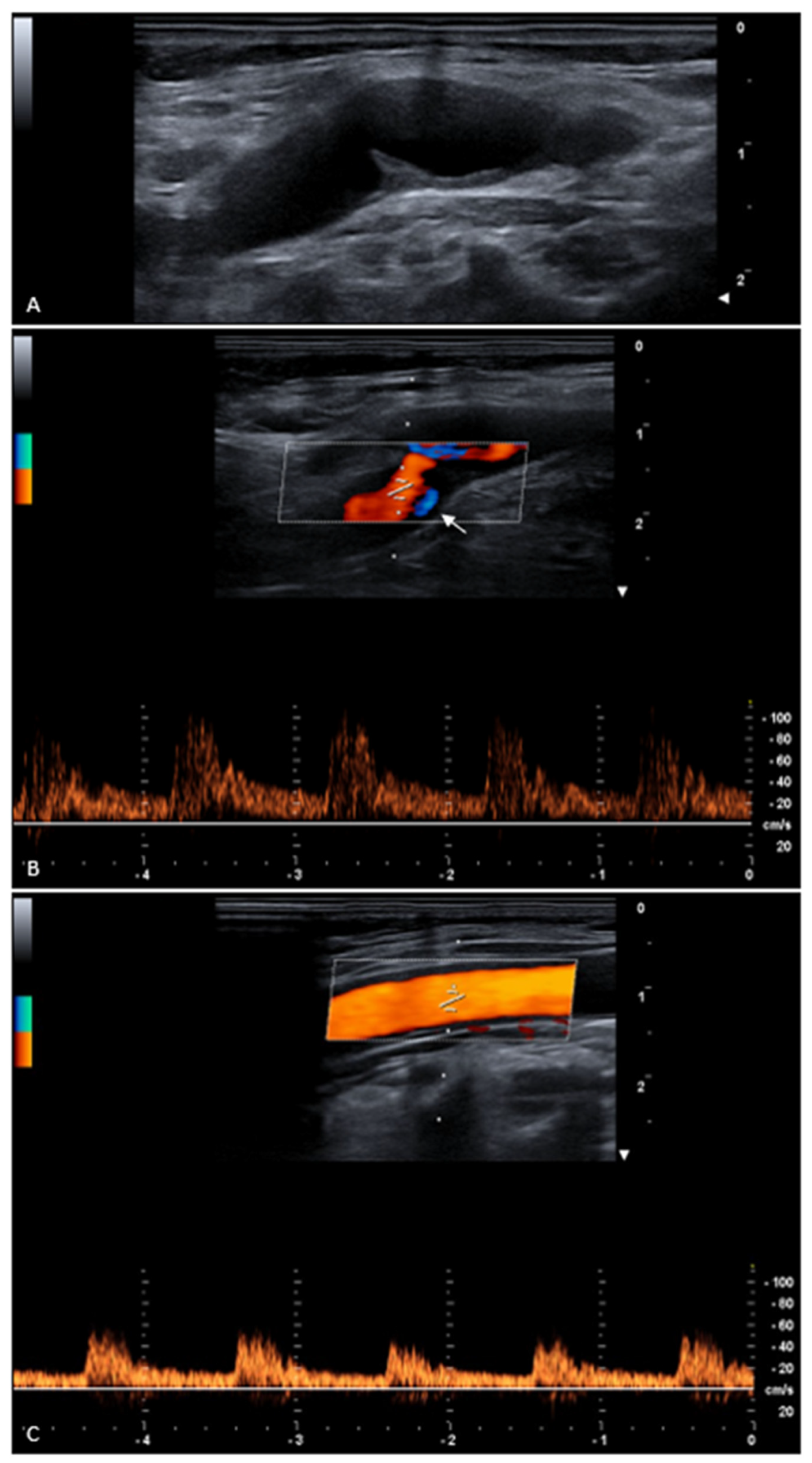
- In our patient, the carotid web length was 4 mm, the degree of stenosis was 50%, the common carotid artery web-pouch angle was 67°, and the common carotid artery pouch-tip angle was 93°, which are potential morphological high-risk features.
- Intima–media thickening is not a normal finding, even in older patients. It is true that intima–media thickness increases with age, but at the age 70 it is considered normal up to 0.75 mm. Therefore, 1.3 mm is definitely abnormal, as in our patient. Intima–media thickening could cause an increase in carotid web length, as carotid web is an intimal lesion. Therefore, as statin treatment can reduce the increase in intima–media thickening and reduce the risk of plaque development, statins could be justified in this setting [14].
- We performed B-mode and Doppler ultrasound to diagnose the carotid web, and these techniques are recognized as valuable tools for carotid web diagnosis, particularly the longitudinal view [1,15,16]. Microvascular imaging can be useful for carotid web detection, particularly for very thin ones [14]. Contrast-enhanced ultrasound can help detect atherosclerotic plaques and thrombosis associated with carotid webs [17]. However, other previously published studies suggested that computed tomography and magnetic resonance imaging could detect carotid webs better [18]. Digital subtraction angiography has been reported by some as the gold-standard imaging modality for carotid web detection, and it can show contrast stagnation and its duration, but it is an invasive technique not recommended for diagnosis [18,19]. However, the lack of knowledge could lead to misdiagnosis and missed diagnosis with all diagnostic techniques [20].
- Not as many carotid webs have been described in the literature with hemodynamic effects. In fact, in a case series of 24 patients with carotid webs, no one showed those effects [15]. Another study reported that, in 94% of patients, the carotid stenosis was less than 50% [21]. Moreover, a previously published article reported that 7 out of 68 carotid webs showed at least 50% stenosis [22].
- Our patient did not show a carotid plaque, but carotid webs with associated atherosclerotic plaques were previously reported [20,22]. It was described that the atherosclerotic plaque can be placed in two different locations, under the carotid web or parallel to it [20]. The same study reported that a higher patient age is significantly associated with carotid webs and concomitant atherosclerotic plaques. Moreover, atherosclerotic plaque presence significantly raises the probability of a stenosis
tohigher than 50% [20], and, in general terms, plaque geometric features such as surface irregularities and the length of upstream segment are associated with plaque vulnerability and the risk of stroke [23]. Furthermore, atherosclerotic plaque can raise the probability of carotid web misdiagnosis [24]. However, to the best of our knowledge, this is the first case of carotid web with abnormal intima–media thickness, and no previous articles have discussed about potential high-risk carotid webs associated with intima–media thickening. - Atypical carotid webs, characterized by unusual location, abnormal morphology, or association with concurrent diseases, such as intraluminal thrombus, dissection, or atherosclerotic plaques, can make carotid webs particularly challenging to diagnose, leading to potential misdiagnosis and missed diagnosis [26,27,28]. A case report showed that Doppler ultrasound revealed a carotid web with a lodged thrombus, whereas digital subtraction angiography and computed tomography were not able to diagnose it [28]. Sometimes, only subsequent examinations can reveal that a carotid web with a lodged thrombus was at the base of carotid stenosis [29]. At times, in symptomatic patients, the definitive diagnoses of carotid webs can be reached only after carotid endoarterectomy or after the re-evaluation of previously acquired images [27,28,30]. However, a case report showed that, in a patient with an acute vestibular syndrome and an incidental diagnosis of carotid webs, the latter was not present in a computed tomography angiogram examination performed seven years earlier [31]. Therefore, radiologists and clinicians cannot always trust a previous examination with normal carotid arteries to exclude a carotid web in a symptomatic patient with atypical carotid findings.
- Carotid web treatment is suggested in symptomatic carotid webs after a stroke or a transient ischemic attack, where carotid endarterectomy or stenting are the preferred treatments to prevent future ischemic events [17,32,33]. However, our patient was asymptomatic, and no consensus guidelines exist about asymptomatic carotid web treatment; therefore, future studies need to determine what is the course of action in these not so rare patients [34,35]. Is antiplatelet treatment recommended in all asymptomatic patients with a carotid web? Is antiplatelet treatment enough in asymptomatic patients with carotid webs with high-risk features? How long does the follow-up need to be after carotid web diagnosis to be sure that the patient is really asymptomatic?
- In conclusion, we describe a case of an asymptomatic 70-year-old female patient on whom a carotid ultrasound examination was performed that showed intima–media thickening and a carotid web with high-risk features; therefore, antiplatelet treatment was suggested to reduce the risk of possible cerebrovascular accidents. Future research and guidelines need to guide clinicians on how to manage asymptomatic patients with carotid webs.
Supplementary Materials
Author Contributions
Funding
Institutional Review Board Statement
Informed Consent Statement
Data Availability Statement
Conflicts of Interest
References
- Liang, S.; Qin, P.; Xie, L.; Niu, S.; Luo, J.; Chen, F.; Chen, X.; Zhang, J.; Wang, G. The carotid web: Current research status and imaging features. Front. Neurosci. 2023, 17, 1104212. [Google Scholar] [CrossRef]
- Konieczna-Brazis, M.; Brazis, P.; Switonska, M.; Migdalski, A. Carotid Web as a Cause of Ischemic Stroke: Effective Treatment with Endovascular Techniques. J. Clin. Med. 2025, 14, 2568. [Google Scholar] [CrossRef] [PubMed]
- Park, C.C.; El Sayed, R.; Risk, B.B.; Haussen, D.C.; Nogueira, R.G.; Oshinski, J.N.; Allen, J.W. Carotid webs produce greater hemodynamic disturbances than atherosclerotic disease: A DSA time-density curve study. J. NeuroInterventional Surg. 2022, 14, 729–733. [Google Scholar] [CrossRef] [PubMed]
- Hassani, S.; Nogueira, R.G.; Al-Bayati, A.R.; Kala, S.; Philbrook, B.; Haussen, D.C. Carotid Webs in Pediatric Acute Ischemic Stroke. J. Stroke Cerebrovasc. Dis. 2020, 29, 105333. [Google Scholar] [CrossRef] [PubMed]
- Mei, J.; Chen, D.; Esenwa, C.; Gold, M.; Burns, J.; Zampolin, R.; Slasky, S.E. Carotid web prevalence in a large hospital-based cohort and its association with ischemic stroke. Clin. Anat. 2021, 34, 867–871. [Google Scholar] [CrossRef]
- Coutinho, J.M.; Derkatch, S.; Potvin, A.R.; Tomlinson, G.; Casaubon, L.K.; Silver, F.L.; Mandell, D.M. Carotid artery web and ischemic stroke: A case-control study. Neurology 2017, 88, 65–69. [Google Scholar] [CrossRef]
- Ahmad, M.; Tan, M.; Abuarqoub, M.; Patel, K.; Siracusa, F.; Shalhoub, J.; Davies, A.H. Carotid webs: A review of diagnosis and management strategies in current literature. J. Vasc. Soc. Great Br. Irel. 2025, 4, 99–110. [Google Scholar] [CrossRef]
- Wang, M.; Zhou, R.; Zhao, H.; Niu, L.; Liu, S.; Li, Y.; Liu, X. Imaging and clinical features of cervical artery web: Report of 41 cases and literature review. Acta Neurol. Belg. 2021, 121, 1225–1233. [Google Scholar] [CrossRef]
- Tabibian, B.E.; Parr, M.; Salehani, A.; Mahavadi, A.; Rahm, S.; Kaur, M.; Howell, S.; Jones, J.G.; Liptrap, E.; Harrigan, M.R. Morphological characteristics of symptomatic and asymptomatic carotid webs. J. Neurosurg. 2022, 137, 1727–1732. [Google Scholar] [CrossRef]
- Perry da Camara, C.; Nogueira, R.G.; Al-Bayati, A.R.; Pisani, L.; Mohammaden, M.; Allen, J.W.; Nahab, F.; Olive Gadea, M.; Frankel, M.R.; Haussen, D.C. Comparative analysis between 1-D, 2-D and 3-D carotid web quantification. J. NeuroInterventional Surg. 2023, 15, 153–156. [Google Scholar] [CrossRef]
- Yirmibeş, E.Ö.B.; Şengeze, N.; Gürel, B. The frequency of carotid web in cryptogenic stroke and its association with stroke risk factors. J. Stroke Cerebrovasc. Dis. 2025, 34, 108295. [Google Scholar] [CrossRef] [PubMed]
- von Oiste, G.G.; Sangwon, K.L.; Chung, C.; Narayan, V.; Raz, E.; Shapiro, M.; Rutledge, C.; Nelson, P.K.; Ishida, K.; Torres, J.L.; et al. Use of Carotid Web Angioarchitecture for Stroke Risk Assessment. World Neurosurg. 2024, 182, e245–e252. [Google Scholar] [CrossRef] [PubMed]
- Negash, B.; Wiggan, D.D.; Grin, E.A.; Sangwon, K.L.; Chung, C.; Gutstadt, E.; Sharashidze, V.; Raz, E.; Shapiro, M.; Ishida, K.; et al. Use of carotid web angioarchitecture in stratification of stroke risk. J. NeuroInterventional Surg. 2025, jnis-2025-023368. [Google Scholar] [CrossRef] [PubMed]
- Lind, L. Effect of new statin treatment on carotid artery intima-media thickness: A real-life observational study over 10 years. Atherosclerosis 2020, 306, 6–10. [Google Scholar] [CrossRef]
- Fontaine, L.; Guidolin, B.; Viguier, A.; Gollion, C.; Barbieux, M.; Larrue, V. Ultrasound characteristics of carotid web. J. Neuroimaging 2022, 32, 894–901. [Google Scholar] [CrossRef]
- Rashid, S.A. Ultrasound Assessment of Carotid Intima-Media Thickness: Comparison between Diabetes and Nondiabetes Subjects, and Correlation with Serum Vitamin D. Radiol. Res. Pract. 2024, 2024, 7178920. [Google Scholar] [CrossRef]
- Zhou, Q.; Li, R.; Feng, S.; Qu, F.; Tao, C.; Hu, W.; Zhu, Y.; Liu, X. The Value of Contrast-Enhanced Ultrasound in the Evaluation of Carotid Web. Front. Neurol. 2022, 13, 860979. [Google Scholar] [CrossRef]
- Chen, H.; Colasurdo, M.; Costa, M.; Nossek, E.; Kan, P. Carotid webs: A review of pathophysiology, diagnostic findings, and treatment options. J. NeuroInterventional Surg. 2024, 16, 1294–1298. [Google Scholar] [CrossRef]
- Damiani Monteiro, M.; Tarek, M.A.; Martins, P.N.; Allen, J.W.; Nogueira, R.G.; Landzberg, D.; Dolia, J.; Park, C.C.; Liberato, B.; Frankel, M.R.; et al. Carotid web catheter angiography hemodynamic parameters. J. NeuroInterventional Surg. 2025, 17, 843–847. [Google Scholar] [CrossRef]
- Ning, B.; Zhang, D.; Sui, B.; He, W. Ultrasound imaging of carotid web with atherosclerosis plaque: A case report. J. Med. Case Rep. 2020, 14, 145. [Google Scholar] [CrossRef]
- Brinster, C.J.; O’Leary, J.; Hayson, A.; Steven, A.; Leithead, C.; Sternbergh, W.C., 3rd; Money, S.R.; Vidal, G. Symptomatic carotid webs require aggressive intervention. J. Vasc. Surg. 2024, 79, 62–70. [Google Scholar] [CrossRef] [PubMed]
- Hou, C.; Li, S.; Zhang, L.; Zhang, W.; He, W. The differences between carotid web and carotid web with plaque: Based on multimodal ultrasonic and clinical characteristics. Insights Imaging 2024, 15, 78. [Google Scholar] [CrossRef] [PubMed]
- Li, L.; Dai, F.; Xu, J.; Dong, J.; Wu, B.; He, S.; Liu, H. Geometric consistency among atherosclerotic plaques in carotid arteries evaluated by multidimensional parameters. Heliyon 2024, 10, e37419. [Google Scholar] [CrossRef] [PubMed]
- Wang, L.; Yang, Y. The diagnostic value of ultrasound in carotid web and the characteristics of misdiagnosed cases. Zhonghua Yi Xue Za Zhi 2022, 102, 2975–2978. [Google Scholar] [CrossRef]
- Takahashi, T.; Yanaka, K.; Aiyama, H.; Saura, M.; Kajita, M.; Takahashi, N.; Marushima, A.; Matsumaru, Y.; Ishikawa, E. Spontaneous asymptomatic common carotid artery dissection resembling a carotid web: Illustrative case. J. Neurosurg. Case Lessons 2024, 8, CASE24344. [Google Scholar] [CrossRef]
- Shrestha, S.; Gu, H.; Xie, W.; He, B.; Zhao, W.; Tang, Z.; Nie, L.; Li, Z. Assessment of association between the carotid web and dissection in spontaneous internal carotid artery dissection patients using vessel wall MRI. Acta Radiol. 2023, 64, 282–288. [Google Scholar] [CrossRef]
- Grin, E.A.; Raz, E.; Shapiro, M.; Sharashidze, V.; Negash, B.; Wiggan, D.D.; Belakhoua, S.; Sangwon, K.L.; Ishida, K.; Torres, J.; et al. Atypical Carotid Webs: An Elusive Etiology of Ischemic Stroke. World Neurosurg. 2025, 196, 123770. [Google Scholar] [CrossRef]
- Yamaguchi, Y.; Miyata, K.; Tomeoka, F.; Ajiki, M.; Takada, T. Lodged Thrombus: A Pitfall for Diagnosing Carotid Web Using Contrast-Angiography. Cureus 2024, 16, e72237. [Google Scholar] [CrossRef]
- Kimura, T.; Yanagawa, T.; Fukumoto, K.; Sato, M.; Ikeda, S.; Yoshikawa, S.; Uesugi, T.; Ikeda, T. Short-term recurrence of stroke following misdiagnosis of carotid web masked by thrombus. Surg. Neurol. Int. 2024, 15, 441. [Google Scholar] [CrossRef]
- Corvino, A.; Lonardo, V.; Tafuri, D.; Cocco, G.; Pizzi, A.D.; Boccatonda, A.; Corvino, F.; Costantino, T.G.; Horer, T.; Catalano, O. Aortic dissection: How to identify it during an abdominal ultrasound examination and achieve a potentially lifesaving diagnosis. J. Clin. Ultrasound 2024, 52, 967–972. [Google Scholar] [CrossRef]
- Singh, R.J.; Mandell, D.M.; Menon, B.K.; Appireddy, R. De novo formation of a carotid web in an adult: A longitudinal observation. J. Stroke Cerebrovasc. Dis. 2024, 33, 108010. [Google Scholar] [CrossRef]
- Talathi, S.; Lipsitz, E.C. Current Therapy for Carotid Webs. Ann. Vasc. Surg. 2025, 113, 415–420. [Google Scholar] [CrossRef]
- Bounajem, M.T.; Liang, A.; Trang, A.; El Baba, B.; Bielinski, T.M.; Sangwon, K.; Zhang, Y.; Wiggan, D.; Grin, E.; Gajjar, A.; et al. Outcomes after carotid revascularization for symptomatic carotid artery web: A multi-institutional cohort study. Interv. Neuroradiol. 2025, 15910199251365529. [Google Scholar] [CrossRef]
- Zedde, M.; Stoenoiu, M.S.; Persu, A.; Pascarella, R. Carotid Web: An Update Focusing on Its Relationship with Fibromuscular Dysplasia and Therapeutic Strategy. J. Stroke 2025, 27, 169–183. [Google Scholar] [CrossRef]
- Wojcik, K.; Milburn, J.; Vidal, G.; Tarsia, J.; Steven, A. Survey of Current Management Practices for Carotid Webs. Ochsner J. 2019, 19, 296–302. [Google Scholar] [CrossRef]
Disclaimer/Publisher’s Note: The statements, opinions and data contained in all publications are solely those of the individual author(s) and contributor(s) and not of MDPI and/or the editor(s). MDPI and/or the editor(s) disclaim responsibility for any injury to people or property resulting from any ideas, methods, instructions or products referred to in the content. |
© 2025 by the authors. Licensee MDPI, Basel, Switzerland. This article is an open access article distributed under the terms and conditions of the Creative Commons Attribution (CC BY) license (https://creativecommons.org/licenses/by/4.0/).
Share and Cite
Tagliati, C.; Quaranta, A.; Fogante, M.; Ventura, C.; Lamja, S.; Matarrese, A.A.; Palumbo, P.; Carbone, I.; Di Cesare, E.; Polonara, G.; et al. Intima–Media Thickening with Carotid Webs: A Case Report of a Potentially High-Risk Association. Diagnostics 2025, 15, 2756. https://doi.org/10.3390/diagnostics15212756
Tagliati C, Quaranta A, Fogante M, Ventura C, Lamja S, Matarrese AA, Palumbo P, Carbone I, Di Cesare E, Polonara G, et al. Intima–Media Thickening with Carotid Webs: A Case Report of a Potentially High-Risk Association. Diagnostics. 2025; 15(21):2756. https://doi.org/10.3390/diagnostics15212756
Chicago/Turabian StyleTagliati, Corrado, Alessia Quaranta, Marco Fogante, Claudio Ventura, Stefania Lamja, Alfonso Alberto Matarrese, Pierpaolo Palumbo, Iacopo Carbone, Ernesto Di Cesare, Gabriele Polonara, and et al. 2025. "Intima–Media Thickening with Carotid Webs: A Case Report of a Potentially High-Risk Association" Diagnostics 15, no. 21: 2756. https://doi.org/10.3390/diagnostics15212756
APA StyleTagliati, C., Quaranta, A., Fogante, M., Ventura, C., Lamja, S., Matarrese, A. A., Palumbo, P., Carbone, I., Di Cesare, E., Polonara, G., & Schicchi, N. (2025). Intima–Media Thickening with Carotid Webs: A Case Report of a Potentially High-Risk Association. Diagnostics, 15(21), 2756. https://doi.org/10.3390/diagnostics15212756


