Review

A Mutational Landscape in Acute Myeloid Leukemia: Overview and Prognostic Impacts

1
The International Circle of Genetic Studies Project, Stony Brook, NY 11794, USA
2
Department of Biology, Stony Brook University, Stony Brook, NY 11794, USA
3
Department of Pathology, Renaissance School of Medicine at Stony Brook University, Stony Brook, NY 11794, USA
*
Author to whom correspondence should be addressed.
These authors contributed equally to this work.
Diagnostics 2025, 15(19), 2537; https://doi.org/10.3390/diagnostics15192537
Submission received: 29 August 2025 / Revised: 30 September 2025 / Accepted: 6 October 2025 / Published: 8 October 2025

Abstract

Acute myeloid leukemia (AML) comprises 15−20% of pediatric leukemia and 35% of adult leukemia cases, requiring insights into prognostic factors of this disease to be an important aspect of diagnosis and treatment. A mutational profile of patients with AML is a crucial predictor of their outcome. Discernment of present mutations, co-mutation combinations, and variations in the mutations in a single gene requires proper research and analysis to determine their impact on a patient’s prognosis. Common and infrequent mutations are continuously investigated and analyzed in different patient cohorts, bringing new insights that lead to changes in classifications, treatments, and diagnoses. For instance, mutations in NPM1, FLT3, and DNMT3A, three frequent driver mutations, have high incident rates with differing prognoses and treatments in pediatric and adult patients. AML patients with MECOM face particularly dire outcomes, as well as those with ASXL1 and TP53, making their mutational analysis crucial for review in developing a prognosis.

1. Introduction

AML is characterized by hematopoietic stem cell malignancies and uncontrolled immature myeloid proliferation, and it is present in 15–20% of pediatric leukemia and 35% of adult leukemia cases [1]. The disease is a complex and harsh mutational landscape that necessitates understanding its development, prognosis, and treatment strategies. Most studies of AML and its abnormalities either precede the latest European Leukemia Network (ELN) 2022 and ELN 2024 guidelines or focus on limited patient populations [2,3]. Thus, a gap remains for an updated mutational report that integrates the ELN 2022 and ELN 2024 recommendations with recent patient studies. This study uniquely addresses the gap by combining updated genomic data, pediatric and adult comparisons, and evolving guideline-based stratifications.
Firstly, it is necessary to clearly differentiate between pediatric (typically between 0 and 18 years) and adult (>18 years) AML cases, as the disease’s biology, clinical presentation, and treatment responses vary significantly between the two. Similarly, recent advancements in the treatment of AML have led to redefined risk stratifications with varying intensities of treatment. Along with the updated ELN guidelines, it has become evident that it is also necessary to differentiate between intensively treated and non-intensively treated AML in adult cases [2,3]. In pediatric AML, the disease typically presents de novo compared to adult AML, which is usually preceded by myelodysplastic syndromes (MDSs). Therapy typically is more intensive in pediatric AML than in adult AML, as pediatric AML survival rates plateaued around 60% [4,5]. On the gene spectrum, multiple differences are visible, such as KMT2A-AML being much more prevalent (38% vs. 2% in adult AML), or mutations in DNMT3A and TP53, common in adult AML, being almost unseen in pediatric AML [6]. In the ELN 2022 recommendations, comprehensive frameworks for risk stratification are provided for adults undergoing intense chemotherapy for AML with updates and information on genetic markers [2]. Then, in the ELN 2024 update, a separate risk stratification was introduced for patients receiving less-intensive therapies, most likely due to old age or overall health [3].
Mutations causing aberrations of cellular signaling pathways make up two-thirds of mutations in AML and consist of mutations such as FLT3 and KIT [7]. FLT3, a receptor tyrosine kinase (RTK) expressed in immature hematopoietic cells, can express mutations involving internal tandem duplications (ITDs) and the tyrosine kinase domain (TKD). This mutation has one of the highest incidence rates in both pediatric (15%) and adult (20–35%) patients (Table 1). Meanwhile, the most common mutation in adult AML, occurring in 30–35% of cases of AML and more than 50% of AML with normal karyotypes, is NPM1 (Table 1) [8]. NPM1 encodes a shuttling protein for multiple purposes, including regulating histone and ribosome activity and inhibiting centrosome duplication [9]. Patients with AML-NPM1 mutations typically have a favorable prognosis and better overall survival in the absence of FMS-like tyrosine kinase 3 mutations or FLT3-ITD [2]. As of the 2022 ELN guidelines, favorable risk classification mutations of AML include NPM1 without FLT3-ITD and bZIP in-frame mutated CEBPA, intermediate risk classification mutations include NPM1 with FLT3-ITD, and adverse risk classification mutations include ASXL1, TP53, RUNX1, etc. [2].
Cytogenetic analysis remains a cornerstone in classifying AML, as it effectively identifies large structural variants and chromosomal abnormalities, allowing patients to be categorized based on their karyotype into different prognostic groups. Each group correlates with distinct overall survival outcomes. However, modern techniques such as next-generation sequencing (NGS), the parallel sequencing of millions of DNA fragments simultaneously, have revealed novel mutations that carry their own diagnostics and prognosis [7,10]. NGS can provide a rapid read of multiple genomes or just a few genes at an incredibly accurate rate compared to earlier sequencing technologies. Furthermore, optical genome mapping (OGM), a newer tool in monitoring and detecting genomic aberrations in AML, has been shown to be even more effective in scenarios where standard cytogenetic techniques would have missed critical clinical information and recommended alternative treatments [11]. OGM provides precise observation of structural variants (SVs) by linearizing and imaging DNA through linear nano-channels and can thoroughly analyze SVs from 500 bp to multiple megabase pairs [12].
Furthermore, the presence of different genetic translocations in AML, particularly t(8;21)(q22,q22.1)/RUNX1::RUNX1T1, t(9;22)(q34.1;q11.2)/BCR::ABL1, and KMNT2A (MLL) rearrangements, plays a significant role in patient risk prognosis [2]. Each of these translocations is among the most recurrent and well-characterized abnormalities in AML, along with each presenting with a differing prognostic impact. As of the 2022 ELN recommendations, a t(8;21) translocation carries a favorable prognosis compared to the adverse prognosis of t(9;22) and an intermediate prognosis of MLLT3::KMNT2A [2]. Of these, t(8;21) is the most common, in approximately 15% of AML cases [Table 1]. The RUNX1::RUNX1T1 fusion protein impairs the normal RUNX1 pathway and alters early myeloid differentiation, leading to leukemic conditions with cooperating mutations, such as FLT3 [13].
AML as a disease has a varied prognosis, depending largely on the associated mutations of each patient, as well as a limited understanding of many of its unfavorable mutations. Though some of these mutations, such as NPM1 and FLT3, are fairly common, with well-studied therapeutic options, the overall genetic landscape of the disease demands further advancements and analysis.
In this review, we aim to synthesize an updated, extensive mutational landscape of AML that thoroughly reflects the adjustments from the ELN 2022 and 2024 guidelines, as well as modern technological advancements. We hypothesize that our integration of recent patient data with a discussion on current diagnostic modalities and the future of clinical care and research in the field will yield a clinically relevant framework for AML risk stratification and treatment.
We worked to complete a targeted search across PubMed/MEDLINE and Embase to inform all sections appropriately. The examination combined AML with diagnostic modalities, molecular/risk terms, and treatment concepts, with citation chaining of key papers. The inclusion of AML guidelines/consensus, meta-analyses, pivotal trials, and major studies was managed within each respective section. We excluded single-patient case reports, non-peer-reviewed items, and abstracts without full manuscripts. As a narrative review, we did not perform dual independent screening, formal quality scoring, or PRISMA registration.
Table 1. Incidence rate and functional groups of genetic mutations in AML by the ELN recommendations, 2024.
MutationsIncidence Rate in AMLFunctional Group
PediatricAdult
NPM1
[1,8]
8–10%30–35%Nucleophosmin
FLT3-ITD
[14]
15%20–35%Signaling/Kinase Pathway
DNMT3A
[1]
2.1%20%DNA Methylation
MECOM
[15,16]
<1%4%Transcription Factor
ASXL1
[17,18]
10.81%14.4–19.1%Chromatin modification
TP53
[1]
2.1%10%Transcription Factor
CEBPA
[1]
18%~10%Transcription Factor
KMT2A
[15]
10–15%3%Chromatin modification
RUNX1
[1]
2.8%10–15%Transcription Factor
TET2
[19]
6%7.6%DNA Methylation
GATA2
[1]
2.6%5%Transcription Factor
BCOR
[20,21]
1.7%3.8–5%Transcriptional Corepressor

2. Contemporary Diagnostic Modalities in AML

An accurate detection of the genetic mutations in AML is critical in guiding proper patient diagnosis, risk stratification, and therapeutic decision-making. The complexity and heterogeneity of the disease require integrated cytogenetic and molecular approaches to capture clinically relevant alterations. Furthermore, sequencing-technology advancements in the last few decades have redefined AML prognosis and classification altogether [22]. As such, many of the genetic mutations mentioned are currently detected primarily through molecular and cytogenetic testing. NGS was developed in the early 2000s but initially remained a high-cost method. In 2011, the cost of sequencing a raw megabase dropped dramatically—from around $10,000 to less than 10 cents [23]. Today, NGS, along with various forms of cytogenetic testing, serves as a primary method for identifying genetic mutations in AML. To aid method selection and interpretation, we summarize the key performance characteristics of NGS, FISH, and OGM (Table 2).
Assay choice directly affects risk assignment and therapy. Hotspot-only NGS can miss non-hotspot DNMT3A or atypical/long FLT3-ITD (or under-call allelic ratio), leading to wrong ELN/ICC risk and missed FLT3-inhibitor use; incomplete IDH1/2 or NPM1 coverage can undercut IDH-inhibitor eligibility and MRD-based decisions. FISH works to rapidly confirm class-defining rearrangements (e.g., PML::RARA, MECOM), which helps to prevent delays in treatment and enable the movement to a higher-risk category when indicated. OGM reveals cryptic SVs (e.g., KMT2A, inv(3)), helping to refine prognosis and aid in clinical trial decisions. Accurate TP53 multi-hit status (SNV + CN/LOH) is critical; panels lacking CN/LOH or with low depth can underestimate the adverse risk.
Examples of major, commonly used myeloid NGS panels include Qiagen Human Myeloid Neoplasms Panel, Illumina AmpliSeq Myeloid Panel, Quest Diagnostics LeukoVantage Panel, and Oxford Gene Technology SureSeq myPanel (NGS Custom AML), which all provide sequencing of exons for TP53, ASXL1, CEBPA, DNMT3A, FLT3, and NPM1 [22,23]. However, the extent of coverage per exon varies among platforms. The Illumina AmpliSeq Myeloid Panel provides full exon sequencing for ASXL1, CEBPA, and TP53 but only hotspot exon sequencing for DNMT3A, FLT3, and NPM1 [22]. It specifically focuses on 50 hotspot mutations, though this leaves room for error in non-hotspot mutations [24]. Qiagen’s panel covers regions of genes ±5–10 bases of exon/intron boundaries to allow for a broader coverage [29]. Quest Diagnostics’ panel identifies mutations in key genes, though detailed information on specific exon coverage is not as readily available, while Oxford Gene Technology offers customizable panels that allow for a variability in coverage [25,26]. Differences in these panels may introduce bias in detection and, as such, should be considered when comparing data across platforms.
For example, MECOM rearrangements utilize cytogenetic methods such as FISH rather than identification with NGS. MECOM FISH assays typically utilize break-apart probes, in which a normal cell displays the two probe signals (5′ and 3′) as fused or overlapping, producing a single signal per allele. In cells with a rearrangement, these signals split apart. A separation of the probe signals (rather than a fusion) indicates a MECOM rearrangement [28].
In OGM technology, it is possible to detect multiple forms of SVs to 5% Variant Allele Fraction (VAF) for mosaic/heterogeneous cancer samples, repeats, complex rearrangements, and fusions with fusion partners [12]. In comparison to FISH, it has a whole-genome coverage at a higher resolution and the ability to detect all classes of SVs in a single assay [11]. VAF cutoff value interpretation, however, cannot be universally applied to all cases. Alternative genetic events, such as duplications or multiple hits in the same gene, can impact the specificity and accuracy of VAF percentages. When clinicians consider cutoff values for VAF, it must be under the consideration of such factors. For instance, as denoted by the ICC, certain MDS cases involving TP53 mutations typically require two mutations in a lower percentage of evaluated alleles (>10% VAF) or a single higher frequency mutation (>50% VAF). Fundamentally, the clinical significance of a VAF value depends on disease-specific guidelines as set by organizations such as the ICC [32]. Though OGM can detect multiple SVs that are typically undetectable or difficult to detect by short-read methods, short-read sequencing remains the dominant method of genetic research today due to its reliability, accuracy, and wide application [29,30].
In this review, the following sections will explore the most prevalent and comprehensively analyzed functional groups in AML, focusing on defining characteristics, impacts on disease progression, and relevance in risk factors.

3. NPM1

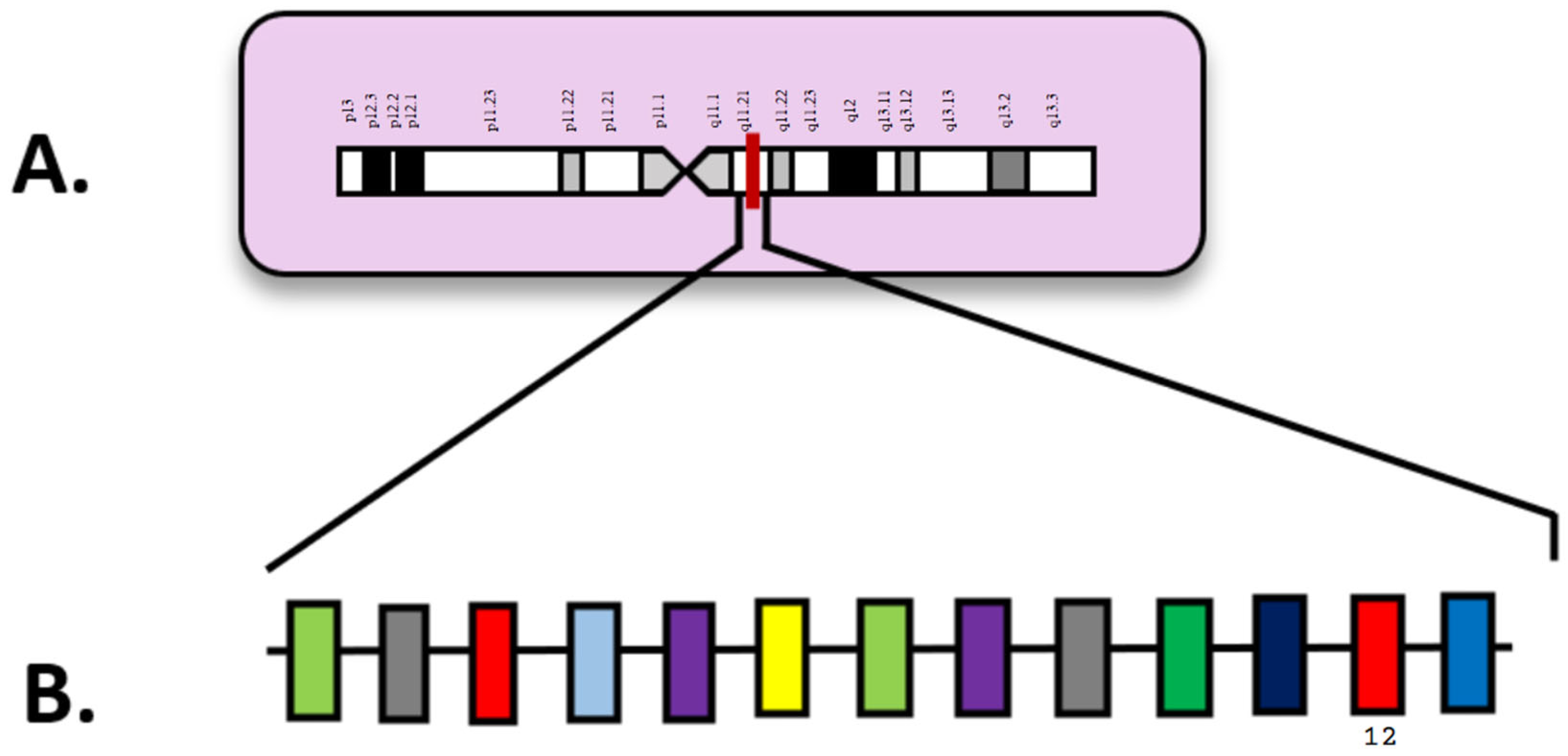
NPM1, located on chromosome 5q35, is the most commonly mutated gene in AML. Mutations are commonly seen in exon 12, accounting for 75–80% of these cases, and are considered an “initial step” for complete leukemia (Figure 1) [33]. Figure 1 depicts a magnified schematic of NPM1 on chromosome 5 (Figure 1A), along with all the exons, especially exon 12, where mutations occur (Figure 1B). Wild-type NPM1 encodes the multifunctional shuttling protein nucleophosmin that works in ribosome biogenesis, DNA repair, and cell cycle regulation. Typically, NPM1 binds to nucleus-localized tumor suppressor protein ARF, but in mutated NPM1, nucleophosmin is unable to bind to ARF and is instead localized in the cytoplasm. As a result, ARF degrades and is destabilized, unable to regulate the p53 pathway. This results in an accumulation of leukemic blasts and eventually leukocytosis [34].
AML with mutated NPM1 has been classified as a distinct entity in the fifth World Health Organization classification of hematopoietic malignancies, with diagnosis not requiring a specific percentage of blast cells [9]. Mutated NPM1 can be identified by heterozygous mutations at the C-terminus that can cause unwinding of the three-helix bundle structure and create new nuclear export signals (NESs). Together, these changes lead to a cytoplasmic shift [33]. Patients with NPM1-mutated AML experience low/moderate white blood cell (WBC) counts in the absence of FLT3-ITD, no/low CD34, and typically favorable responses to intense chemotherapy [8].
Figure 1. (A) Locus of NPM1 on 5q35; (B) NPM1 diagram with exons. Numbered exons represent typical sites of mutation. Modified from McGowan-Jordan et al., 2020 [35].
Diagnostics 15 02537 g001
AML with mutated NPM1 without FLT3-ITD has been classified with a favorable prognosis by the ELN, though it is observed that older patients (>60 years) typically carry an inferior outcome [36]. OS is observed at 40% with complete remission (CR) around 80%. However, >50% of patients typically relapse [37,38]. A 2023 study of wild-type and mutated NPM1 patients revealed that an NPM1 mutation led to higher rates of CR with no effect on relapse-free survival [39]. In a 2020 pediatric NPM1 AML study, Xu et al. saw 96.9% of patients achieving CR after two rounds of therapy and 87.7% after the first round alone. This is compared to 86.4% without NPM1 mutations achieving CR after two rounds and 75.3% after one round. A better 5-year event-free survival (EFS) was observed in NPM1 patients as well (66.4 ± 6.0%) compared to wild-type patients (45.5 ± 1.8%) [40]. Much more common in adult AML than pediatric AML, NPM1 mutations in adults are very age-dependent in terms of prognostic benefit (Table 1) [41]. Patients <65 years typically experience very favorable survival outcomes, though in older patients, this benefit does not exist. In older patients, it is common to find complex karyotypes and a less intensive therapy treatment [41]. In general, non-intensively treated patients with NPM1-mutated AML do not see a prognostic benefit as beneficial, especially in terms of OS, compared to intensively treated patients [42]. Overall, NPM1 in AML is characterized by a higher remission rate following intensive induction chemotherapy, although its prognosis in long-term trials is variable [43].
Instances involving FLT3-ITD and/or DNMT3A mutations often occur, and patients with these three simultaneous mutations portray a poor prognosis. Oñate et al., in 2022 [44], found that DNMT3A mutations appeared in 73% of AML-NPM1 patients, along with a correlation of DNMT3A mutations in younger patients. Additionally, a separate 2022 study by Borlenghi et al. [45] found that 19.3% of 366 AML patients carried both NPM1 and FLT3 mutations. NPM1-mutated patients made up 40.3% of the patients with a CR rate of 93.9% and 3-year and 5-year OS rates of 72% and 64%, respectively. In this study, AML patients with NPM1, FLT3, or both were treated for over 17 years with an idarubicin-based induction regimen, followed by the addition of idarubicin to high-dose cytarabine (HD-ARAC) for consolidation. Compared to NPM1-mutated patients who received conventional chemotherapy, those treated with this regimen fared significantly better.
In multiple studies, it has been shown that venetoclax with or without a hypomethylating agent (HMA) is an effective treatment for NPM1-mutated AML, with high response rates and durable remissions [46,47]. Venetoclax by itself can target the NPM1 apoptotic pathway through the inhibition of B cell leukemia (BCL-2) [46]. Though recent drugs in FLT3 and BCL-2 inhibitors have recently been approved, the standard treatment remains standard induction “7 + 3” chemotherapy with or without FLT3 inhibitors and consolidation with high/medium doses of cytarabine [48]. Menin inhibitors, which target HOX and MEIS1 genes and disruption of the menin-KMT2A complex that led to leukemogenesis, have been shown, in recent clinical trials, to have promising results in response rates [49]. Similarly, they are still in clinical development, and XPO1 inhibitors have also been seen to downregulate HOX genes [50]. In one study, co-treatment of both menin and XPO1 inhibitors showed an improved increase in apoptotic cell death compared to either agent alone [51].

4. FLT3

FLT3 (fms-like tyrosine kinase 3), located on 13q12 encodes for a transmembrane RTK belonging to the class III family of RTKs. Normal activation of FLT3 plays an important role in several cellular processes, including phospholipid metabolism, transcriptional regulation, cell proliferation, apoptosis, and maintaining a close association with the RAS signaling pathway [52]. Wild-type FLT3 physically associates with PI3K, the RAS GTPase, Grb2/shc, PLC-γ, and SRC kinases, leading to downstream signaling, resulting in controlled hematopoiesis, regulated proliferation, and normal cell survival (Figure 2) [53]. Mutations in the FLT3 gene create a protein that lacks regulation and participates in autophosphorylation, leading to enhanced signaling and irregular activation of STAT5, which activates transcription for Cyclin D1, c-Myc, p21, Pim-1, and Pim-2; these irregularities cause uncontrolled proliferation in AML and prolonged cell survival (Figure 2) [53].
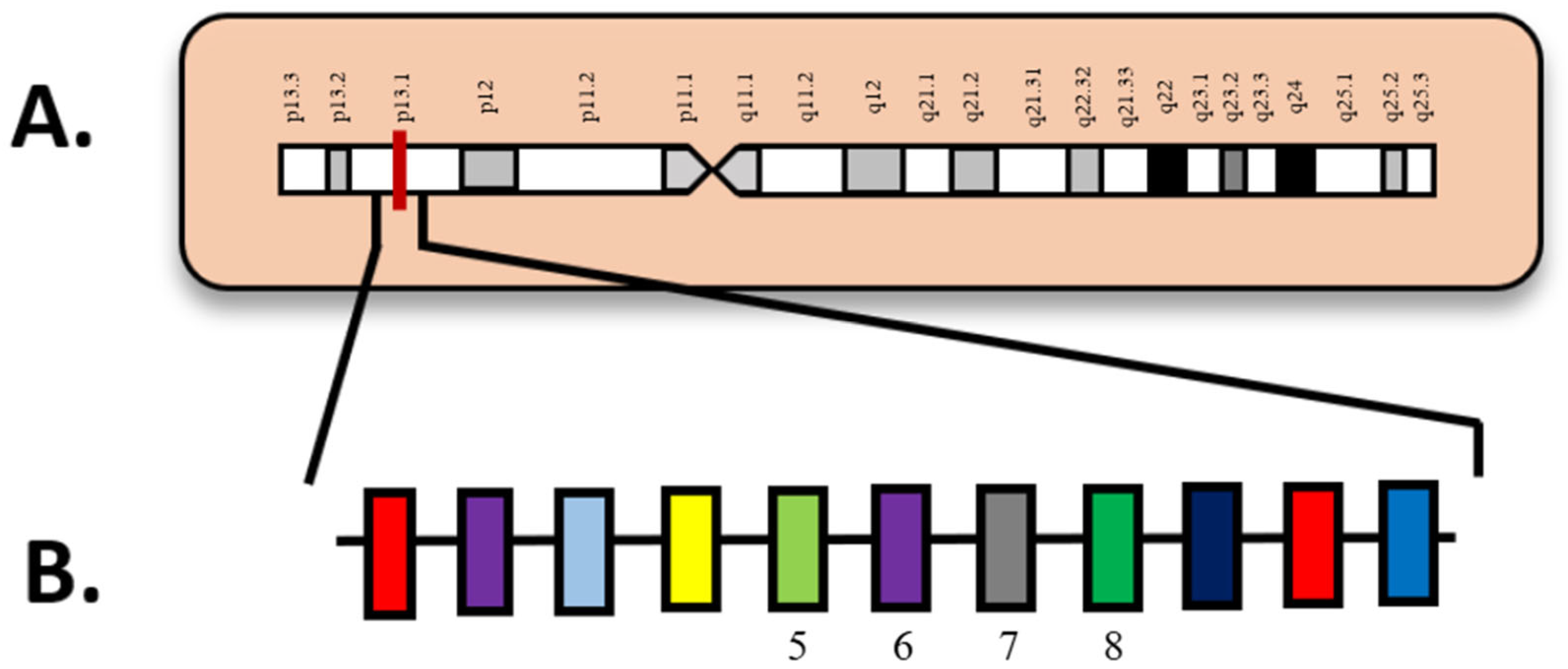
FLT3 internal tandem duplication (FLT3-ITD) mutations are characterized by in-frame insertions of duplicated sequences within exons 14 and 15 (Figure 3) [54]. In Figure 3, exons 14 and 15 are visualized in a larger schematic (Figure 3B) involving all the exons in FLT3, as well as an overview of chromosome 13 with key locations labeled (Figure 3A). These mutations allow for uninterrupted activation of the protein by negatively affecting the tyrosine kinase domain. As a result, FLT3-ITD mutations promote autophosphorylation of the tyrosine kinase domain, which induces continuous proliferative signaling in immature hematopoietic cells [54]. This dysregulation can contribute to hyperleukocytosis, which is an abnormality commonly observed in AML patients. Hyperleukocytosis is defined by elevated WBC counts, typically above 100,000 cells/µL, although counts above 50,000 cells/µL can also have significant clinical implications and likely require immediate medical intervention. Furthermore, FLT3-ITD mutations have been consistently associated with adverse prognostic outcomes in AML, corresponding with increased disease aggressiveness and higher relapse rates.
The second type of mutation that can occur in the FLT3 gene is FLT3 tyrosine kinase domain (FLT3-TKD) mutations. These are typically caused by point mutations at codon D835 or deletions at codon I836 within the activation loop of the FLT3 receptor. These FLT3-TKD mutations disrupt the autoinhibitory mechanisms of the protein, resulting in its constitutive activation and subsequent increased proliferation of immature hematopoietic cells [55]. In reference to the prognostic implications, FLT3-TKD mutations are generally associated with a more favorable outcome in patients with AML compared to those with FLT3-ITD mutations [55]. However, the overall prognostic significance of FLT3-TKD mutations remains a topic of ongoing debate among researchers and clinicians.
A multivariable study by Shimony et al. (2023) [56] examined the overall survival outcomes of patients aged 60 to 75 years with AML, comparing those with FLT3-ITD mutations (FLT3-ITD+) to those without (FLT3-ITD-). The cohort consisted of 378 patients; however, only 53 were identified as FLT3-ITD+. The study found that FLT3-ITD+ patients did not experience significant differences in overall survival when treated with either intensive chemotherapy or HMAs [56]. Furthermore, a 2022 study by Konopleva et al. [57] ratified the efficacy of venetoclax by analyzing its effectiveness in combination with azacitidine in patients with FLT3-mutated (FLT3mt) versus wild-type (FLT3wt) AML. The findings indicated that the overall survival for the FLT3wt group receiving venetoclax and azacitidine was 12.5 months, in comparison to 8.6 months for those who received only azacitidine. In the FLT3mt group, the overall survival was reported as 14.7 months with venetoclax and azacitidine versus 10.1 months with azacitidine only [57]. These results provide evidence of the success of incorporating venetoclax into treatment regimens, especially when compared to azacitidine alone. They also demonstrate comparable overall survival outcomes between the FLT3mt and FLT3wt groups undergoing similar treatments.
The first FLT3 inhibitor, midostaurin, showed significant improvement in the prognosis of FLT3 patients, helping move FLT3-ITD mutations from an adverse risk classification in the 2018 ELN guidelines to an intermediate risk classification in the 2022 ELN guidelines [2,58,59]. In general, FLT3 inhibitors are now standard in initial therapy for FLT3 mutant AML patients and followed by consolidation with intermediate-dose cytarabine. Second-generation inhibitor, quizartinib, has been studied to have an improved median OS as well as being a generally well-tolerated and effective treatment in newly diagnosed FLT3-mutated AML patients [59]. Meanwhile, for non-intensive-treatment patients, combining FLT3 inhibitors with HMAs has been standard, with improved CR rates and negative molecular residual disease (MRD) [60]. However, with the introduction of several FLT3 inhibitors, drug resistance has also been well documented. The clonal evolution of FLT3-ITD mutations seems responsible for resistance and disease progression in particular. As seen in one study by Stone et al., 59% of patients treated with midostaurin achieved CR, after which half of them relapsed [60].

5. DNMT3A

DNMT3A is located on chromosome 2p23 and encodes for DNA methyltransferase 3 alpha, a DNA methylation protein that plays a key role in embryogenesis and gene regulation in de novo methylation. It is especially important for CpG methylation, which is critical during embryonic development. (Figure 4). By encoding a DNA methyltransferase enzyme to regulate gene expression, hematopoietic cell differentiation is guided primarily into granulocytes [61]. Meanwhile, DNMT3A mutations have been observed in 20% of AML cases and 30% of cytogenetically normal AML (CN-AML) cases, with a frequent association with a lack of tumor suppressor cell silencing typically present in standard DNA methylation [61]. The DNMT3A-R882 missense mutation is the most common mutation in DNMT3A, accounting for about 60% of cases. In Figure 4, R882 is present on exon 23 (Figure 4B) of DNMT3A with its location on chromosome 2 (Figure 4A). DNMT3A-R882 missense mutations often occur alongside NPM1 and/or FLT3 mutations. This mutation typically prevents methyltransferase activity/DNA binding and hematopoietic activity, resulting in focal methylation at CpGs in the genome [62,63,64]. In 2020, Bezerra et al. [63] observed 507 patients with de novo AML, of which 64 (13%) were identified with a DNMT3A-R882 mutation. Of these 64, 35 patients (~55%) were “triple-mutated,” meaning they had mutations in NPM1, FLT3, and DNMT3A. This group represented about 7% of the total 507 patients. Furthermore, the age group 41–60 years had the highest percentage of DNMT3A patients, with 34 of the 64 DNMT3A patients (53.1%) and 21 of the 35 (60%) “triple-mutated” patients.
The prognostic impact of DNMT3A mutations occurring with NPM1 and FLT3 mutations has had conflicting outcomes in past findings. Due to DNMT3A mutations typically occurring in patients presenting with NPM1 and FLT3 mutations simultaneously, which are very telling predictors of patient prognosis, the independent prognostic impact of this mutation is oftentimes misrepresented or underestimated [65]. A 2023 analysis found high levels of DNMT3A mutations in NPM1mut AML patients, with 47.7% of a 174-patient cohort showing these mutations. Within this group, the DNMT3A-R882 mutation was identified as a strong predictor of poor prognosis, as it was associated with shorter overall survival [65]. This study failed to highlight any clinical significance of DNMT3A non-R882 mutations.
Additionally, DNMT3A plays a vital role in clonal hematopoiesis (CH), an age-related disease that appears frequently in the average population [66]. In clonal hematopoiesis of indeterminate potential (CHIP), patients with a single DNMT3A mutation have the lowest risk of progression to myeloid neoplasms [67]. Given their frequent status as preleukemic clones, patients with DNMT3A mutations may also require additional monitoring for minimal residual disease (MRD). In one 2022 study of AML-NPM1 patients, those with tandem DNMT3A mutations were all MRD-positive after the first cycle of chemotherapy and had significant DNMT3A persistence in further cycles [44]. However, DNMT3A mutations commonly remain detectable in patients even after successful treatment, leading to false MRD positives. Instead, it may be an indication of maintained CH and not relapse/refractory disease. As of the ELN 2022 guidelines, it has been recommended to exclude mutations such as DNMT3A, TET2, and ASXL1 (DTA) from MRD assessment, given their prevalence in CHIP carriers [68,69]. TET2 appears in 7.6% of adult AML patients, typically as loss-of-function variants, and when occurring with ASXL1, is generally linked with an adverse prognosis (Table 1) [19,70]. TET2 itself has a typically adverse outlook, with a 2025 study finding a shorter median OS, lower CR rates, and a decreased response to allo-HSCT treatment [70].

6. MECOM

Appearing in approximately 4% of AML patients, rearrangements of the myelodysplasia syndrome 1 (MDS1) and ecotropic viral integration site 1 (EVI1) complex locus (MECOM) are rare but notable [15,71]. Located on chromosome 3q26.2, MECOM encodes a protein involved in transcriptional regulation with roles in hematopoiesis, apoptosis, and cell differentiation (Figure 5). In Figure 3, exon 3 is highlighted as the location of the first full in-frame ATG start codon (Figure 5B) and its location on chromosome 3 (Figure 5A). As an oncogene, mutated MECOM has a very unfavorable prognosis and is heavily linked to bone marrow fibrosis and thrombocytopenia [2]. MECOM rearrangements (MECOM-R) are a critical biomarker in prognosis in AML and a defining genetic abnormality in AML regardless of blast count by the fifth World Health Organization classification of hematologic malignancies [2]. MECOM translocations at 3q26.2 are well represented in the literature surrounding myeloid disorders, including AML, MDS, and chronic myelogenous leukemia (CML) [72].
Mutations inv(3)(q21q26.2) and t(3;3)(q21;q26.2) occur in 30–50% of MECOM-R in AML. These cause the super-enhancer of GATA-binding protein 2 (GATA2) found at 3q21 to be affected by MECOM at 3q26.2, leading to transcriptional errors [15]. Otherwise, MECOM-R can be associated with a variety of possible chromosomal abnormalities, which are collectively known as “atypical 3q26.2/MECOM-R” [15].
Chantana et al., in 2023 [71], observed MECOM rearrangements in AML/MDS patients, comparing wild-type and mutated MECOM patients who were subsequently categorized into favorable, intermediate, and adverse prognosis subgroups. It was found that AML patients with MECOM mutations experienced low WBC counts and, interestingly, high platelet counts. Furthermore, a female predominance was observed in this group. Among the six patients studied, the 1-year survival rate was 16.7%, 0% CR, and no survivors after two years. In recent cases, MECOM rearrangements have also been documented in lymphoid disorders, such as acute lymphoblastic leukemia (ALL) and persistent polyclonal binucleated B-cell lymphoma (PBBL) [72]. Overall, MECOM rearrangements remain relatively rare, particularly in the context of lymphoid malignancies. More research and documentation are needed, especially in areas such as therapeutic approaches and treatment strategies.

7. ASXL1

ASXL transcription regulator 1 or ASXL1 (additional sex combs like 1), located on chromosome 20q11.21 (Figure 6), is a gene that normally provides instructions for proteins involved in chromatin remodeling. As seen in Figure 6, exon 12 is highlighted as the most frequently mutated exon (Figure 6B) in ASXL1 on chromosome 20 (Figure 6A), especially those with intermediate-risk karyotypes [73]. Through this mechanism, ASXL1 plays a role in the regulation of HOX gene expression through activation and repression. In acute myeloid leukemia, ASXL1 mutations typically occur as somatic nonsense or insertion/deletion mutations adjacent to the plant homeodomain (PHD) [74]. Homozygous frameshift/nonsense mutations of the gene are shown to completely silence the protein expression, as demonstrated by a Western blot analysis [74].
ASXL1 mutations have a relatively high incidence rate of 10.8% in adults and 14.4–19.1% in pediatric patients (Table 1). Though ASXL1 is typically associated with a worse prognosis, due to the mutation’s role in gene dysregulation triggering the occurrence of AML, this notion is still considered relatively controversial. AML with mutant ASXL1 is shown to have a worse 5-year overall survival and event-free survival (EFS) in patients presenting with a WBC count 50 × 109/L [17]. However, patients receiving hematopoietic stem cell transplantation experienced both a higher 5-year overall survival and EFS [17].
A 2023 analysis with a sample of 219 adult AML and MDS patients showed that >94% of patients present with mutant ASXL1 had concomitant mutations, noting that wild-type NPM1, FLT3, and CEBPA were associated with the presence of mutant ASXL1 [42]. The presence of ASXL1 mutations implicated an overall worse CR rate in patients. Notably, co-mutations with RUNX1 were linked to an even worse prognosis [75]. RUNX1 mutations themselves are typically associated with a poor prognosis, lower CR, and OS [31]. RUNX1::RUNX1T1, from t(8;21)(q22;q22.1), occurs in 10–15% of adult AML but is associated with a favorable prognosis as well as being a key MRD marker [1,2].
A 2022 meta-analysis by Zhang et al. (2023) [76] reviewed studies from three databases (PubMed, Embase, and Cochrane Library), in which a final 10 studies were included in the analysis regarding ASXL1 mutations in AML. Across all analyzed studies, patients with AML and ASXL1 mutations consistently showed lower overall survival. The prognostic impact of ASXL1 mutations was especially significant in patients with cytogenetically normal AML (CN-AML), with a hazard ratio of 1.78 for overall survival at a 95% confidence interval [76]. It was also indicated that patients with MDS and ASXL1 mutations were more likely to develop AML and that these mutations occurred more frequently in patients with an older age of onset [76].

8. TP53

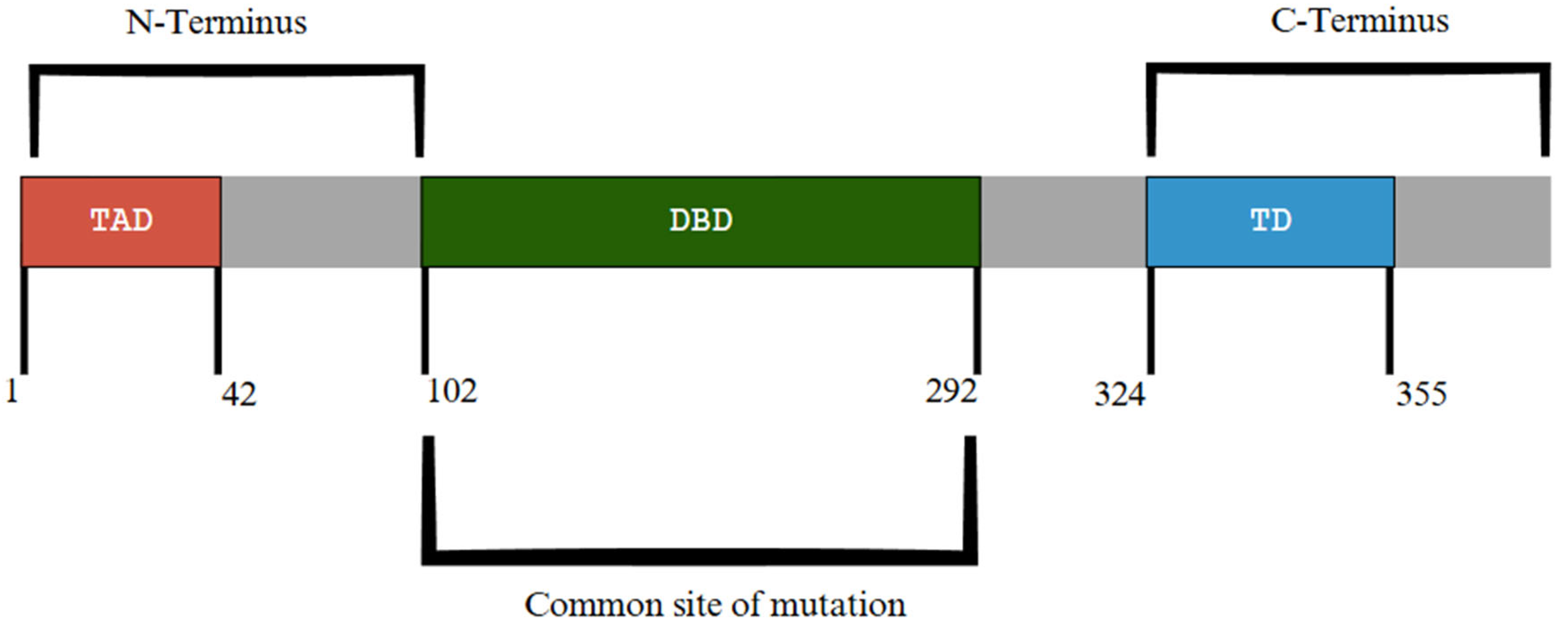
TP53 (tumor protein 53), located on chromosome 17p13.1, encodes the tumor protein p53, which serves a critical role in regulating genomic stability and tumor suppression, as seen in Figure 7A Further represented in Figure 8, exons 5–8 of TP53 encode the DNA-binding domain where the majority of TP53 mutations occur (Figure 8) [77]. Other functional domains of p53 include a tetramerization domain and transactivation domain in the N-terminus and C-terminus ends, respectively (Figure 8) [77]. TP53 plays a significant part in the regulation of several functions, including DNA damage repair, cell cycle arrest, apoptosis, gluconeogenesis and glycolysis, differentiation, and other cellular processes [78,79]. Mutations are extremely heterogeneous among patients but primarily occur within the gene’s DNA-binding domain, which leads to a loss in the DNA-binding capability [79]. TP53 mutations typically represent 5–10% of de novo AML cases and 20–35% of therapy-related AML. In mutant p53 proteins, they are unable to effectively bind to DNA, causing atypical target gene transcription [78,80]. Additionally, mutant p53 proteins may form hetero-oligomers with wild-type p53, which can further prevent regulatory functions [78].
TP53-mutated AML is classified as a distinct subgroup of AML via the ELN and is associated with a poor prognosis, complex karyotypes, poor response to intensive chemotherapy, and short median OS rates [2,80]. Grob et al. in 2022 [81] studied 2200 patients with either AML or myelodysplastic syndrome with excess blasts (MDS-EB) and detected that 10.5% of them had TP53 mutations and 76% had bi-allelic TP53 status. As established in the ELN 2017 adverse-risk category, TP53-mutated AML patients see a 2-year OS of 12.8% in TP53-mutated AML. Meanwhile, Nong et al. 2024 [78] stated biallelic TP53 mutations were associated with an extremely poor prognosis, with a 2-year OS of 4% compared to monoallelic mutations with a 2-year OS of 43%. However, both variants are associated with very poor outcomes due to resistance to intensive chemotherapy and HMAs. In general, patients with TP53-mutated AML have a median OS of less than six months. The frequent co-mutations of TP53 with other aberrations further complicate the issue. TP53 mutations are often associated with specific copy number alterations and the presence of a monosomal karyotype [6]. Subclonal mutations in TP53, defined by variant allele frequencies <20%, serve as a significant adverse prognostic marker for patients [82]. In previous studies, patients with subclonal TP53 in AML were seen to exhibit negative trends in CR, OS, and EFS rates, along with increased resistance to chemotherapy [82,83].
TP53 mutations carry a poor prognosis because of their resistance to many conventional and standard therapies, including a 10-day regimen of decitabine (DEC10) with venetoclax. In 2021, Kim et al. [80] found that the mutation caused lower response rates and shorter survival. Furthermore, traditional techniques, such as allo-HCT, led to long-term remission in only 10–15% of eligible patients, necessitating the development of novel therapeutic treatments. One prospective novel therapy is APR-246 (eprenetapopt), which works by refolding mutant p53 proteins back into their wild-type conformation. In preclinical trials, it has been shown to induce apoptosis and restrict tumorigenesis in TP53 mutant leukemic cells [78]. In clinical trials, TP53-mutated MDS and AML patients treated with APR-246 and azacitidine saw a CR of 50% and a median OS of 10.8 months, though subsequent studies have not confirmed long-term results, leading to a discontinuation of many trials with this combination [78]. Other therapies are also being explored, including MDM2 inhibitors, which reactivate the p53 pathway by blocking its negative regulator. Combination therapies and immune checkpoint inhibitors, such as pembrolizumab, are also being tested. Despite these efforts, the need for new and effective treatments for TP53-mutated AML continues to grow [78].

9. CEBPA

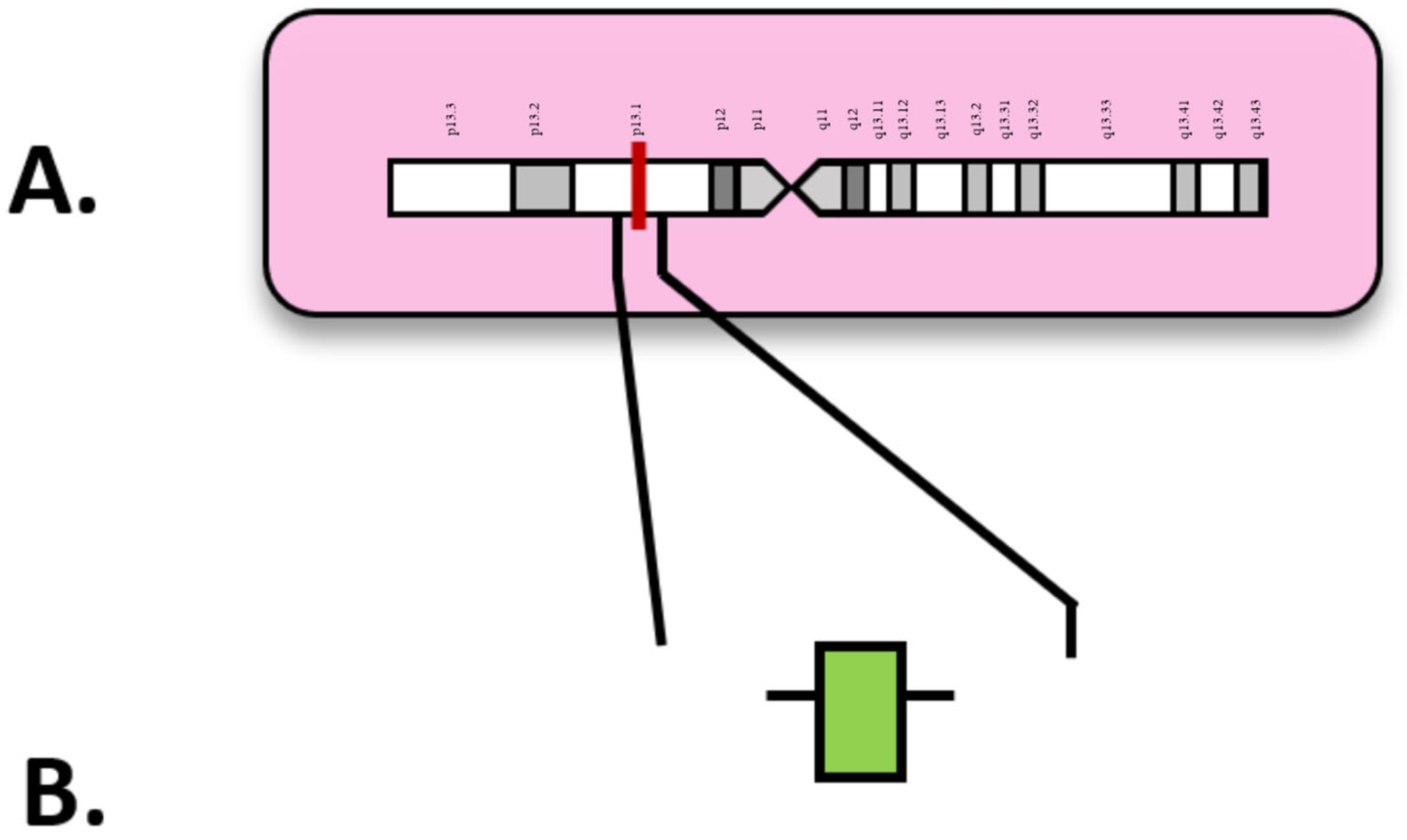
CEBPA (CCAAT enhancer binding protein alpha) gene mutations are relatively common in patients with AML, with a higher incidence rate observed in Asian countries [84]. The gene itself is located on chromosome 19 (19q13.1) (Figure 9A) and is an important transcription factor involved in granulocyte differentiation, along with roles in metabolism regulation [84]. Also, as seen in Figure 9B, CEBPA is made up of just one exon. Incidence rates are 18% in pediatric patients and 10% in adults (Table 1). CEBPA mutations frequently take on two main forms, N-terminal frameshift and C-terminal in-frame deletions/insertions, although mutations are not limited to these areas [84]. Both double mutations affecting both the N and C-terminus, as well as single mutations affecting only one terminal, exhibit favorable outcomes. This is particularly true in patients who also display in-frame mutations within the basic leucine zipper (bZIP) domain of CEBPA, which is in the C-terminal (Figure 10). This may allow for classifications of AML with CEBPA to place greater emphasis on alterations in the bZIP domain rather than single or double mutation categories [84]. Two transactivation domains (TADs) are in the N-terminal; mutations affecting these domains typically show worse outcomes in contrast to bZIP-affected counterparts (Figure 10).
A 2024 multivariable study analyzing both site and allelic status of mutation observed that patients with double-mutation CEBPA (dmCEBPA) exhibited alterations in both the bZIP and TADs. In rare cases, the double mutation involved two alterations within the bZIP domain [85]. Meanwhile, mutated GATA2 was found in only 4–12% of patients within subgroups that had non-bZIP insertion/deletion mutations (bZIP indel). In contrast, it was present in 33–39% of patients in other mutation subgroups. Groups defined by the presence of bZIP indel mutations were also observed to have higher rates of Wilms’ tumor gene 1 (WT1) co-mutations. This same subgroup was categorized not only by a striking co-mutation profile but also by the younger age of the patient sample.
Figure 10. Relative domain locations in C/EBPα protein, with amino acid range for each domain presented. Location of DNA-binding domain (DBD) and the leucine zipper (ZIP) components of bZIP are presented with the transactivation domain (TAD) domain located at the opposite N-terminal end [86]. Modified from Tian and Graf et al., 2014 [86].
Diagnostics 15 02537 g010
A study by Wakita et al. (2022) [87] corroborated the conclusion of dmCEBPA exhibiting combinations of bZIP mutations and non-bZIP mutations. Though the analysis of this cohort did not include a TAD analysis, the findings of Georgi et al. (2024) [85] may fill in the non-bZIP category. NPM1 mutations were found in 5.2% of AML patients with CEBPAmu in bZIP, and FLT3-ITD mutations were present in 4.3% of AML patients with CEBPAmu in bZIP. In AML patients with CEBPA mutations in the bZIP domain, the co-occurrence of NPM1 and FLT3-ITD mutations was less frequent compared to their rates in the overall AML population.

10. Therapeutic Approaches and Treatment Limitations in AML

Given the ELN 2022 and 2024 guidelines, the management of AML has improved significantly with the surfacing of new genetic screening protocols and high-resolution techniques. At the time of diagnosis, considerable care should be taken in conducting comprehensive genetic profiling of the patient to identify defining mutations and characteristics [2]. These include techniques such as conventional cytogenetic techniques and fluorescent in situ hybridization (FISH); NGS in determining mutations in key genes such as NPM1, FLT3, TP53, RUNX1, etc.; and OGM to detect structural variants and copy number variants that cannot be found through conventional methods [2,11,23].
The ELN 2022 guidelines categorize patients into favorable, intermediate, or adverse risk groups based on detected cytogenetic abnormalities and mutations [2]. In the ELN 2024 update, multiple revisions can be found regarding older/unfit patients that require less intensive therapies [3]. Given the ELN risk stratification and overall patient fitness, treatment selection is tailored to each patient. In the favorable risk group, where patients are more fit, standard induction therapy is typically chosen. Intensive chemotherapy, for example, 7 + 3, is followed by consolidation and maintenance as needed [2]. For patients receiving less intensive therapies, HMAs such as azacitidine or decitabine combined with venetoclax and standard therapy have shown positive results in response rates and OS [3,46].
Meanwhile, monitoring MRD is vital in determining CR or CR with incomplete hematologic recovery (CRi) for patients of both intensive and non-intensive therapies. For many AML patients, MRD assessment provides a reliable and quantitative method of determining remission status and risk of relapse [2,88]. Multiparametric flow cytometry (MFC) and molecular MRD assessed by qPCR for specific fusion transcripts are two widely used methods of MRD assessment, with MRD detection before allo-HCT being an extremely accurate predictor of relapse [88,89]. In 2025, Ali et al. found that patients who did not experience relapse by day 100 after HCT typically did not display MRD, but those who did were accurately identified at an increased risk of relapse and worse survival. Furthermore, those with pre-HCT MRD cleared MRD by days 70–100 after HCT, but with significantly worse outcomes than MRD-negative patients. Interestingly, more than half of the patients studied ended up relapsing after HCT within 100 days of HCT. However, as expected, these relapses were much more common in pre-HCT MRD patients than those without [89].
Though MRD is a valuable prognostic tool, it is not without its flaws. Relapses are still possible in MRD-negative patients, though rare. As a result, patients may not have complete disease eradication but a level below the threshold of MRD. Inversely, not all MRD-positive patients will relapse. Apart from prognosis, MRD can also serve as a therapeutic tool to determine stem cell transplantation for patients. It has been seen in NPM1-mutated AML patients that receiving HCT had no significant benefit for patients who did not display detectable MRD or a >4 log reduction in MRD prior to HCT [90]. As a result, MRD detection can be considered in deciding on HCT for patients with NPM1-mutated AML, as well as reserving HCT for patients with an MRD response. MRD remains limited by technical variability, heterogeneous cutoffs, and relapses, even in MRD-negative patients. Overall, these issues complicate standardizations and stress the need for more homogenized assays before MRD can fully guide any therapeutic decision-making.
In less-intensively treated patients, MRD serves as an extremely valuable prognostic tool [47]. Patients treated with azacitidine and venetoclax who then achieved CR or CRi saw that negative MFC-MRD was associated with a significantly better outcome than their counterparts. In an NPM1-mutated AML study of patients treated with HMA + venetoclax, MRD negativity saw a better OS, EFS, and lower incidence of relapse than MRD positivity [47,91].
Post-remission strategies are decided by MRD status and initial risk stratification, with patients achieving CR typically consolidated with intermediate-to-high-dose cytarabine [2,91]. Patients with intermediate/adverse risk or positive MRD can benefit from consolidation with allo-HCT. In patients not undergoing transplant or in older patients, maintenance therapy with oral azacitidine (CC-486) is associated with longer OS and relapse-free survival (RFS) [92]. In relapsed/refractory disease management, molecular re-evaluation is important to identify patients suitable for salvage regimens with clinical trial enrollment encouraged, especially those with TP53 mutations or complex karyotypes [2,93]. Patients with primary refractory AML, defined as a lack of remission after two cycles of induction with at least one cycle of intermediate-dose cytarabine, are unlikely to benefit from further chemotherapy. It is instead encouraged to undergo allo-HCT or clinical trial enrollment [2]. It has been studied that patients who are older, MRD-positive, have a non-favorable risk karyotype, or have incomplete recovery of peripheral counts are all at an increased risk of relapse [2,94].
In patients with relapsed/refractory FLT3 mutations, gilteritinib showed significantly improved CR rates, OS, and rates of remission than salvage chemotherapy [95]. Enasidenib and ivosidenib are associated with durable remissions and favorable outcomes in IDH1- and IDH2-mutated relapsed/refractory AML patients. In IDH1-mutated patients treated with ivosidenib monotherapy, CR or complete remission with partial hematologic recovery (CRh) is detected at 30.4%, and among those that achieve CR or CRh, 21% saw no residual IDH1 detection [2,95]. For most patients, after cytoreduction, allo-HCT is encouraged. In older patients, less-intensive treatment, such as HMA with/without venetoclax, is favored [2].
Furthermore, AML exhibits an extensive clonal heterogeneity, which requires different treatment methods as well as the potential for relapses. Multiple studies have shown this clonal structure evolving throughout therapeutic treatment, with NGS studies suggesting that relapsed patients disproportionately have higher mutational risk stratification at diagnosis than those without relapse. This complicates and limits the prognosis and durability of therapeutic response, even in patients in a favorable-risk category [96].
With the expansion of novel combinations of therapies in recent years, multiple adverse events (AEs) have been associated with these agents. Understanding these AEs is thus critical for managing the effects of AML therapies in delivering patient care [97]. In the case of HMA/venetoclax, common grade ≥3 AEs included thrombocytopenia, neutropenia, and infections. Modifications to address these issues include delaying cycles, decreasing venetoclax administration duration per cycle, and dose adjustment of venetoclax. In cases such as prolonged cytopenia, where treatment length is indefinite, the length of cycles may be extended to 5–6 weeks, and the duration and dosage of HMAs decreased [97,98]. Also reported, but rare, is tumor lysis syndrome (TLS) with HMA/venetoclax in 1% of patients. Modifications can include a reduction in WBC <25 × 109/L with hydroxyurea before treatment and administering venetoclax with a ramp-up to target dose approach.
In IDH inhibitors enasidenib and ivosidenib, AEs can include differentiation syndrome (DS), leukocytosis, and QTc prolongation [99]. DS is characterized by dyspnea, fever/hypotension, weight gain, acute renal failure, or pleuropericardial effusions and is an extremely important, and potentially fatal, AE. The incidence of DS with IDH inhibitors ranged from 13 to 25%, though fatalities were rarely recorded [97]. In FLT3 inhibitor midostaurin, GI toxicities such as nausea, vomiting, and diarrhea, as well as cardiac toxicities (primarily in older patients), have been recorded [100,101]. Gilteritinib has also been reported to have AEs of DS, QTc prolongation, and posterior reversible encephalopathy syndrome (PRES). DS was seen in 3% of patients, with PRES in 1% of patients necessitating the immediate discontinuation of gilteritinib once confirmed [97,102].

11. Future Directions of AML Research

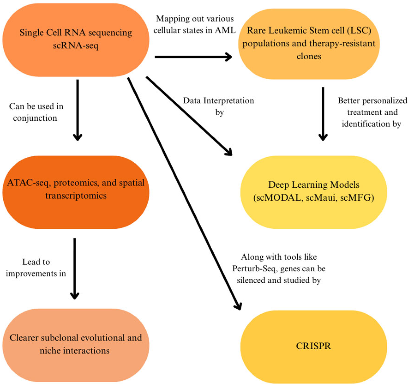
In the next few years, AML research is expected to greatly benefit from advances in high-resolution genomics, functional screening, and computational analysis. One of the most promising developments is in the growth of single-cell sequencing in the heterogeneity of AML (Figure 11). One example, single-cell RNA sequencing (scRNA-seq), is an established technique that has already been able to map out various cellular states in AML, such as rare leukemic stem cell (LSC) populations and therapy-resistant clones [103]. The next step will likely involve combining scRNA-seq with further methods, such as ATAC-seq, proteomics, and spatial transcriptomics [104]. As a result, the integration could offer a much clearer image of how subclones evolve over time and how they interact in their native microenvironment.
Furthermore, it will most likely be seen that computational tools will play an increasingly important role in interpreting the data generated by these experiments. New deep learning models have been developed specifically for single-cell and multi-omics data, enabling researchers to find meaningful biological patterns while minimizing batch effects [105]. scMODAL, a deep learning model, combines multi-modal single-cell datasets like scRNA-seq and proteomics through neural networks and generative adversarial networks to eliminate variation while preserving biological signals across modalities [105]. Another model, scMaui, has been found to effectively balance transcriptomic and proteomic modalities while simultaneously correcting for batch effects and enhancing downstream analyses like cell-type classifications [106]. Meanwhile, the scMFG model employs feature grouping and interpretable matrix-factorization in integrating multi-omics datasets, retaining biological interpretability and revealing rare cell types even in technically noisy datasets [107]. Using single-cell multi-omics data, these models now allow researchers to better identify LSC populations, monitor clonal evolution, and ultimately facilitate a more personalized and data-driven treatment strategy for individual patients.
Integrated single-cell multi-omics that merge scRNA-seq, ATAC-seq, mitochondrial genotyping, and clonal tracking continue to improve our understanding of AML subclones and LSCs. A 2024 review highlighted how platforms like CITE-seq and TARGET-seq enable high-resolution profiling of transcriptional, surface protein, and mutational states within individual cells, and offer a critical insight into therapy resistance mechanisms [108]. In a different 2024 study focusing on spatial multi-omic profiling, researchers applied spatial transcriptomics in studying post-transplant AML bone marrow, revealing distinct “immune hubs” in tumor microenvironments, opening a valuable pathway into the mechanisms of relapse and remission maintenance after donor lymphocyte infusion [109]. Work with co-detection by indexing (CODEX), especially with scRNA-seq, has further mapped out how mesenchymal stromal cells (MSCs) and leukemic blasts co-localize within spatially organized microenvironments, creating niches that may fuel resistance and persistence (Figure 11) [110]. Single-cell and spatial multi-omics thus offer a straightforward way to resolve the challenge of clonal heterogeneity by enabling the high-resolution tracking of resistant subclones through multiple modalities of detection and treatment (Figure 11).
However, single-cell and spatial multi-omics remain a costly tool, technically restraining, and limited to a few research centers, confining its clinical impact [103,104,105,106,107,108,109,110]. Their successful translation will rely on improvements in both affordability and scalability. Concurrently, the growth of genome-wide functional screens using CRISPR has become much more accessible. Tools like Perturb-seq, which combine CRISPR with scRNA-seq, allow for the visualization of primary AML cells, allowing researchers to systematically silence and activate genes to study their impact on cell survival and drug sensitivity [111]. Meanwhile, non-invasive monitoring procedures are improving every year, like the development of circulating tumor DNA (ctDNA)-based AML assays to detect minimal residual disease (MRD) with high sensitivity. Tumor-informed methods, such as AML-CAPP-Seq, can illuminate mutations in tracking ctDNA progression and have demonstrated detection thresholds < 0.01%, and even tumor-agnostic approaches, like methylation-based assays and fixed mutation panels, show potential in routine MRD monitoring [112]. Collectively, these approaches can eventually reduce the need for repeated bone marrow biopsies as well as earlier detection of relapse. Looking forward, these advances are expected to thoroughly refine and deepen our understanding of the landscape of AML and the ability to identify new therapeutic targets.

12. Clinical Implications and Changing Approaches to Patient Care

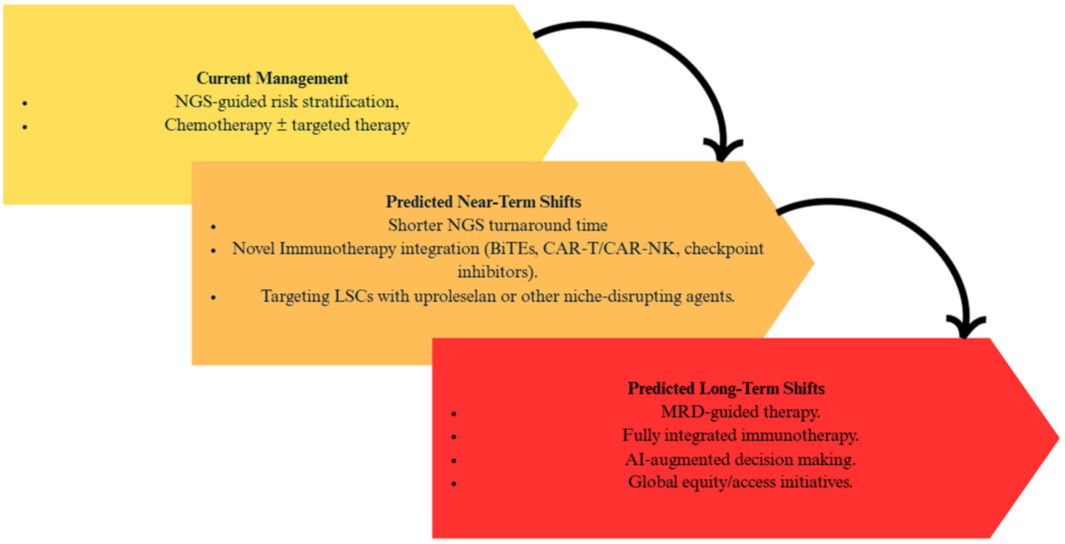
As genomic technologies develop and become faster, more affordable, and further incorporated into clinical practice, the management of AML is likely to undergo substantial changes in the next three to five years. One of the most immediate shifts will be in the timing and use of genomic data. Turnaround times for NGS now approach 48–72 h in some institutions; clinicians may soon have full mutational profiles in hand before even starting induction therapy (Figure 12) [113]. This may support earlier, more precise treatment decisions, for example, starting a FLT3 inhibitor up front in a newly diagnosed FLT3-ITD-positive patient, or avoiding standard 7 + 3 chemotherapy altogether in favor of a venetoclax-based regimen in older adults or those with poor-risk mutations [2]. These shifts are consistent with previously discussed evidence where the ability to profile mutational abnormalities early is shown to influence both prognosis and initial treatment selection, justifying prioritization of faster, clinically actionable NGS (Figure 12).
Immunotherapy, more broadly, is also gaining ground in AML, although progress has been slower than in other forms of cancer. Bispecific T-cell engagers (BiTEs), antibody–drug conjugates, chimeric antigen receptor (CAR)-T and CAR-NK cells, and checkpoint inhibitors are all being investigated for use in relapsed/refractory AML (Figure 12). Recent efforts have focused on improving the specificity and safety of these therapies, for example, by designing dual antigen CARs or by incorporating logic-gated controls to prevent off-target toxicity [114]. These novel immunotherapy strategies place a particular emphasis on the integration of modern immune-based strategies to improve outcomes in adverse-risk and MRD-positive patients. This corresponds with our discussion that MRD detection before all-HSCT is an extremely accurate predictor of relapse and poor survival rates [88,89]. Therefore, the prioritizing of immunotherapy approaches aimed at clearing residual disease directly addresses these needs. Over the next few years, these agents may find themselves in a more front-stage role in combination regimens, especially for clearing MRD after initial therapy or treating patients with high-risk molecular profiles.
Fresh endeavors to target the leukemia stem cell (LSC) population, a key driver of relapse, are also gaining momentum. Agents such as uproleselan, an E-selectin inhibitor, focus on disrupting the protective bone marrow that shelters LSCs from chemotherapy. A phase 3 trial evaluating uproleselan jointly with cytarabine-based chemotherapy is currently underway and has the possibility to shift the model for addressing minimal residual disease and chemoresistance [115]. Targeting LSC-specific pathways may better improve the elimination of residual disease post-remission and reduce the risk of relapse, especially when integrated into immunotherapy regimens or MRD-guided treatment strategies (Figure 12).
Looking ahead, equity in accessing these innovations proves a significant concern. The most advanced diagnostic and therapeutic technologies are still limited to academic centers in high-income countries. However, several groups are working on simplified, lower-cost MRD assays and sequencing protocols that could be enacted in community hospitals or lower-resource settings. Further advancements in automation, cloud-based AI interpretation, and decentralized testing could aid in making precision medicine a reality for more patients worldwide [116].
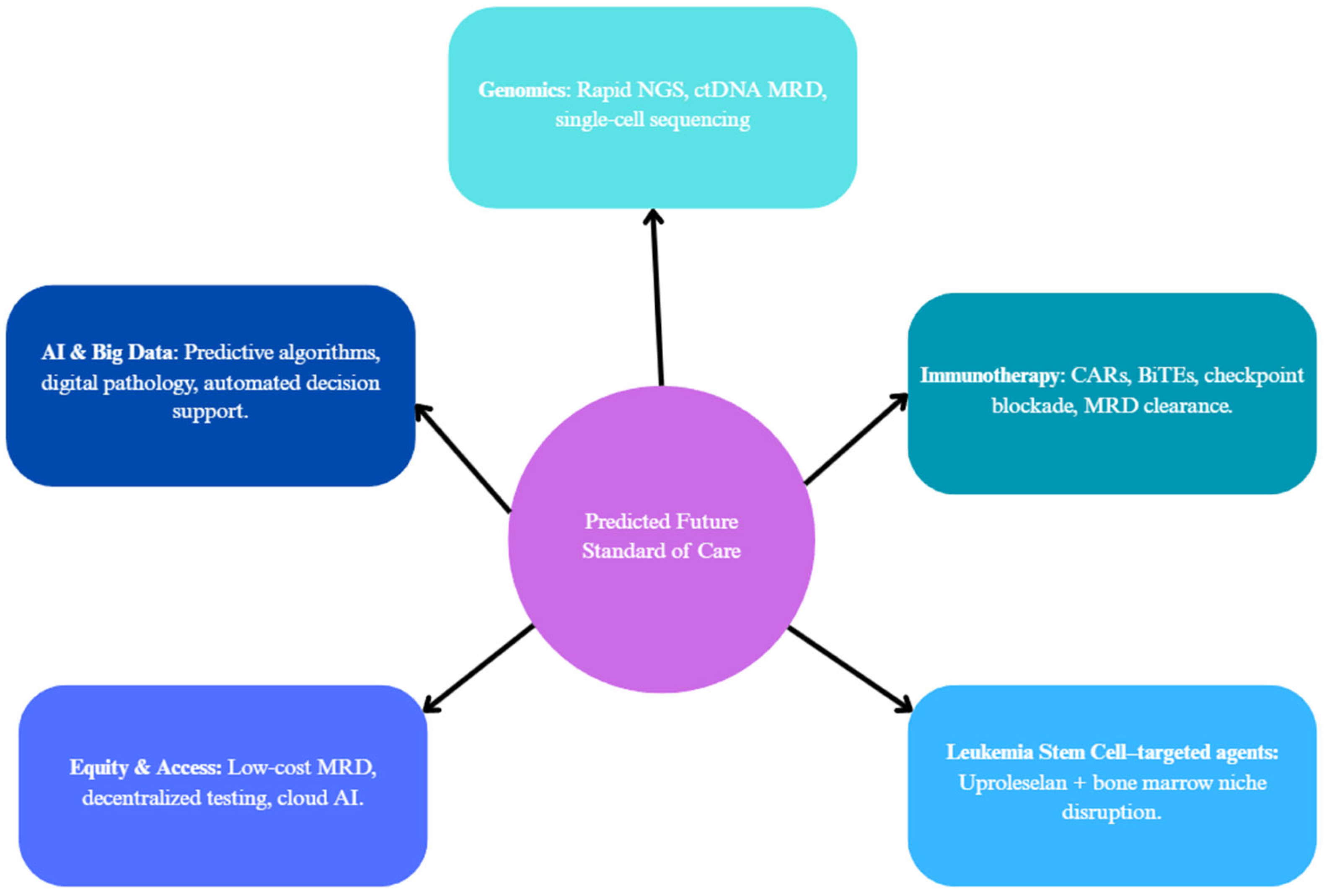
Implementation of big data and artificial intelligence into AML management promises to further improve efforts in diagnostics and personalized therapy. Machine learning algorithms trained on large genomic and clinical datasets have already demonstrated their potential to predict treatment response and survival outcomes, even outperforming traditional risk scores in some models (Figure 13) [117]. Furthermore, digital pathology tools utilizing deep learning can aid in a variety of aspects, such as helping to quantify blast burden, assess bone marrow morphology, or even detect early signs of relapse [118]. As these systems develop further and become more accessible, they may allow for a wider outreach of precision oncology by reducing the burden on overextended human expertise, particularly in community or resource-limited settings. These proposed shifts are justified by the complexity and heterogeneity of AML treatment responses previously outlined in this review, where mutational risk stratification and MRD monitoring alone were insufficient in a clear outcome prediction for many patients [2,88,90]. Thus, modern technologies, such as AI, show potential in improving predictive accuracies, especially in lower-income and resource-limited settings [Figure 13].
To prepare for these shifts in practice, the field should increase its focus on prioritizing a set of critical research goals. These can include the standardization and clinical validity of ctDNA MRD assays, integration of single-cell technologies into decision-making workflows, and the expansion of immunotherapy trials into maintenance and frontline settings (Figure 13). At the same time, current AI and digital pathology models are trained primarily in high-income regions, requiring broader validation to minimize algorithmic bias and maximize applicability to more diverse and clinical settings. Addressing infrastructure and training gaps in underdeveloped regions is also of high importance in ensuring that these tools reach all patients, not just those treated at top-tier institutions.

13. Discussions and Conclusions

This review functionally integrates AML genomics with detection modalities (NGS, FISH, and OGM) and different care pathways. Going over all mutation groups, evidence for clinical utility in the changing of risk groups is strong for NPM1 (including MRD), FLT3 (ITD allelic ratio), and IDH1/2 (targeted inhibitors); moderate for ASXL1, RUNX1, and adverse structural lesions (KMT2A, NUP98, inv(3)/MECOM); and limited/heterogeneous for several rarer alterations. Our unique contribution is a modality to management map, linking assay design (panel coverage, hotspot bias, ITD allelic-ratio detection, CN/LOH assessment for TP53) to concrete risks of misclassification and missed therapeutic eligibility, which is summarized in Table 2, applied across the gene-specific sections, and operationalized in the clinical workflow sections. These conclusions align with recent ELN/ICC recommendations and contemporary reviews, while also extending the prior work by recognizing where detection gaps most often alter the ELN/ICC category, MRD interpretation, and therapy selection (see Figure 11, Figure 12 and Figure 13).
In this review, while an overview of recent advances in risk stratification and therapeutic approaches was addressed, several limitations of the studies should be acknowledged. Many referenced studies included relatively small patient populations and often within single-center settings, limiting statistical power and the generalizability of results. Furthermore, much available evidence is derived from retrospective analyses of heterogeneous populations and variable treatment administrations, causing potential partiality compared to prospective randomized trials.
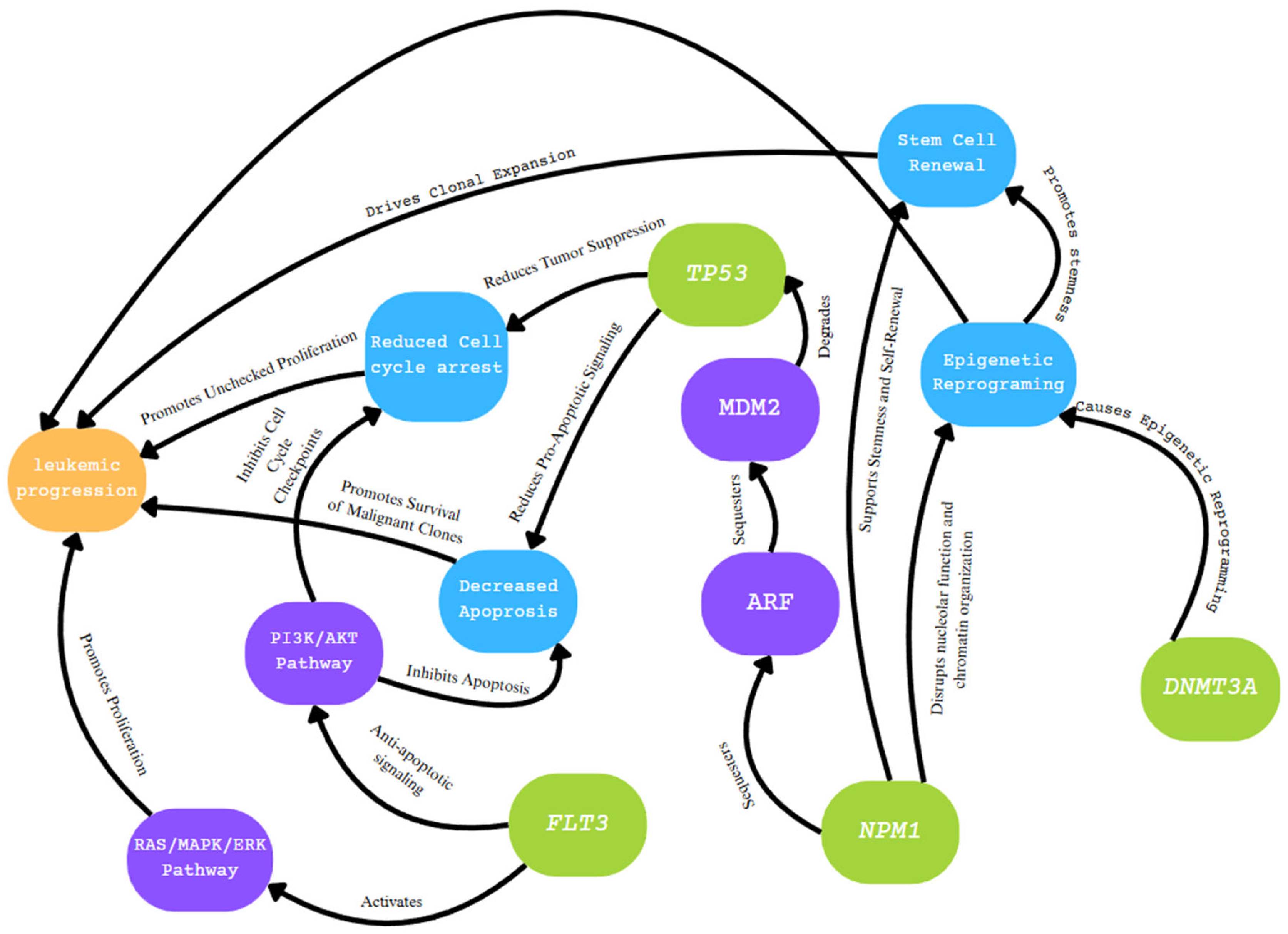
AML remains a complex and heterogeneous hematologic malignancy that frequently spurs amended and improved clinical updates. Many mutations that are present within varying cases of AML include overlapping pathways that induce negative effects in regard to leukemic progression (Figure 11). Previously mentioned pathways, such as the RAS pathway, PI3K/AKT pathways, and others, have downstream consequences that are shared between irregularities in certain genes, such as NPM1, FLT3, DNMT3A, and TP53 (Figure 14). The introduction of techniques such as NGS and OGM in recent decades has allowed for clinical diagnoses of AML to be conducted more efficiently and accurately, along with significant progress in treatment and overall prognosis. Understanding the importance of genetic mutations in AML can definitively shape the prognosis of a patient and inform critical therapeutic decisions.

Author Contributions

Conceptualization, J.C. and F.H.; methodology, J.C. and F.H.; software, J.C. and F.H.; validation, F.H., J.C. and C.A.T.; formal analysis, J.C.; investigation, F.H.; resources, C.A.T.; data curation, J.C. and F.H.; writing—original draft preparation, J.C. and F.H.; writing—review and editing, J.C. and F.H.; visualization, F.H.; supervision, C.A.T.; project administration, C.A.T.; funding acquisition, C.A.T. All authors have read and agreed to the published version of the manuscript.

Funding

This research received no external funding.

Institutional Review Board Statement

Not applicable.

Informed Consent Statement

Not applicable.

Data Availability Statement

Not applicable.

Conflicts of Interest

The authors declare no conflicts of interest.

References

  1. Makkar, H.; Majhi, R.K.; Goel, H.; Gupta, A.K.; Chopra, A.; Tanwar, P.; Seth, R. Acute Myeloid Leukemia: Novel Mutations and Their Clinical Implications. Am. J. Blood Res. 2023, 13, 12–27. [Google Scholar]
  2. Döhner, H.; Wei, A.H.; Appelbaum, F.R.; Craddock, C.; DiNardo, C.D.; Dombret, H.; Ebert, B.L.; Fenaux, P.; Godley, L.A.; Hasserjian, R.P.; et al. Diagnosis and Management of AML in Adults: 2022 Recommendations from an International Expert Panel on Behalf of the ELN. Blood 2022, 140, 1345–1377. [Google Scholar] [CrossRef] [PubMed]
  3. Döhner, H.; DiNardo, C.D.; Appelbaum, F.; Craddock, C.; Dombret, H.; Ebert, B.L.; Fenaux, P.; Godley, L.A.; Hasserjian, R.P.; Larson, R.A.; et al. Genetic Risk Classification for Adults with AML Receiving Less-Intensive Therapies: The 2024 ELN Recommendations. Blood 2024, 144, 2169–2173. [Google Scholar] [CrossRef] [PubMed]
  4. de Rooij, J.; Zwaan, C.; van den Heuvel-Eibrink, M. Pediatric AML: From Biology to Clinical Management. J. Clin. Med. 2015, 4, 127–149. [Google Scholar] [CrossRef] [PubMed]
  5. Bolouri, H.; Farrar, J.E.; Triche, T.; Ries, R.E.; Lim, E.L.; Alonzo, T.A.; Ma, Y.; Moore, R.; Mungall, A.J.; Marra, M.A.; et al. The Molecular Landscape of Pediatric Acute Myeloid Leukemia Reveals Recurrent Structural Alterations and Age-Specific Mutational Interactions. Nat. Med. 2017, 24, 103–112. [Google Scholar] [CrossRef]
  6. Aung, M.M.K.; Mills, M.L.; Bittencourt-Silvestre, J.; Keeshan, K. Insights into the Molecular Profiles of Adult and Paediatric Acute Myeloid Leukaemia. Mol. Oncol. 2021, 15, 2253–2272. [Google Scholar] [CrossRef]
  7. DiNardo, C.D.; Cortes, J.E. Mutations in AML: Prognostic and Therapeutic Implications. Hematol. Am. Soc. Hematol. Educ. Program 2016, 2016, 348–355. [Google Scholar] [CrossRef]
  8. Ranieri, R.; Pianigiani, G.; Sciabolacci, S.; Perriello, V.M.; Marra, A.; Cardinali, V.; Pierangeli, S.; Milano, F.; Gionfriddo, I.; Brunetti, L.; et al. Current Status and Future Perspectives in Targeted Therapy of NPM1-Mutated AML. Leukemia 2022, 36, 2351–2367. [Google Scholar] [CrossRef]
  9. Falini, B.; Brunetti, L.; Sportoletti, P.; Martelli, M.P. NPM1-Mutated Acute Myeloid Leukemia: From Bench to Bedside. Blood 2020, 136, 1707–1721. [Google Scholar] [CrossRef]
  10. Behjati, S.; Tarpey, P.S. What Is next Generation Sequencing? Arch. Dis. Child. - Educ. Pract. Ed. 2013, 98, 236–238. [Google Scholar] [CrossRef]
  11. Levy, B.; Baughn, L.B.; Akkari, Y.; Chartrand, S.; LaBarge, B.; Claxton, D.; Lennon, P.A.; Cujar, C.; Kolhe, R.; Kroeger, K.; et al. Optical Genome Mapping in Acute Myeloid Leukemia: A Multicenter Evaluation. Blood Adv. 2023, 7, 1297–1307. [Google Scholar] [CrossRef]
  12. Bionano Optical Genome Mapping. Available online: https://www.mdanderson.org/research/research-resources/core-facilities/advanced-technology-genomics-core/services-and-fees/bionano-optical-genome-mapping.html (accessed on 2 April 2025).
  13. Han Yu, P.; Yan Zhang, Z.; Yuan Kang, Y.; Huang, P.; Yang, C.; Naranmandura, H. Acute Myeloid Leukemia with T(8;21) Translocation: Molecular Pathogenesis, Potential Therapeutics and Future Directions. Biochem. Pharmacol. 2025, 233, 116774. [Google Scholar] [CrossRef]
  14. Qiu, K.-Y.; Liao, X.-Y.; Liu, Y.; Huang, K.; Li, Y.; Fang, J.-P.; Zhou, D.-H. Poor Outcome of Pediatric Patients with Acute Myeloid Leukemia Harboring High FLT3/ITD Allelic Ratios. Nat. Commun. 2022, 13, 3679. [Google Scholar] [CrossRef] [PubMed]
  15. Tang, Z.; Wang, W.; Yang, S.; El Achi, H.; Fang, H.; Nahmod, K.A.; Toruner, G.A.; Xu, J.; Thakral, B.; Ayoub, E.; et al. 3q26.2/MECOM Rearrangements by Pericentric Inv(3): Diagnostic Challenges and Clinicopathologic Features. Cancers 2023, 15, 458. [Google Scholar] [CrossRef] [PubMed]
  16. Ho, P.A.; Kutny, M.A.; Alonzo, T.A.; Gerbing, R.B.; Joaquin, J.; Raimondi, S.C.; Gamis, A.S.; Meshinchi, S. Leukemic Mutations in the Methylation-Associated Genes DNMT3A and IDH2 Are Rare Events in Pediatric AML: A Report from the Children’s Oncology Group. Pediatr. Blood Cancer 2011, 57, 204–209. [Google Scholar] [CrossRef] [PubMed]
  17. Zeng, M.; Chen, K.; Tian, X.; Zou, R.; Feng, X.; Li, C.; Li, J.; Zheng, M.; Mai, H.; Yang, L.; et al. Clinical Characteristics and Prognosis Analysis of Patients with de Novo ASXL1-Mutated AML Treated with the C-HUNAN-AML-15 Protocol: A Multicenter Study by the South China Pediatric AML Collaborative Group. Cancer Med. 2023, 12, 13182–13192. [Google Scholar] [CrossRef]
  18. Lipilkin, P.V.; Kulaeva, E.D.; Mashkina, E.V. Prognostic Value of ASXL1 Mutations in Acute Myeloid Leukemia: A Meta-Analysis. Leuk. Res. 2022, 120, 106910. [Google Scholar] [CrossRef]
  19. Yao, Y.; Lin, X.; Wang, C.; Gu, Y.; Jin, J.; Zhu, Y.; Wang, H. Identification of a Novel NPM1 Mutation in Acute Myeloid Leukemia. Exp. Hematol. Oncol. 2023, 12, 87. [Google Scholar] [CrossRef]
  20. de Rooij, J.D.E.; van den Heuvel-Eibrink, M.M.; Hermkens, M.C.H.; Verboon, L.J.; Arentsen-Peters, S.T.C.J.M.; Fornerod, M.; Baruchel, A.; Stary, J.; Reinhardt, D.; de Haas, V.; et al. BCOR and BCORL1 Mutations in Pediatric Acute Myeloid Leukemia. Haematologica 2015, 100, e194–e195. [Google Scholar] [CrossRef]
  21. Sportoletti, P.; Sorcini, D.; Falini, B. BCOR Gene Alterations in Hematologic Diseases. Blood 2021, 138, 2455–2468. [Google Scholar] [CrossRef]
  22. Chaudhary, S.; Chaudhary, P.; Ahmad, F.; Arora, N. Acute Myeloid Leukemia and Next-Generation Sequencing Panels for Diagnosis: A Comprehensive Review. J. Pediatr. Hematol./Oncol. 2024, 46, 125–137. [Google Scholar] [CrossRef]
  23. Leisch, M.; Jansko, B.; Zaborsky, N.; Greil, R.; Pleyer, L. Next Generation Sequencing in AML-on the Way to Becoming a New Standard for Treatment Initiation and/or Modulation? Cancers 2019, 11, 252. [Google Scholar] [CrossRef] [PubMed]
  24. Illumina AmpliSeq for Illumina Myeloid Panel. Available online: https://www.illumina.com/products/by-type/sequencing-kits/library-prep-kits/ampliseq-myeloid-panel.html? (accessed on 14 May 2025).
  25. QIAGEN QIAseq Targeted DNA Human Myeloid Neoplasms Panel|GeneGlobe. Available online: https://geneglobe.qiagen.com/us/product-groups/qiaseq-targeted-dna-panels/DHS-003Z? (accessed on 14 May 2025).
  26. Quest Diagnostics LeukoVantage Myeloid Neoplasm Mutation Panels|Quest Diagnostics. Available online: https://www.questdiagnostics.com/healthcare-professionals/clinical-education-center/faq/faq208? (accessed on 14 May 2025).
  27. OGT SureSeq MyPanel Custom AML NGS Panel|OGT. Available online: https://www.ogt.com/products/product-search/sureseq-mypanel-custom-aml-next-generation-sequencing-ngs-panel/ (accessed on 15 May 2025).
  28. Zhao, M.; Medeiros, L.J.; Wang, W.; Tang, G.; Jung, H.S.; Sfamenos, S.M.; Fang, H.; Toruner, G.A.; Hu, S.; Yin, C.C.; et al. Newly Designed Breakapart FISH Probe Helps to Identify Cases with True MECOM Rearrangement in Myeloid Malignancies. Cancer Genet. 2022, 262–263, 23–29. [Google Scholar] [CrossRef] [PubMed]
  29. Schrauwen, I.; Rajendran, Y.; Acharya, A.; Öhman, S.; Arvio, M.; Paetau, R.; Siren, A.; Avela, K.; Granvik, J.; Leal, S.M.; et al. Optical Genome Mapping Unveils Hidden Structural Variants in Neurodevelopmental Disorders. Sci. Rep. 2024, 14, 11239. [Google Scholar] [CrossRef] [PubMed]
  30. Polonis, K.; Blommel, J.H.; Hughes, A.E.O.; Spencer, D.; Thompson, J.A.; Schroeder, M.C. Innovations in Short-Read Sequencing Technologies and Their Applications to Clinical Genomics. Clin. Chem. 2025, 71, 97–108. [Google Scholar] [CrossRef]
  31. Ran, W.-J.; Xu, L.-P.; Zhang, X.-H.; Chang, Y.-J.; Mo, X.-D.; Sun, Y.-Q.; Huang, X.-J.; Wang, Y. Clinical Effects of RUNX1 Mutations on the Outcomes of Patients with Acute Myeloid Leukemia Treated with Allogeneic Hematopoietic Stem-Cell Transplantation. Curr. Oncol. 2025, 32, 294. [Google Scholar] [CrossRef]
  32. Smith, A.C.; Tsui, H.; Usta, S.; Capo-Chichi, J.-M. What the VAF? A Guide to the Interpretation of Variant Allele Fraction, Percent Mosaicism, and Copy Number in Cancer. Mol. Cytogenet. 2025, 18, 13. [Google Scholar] [CrossRef]
  33. Zarka, J.; Short, N.J.; Kanagal-Shamanna, R.; Issa, G.C. Nucleophosmin 1 Mutations in Acute Myeloid Leukemia. Genes 2020, 11, 649. [Google Scholar] [CrossRef]
  34. Khoury, J.D.; Solary, E.; Abla, O.; Akkari, Y.; Alaggio, R.; Apperley, J.F.; Bejar, R.; Berti, E.; Busque, L.; Chan, J.K.C.; et al. The 5th Edition of the World Health Organization Classification of Haematolymphoid Tumours: Myeloid and Histiocytic/Dendritic Neoplasms. Leukemia 2022, 36, 1703–1719. [Google Scholar] [CrossRef]
  35. McGowan-Jordan, J.; Hastings, R.J.; Moore, S. ISCN 2020: An International System for Human Cytogenomic Nomenclature (2020). Reprint of: Cytogenetic and Genome Research 2020, Vol. 160, No. 7-8; S Karger AG: Basel, Switzerland, 2020; pp. 17–27. [Google Scholar]
  36. Jentzsch, M.; Grimm, J.; Bill, M.; Goldmann, K.; Schulz, J.; Niederwieser, D.; Platzbecker, U.; Schwind, S. Outcomes of Older Patients with NPM1 Mutated and FLT3-ITD Negative Acute Myeloid Leukemia Receiving Allogeneic Transplantation. HemaSphere 2020, 4, e326. [Google Scholar] [CrossRef]
  37. Wang, R.; Xu, P.; Chang, L.-L.; Zhang, S.-Z.; Zhu, H.-H. Targeted Therapy in NPM1-Mutated AML: Knowns and Unknowns. Front. Oncol. 2022, 12, 972606. [Google Scholar] [CrossRef]
  38. Récher, C. Transplant in AML: Just Follow the NPM1 Guide! Blood 2024, 143, 1881–1882. [Google Scholar] [CrossRef]
  39. Issa, G.C.; Bidikian, A.; Venugopal, S.; Konopleva, M.; DiNardo, C.D.; Kadia, T.M.; Borthakur, G.; Jabbour, E.; Pemmaraju, N.; Yilmaz, M.; et al. Clinical Outcomes Associated with NPM1 Mutations in Patients with Relapsed or Refractory AML. Blood Adv. 2023, 7, 933–942. [Google Scholar] [CrossRef]
  40. Xu, L.-H.; Fang, J.-P.; Liu, Y.-C.; Jones, A.I.; Chai, L. Nucleophosmin Mutations Confer an Independent Favorable Prognostic Impact in 869 Pediatric Patients with Acute Myeloid Leukemia. Blood Cancer J. 2020, 10, 1. [Google Scholar] [CrossRef] [PubMed]
  41. Dhillon, V.; Khan, A.M.; Justin, J.; Reddy, S.N.; Aly, M.M.; Kewan, T.; Bahaj, W.; Gurnari, C.; Visconte, V.; Carr, D.; et al. Comprehensive Age-Stratified Impact of NPM1 Mutation in Acute Myeloid Leukemia: A Real-World Experience. Cancers 2025, 17, 1020. [Google Scholar] [CrossRef] [PubMed]
  42. Prata, P.H.; Bally, C.; Prebet, T.; Recher, C.; Venton, G.; Thomas, X.; Raffoux, E.; Pigneux, A.; Cluzeau, T.; Desoutter, J.; et al. NPM1 Mutation Is Not Associated with Prolonged Complete Remission in Acute Myeloid Leukemia Patients Treated with Hypomethylating Agents. Haematologica 2018, 103, e455–e457. [Google Scholar] [CrossRef] [PubMed]
  43. Ishikawa, Y.; Ushijima, Y.; Kiyoi, H. Recent Advances in AML with Mutated NPM1. Int. J. Hematol. 2024, 120, 556–565. [Google Scholar] [CrossRef]
  44. Oñate, G.; Bataller, A.; Garrido, A.; Hoyos, M.; Arnan, M.; Vives, S.; Coll, R.; Tormo, M.; Sampol, A.; Escoda, L.; et al. Prognostic Impact of DNMT3A Mutation in Acute Myeloid Leukemia with Mutated NPM1. Blood Adv. 2022, 6, 882–890. [Google Scholar] [CrossRef]
  45. Borlenghi, E.; Cattaneo, C.; Bertoli, D.; Cerqui, E.; Archetti, S.; Passi, A.; Oberti, M.; Zollner, T.; Giupponi, C.; Pagani, C.; et al. Prognostic Relevance of NPM1 and FLT3 Mutations in Acute Myeloid Leukaemia, Longterm Follow-up-A Single Center Experience. Cancers 2022, 14, 4716. [Google Scholar] [CrossRef]
  46. DiNardo, C.D.; Jonas, B.A.; Pullarkat, V.; Thirman, M.J.; Garcia, J.S.; Wei, A.H.; Konopleva, M.; Döhner, H.; Letai, A.; Fenaux, P.; et al. Azacitidine and Venetoclax in Previously Untreated Acute Myeloid Leukemia. N. Engl. J. Med. 2020, 383, 617–629. [Google Scholar] [CrossRef]
  47. Shimony, S.; Stahl, M.; Stone, R.M. Acute Myeloid Leukemia: 2025 Update on Diagnosis, Risk-Stratification, and Management. Am. J. Hematol. 2025, 100, 860–891. [Google Scholar] [CrossRef]
  48. Greiner, J.; Mohamed, E.; Fletcher, D.M.; Schuler, P.J.; Schrezenmeier, H.; Götz, M.; Guinn, B. Immunotherapeutic Potential of Mutated NPM1 for the Treatment of Acute Myeloid Leukemia. Cancers 2024, 16, 3443. [Google Scholar] [CrossRef]
  49. Candoni, A.; Coppola, G. A 2024 Update on Menin Inhibitors. A New Class of Target Agents against KMT2A-Rearranged and NPM1-Mutated Acute Myeloid Leukemia. Hematol. Rep. 2024, 16, 244–254. [Google Scholar] [CrossRef] [PubMed]
  50. Shimosato, Y.; Yamamoto, K.; Jia, Y.; Zhang, W.; Shiba, N.; Hayashi, Y.; Ito, S.; Kitamura, T.; Goyama, S. NPM1-Fusion Proteins Promote Myeloid Leukemogenesis through XPO1-Dependent HOX Activation. Leukemia 2024, 39, 75–86. [Google Scholar] [CrossRef] [PubMed]
  51. Dhiman, S.; Uddin, M.H.; Dhillon, V.; Aguilar, J.J.; Aboukameel, A.; Khan, H.; Bannoura, S.; Yang, J.; Dyson, G.; Buck, S.; et al. Combined Use of Ziftomenib and Selinexor Is Effective in NPM1 Mutant Acute Myeloid Leukemia. Blood 2024, 144, 2768. [Google Scholar] [CrossRef]
  52. Grafone, T.; Palmisano, M.; Nicci, C.; Storti, S. An Overview on the Role of FLT3-Tyrosine Kinase Receptor in Acute Myeloid Leukemia: Biology and Treatment. Oncol. Rev. 2012, 6, e8. [Google Scholar] [CrossRef]
  53. Takahashi, S. Downstream Molecular Pathways of FLT3 in the Pathogenesis of Acute Myeloid Leukemia: Biology and Therapeutic Implications. J. Hematol. Oncol. 2011, 4, 13. [Google Scholar] [CrossRef]
  54. Schlenk, R.F.; Kayser, S.; Bullinger, L.; Kobbe, G.; Casper, J.; Ringhoffer, M.; Held, G.; Brossart, P.; Lübbert, M.; Salih, H.R.; et al. Differential Impact of Allelic Ratio and Insertion Site in FLT3-ITD-Positive AML with Respect to Allogeneic Transplantation. Blood 2014, 124, 3441–3449. [Google Scholar] [CrossRef]
  55. Bacher, U.; Haferlach, C.; Kern, W.; Haferlach, T.; Schnittger, S. Prognostic Relevance of FLT3-TKD Mutations in AML: The Combination Matters--an Analysis of 3082 Patients. Blood 2008, 111, 2527–2537. [Google Scholar] [CrossRef]
  56. Shimony, S.; Fell, G.; Chen, E.C.; Tsai, H.K.; Wadleigh, M.; Winer, E.S.; Garcia, J.S.; Luskin, M.R.; Stahl, M.; Neuberg, D.S.; et al. FLT3-ITD Does Not Predict Inferior Prognosis in Acute Myeloid Leukemia Patients Aged ≥60 Years. Blood Adv. 2023, 7, 5354–5358. [Google Scholar] [CrossRef]
  57. Konopleva, M.; Thirman, M.J.; Pratz, K.W.; Garcia, J.S.; Recher, C.; Pullarkat, V.; Kantarjian, H.M.; DiNardo, C.D.; Dail, M.; Duan, Y.; et al. Impact of FLT3 Mutation on Outcomes after Venetoclax and Azacitidine for Patients with Treatment-Naïve Acute Myeloid Leukemia. Clin. Cancer Res. 2022, 28, 2744–2752. [Google Scholar] [CrossRef]
  58. Döhner, H.; Dolnik, A.; Tang, L.; Seymour, J.F.; Minden, M.D.; Stone, R.M.; del Castillo, T.B.; Al-Ali, H.K.; Santini, V.; Vyas, P.; et al. Cytogenetics and Gene Mutations Influence Survival in Older Patients with Acute Myeloid Leukemia Treated with Azacitidine or Conventional Care. Leukemia 2018, 32, 2546–2557. [Google Scholar] [CrossRef]
  59. Rataj, J.; Gorecki, L.; Muthna, D.; Sorf, A.; Krystof, V.; Klener, P.; Ceckova, M.; Rezacova, M.; Korabecny, J. Targeting FMS-like Tyrosine Kinase 3 (FLT3) in Acute Myeloid Leukemia: Novel Molecular Approaches and Therapeutic Challenges. Biomed. Pharmacother. 2024, 182, 117788. [Google Scholar] [CrossRef]
  60. Stone, R.M.; Mandrekar, S.J.; Sanford, B.L.; Laumann, K.; Geyer, S.; Bloomfield, C.D.; Thiede, C.; Prior, T.W.; Döhner, K.; Marcucci, G.; et al. Midostaurin plus Chemotherapy for Acute Myeloid Leukemia with a FLT3 Mutation. N. Engl. J. Med. 2017, 377, 454–464. [Google Scholar] [CrossRef] [PubMed]
  61. Khattab, A.M.T.; Ghaffar, A.A.A.A.; El-Sewefy, D.A.; ElSakhawy, Y.N.; Salem, R.M.; Omar, H.S.A. Characteristics of DNMT3A Mutation in Acute Myeloid Leukemia and Its Prognostic Implication. Egypt. J. Med. Hum. Genet. 2024, 25, 17–26. [Google Scholar] [CrossRef]
  62. Park, D.J.; Kwon, A.; Cho, B.-S.; Kim, H.-J.; Hwang, K.-A.; Kim, M.; Kim, Y. Characteristics of DNMT3A Mutations in Acute Myeloid Leukemia. Blood Res. 2020, 55, 17–26. [Google Scholar] [CrossRef] [PubMed]
  63. Bezerra, M.F.; Lima, A.S.; Piqué-Borràs, M.-R.; Silveira, D.R.; Coelho-Silva, J.L.; Pereira-Martins, D.A.; Weinhäuser, I.; Franca-Neto, P.L.; Quek, L.; Corby, A.; et al. Co-Occurrence of DNMT3A, NPM1, FLT3 Mutations Identifies a Subset of Acute Myeloid Leukemia with Adverse Prognosis. Blood 2020, 135, 870–875. [Google Scholar] [CrossRef]
  64. Chaudry, S.F.; Chevassut, T.J.T. Epigenetic Guardian: A Review of the DNA Methyltransferase DNMT3A in Acute Myeloid Leukaemia and Clonal Haematopoiesis. Biomed Res. Int. 2017, 2017, 5473197. [Google Scholar] [CrossRef]
  65. Wakita, S.; Marumo, A.; Morita, K.; Kako, S.; Toya, T.; Najima, Y.; Doki, N.; Kanda, J.; Kuroda, J.; Mori, S.; et al. Mutational Analysis of DNMT3A Improves the Prognostic Stratification of Patients with Acute Myeloid Leukemia. Cancer Sci. 2023, 114, 1297–1308. [Google Scholar] [CrossRef]
  66. Buscarlet, M.; Provost, S.; Zada, Y.F.; Barhdadi, A.; Bourgoin, V.; Lépine, G.; Mollica, L.; Szuber, N.; Dubé, M.-P.; Busque, L. DNMT3A and TET2 Dominate Clonal Hematopoiesis and Demonstrate Benign Phenotypes and Different Genetic Predispositions. Blood 2017, 130, 753–762. [Google Scholar] [CrossRef]
  67. Weeks, L.D.; Ebert, B.L. Causes and Consequences of Clonal Hematopoiesis. Blood 2023, 142, 2235–2246. [Google Scholar] [CrossRef]
  68. Murphy, T.; Zou, J.; Arruda, A.; Wang, T.T.; Zhao, Z.; Zheng, Y.; Gupta, V.; Maze, D.; McNamara, C.; Minden, M.D.; et al. Exclusion of Persistent Mutations in Splicing Factor Genes and Isocitrate Dehydrogenase 2 Improves the Prognostic Power of Molecular Measurable Residual Disease Assessment in Acute Myeloid Leukemia. Haematologica 2023, 109, 671–675. [Google Scholar] [CrossRef] [PubMed]
  69. Heuser, M.; Freeman, S.D.; Ossenkoppele, G.J.; Buccisano, F.; Hourigan, C.S.; Ngai, L.L.; Tettero, J.M.; Bachas, C.; Baer, C.; Béné, M.-C.; et al. 2021 Update on MRD in Acute Myeloid Leukemia: A Consensus Document from the European LeukemiaNet MRD Working Party. Blood 2021, 138, 2753–2767. [Google Scholar] [CrossRef] [PubMed]
  70. Iyama, S.; Chi, S.; Idogawa, M.; Ikezoe, T.; Fukushima, K.; Utsu, Y.; Kanda, J.; Yoshimoto, G.; Kobayashi, T.; Hosono, N.; et al. Prognostic Impact of TET2 Mutations in Patients with Acute Myeloid Leukemia: HM-SCREEN-Japan 01 and 02 Study. Ann. Hematol. 2025, 104, 275–284. [Google Scholar] [CrossRef] [PubMed]
  71. Chantana, P.; Chantiya, C.; Weerapat, O.; Smith, K.; Ekarat, R.; Thanawat, R.; Wasithep, L.; Supawee, S.; Pimjai, N.; Teeraya, P.; et al. Clinical Characteristics and Outcomes of Myeloid Neoplasms with MECOM Rearrangement: Results from a Nationwide Multicenter Study. Blood 2023, 142, 4213. [Google Scholar] [CrossRef]
  72. Liu, K.; Tirado, C.A. MECOM: A Very Interesting Gene Involved Also in Lymphoid Malignancies. J. Assoc. Genet. Technol. 2019, 45, 109–114. [Google Scholar]
  73. Schnittger, S.; Eder, C.; Jeromin, S.; Alpermann, T.; Fasan, A.; Grossmann, V.; Kohlmann, A.; Illig, T.; Klopp, N.; Wichmann, H.-E.; et al. ASXL1 Exon 12 Mutations Are Frequent in AML with Intermediate Risk Karyotype and Are Independently Associated with an Adverse Outcome. Leukemia 2012, 27, 82–91. [Google Scholar] [CrossRef]
  74. Abdel-Wahab, O.; Adli, M.; LaFave, L.M.; Gao, J.; Hricik, T.; Shih, A.H.; Pandey, S.; Patel, J.P.; Chung, Y.R.; Koche, R.; et al. ASXL1 Mutations Promote Myeloid Transformation through Loss of PRC2-Mediated Gene Repression. Cancer Cell 2012, 22, 180–193. [Google Scholar] [CrossRef]
  75. Yang, L.; Wei, X.; Gong, Y. Prognosis and Risk Factors for ASXL1 Mutations in Patients with Newly Diagnosed Acute Myeloid Leukemia and Myelodysplastic Syndrome. Cancer Med. 2024, 13, e6871. [Google Scholar] [CrossRef]
  76. Zhang, A.; Wang, S.; Ren, Q.; Wang, Y.; Jiang, Z. Prognostic Value of ASXL1 Mutations in Patients with Myelodysplastic Syndromes and Acute Myeloid Leukemia: A Meta-Analysis. Asia Pac. J. Clin. Oncol. 2023, 19, e183–e194. [Google Scholar] [CrossRef]
  77. Chen, X.; Zhang, T.; Su, W.; Dou, Z.; Zhao, D.; Jin, X.; Lei, H.; Wang, J.; Xie, X.; Cheng, B.; et al. Mutant P53 in Cancer: From Molecular Mechanism to Therapeutic Modulation. Cell Death Dis. 2022, 13, 974. [Google Scholar] [CrossRef] [PubMed]
  78. Nong, T.; Mehra, S.; Taylor, J. Common Driver Mutations in AML: Biological Impact, Clinical Considerations, and Treatment Strategies. Cells 2024, 13, 1392. [Google Scholar] [CrossRef] [PubMed]
  79. Shin, D.-Y. TP53 Mutation in Acute Myeloid Leukemia: An Old Foe Revisited. Cancers 2023, 15, 4816. [Google Scholar] [CrossRef] [PubMed]
  80. Kim, K.; Maiti, A.; Loghavi, S.; Pourebrahim, R.; Kadia, T.M.; Rausch, C.R.; Furudate, K.; Daver, N.G.; Alvarado, Y.; Ohanian, M.; et al. Outcomes of TP53-Mutant Acute Myeloid Leukemia with Decitabine and Venetoclax. Cancer 2021, 127, 3772–3781. [Google Scholar] [CrossRef]
  81. Grob, T.; Al Hinai, A.S.A.; Sanders, M.A.; Kavelaars, F.G.; Rijken, M.; Gradowska, P.L.; Biemond, B.J.; Breems, D.A.; Maertens, J.; van Marwijk Kooy, M.; et al. Molecular Characterization of Mutant TP53 Acute Myeloid Leukemia and High-Risk Myelodysplastic Syndrome. Blood 2022, 139, 2347–2354. [Google Scholar] [CrossRef]
  82. Prochazka, K.T.; Pregartner, G.; Rücker, F.G.; Heitzer, E.; Pabst, G.; Wölfler, A.; Zebisch, A.; Berghold, A.; Döhner, K.; Sill, H. Clinical Implications of Subclonal TP53 Mutations in Acute Myeloid Leukemia. Haematologica 2018, 104, 516–523. [Google Scholar] [CrossRef]
  83. Yan, B.; Claxton, D.; Huang, S.; Qiu, Y. AML Chemoresistance: The Role of Mutant TP53 Subclonal Expansion and Therapy Strategy. Exp. Hematol. 2020, 87, 13–19. [Google Scholar] [CrossRef]
  84. Su, L.; Shi, Y.-Y.; Liu, Z.-Y.; Gao, S.-J. Acute Myeloid Leukemia with CEBPA Mutations: Current Progress and Future Directions. Front. Oncol. 2022, 12, 806137. [Google Scholar] [CrossRef]
  85. Georgi, J.-A.; Stasik, S.; Kramer, M.; Meggendorfer, M.; Röllig, C.; Haferlach, T.; Valk, P.; Linch, D.; Herold, T.; Duployez, N.; et al. Prognostic Impact of CEBPA Mutational Subgroups in Adult AML. Leukemia 2024, 38, 281–290. [Google Scholar] [CrossRef]
  86. Tian, T.V.; Graf, T. CEBPA (CCAAT/Enhancer Binding Protein (C/EBP), Alpha). Available online: https://atlasgeneticsoncology.org/gene/40050/cebpa-%28ccaat-enhancer-binding-protein-%28c-ebp%29-alpha%29 (accessed on 15 May 2025).
  87. Wakita, S.; Sakaguchi, M.; Oh, I.; Kako, S.; Toya, T.; Najima, Y.; Doki, N.; Kanda, J.; Kuroda, J.; Mori, S.; et al. Prognostic Impact of CEBPA bZIP Domain Mutation in Acute Myeloid Leukemia. Blood Adv. 2022, 6, 238–247. [Google Scholar] [CrossRef]
  88. Dillon, R.; Potter, N.; Freeman, S.; Russell, N. How We Use Molecular Minimal Residual Disease (MRD) Testing in Acute Myeloid Leukaemia (AML). Br. J. Haematol. 2020, 193, 231–244. [Google Scholar] [CrossRef] [PubMed]
  89. Ali, N.; Othus, M.; Rodríguez-Arbolí, E.; Orvain, C.; Milano, F.; Sandmaier, B.M.; Davis, C.; Basom, R.; Appelbaum, F.R.; Walter, R.B. Measurable Residual Disease as Predictor of Post-Day +100 Relapses Following Allografting in Adult AML. Blood Adv. 2024, 9, 558–570. [Google Scholar] [CrossRef] [PubMed]
  90. Aitken, M.J.L.; Ravandi, F.; Patel, K.P.; Short, N.J. Prognostic and Therapeutic Implications of Measurable Residual Disease in Acute Myeloid Leukemia. J. Hematol. Oncol. 2021, 14, 137. [Google Scholar] [CrossRef] [PubMed]
  91. Löwenberg, B. Sense and Nonsense of High-Dose Cytarabine for Acute Myeloid Leukemia. Blood 2013, 121, 26–28. [Google Scholar] [CrossRef]
  92. Wei, A.H.; Döhner, H.; Pocock, C.; Montesinos, P.; Afanasyev, B.; Dombret, H.; Ravandi, F.; Sayar, H.; Jang, J.-H.; Porkka, K.; et al. Oral Azacitidine Maintenance Therapy for Acute Myeloid Leukemia in First Remission. N. Engl. J. Med. 2020, 383, 2526–2537. [Google Scholar] [CrossRef]
  93. Zawacka, J.E. P53 Biology and Reactivation for Improved Therapy in MDS and AML. Biomark. Res. 2024, 12, 34. [Google Scholar] [CrossRef]
  94. Lim, J.J.; Othus, M.; Shaw, C.M.; Russell, K.; Halpern, A.B.; Appelbaum, J.S.; Hendrie, P.; Walter, R.B.; Estey, E.H.; Percival, M.-E.M. Time Independent Factors That Predict Relapse in Adults with Acute Myeloid Leukemia. Blood Cancer J. 2024, 14, 5. [Google Scholar] [CrossRef]
  95. Perl, A.E.; Martinelli, G.; Cortes, J.E.; Neubauer, A.; Berman, E.; Paolini, S.; Montesinos, P.; Baer, M.R.; Larson, R.A.; Ustun, C.; et al. Gilteritinib or Chemotherapy for Relapsed or Refractory FLT3-Mutated AML. N. Engl. J. Med. 2019, 381, 1728–1740. [Google Scholar] [CrossRef]
  96. Romer-Seibert, J.S.; Meyer, S.E. Genetic Heterogeneity and Clonal Evolution in Acute Myeloid Leukemia. Curr. Opin. Hematol. 2021, 28, 64–70. [Google Scholar] [CrossRef]
  97. Abaza, Y.; McMahon, C.; Garcia, J.S. Advancements and Challenges in the Treatment of AML. Am. Soc. Clin. Oncol. Educ. Book 2024, 44, e438662. [Google Scholar] [CrossRef]
  98. Wang, E.S.; Baron, J. Management of Toxicities Associated with Targeted Therapies for Acute Myeloid Leukemia: When to Push through and When to Stop. Hematology 2020, 2020, 57–66. [Google Scholar] [CrossRef]
  99. Roboz, G.J.; DiNardo, C.D.; Stein, E.M.; de Botton, S.; Mims, A.S.; Prince, G.T.; Altman, J.K.; Arellano, M.L.; Donnellan, W.; Erba, H.P.; et al. Ivosidenib Induces Deep Durable Remissions in Patients with Newly Diagnosed IDH1-Mutant Acute Myeloid Leukemia. Blood 2020, 135, 463–471. [Google Scholar] [CrossRef] [PubMed]
  100. Schlenk, R.F.; Weber, D.; Fiedler, W.; Salih, H.R.; Wulf, G.; Salwender, H.; Schroeder, T.; Kindler, T.; Lübbert, M.; Wolf, D.; et al. Midostaurin Added to Chemotherapy and Continued Single-Agent Maintenance Therapy in Acute Myeloid Leukemia with FLT3-ITD. Blood 2019, 133, 840–851. [Google Scholar] [CrossRef] [PubMed]
  101. Stone, R.M.; Fischer, T.; Paquette, R.; Schiller, G.; Schiffer, C.A.; Ehninger, G.; Cortes, J.; Kantarjian, H.M.; DeAngelo, D.J.; Huntsman-Labed, A.; et al. Phase IB Study of the FLT3 Kinase Inhibitor Midostaurin with Chemotherapy in Younger Newly Diagnosed Adult Patients with Acute Myeloid Leukemia. Leukemia 2012, 26, 2061–2068. [Google Scholar] [CrossRef]
  102. Pulte, E.D.; Norsworthy, K.J.; Wang, Y.; Xu, Q.; Qosa, H.; Gudi, R.; Przepiorka, D.; Fu, W.; Okusanya, O.O.; Goldberg, K.B.; et al. FDA Approval Summary: Gilteritinib for Relapsed or Refractory Acute Myeloid Leukemia with a FLT3 Mutation. Clin. Cancer Res. 2021, 27, 3515–3521. [Google Scholar] [CrossRef] [PubMed]
  103. Zeng, A.G.X.; Iacobucci, I.; Shah, S.; Mitchell, A.; Wong, G.; Bansal, S.; Chen, D.; Gao, Q.; Kim, H.; Kennedy, J.A.; et al. Single-Cell Transcriptional Atlas of Human Hematopoiesis Reveals Genetic and Hierarchy-Based Determinants of Aberrant AML Differentiation. Blood Cancer Discov. 2025, 6, 307–324. [Google Scholar] [CrossRef]
  104. Wu, X.; Yang, X.; Dai, Y.; Zhao, Z.; Zhu, J.; Guo, H.; Yang, R. Single-Cell Sequencing to Multi-Omics: Technologies and Applications. Biomark. Res. 2024, 12, 110. [Google Scholar] [CrossRef]
  105. Wang, G.; Zhao, J.; Lin, Y.; Liu, T.; Zhao, Y.; Zhao, H. ScMODAL: A General Deep Learning Framework for Comprehensive Single-Cell Multi-Omics Data Alignment with Feature Links. Nat. Commun. 2025, 16, 4994. [Google Scholar] [CrossRef]
  106. Jeong, Y.; Ronen, J.; Kopp, W.; Lutsik, P.; Akalin, A. ScMaui: A Widely Applicable Deep Learning Framework for Single-Cell Multiomics Integration in the Presence of Batch Effects and Missing Data. BMC Bioinform. 2024, 25, 257. [Google Scholar] [CrossRef]
  107. Ma, L.; Liu, J.; Sun, W.; Zhao, C.; Yu, L. ScMFG: A Single-Cell Multi-Omics Integration Method Based on Feature Grouping. BMC Genom. 2025, 26, 132. [Google Scholar] [CrossRef]
  108. Zhang, Z.; Huang, J.; Zhang, Z.; Shen, H.; Tang, X.; Wu, D.; Bao, X.; Xu, G.; Chen, S. Application of Omics in the Diagnosis, Prognosis, and Treatment of Acute Myeloid Leukemia. Biomark. Res. 2024, 12, 60. [Google Scholar] [CrossRef]
  109. Maurer, K.; Raths, F.; Gouin, K.; Park, C.Y.; Lawson, M.; Koh, J.; Fabani, M.; Livak, K.J.; Ritz, J.; Soiffer, R.J.; et al. Deconvolving Immunologic Networks of the AML Marrow Microenvironment with Spatial Multi-Omic Profiling. Blood 2024, 144, 928. [Google Scholar] [CrossRef]
  110. Bandyopadhyay, S.; Duffy, M.P.; Ahn, K.J.; Sussman, J.H.; Pang, M.; Smith, D.; Duncan, G.; Zhang, I.; Huang, J.; Lin, Y.; et al. Mapping the Cellular Biogeography of Human Bone Marrow Niches Using Single-Cell Transcriptomics and Proteomic Imaging. Cell 2024, 187, 3120–3140. [Google Scholar] [CrossRef] [PubMed]
  111. Replogle, J.M.; Saunders, R.A.; Pogson, A.N.; Hussmann, J.A.; Lenail, A.; Guna, A.; Mascibroda, L.; Wagner, E.J.; Adelman, K.; Lithwick-Yanai, G.; et al. Mapping Information-Rich Genotype-Phenotype Landscapes with Genome-Scale Perturb-Seq. Cell 2022, 185, 2559–2575. [Google Scholar] [CrossRef] [PubMed]
  112. Gunaratne, R.; Zhou, C.; Rajaram, S.; Tai, J.W.; Kim, S.; Tanaka, K.; Tiwari, C.; Gao, G.; Carleton, M.; Sworder, B.J.; et al. Abstract 3252: Circulating Tumor DNA (CtDNA) Enables Enhanced Measurable Residual Disease (MRD) Detection and Non-Invasive Clonal Profiling in Acute Myeloid Leukemia (AML). Cancer Res. 2025, 85, 3252. [Google Scholar] [CrossRef]
  113. Ilié, M.; Hofman, V.; Bontoux, C.; Heeke, S.; Lespinet-Fabre, V.; Bordone, O.; Lassalle, S.; Lalvée, S.; Tanga, V.; Allegra, M.; et al. Setting up an Ultra-Fast Next-Generation Sequencing Approach as Reflex Testing at Diagnosis of Non-Squamous Non-Small Cell Lung Cancer; Experience of a Single Center (LPCE, Nice, France). Cancers 2022, 14, 2258. [Google Scholar] [CrossRef]
  114. Wang, Y.; Yang, X.; Liu, Y.; Li, Y. A Review of Common Immunotherapy and Nano Immunotherapy for Acute Myeloid Leukemia. Front. Immunol. 2025, 16, 1505247. [Google Scholar] [CrossRef]
  115. DeAngelo, D.J.; Jonas, B.A.; Liesveld, J.L.; Bixby, D.L.; Advani, A.S.; Marlton, P.; Magnani, J.L.; Thackray, H.M.; Feldman, E.J.; O’Dwyer, M.E.; et al. Phase 1/2 Study of Uproleselan Added to Chemotherapy in Patients with Relapsed or Refractory Acute Myeloid Leukemia. Blood 2022, 139, 1135–1146. [Google Scholar] [CrossRef]
  116. World Health Organization. Advancing Equity and Resilience in a Turbulent World a Global Health Strategy for 2025–2028 Fourteenth General Programme of Work; World Health Organization: Geneva, Switzerland, 2025. [Google Scholar]
  117. Lee, H.; Han, J.H.; Kim, J.K.; Yoo, J.; Yoon, J.-H.; Cho, B.S.; Kim, H.-J.; Lim, J.; Jekarl, D.W.; Kim, Y. Machine Learning Predicts 30-Day Outcome among Acute Myeloid Leukemia Patients: A Single-Center, Retrospective, Cohort Study. J. Clin. Med. 2023, 12, 5940. [Google Scholar] [CrossRef]
  118. Nazari, E.; Farzin, A.H.; Aghemiri, M.; Avan, A.; Tara, M.; Tabesh, H. Deep Learning for Acute Myeloid Leukemia Diagnosis. J. Med. Life 2020, 13, 382–387. [Google Scholar] [CrossRef]
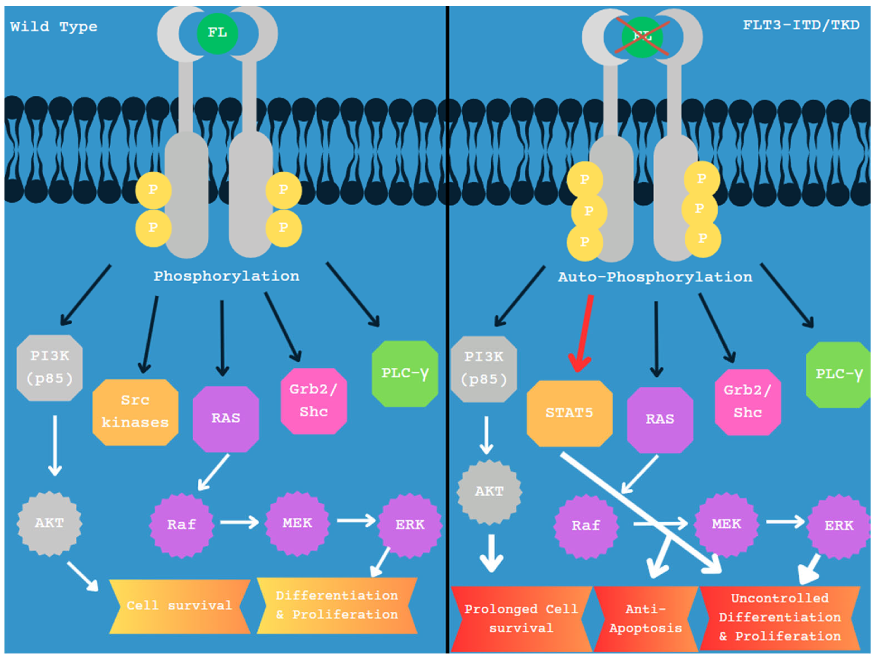
Figure 2. Wild-type vs. mutated FLT3. Black arrows indicate direct interaction with the FLT3 protein, and white arrows indicate downstream effects and related pathways. STAT5 proteins are not as effectively activated by wild-type FLT3 and are not substantially observed. FLT3-ITD mutated protein efficiently activates STAT5 and is therefore included on the right panel, with a red arrow designation. STAT5 increases transcription of Cyclin D1, c-Myc, p21, Pim-1, and Pim-2, leading to factors for anti-apoptosis and uncontrolled proliferation, as outlined in panel 2 [53].
Diagnostics 15 02537 g002
Figure 3. (A) Locus of FLT3 on 13q12; (B) FLT3 diagram with exons. Numbered exons represent typical sites of mutation. Modified from McGowan-Jordan, et al., 2020 [35].
Diagnostics 15 02537 g003
Figure 4. (A) Locus of DNMT3A on 2p23; (B) DNMT3A diagram with exons. Numbered exons represent typical sites of mutation. Modified from McGowan-Jordan, et al., 2020 [35].
Diagnostics 15 02537 g004
Figure 5. (A) Locus of MECOM on 3q26.2; (B) MECOM diagram with exons. Numbered exons represent typical sites of mutation. Modified from McGowan-Jordan, et al., 2020 [35].
Diagnostics 15 02537 g005
Figure 6. (A) Locus of ASXL1 on 20q11.21; (B) ASXL1 diagram with exons. Numbered exons represent typical sites of mutation. Modified from McGowan-Jordan, et al., 2020 [35].
Diagnostics 15 02537 g006
Figure 7. (A) Locus of TP53 on 17p13.1; (B) TP53 diagram with exons. Numbered exons represent typical sites of mutation. Modified from McGowan-Jordan, et al., 2020 [26].
Diagnostics 15 02537 g007
Figure 8. Relative domain locations in p53 protein, with amino acid range for each domain presented. Location of DNA-binding domain (DBD), in which most mutations occur, and carboxy-terminal tetramerization domain (TD), with amino-terminal transactivation domain (TAD) located at opposite N-terminal end [77]. Modified from Chen et al., 2022 [77].
Diagnostics 15 02537 g008
Figure 9. (A) Locus of CEBPA on 19q13.1; (B) CEBPA diagram of exon. Modified from McGowan-Jordan, et al., 2020 [35].
Diagnostics 15 02537 g009
Figure 11. Technological advancements in AML research, with predictions on how techniques can be consolidated. Incorporation of multiple technologies in improving patient health, identification, and personal treatment.
Diagnostics 15 02537 g011
Figure 12. Evolving paradigm of AML management. From current methods of management to insights into long-term shifts in treatment and disease management.
Diagnostics 15 02537 g012
Figure 13. Future standard of care in AML: integration of genomics, immunotherapy, AI, and equity. Predicted directions of change in major future components of care.
Diagnostics 15 02537 g013
Figure 14. Overview of overlapping and related effects caused by dysfunction in NPM1, FLT3, TP53, and DNMT3A and related pathways. Irregular gene types are labeled in green; associated pathways and proteins are labeled in purple; induced effects leading to leukemic progression are labeled in blue [33,53,61].
Diagnostics 15 02537 g014
Table 2. Comparative performance and clinical implications of NGS, FISH, and optical genome mapping in AML.
AttributeNGS (Short-Read Targeted Panels/WES/WGS)FISHOptical Genome Mapping (OGM)
Primary targetsSNVs, small indels; some CNVs/SVs depending on designKnown rearrangements, aneuploidies, and copy number at targeted lociLarge/complex SVs (translocations, inversions, insertions, deletions, CNVs); repeat expansions; genome-wide
SensitivityHigh for SNVs/indels; VAF often ≥1–5% for panels; SV/CNV sensitivity depends on assay/bioinformatics [22,23,24,25,26,27,28,29,30]High for targeted loci if probe exists; detects mosaicism to ~5–10% depending on lab [28]Genome-wide SV detection; claims down to ~5% VAF for mosaic/heterogeneous samples (assay-dependent) [11,12]
SpecificityHigh with orthogonal confirmation; false positives from artifacts/low-complexity regions; panel design matters [22,23,24,25,26,27,28,29,30]High for predefined targets; interpretation can be confounded by signal overlap/signal noise [28]High for large SVs; specificity impacted by complex duplications/repeats; orthogonal confirmation recommended [11,12]
ResolutionSingle-base for SNVs/indels; exon-level to gene-level for CNVs/SVs depending on depth/algorithm [22,23,24,25,26,27,28,29,30]Probe-level (typically 100–200 kb around target; break-apart or fusion probes) [28]Kilobase-scale mapping of long molecules; resolves structure/orientation and partners for many SVs [11,12]
Prior knowledge neededNot for broad panels/WES/WGS; targeted panels predefine genes/hotspotsYes—probe must be designed for suspected locus/rearrangementNo (genome-wide), unbiased for SV discovery
AdvantagesDetects recurrent AML SNVs/indels (e.g., NPM1, DNMT3A, FLT3); scalable; actionable variant interpretation frameworks exist [22,23,24,25,26,27,28,29,30]Rapid, cost-effective for confirming known rearrangements (e.g., MECOM, RUNX1); single-cell resolutionSingle assay for all major SV classes; clarifies complex/cryptic rearrangements and fusions; complements NGS [11,12]
LimitationsShort reads miss/under-resolve complex SVs, repeats; panel bias and variable exon coverage; SV calling requires optimized pipelines [22,23,24,25,26,27,28,29,30]Limited scope (only where probes exist); cannot genome-wide screen; lower structural detailLimited for small variants (SNVs/indels); VAF cutoffs need clinical context; orthogonal confirmation often needed [11,12,31]
Typical VAF/abundance limitsPanels often call ≥1–5% VAF for SNVs (lab-dependent); SV/CNV thresholds vary [22,23,24,25,26,27,28,29,30]Mosaic detection depends on probe/assay (~5–10% typical) [28]Reported ~5% for SVs in cancer samples; interpret with disease-specific rules (e.g., TP53 in ICC) [12,31]
Best use cases in AMLMutational profiling for diagnosis/risk (e.g., NPM1, FLT3-ITD/TKD, DNMT3A, ASXL1, TP53); MRD with specialized assaysRapid confirmation of specific rearrangements (e.g., MECOM break-apart), aneuploidies, and hallmark fusionsDiscovery/refinement of karyotype-scale abnormalities, cryptic/complex rearrangements, fusion partners; complement to NGS
Turnaround (typical)Days (panels) to 1–2 weeks (WES/WGS); lab-specific24–72 h for targeted probesDays to ~1 week; lab-specific
Notes on validationConfirm key calls (esp. low VAF or SVs) with orthogonal method (ddPCR, Sanger, FISH)Use with orthogonal methods for breakpoint/partner resolutionConfirm clinically actionable SVs with NGS/FISH or PCR, per lab policy
Disclaimer/Publisher’s Note: The statements, opinions and data contained in all publications are solely those of the individual author(s) and contributor(s) and not of MDPI and/or the editor(s). MDPI and/or the editor(s) disclaim responsibility for any injury to people or property resulting from any ideas, methods, instructions or products referred to in the content.