Case Report

A Rare Case of Exophiala Dermatitidis Isolation in a Patient with Non-Cystic Fibrosis Bronchiectasis: Colonization or True Infection?

by
Francesco Rocco Bertuccio
1,2,
Nicola Baio
3,
Simone Montini
1,2,
Valentina Ferroni
1,2,
Vittorio Chino
4,
Lucrezia Pisanu
1,2,
Marianna Russo
1,2,
Ilaria Giana
1,2,
Elisabetta Gallo
1,2,
Lorenzo Arlando
1,2,
Klodjana Mucaj
1,2,
Mitela Tafa
1,2,
Maria Arminio
1,2,
Emanuela De Stefano
1,2,
Alessandro Cascina
1,2,
Angelo Guido Corsico
1,2,
Giulia Maria Stella
1,2,* and
Valentina Conio
1,2,*
1
Unit of Respiratory Disease, Cardiothoracic and Vascular Department, IRCCS Policlinico San Matteo, Viale Golgi 19, 27100 Pavia, Italy
2
Department of Internal and dicine and Pharmacology, University of Pavia, 27100 Pavia, Italy
3
Ospedale Maggiore, ASST Crema, 26013 Crema, Italy
4
Ospedale Pederzoli, Peschiera del Garda, 37121 Verona, Italy
*
Authors to whom correspondence should be addressed.
Diagnostics 2025, 15(13), 1661; https://doi.org/10.3390/diagnostics15131661
Submission received: 31 May 2025 / Revised: 24 June 2025 / Accepted: 28 June 2025 / Published: 29 June 2025
(This article belongs to the Section Diagnostic Microbiology and Infectious Disease)

Abstract

Background: Exophiala dermatitidis is a dematiaceous, thermotolerant, yeast-like fungus increasingly recognized as an opportunistic pathogen in chronic airway diseases. While commonly associated with cystic fibrosis, its clinical significance in non-cystic fibrosis bronchiectasis (NCFB) remains unclear. Case Presentation: We report the case of a 66-year-old immunocompetent woman with a history of breast cancer in remission and NCFB, who presented with chronic cough and dyspnea. Chest CT revealed bilateral bronchiectasis with new pseudonodular opacities. Bronchoalveolar lavage cultures identified E. dermatitidis, along with Pseudomonas aeruginosa and methicillin-sensitive Staphylococcus aureus. Given clinical stability and the absence of systemic signs, initial therapy included oral voriconazole, levofloxacin, doxycycline, and inhaled amikacin. Despite persistent fungal isolation on repeat bronchoscopy, the patient remained asymptomatic with stable radiologic and functional findings. Antifungal therapy was discontinued, and the patient continued under close monitoring. The patient exhibited clinical and radiological stability despite repeated fungal isolation, reinforcing the hypothesis of persistent colonization rather than active infection. Discussion: This case underscores the diagnostic challenges in distinguishing fungal colonization from true infection in structurally abnormal lungs. In NCFB, disrupted mucociliary clearance and microbial dysbiosis may facilitate fungal persistence, even in the absence of overt immunosuppression. The detection of E. dermatitidis should prompt a comprehensive evaluation, integrating clinical, radiologic, and microbiologic data to guide management. Voriconazole is currently the antifungal agent of choice, though therapeutic thresholds and duration remain undefined. Conclusions: This report highlights the potential role of E. dermatitidis as an under-recognized respiratory pathogen in NCFB and the importance of a multidisciplinary, individualized approach to diagnosis and treatment. This case underscores the need for further research on fungal colonization in NCFB and the development of evidence-based treatment guidelines. Further studies are needed to clarify the pathogenic significance, optimal management, and long-term outcomes of E. dermatitidis in non-CF chronic lung diseases.

1. Introduction

Exophiala dermatitidis is a dematiaceous, thermotolerant fungus increasingly recognized for its opportunistic potential in the human respiratory tract. Though traditionally viewed as an environmental saprophyte, it has emerged as a pathogen of interest, particularly in patients with chronic pulmonary diseases such as cystic fibrosis (CF), where its prevalence may reach up to 19% in respiratory samples. Despite this, its clinical significance outside of CF remains poorly understood, with very few cases reported in non-cystic fibrosis bronchiectasis (NCFB) or structurally abnormal lungs [1,2,3].
NCFB is a chronic airway disorder characterized by irreversible bronchial dilatation, impaired mucociliary clearance, and persistent microbial colonization. These factors create a permissive niche not only for typical bacterial pathogens such as Pseudomonas aeruginosa and Staphylococcus aureus but also for less common fungal organisms. Repeated antibiotic exposure, mucus retention, and local immune dysregulation further disrupt the airway microbiome, potentially facilitating fungal persistence and pathogenesis [4,5,6].
In this context, the isolation of E. dermatitidis raises a critical diagnostic dilemma: does it represent mere colonization or an active infection warranting antifungal treatment? This distinction is essential, as treatment protocols are not standardized, susceptibility testing is limited, and prolonged azole therapy may carry significant toxicity. Voriconazole is generally considered the first-line agent due to its antifungal spectrum and good pulmonary bioavailability, although the duration and indications for therapy remain debated and largely based on expert opinion or case series [7,8,9].
Here, we report a rare case of E. dermatitidis isolated from the bronchoalveolar lavage of an immunocompetent woman with NCFB alongside P. aeruginosa and S. aureus co-colonization. The case underscores the importance of integrating clinical, radiologic, and microbiologic data to guide individualized decision-making, and it contributes to the growing body of literature on emerging fungal pathogens in non-CF airway diseases. To date, only a limited number of cases of Exophiala dermatitidis pulmonary isolation have been reported in patients with non-cystic fibrosis bronchiectasis, especially among immunocompetent individuals. This report contributes to the growing but still scarce body of literature on the clinical implications of rare fungal colonization in structurally abnormal lungs.

2. Case Presentation

A 66-year-old woman presented to the respiratory disease outpatient clinic with a prolonged history of persistent cough and shortness of breath during exertion. Her symptoms had gradually developed over the past months. She denied sputum production, wheezing, night sweats, weight loss, or fever. Her history was notable for a remote history of cigarette smoking (10 pack-years), having quit five years earlier. She denied any known exposure to birds, hot tubs, or mold and reported no known allergies. The patient was a retired office employee, without recent travel or relevant occupational/environmental exposures. She was not taking chronic medications at the time of presentation.
The patient had a significant oncologic history. Nine years ago, she was diagnosed with left-sided breast cancer and underwent neoadjuvant chemotherapy followed by a left radical mastectomy. Five years later, she experienced a local recurrence and began another chemotherapy treatment with docetaxel, trastuzumab, and pertuzumab. Restaging PET showed no evidence of distant metastases or recurrence. As of the most recent follow-up, her oncologic disease remains in remission.
Three years earlier, she had begun experiencing similar respiratory symptoms, and a workup at that time revealed bronchiectasis with mucoid impactions and multiple tiny pulmonary nodules. Bronchoalveolar lavage (BAL) cultures identified Mycobacterium chimaera, sensitive to amikacin, clarithromycin, and linezolid. A 12-month course of targeted therapy led to the eradication of the mycobacterial infection.

2.1. Physical Examination Findings

At presentation, the patient appeared in no acute distress. Her vital signs were stable: no fever, heart rate 78 bpm, normal blood pressure and respiratory rate, and oxygen saturation of 97% on room air. Lung auscultation revealed fine bilateral inspiratory crackles, without wheezes or rhonchi. No digital clubbing or peripheral edema was observed. Cardiac, abdominal, and neurologic examinations were unremarkable.

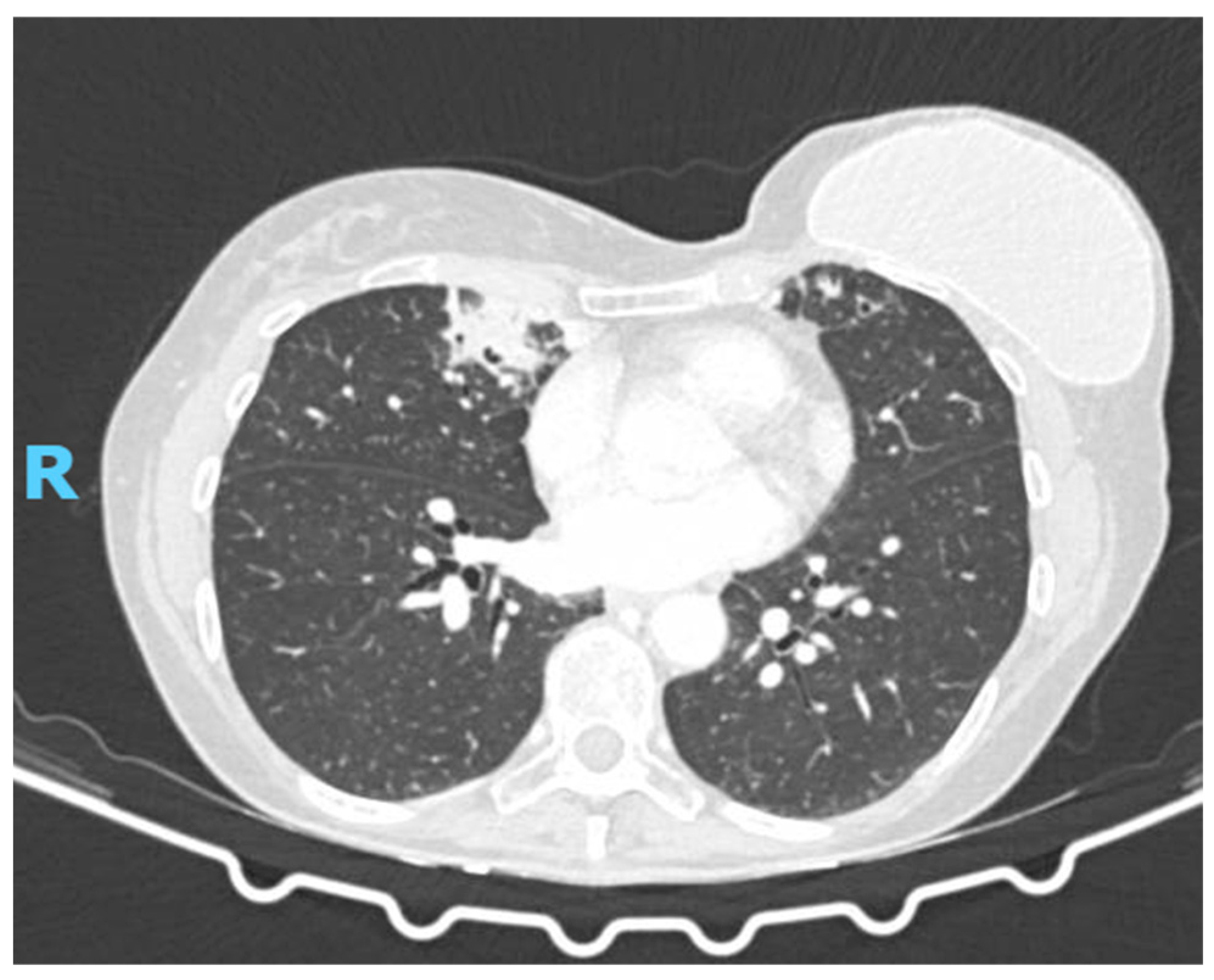
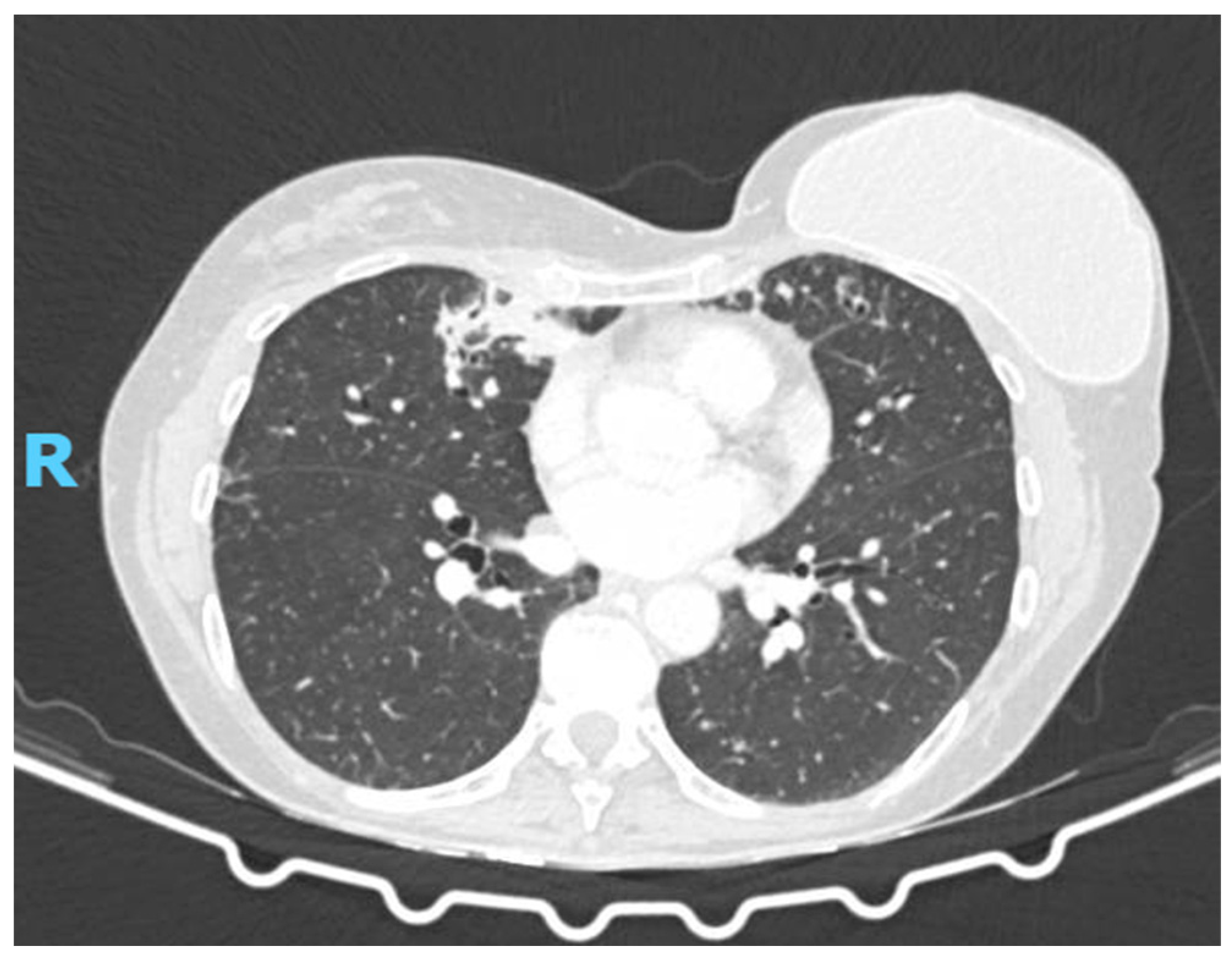
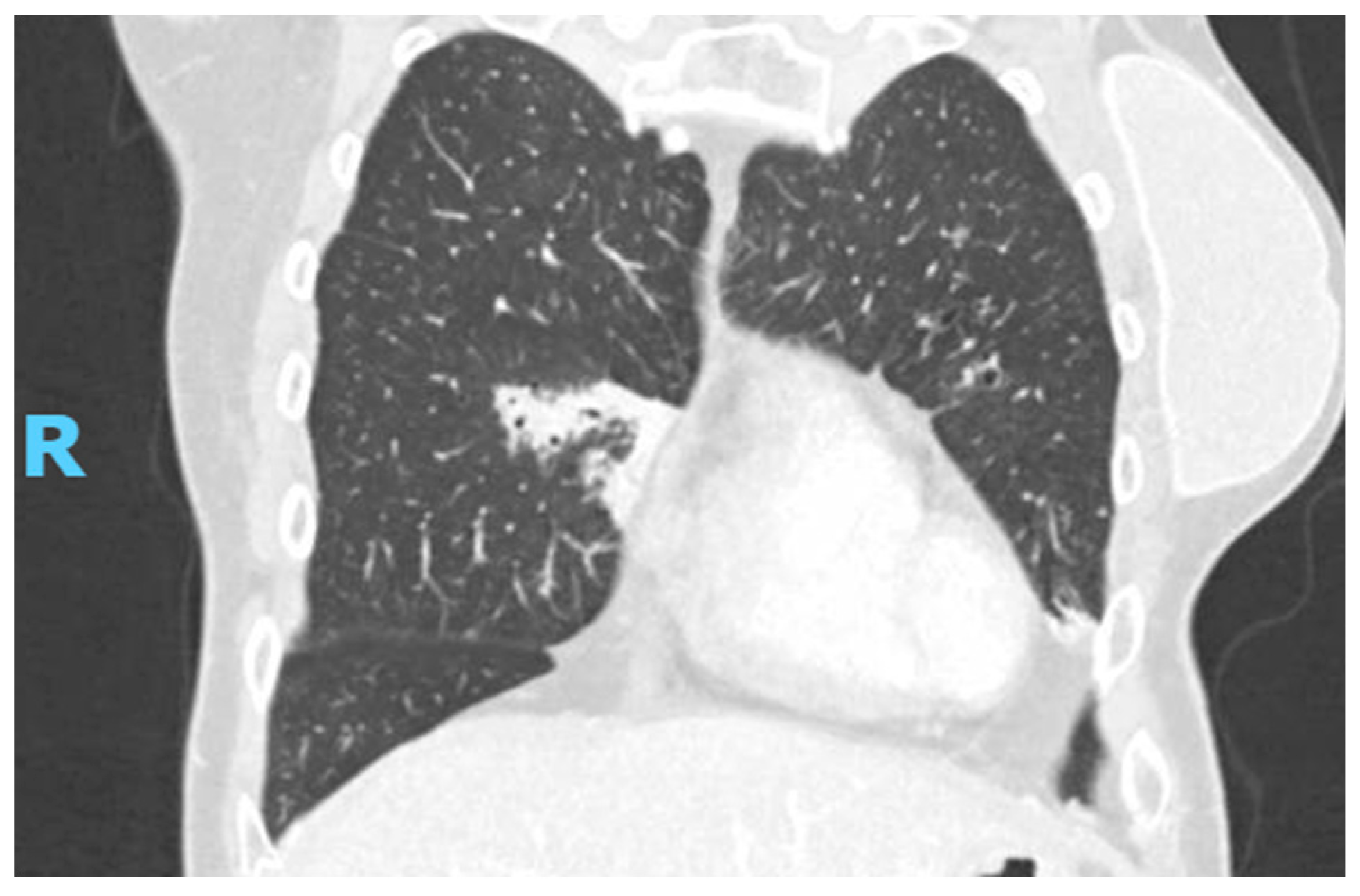
2.2. Diagnostic Studies

Initial blood tests, including complete blood count, liver and kidney function tests, electrolytes, and glucose, were all within normal limits. Inflammatory markers were not elevated (C-reactive protein, erythrocyte sedimentation rate, and procalcitonin were within normal ranges). The serologic workup for connective tissue diseases was negative, including ANA, ANCA, and anti-SSA/SSB antibodies. HIV and viral hepatitis screening were negative. Immunoglobulin levels (IgA, IgE, IgG, IgM) and alpha-1 antitrypsin were within the normal range. Serum β-D-glucan was not performed, while serum galactomannan was borderline (0.19), not consistent with invasive fungal infection.
Pulmonary function tests showed normal static and dynamic lung volumes with a preserved diffusing capacity for carbon monoxide: FVC 90% of predicted; RV 102% of predicted; TLC 95 of predicted %; FEV1 93% of predicted; DLCO 83% of predicted.
Repeat high-resolution chest CT (Figure 1) revealed new pseudonodular opacities in the middle lobe and left lower lobe, randomly distributed with an inflammatory appearance, without signs of neoplastic recurrence. Background findings of bilateral bronchiectasis, mucus impaction, and bronchial wall thickening were unchanged. Bronchoscopy with bronchoalveolar lavage was executed.

2.3. What Is the Diagnosis?

The diagnosis was pulmonary infection by Exophiala dermatitidis in a patient with non-cystic fibrosis bronchiectasis.

3. Discussion

Exophiala dermatitidis is a dematiaceous (black) yeast-like fungus ubiquitously found in the environment, including decaying organic matter and moist indoor surfaces. Belonging to the Herpotrichiellaceae family, it exhibits thermotolerance and thrives in humid habitats. Though occasionally isolated from respiratory specimens, clinically relevant pulmonary infections by Exophiala species are rare, particularly among immunocompetent hosts [7,10,11,12].
Invasive and disseminated infections are mostly reported in immunocompromised patients or those with hematologic malignancies, organ or stem cell transplants, or prolonged neutropenia [7,13]. However, an increasing number of case reports have identified E. dermatitidis as an emerging opportunistic pathogen in patients with chronic respiratory diseases, particularly cystic fibrosis (CF) and, more recently, bronchiectasis [9]. In non-CF bronchiectasis, the organism’s role remains poorly defined, with colonization versus infection often challenging to distinguish.
Predisposing factors such as impaired mucociliary clearance, structural airway damage, mucus retention, and frequent antibiotic exposure likely facilitate colonization and, in certain cases, progression to infection. While the presence of E. dermatitidis in sputum may represent harmless colonization in some, it can trigger an inflammatory response, exacerbate respiratory symptoms, or evolve into invasive disease in others [14,15].
Bronchiectasis is characterized by permanent bronchial dilation, disrupted mucociliary function, and chronic mucus retention. This creates a nutrient-rich, hypoxic microenvironment conducive to microbial persistence [4]. Dematiaceous fungi such as Exophiala, with biofilm-forming capacity and thermotolerance, can exploit this niche. Additionally, repeated use of broad-spectrum antibiotics disrupts the microbial balance of the airway, potentially promoting fungal overgrowth and dysbiosis [16].
Notably, interactions between E. dermatitidis and common bacterial pathogens may influence fungal behavior and host immune responses. P. aeruginosa is known to secrete quorum-sensing molecules and iron-chelating agents that can inhibit or modulate fungal growth, while S. aureus may enhance fungal persistence through biofilm co-aggregation and immune evasion. These interactions may shift the microbial community toward chronic inflammation or facilitate transitions from colonization to infection. In particular, coinfection or sequential colonization with these bacteria might predispose the lung to fungal persistence or pathogenesis [16,17].
Although often asymptomatic, fungal colonization may elicit an aberrant immune response in certain patients, manifesting as increased cough, sputum production, radiologic changes, and clinical deterioration. In rare cases, especially with additional risk factors such as prior cancer therapy or subtle immunologic defects, E. dermatitidis can invade pulmonary tissue, leading to bronchopneumonia, necrotizing infections, or systemic dissemination [16].
Recognizing this potential transition is critical for timely diagnosis and management. Increasing awareness of such fungal pathogens in non-CF bronchiectasis is essential for guiding future therapeutic strategies and for refining our understanding of host–pathogen–microbiome interactions in chronic airway diseases.
Symptoms are often nonspecific, cough, sputum, and dyspnea, and overlap with baseline bronchiectasis manifestations. Imaging findings are also variable. In our case, chest CT showed bronchiectasis, mucus plugging, and new pseudonodular inflammatory opacities, raising suspicion for fungal involvement [18]. These were not consistent with cancer recurrence or NTM reactivation, both of which had been previously addressed.
The isolation of E. dermatitidis from respiratory samples confirms diagnosis, ideally supported by repeat cultures, growth on Sabouraud dextrose agar, and molecular identification. Histopathological confirmation is rare and usually reserved for suspected invasive disease [19,20,21]. Given the overlap with colonization, clinical judgment remains essential in deciding whether to initiate antifungal therapy.
Treatment remains non-standardized due to the rarity of cases. In vitro susceptibility profiles show variable responses, but azoles, particularly voriconazole and posaconazole, demonstrate consistent activity. Amphotericin B shows less efficacy and is usually reserved for severe infections or as induction therapy. Voriconazole is the most commonly used agent, likely due to its excellent lung penetration and activity against melanized fungi. It is employed as monotherapy or following initial induction with amphotericin B or itraconazole. Itraconazole is often used orally in milder cases or as a step-down agent. Posaconazole is generally reserved for cases with azole intolerance or resistance [9,18,22,23,24,25,26,27,28,29,30,31].
Although specific treatment durations are seldom reported, available evidence suggests regimens typically last 3 to 6 months, with prolonged therapy (>6 months) in patients with structural lung disease or coinfections (e.g., NTM). In a few cases, surgical resection was performed, either due to localized disease or inadequate response to medical therapy, highlighting the potential utility of a combined medical–surgical approach in selected patients [9,18,22,23,24,25,26,27,28,29,30,31].
Clinical outcomes are generally favorable, particularly in immunocompetent individuals. The only fatality in the reviewed literature involved a patient with multiple comorbidities and likely immunosuppression. Thus, individualized therapeutic strategies based on clinical severity, host status, radiologic progression, and microbiological findings are essential [32,33,34,35].
In our case, the patient received a 12-week course of oral voriconazole, combined with levofloxacin and doxycycline targeting concurrent bacterial pathogens. Inhaled amikacin was added to address chronic Pseudomonas colonization. Voriconazole was initiated despite the absence of systemic symptoms, given the presence of new inflammatory opacities on imaging, the co-isolation of multiple pathogens, and the patient’s prior history of opportunistic infection (NTM infection).
Follow-up imaging showed stable disease, and repeat BAL continued to yield E. dermatitidis without clinical worsening. Given the absence of deterioration, further antifungal therapy was withheld in agreement with infectious disease specialists, suggesting a colonization phenotype rather than active infection.
This case is of particular scientific and clinical relevance due to the rarity of E. dermatitidis infection in immunocompetent patients with non-CF bronchiectasis. While its role as an opportunistic pathogen is increasingly recognized, most cases involve individuals with CF, hematologic malignancy, or immunosuppression. Reports in structurally abnormal but immunocompetent lungs remain scarce. In our case, it could be plausible that the patient’s prior exposure to systemic chemotherapy and monoclonal antibodies may have caused long-lasting subclinical immune dysregulation, thereby promoting fungal persistence. In Table 1, we describe briefly the main case reports published in the literature despite the fact that they are rare. Describing such cases contributes to expanding our understanding of the potential role of dematiaceous fungi in chronic respiratory conditions and highlights the diagnostic and therapeutic uncertainties they present.
Moreover, this report underscores the need for heightened awareness of atypical fungal pathogens in patients with bronchiectasis, especially when standard therapies fail or when radiologic findings are unusual. Reporting rare but well-characterized infections enhances evidence-based decision-making, reveals emerging susceptibility patterns, and helps define the natural history of fungal colonization in chronic airway disease.
Rather than merely presenting an unusual isolation, our aim is to stimulate broader interest in under-recognized fungal pathogens, emphasize the complexity of host–microbe interactions in bronchiectasis, and support future research aimed at developing tailored diagnostic tools and antifungal treatment protocols. Ultimately, this case highlights the critical role of multidisciplinary collaboration, spanning pulmonology, infectious diseases, microbiology, and radiology, in managing complex airway infections and improving patient outcomes.

Clinical Course

The patient’s bronchoscopy revealed isolation of Exophiala dermatitidis, along with Pseudomonas aeruginosa and methicillin-sensitive Staphylococcus aureus. She was initially treated with oral levofloxacin for 14 days, doxycycline for 7 days, and aerosolized amikacin for 15 days. A 12-week course of voriconazole (200 mg twice per day) was also initiated. The follow-up chest CT scan revealed persistent inflammatory opacities without new consolidations (Figure 2). Repeat BAL again grew Exophiala dermatitidis, and galactomannan was borderline (0.19) without other bacterial isolations. Given clinical stability and the absence of new respiratory symptoms, further antifungal therapy was withheld. At her most recent evaluation, she remained asymptomatic, with stable pulmonary function and imaging.

4. Conclusions

This case highlights the emerging relevance of Exophiala dermatitidis as a potential respiratory pathogen in patients with chronic lung disease, even in the absence of overt immunosuppression. Although rare, E. dermatitidis should be considered in the differential diagnosis of new pulmonary infiltrates, particularly in individuals with structural airway abnormalities such as non-cystic fibrosis bronchiectasis (NCFB).
The distinction between colonization and active infection remains a significant clinical challenge. As shown in this case, a comprehensive, longitudinal evaluation incorporating clinical symptoms, radiological evolution, and repeated microbiological findings is often necessary to guide therapeutic decisions and avoid both under- and overtreatment.
When antifungal therapy is deemed appropriate, voriconazole currently represents the first-line agent due to its demonstrated efficacy and favorable pharmacokinetics in pulmonary tissue. However, standardized treatment protocols are lacking, and the optimal duration of therapy remains uncertain, requiring individualized assessment based on disease severity, response to treatment, and host factors.
Importantly, NCFB itself constitutes a predisposing condition for unusual infections, as impaired mucociliary clearance, mucus stasis, and frequent antibiotic exposure create a permissive environment for opportunistic fungal colonization and, potentially, pathogenic invasion—even in immunocompetent patients.
This report is limited by the absence of histopathological confirmation and the relatively short duration of follow-up. Moreover, the lack of standardized guidelines for managing fungal infections in non-CF bronchiectasis represents a significant challenge in clinical decision-making.
This case underscores the value of a multidisciplinary approach encompassing pulmonology, infectious diseases, microbiology, and radiology in the management of rare pulmonary fungal infections. Collaboration among specialties not only facilitates timely diagnosis and tailored treatment but also contributes to the growing body of knowledge required to better characterize the clinical significance and natural history of under-recognized fungal pathogens in chronic airway diseases. Future studies should explore host immune mechanisms and microbiome interactions that may underlie chronic fungal colonization in patients with bronchiectasis.

Author Contributions

Conceptualization: F.R.B. and V.C. (Valentina Conio).; Writing and Methodology: F.R.B., V.C. (Valentina Conio)., G.M.S. and A.G.C.; Review and editing: N.B., S.M., L.P., E.G., E.D.S., I.G., M.A., L.A., K.M., M.T., V.C. (Vittorio Chino), A.C., V.F. and M.R.; Validation: N.B., S.M., L.P., E.G., E.D.S., I.G., M.A., L.A., K.M., M.T., V.C. (Vittorio Chino), A.C., V.F. and M.R.; Supervision: A.C., V.C. (Vittorio Chino), A.G.C. and G.M.S. All authors have read and agreed to the published version of the manuscript.

Funding

This study received no external funding.

Institutional Review Board Statement

The research was conducted according to the principles of the World Medical Association Declaration of Helsinki. The study was submitted as part of a main project that was approved by the local Ethical Commission, and each enrolled patient gave written informed consent before enrollment (Comitato di Bioetica, Fondazione IRCCS Policlinico San Matteo, approval numbers: protocol #20090002344; procedure # 20090019080; date of approval: 3 June 2009).

Informed Consent Statement

Written informed consent has been obtained from the patient to publish this paper.

Data Availability Statement

The data that support the findings of this study are available on request from the corresponding author.

Conflicts of Interest

The authors declare that they have no conflicts of interest.

References

  1. Bahadori, T.; Didehdar, M.; Khansarinezhad, B.; Shokohi, T. Identification of Opportunistic and Nonopportunistic Exophiala Species Using High Resolution Melting Analysis. Med. Mycol. 2020, 58, 938–945. [Google Scholar] [CrossRef]
  2. Babič, M.N.; Zupančič, J.; Gunde-Cimerman, N.; de Hoog, S.; Zalar, P. Ecology of the Human Opportunistic Black Yeast Exophiala Dermatitidis Indicates Preference for Human-Made Habitats. Mycopathologia 2018, 183, 201–212. [Google Scholar] [CrossRef]
  3. de Jong, C.C.M.; Slabbers, L.; Engel, T.G.P.; Yntema, J.B.; van Westreenen, M.; Croughs, P.D.; Roeleveld, N.; Brimicombe, R.; Verweij, P.E.; Meis, J.F.; et al. Clinical Relevance of Scedosporium Spp. and Exophiala Dermatitidis in Patients with Cystic Fibrosis: A Nationwide Study. Med. Mycol. 2020, 58, 859–866. [Google Scholar] [CrossRef] [PubMed]
  4. Chalmers, J.D.; Chang, A.B.; Chotirmall, S.H.; Dhar, R.; McShane, P.J. Bronchiectasis. Nat. Rev. Dis. Prim. 2018, 4, 45. [Google Scholar] [CrossRef]
  5. Polverino, E.; Goeminne, P.C.; McDonnell, M.J.; Aliberti, S.; Marshall, S.E.; Loebinger, M.R.; Murris, M.; Cantón, R.; Torres, A.; Dimakou, K.; et al. European Respiratory Society Guidelines for the Management of Adult Bronchiectasis. Eur. Respir. J. 2017, 50, 1700629. [Google Scholar] [CrossRef]
  6. Richardson, H.; Dicker, A.J.; Barclay, H.; Chalmers, J.D. The Microbiome in Bronchiectasis. Eur. Respir. Rev. 2019, 28, 190048. [Google Scholar] [CrossRef] [PubMed]
  7. Usuda, D.; Higashikawa, T.; Hotchi, Y.; Usami, K.; Shimozawa, S.; Tokunaga, S.; Osugi, I.; Katou, R.; Ito, S.; Yoshizawa, T.; et al. Exophiala Dermatitidis. World J. Clin. Cases 2021, 9, 7963–7972. [Google Scholar] [CrossRef] [PubMed]
  8. Revankar, S.G.; Sutton, D.A. Melanized Fungi in Human Disease. Clin. Microbiol. Rev. 2010, 23, 884–928. [Google Scholar] [CrossRef]
  9. Miyoshi, S.; Tanabe, M.; Semba, M.; Sato, C.; Aoyama, S.; Watanabe, A.; Ito, R.; Hamada, K.; Watanabe, A.; Abe, M. Exophiala Dermatitidis Coinfection with Nontuberculous Mycobacteria: A Case Report and Literature Review. Respirol. Case Rep. 2023, 11, e01221. [Google Scholar] [CrossRef]
  10. Horré, R.; Schaal, K.P.; Siekmeier, R.; Sterzik, B.; de Hoog, G.S.; Schnitzler, N. Isolation of Fungi, Especially Exophiala Dermatitidis, in Patients Suffering from Cystic Fibrosis. A Prospective Study. Respiration. 2004, 71, 360–366. [Google Scholar] [CrossRef]
  11. Haase, G.; Skopnik, H.; Groten, T.; Kusenbach, G.; Posselt, H.G. Long-Term Fungal Cultures from Sputum of Patients with Cystic Fibrosis. Mycoses 1991, 34, 373–376. [Google Scholar] [CrossRef] [PubMed]
  12. Diemert, D.; Kunimoto, D.; Sand, C.; Rennie, R. Sputum Isolation of Wangiella Dermatitidis in Patients with Cystic Fibrosis. Scand. J. Infect. Dis. 2001, 33, 777–779. [Google Scholar] [CrossRef] [PubMed]
  13. Matsumoto, T.; Matsuda, T.; McGinnis, M.R.; Ajello, L. Clinical and Mycological Spectra of Wangiella Dermatitidis Infections. Mycoses 1993, 36, 145–155. [Google Scholar] [CrossRef] [PubMed]
  14. Chalmers, J.D.; Ringshausen, F.C.; Harris, B.; Elborn, J.S.; Posthumus, A.; Haworth, C.S.; Pilkington, N.; Polverino, E.; Ruddy, T.; Aliberti, S.; et al. Cross-Infection Risk in Patients with Bronchiectasis: A Position Statement from the European Bronchiectasis Network (EMBARC), EMBARC/ELF Patient Advisory Group and European Reference Network (ERN-Lung) Bronchiectasis Network. Eur. Respir. J. 2018, 51, 1701937. [Google Scholar] [CrossRef]
  15. Chalmers, J.D.; Mall, M.A.; McShane, P.J.; Nielsen, K.G.; Shteinberg, M.; Sullivan, S.D.; Chotirmall, S.H. A Systematic Literature Review of the Clinical and Socioeconomic Burden of Bronchiectasis. Eur. Respir. Rev. 2024, 33, 240049. [Google Scholar] [CrossRef]
  16. Kirchhoff, L.; Olsowski, M.; Rath, P.-M.; Steinmann, J. Exophiala Dermatitidis: Key Issues of an Opportunistic Fungal Pathogen. Virulence 2019, 10, 984–998. [Google Scholar] [CrossRef]
  17. Boral, H.; Metin, B.; Döğen, A.; Seyedmousavi, S.; Ilkit, M. Overview of Selected Virulence Attributes in Aspergillus Fumigatus, Candida Albicans, Cryptococcus Neoformans, Trichophyton Rubrum, and Exophiala Dermatitidis. Fungal Genet. Biol. 2018, 111, 92–107. [Google Scholar] [CrossRef]
  18. Suzuki, K.; Nakamura, A.; Fujieda, A.; Nakase, K.; Katayama, N. Pulmonary Infection Caused by Exophiala Dermatitidis in a Patient with Multiple Myeloma: A Case Report and a Review of the Literature. Med. Mycol. Case Rep. 2012, 1, 95–98. [Google Scholar] [CrossRef]
  19. Myoken, Y.; Sugata, T.; Fujita, Y.; Kyo, T.; Fujihara, M.; Katsu, M.; Mikami, Y. Successful Treatment of Invasive Stomatitis Due to Exophiala Dermatitidis in a Patient with Acute Myeloid Leukemia. J. Oral Pathol. Med. Off. Publ. Int. Assoc. Oral Pathol. Am. Acad. Oral Pathol. 2003, 32, 51–54. [Google Scholar] [CrossRef]
  20. Silva, W.C.; Gonçalves, S.S.; Santos, D.W.C.L.; Padovan, A.C.B.; Bizerra, F.C.; Melo, A.S.A. Species Diversity, Antifungal Susceptibility and Phenotypic and Genotypic Characterisation of Exophiala Spp. Infecting Patients in Different Medical Centres in Brazil. Mycoses 2017, 60, 328–337. [Google Scholar] [CrossRef]
  21. Yoshinouchi, T.; Yamamoto, K.; Migita, M.; Yokoyama, T.; Nakamura, T.; Matsuoka, M. Diagnosis and Clinical Management of Exophiala Dermatitidis Pneumonia in a Patient with Anorexia Nervosa: A Case Report. Med. Mycol. Case Rep. 2023, 42, 100617. [Google Scholar] [CrossRef]
  22. Setoguchi, D.; Iwanaga, N.; Ito, Y.; Ashizawa, N.; Hirayama, T.; Takeda, K.; Ide, S.; Takemoto, S.; Tashiro, M.; Hosogaya, N.; et al. Pulmonary Phaeohyphomycosis Due to Exophiala Dermatitidis in a Patient with Pulmonary Non-Tuberculous Mycobacterial Infection. J. Infect. Chemother. Off. J. Japan Soc. Chemother. 2023, 29, 615–619. [Google Scholar] [CrossRef]
  23. Watanabe, Y.; Sano, H.; Konno, S.; Kamioka, Y.; Hariu, M.; Takano, K.; Yamada, M.; Seki, M. Sinobronchial Syndrome Patients with Suspected Non-Tuberculous Mycobacterium Infection Exacerbated by Exophiala Dermatitidis Infection. Infect. Drug Resist. 2022, 15, 1135–1141. [Google Scholar] [CrossRef]
  24. Li, Z.; Tang, J.; Zhu, J.; Xie, M.; Huang, S.; Li, S.; Zhan, Y.; Zeng, W.; Xu, T.; Ye, F. The Convoluted Process of Diagnosing Pulmonary Mycosis Caused by Exophiala Dermatitidis: A Case Report. BMC Infect. Dis. 2022, 22, 433. [Google Scholar] [CrossRef] [PubMed]
  25. Sekiguchi, R.; Urabe, N.; Sakamoto, S.; Sasaki, M.; Homma, S.; Kishi, K. Exophiala Dermatitidis Pneumonia with Bronchiectasis Required Prolonged Voriconazole Treatment. Respirol. Case Rep. 2021, 9, e00783. [Google Scholar] [CrossRef]
  26. Mukai, Y.; Nureki, S.; Hata, M.; Shigenaga, T.; Tokimatsu, I.; Miyazaki, E.; Kadota, J.; Yarita, K.; Kamei, K. Exophiala Dermatitidis Pneumonia Successfully Treated with Long-Term Itraconazole Therapy. J. Infect. Chemother. Off. J. Japan Soc. Chemother. 2014, 20, 446–449. [Google Scholar] [CrossRef] [PubMed]
  27. Bulloch, M.N. The Treatment of Pulmonary Wangiella Dermatitidis Infection with Oral Voriconazole. J. Clin. Pharm. Ther. 2011, 36, 433–436. [Google Scholar] [CrossRef] [PubMed]
  28. Ozawa, Y.; Suda, T.; Kaida, Y.; Kato, M.; Hasegaw, H.; Fujii, M.; Ida, M.; Nogimura, H.; Nagayama, M.; Chida, K. A case of bronchial infection of Wangiella dermatitidis. Nihon Kokyuki Gakkai Zasshi 2007, 45, 907–911. [Google Scholar]
  29. Taj-Aldeen, S.J.; El Shafie, S.; Alsoub, H.; Eldeeb, Y.; de Hoog, G.S. Isolation of Exophiala Dermatitidis from Endotracheal Aspirate of a Cancer Patient. Mycoses 2006, 49, 504–509. [Google Scholar] [CrossRef]
  30. Mukaino, T.; Koga, T.; Oshita, Y.; Narita, Y.; Obata, S.; Aizawa, H. Exophiala Dermatitidis Infection in Non-Cystic Fibrosis Bronchiectasis. Respir. Med. 2006, 100, 2069–2071. [Google Scholar] [CrossRef]
  31. Barenfanger, J.; Ramirez, F.; Tewari, R.P.; Eagleton, L. Pulmonary Phaeohyphomycosis in a Patient with Hemoptysis. Chest 1989, 95, 1158–1160. [Google Scholar] [CrossRef] [PubMed]
  32. Poyntner, C.; Mirastschijski, U.; Sterflinger, K.; Tafer, H. Transcriptome Study of an Exophiala Dermatitidis PKS1 Mutant on an Ex Vivo Skin Model: Is Melanin Important for Infection? Front. Microbiol. 2018, 9, 1457. [Google Scholar] [CrossRef] [PubMed]
  33. Poyntner, C.; Blasi, B.; Arcalis, E.; Mirastschijski, U.; Sterflinger, K.; Tafer, H. The Transcriptome of Exophiala Dermatitidis during Ex-Vivo Skin Model Infection. Front. Cell. Infect. Microbiol. 2016, 6, 136. [Google Scholar] [CrossRef]
  34. Yazdanparast, S.A.; Mohseni, S.; De Hoog, G.S.; Aslani, N.; Sadeh, A.; Badali, H. Consistent High Prevalence of Exophiala Dermatitidis, a Neurotropic Opportunist, on Railway Sleepers. J. Mycol. Med. 2017, 27, 180–187. [Google Scholar] [CrossRef]
  35. Lang, R.; Minion, J.; Skinner, S.; Wong, A. Disseminated Exophiala Dermatitidis Causing Septic Arthritis and Osteomyelitis. BMC Infect. Dis. 2018, 18, 255. [Google Scholar] [CrossRef]
  36. Kenney, R.T.; Kwon-Chung, K.J.; Waytes, A.T.; Melnick, D.A.; Pass, H.I.; Merino, M.J.; Gallin, J.I. Successful Treatment of Systemic Exophiala dermatitidis Infection in a Patient with Chronic Granulomatous Disease. Clin. Infect. Dis. 1992, 14, 235–242. [Google Scholar] [CrossRef] [PubMed]
  37. Tanamachi, C.; Hashimoto, K.; Nakata, K.; Sagawa, K. A case of pulmonary chromomycosis caused by Exophiala dermatitidis. J. Jpn. Soc. Clin. Microbiol. 2008, 18, 25–30, (In Japanese, Abstract in English). [Google Scholar]
  38. Shintani, R.; Hagiwara, E.; Yamakawa, H.; Ikeda, S.; Kitamura, H.; Baba, T. Pulmonary phaeohyphomycosis caused by Exophiala dermatitidis. JJA Inf. 2017, 91, 785–789. [Google Scholar]
  39. Goto, Y.; Murakami, N.; Yamasaki, Y. Sequelae of pulmonary chromoblastomycosis caused by the viscous species Exophiala dermatitidis in a patient with nontuberculosis mycobacterial disease. Igakukensa 2020, 69, 451–456. [Google Scholar]
  40. Masuo, M.; Hanazawa, S.; Nukui, Y. Pulmonary chromomycosis caused by Exophiala dermatitidis in a patient with pulmonary non-tuberculous mycobacteriosis. J. Jap. Soc. Respir. Endos. 2021, 43, 619–623. [Google Scholar]
Figure 1. The chest CT scan revealed new pseudonodular opacities in the middle lobe.
Diagnostics 15 01661 g001
Figure 2. Follow-up chest CT scan revealed persistent inflammatory opacities without new consolidations.
Diagnostics 15 01661 g002aDiagnostics 15 01661 g002b
Table 1. Main case reports published.
Author (Reference)AgeSexCountryUnderlying Disease(s)SymptomsTreatmentAntifungal Therapy and Estimated DurationNotes
Barenfanger et al. [31]79FUSABronchiectasisHemoptysis, feverAmphotericin B, 5-FCAmphotericin B + 5-FC (IV), ~6–8 weeksLikely IV start, step-down unknown
Kenney et al [36]21FUSAChronic granulomatous diseaseFever, chills, SOBSurgery, Amphotericin B, 5-FC, FluconazoleCombination therapy, ~6–12 weeks + oral FluconazoleImmunocompromised host
Mukaino et al. [30]54FJapanBronchiectasisCough, sputumMiconazole, Amphotericin BLikely topical/oral Miconazole + IV AmB, ~4–6 weeksNon-severe
Taj-Aldeen et al. [29]54FQuatarDiabetes, cervical cancerCough, sputum, hemoptysisFluconazole, Itraconazole, Amphotericin BLikely stepwise approach, total ~2–3 monthsFatal outcome, likely severe
Ozawa et al. [28]81FJapanNoneHemoptysisItraconazoleItraconazole PO, ~3 monthsGood response
Tanamachi et al. [37]53FJapanBronchiectasisSputum, chest painMiconazole, 5-FC, ItraconazoleCombination (oral), likely ~3–6 monthsOral triple therapy
Bulloch et al. [27]86FUSADementiaAsymptomaticVoriconazoleVRCZ PO, likely short course (~4 weeks)Incidental finding
Suzuki et al. [18]65MJapanMultiple myelomaAsymptomaticVoriconazole, SurgeryVRCZ PO, likely ~6 weeks post-surgeryImmunosuppressed
Mukai et al. [26]63FJapanNoneChest painItraconazoleITCZ PO, ~3 monthsStandard regimen
Shintani et al. [38]56FJapanBronchiectasisSputum, feverItraconazoleITCZ PO, ~3–4 monthsBronchiectasis context
Goto et al. [39]70FJapanNTMSputum, chest painVoriconazoleVRCZ PO, ~4–6 monthsNTM co-infection
Masuo et al. [40]58FJapanNTMCoughVoriconazoleVRCZ PO, ~4–6 monthsNTM context
Sekiguchi et al. [25]65FJapanRA, bronchiectasisCough, sputumVoriconazoleVRCZ PO, ~6 monthsAutoimmune comorbidity
Li et al. [24]52MChinaNoneCough, sputum, hemoptysisVRCZ, Amphotericin B, PosaconazoleEscalated therapy, total ~3–4 monthsLikely initial failure
Watanabe et al. [23]65FJapanNTM, sinusitisCough, sputumVoriconazoleVRCZ PO, ~3–4 monthsCoinfection context
Watanabe et al. [23]47FJapanRA, NTM, sinusitisNasal discharge, anosmiaAmphotericin B, VRCZ, ItraconazoleSequential therapy, total ~6 monthsUpper + lower airway involvement
Disclaimer/Publisher’s Note: The statements, opinions and data contained in all publications are solely those of the individual author(s) and contributor(s) and not of MDPI and/or the editor(s). MDPI and/or the editor(s) disclaim responsibility for any injury to people or property resulting from any ideas, methods, instructions or products referred to in the content.

