
The Diagnosis of and Preoperative Planning for Rapidly Progressive Osteoarthritis of the Hip: The Role of Sagittal Spinopelvic Geometry and Anterior Acetabular Wall Deficiency—A Prospective Observational Study

 , , and
Abstract
1. Introduction
2. Materials and Methods
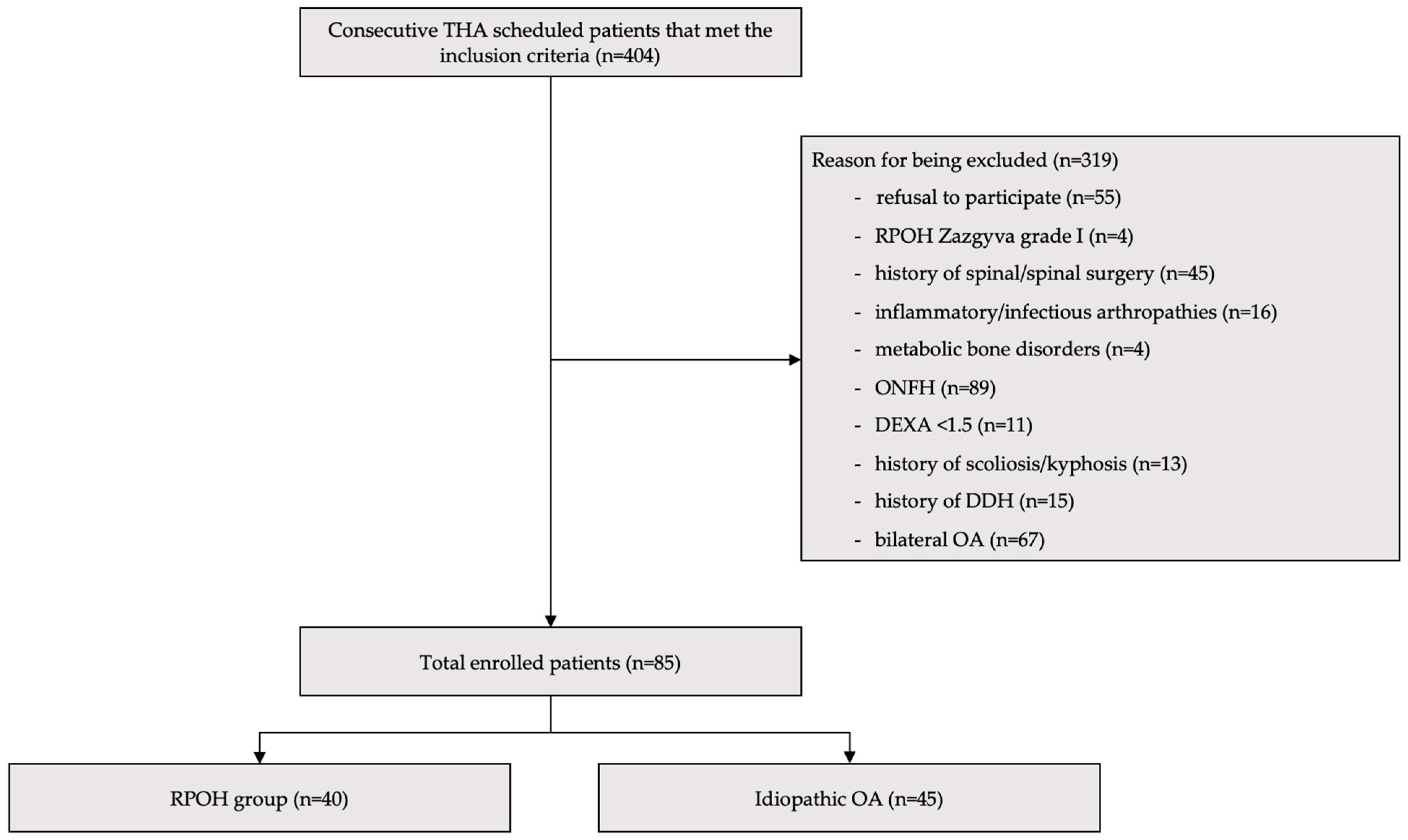
2.1. Design and Enrollment
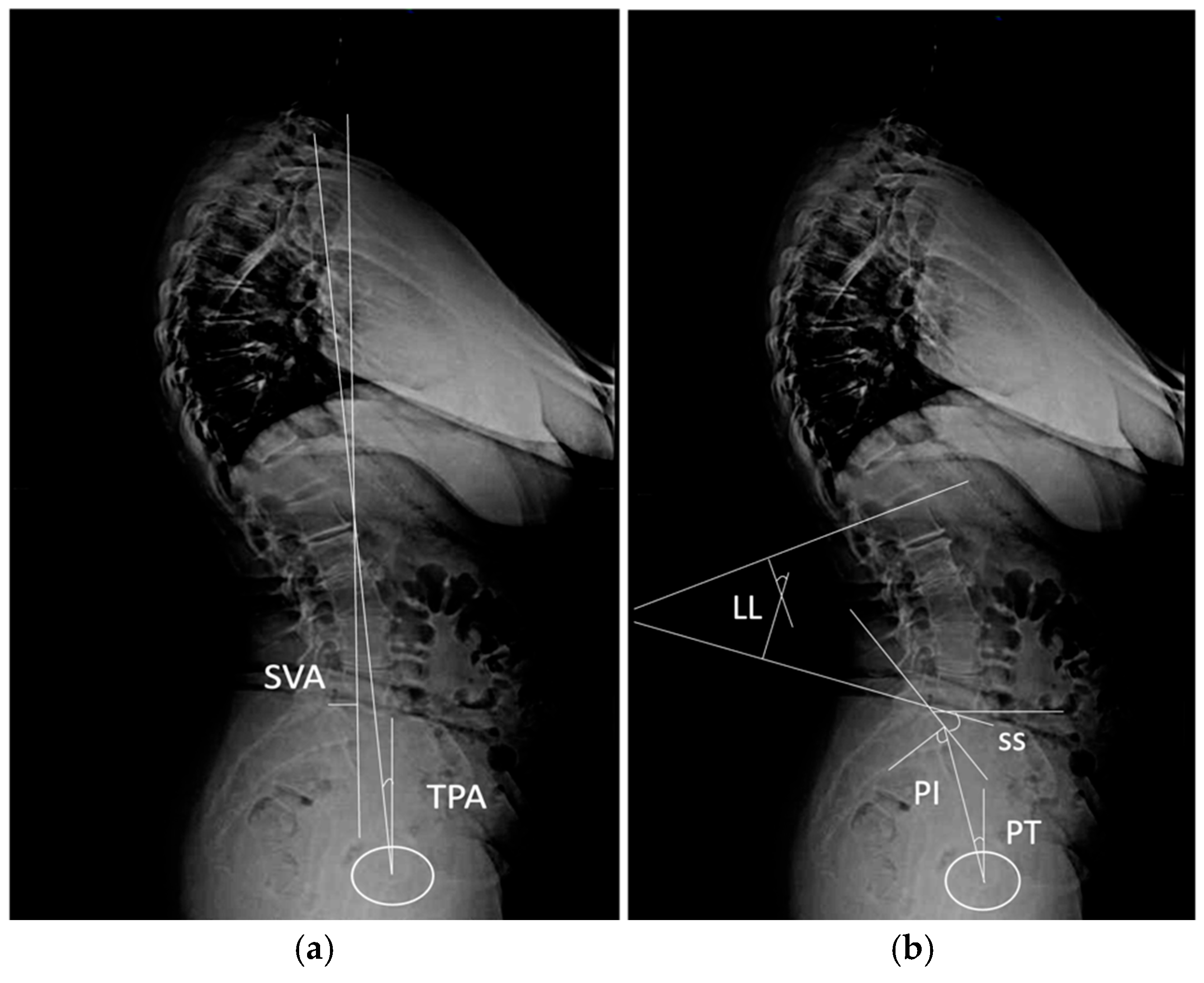
2.2. Imaging Analysis Measurements
2.3. Statistical Analysis
3. Results
4. Discussion
5. Conclusions
Author Contributions
Funding
Institutional Review Board Statement
Informed Consent Statement
Data Availability Statement
Conflicts of Interest
References
- Montero Furelos, L.A.; De Castro Carrasco, A.; Cons Lamas, S.; Sanchez Sierra, F.B.; Caeiro-Rey, J.R. Rapidly Progressive Osteoarthritis of the Hip: A Prospective Study. J. Clin. Med. 2024, 13, 2467. [Google Scholar] [CrossRef]
- Fukui, K.; Kaneuji, A.; Fukushima, M.; Matsumoto, T. Inversion of the Acetabular Labrum Triggers Rapidly Destructive Osteoarthritis of the Hip: Representative Case Report and Proposed Etiology. J. Arthroplast. 2014, 29, 2468–2472. [Google Scholar] [CrossRef]
- Prejbeanu, R.; Mioc, M.L.; Tsiridis, E.; Kenanidis, E.; Valli, F.; Pasquini, A.; Deleanu, B. The Influence of Tranexamic Acid (TXA) on Postoperative Infection Rates Following Total Hip Arthroplasty (THA)—A Systematic Review. J. Clin. Med. 2025, 14, 2910. [Google Scholar] [CrossRef] [PubMed]
- Nakamura, K.; Okamoto, Y.; Wakama, H.; Matsuyama, J.; Ishitani, T.; Otsuki, S.; Neo, M. T1 Pelvic Angle Is Associated with Rapid Progression of Hip Arthrosis. Eur. Spine J. 2023, 32, 1463–1470. [Google Scholar] [CrossRef] [PubMed]
- Morimoto, T.; Kitajima, M.; Tsukamoto, M.; Yoshihara, T.; Sonohata, M.; Mawatari, M. Sagittal Spino-Pelvic Alignment in Rapidly Destructive Coxarthrosis. Eur. Spine J. 2018, 27, 475–481. [Google Scholar] [CrossRef] [PubMed]
- Onishi, E.; Ota, S.; Fujita, S.; Tsukamoto, Y.; Yamashita, S.; Hashimura, T.; Matsunaga, K.; Yasuda, T. Association between Sagittal Spino-Pelvic Alignment and Femoral Head Destruction in the Early Stage of Rapidly Destructive Coxopathy. Bone Jt. Open 2022, 3, 77–84. [Google Scholar] [CrossRef]
- Pivec, R.; Johnson, A.J.; Mont, M.A. Results of Total Hip Arthroplasty in Patients Who Have Rapidly Progressive Hip Disease: A Systematic Review of the Literature. Expert Rev. Med. Devices 2012, 9, 257–262. [Google Scholar] [CrossRef]
- Danaei, B.; McPhee, J. Model-Based Acetabular Cup Orientation Optimization Based on Minimizing the Risk of Edge-Loading and Implant Impingement Following Total Hip Arthroplasty. J. Biomech. Eng. 2022, 144, 111008. [Google Scholar] [CrossRef]
- Okamoto, Y.; Wakama, H.; Nakamura, K.; Ishitani, T.; Otsuki, S.; Neo, M. Worse Patient-Reported Outcomes and Spino-Pelvic Parameters after Total Hip Arthroplasty for Rapidly Progressive Osteoarthritis of the Hip Compared to Osteoarthritis: A Propensity-Matched Cohort Study. J. Arthroplast. 2024, 39, 2303–2310. [Google Scholar] [CrossRef]
- Zazgyva, A.; Gurzu, S.; Gergely, I.; Jung, I.; Roman, C.O.; Pop, T.S. Clinico-Radiological Diagnosis and Grading of Rapidly Progressive Osteoarthritis of the Hip. Medicine 2017, 96, e6395. [Google Scholar] [CrossRef]
- Wu, J.; Wei, F.; Ma, L.; Li, J.; Zhang, N.; Tian, W.; Sun, Y. Accuracy and Reliability of Standing Lateral Lumbar Radiographs for Measurements of Spinopelvic Parameters. Spine 2021, 46, 1033–1038. [Google Scholar] [CrossRef] [PubMed]
- Kokubu, Y.; Kawahara, S.; Kitamura, K.; Hamai, S.; Motomura, G.; Ikemura, S.; Sato, T.; Yamaguchi, R.; Hara, D.; Fujii, M.; et al. Evaluation of the Anterior Acetabular Coverage with a False Profile Radiograph Considering Appropriate Range of Positioning. Sci. Rep. 2023, 13, 8288. [Google Scholar] [CrossRef]
- Siebenrock, K.A.; Kistler, L.; Schwab, J.M.; Büchler, L.; Tannast, M. The Acetabular Wall Index for Assessing Anteroposterior Femoral Head Coverage in Symptomatic Patients. Clin. Orthop. Relat. Res. 2012, 470, 3355–3360. [Google Scholar] [CrossRef] [PubMed]
- Nahal, C.; Slullitel, P.A.; Kamenaga, T.; Payne, E.R.; Nepple, J.J.; Clohisy, J.C.; Pascual-Garrido, C. Acetabular Coverage Analysis of the Proximal Femoral Head Accurately Characterizes Dysplastic Acetabular Morphology. J. Orthop. Res. 2023, 41, 1273–1282. [Google Scholar] [CrossRef] [PubMed]
- Miura, T.; Miyakoshi, N.; Saito, K.; Kijima, H.; Iida, J.; Hatakeyama, K.; Suzuki, K.; Komatsu, A.; Iwami, T.; Matsunaga, T.; et al. Association between Global Sagittal Malalignment and Increasing Hip Joint Contact Force, Analyzed by a Novel Musculoskeletal Modeling System. PLoS ONE 2021, 16, e0259049. [Google Scholar] [CrossRef]
- Zhao, X.; Pan, A.; Hai, Y. Greater Pelvic Obliquity in Adolescent Idiopathic Scoliosis Combined with Hip Dysplasia. Eur. Spine J. 2024, 33, 680–686. [Google Scholar] [CrossRef]
- Oprișan, A.; Feier, A.M.; Zuh, S.G.; Russu, O.M.; Pop, T.S. The Presentation, Clinical Diagnosis, Risk Factors, and Management of Rapidly Progressive Hip Osteoarthritis: A Narrative Literature Review. J. Clin. Med. 2024, 13, 6194. [Google Scholar] [CrossRef]
- Ozawa, Y.; Osawa, Y.; Takegami, Y.; Funahashi, H.; Tanaka, S.; Imagama, S. Characteristics of Pelvic Obliquity in Dysplastic Hip Osteoarthritis. Arch. Orthop. Trauma Surg. 2024, 144, 3813–3821. [Google Scholar] [CrossRef]
- Heckmann, N.; McKnight, B.; Stefl, M.; Trasolini, N.A.; Ike, H.; Dorr, L.D. Late Dislocation Following Total Hip Arthroplasty: Spinopelvic Imbalance as a Causative Factor. J. Bone Jt. Surg. Am. 2018, 100, 1845–1853. [Google Scholar] [CrossRef]
- Kim, W.-D.; Shin, D. Effects of Pelvic-Tilt Imbalance on Disability, Muscle Performance, and Range of Motion in Office Workers with Non-Specific Low-Back Pain. Healthcare 2023, 11, 893. [Google Scholar] [CrossRef]
- van Bosse, H.J.; Lee, D.; Henderson, E.R.; Sala, D.A.; Feldman, D.S. Pelvic Positioning Creates Error in CT Acetabular Measurements. Clin. Orthop. Relat. Res. 2011, 469, 1683–1691. [Google Scholar] [CrossRef] [PubMed]
- Verhaegen, J.C.F.; DeVries, Z.; Horton, I.; Slullitel, P.A.; Rakhra, K.; Beaulé, P.E.; Grammatopoulos, G. Acetabular Sector Angles in Asymptomatic and Dysplastic Hips: Defining Dysplasia and Thresholds to Guide Management. J. Bone Jt. Surg. Am. 2023, 105, 1709–1720. [Google Scholar] [CrossRef]
- Cheng, H.; Zhang, L.; Luo, D.; Ren, N.; Zhang, Z.; Gu, W.; Hu, Y.; Zhang, H. Determining Anterior Hip Coverage in Patients with Hip Dysplasia Using the Anterior Center-Edge Angle on Lequesne’s False-Profile Radiograph and on Computed Tomography. J. Hip Preserv. Surg. 2023, 10, 42–47. [Google Scholar] [CrossRef]
- Hong, K.B.; Lee, W.S.; Kang, K.; Kang, K.T.; Cho, B.W. Evaluation of Lateral and Anterior Center-Edge Angles According to Sex and Anterior Pelvic Plane Tilt Angle: A Three-Dimensional Quantitative Analysis. J. Orthop. Surg. Res. 2023, 18, 280. [Google Scholar] [CrossRef] [PubMed]
- Cheng, H.; Zhang, Z.; Sun, W.; Ren, N.; Luo, D.; Li, Y.; Zhang, J.; Zhang, H. Can We Determine Anterior Hip Coverage from Pelvic Anteroposterior Radiographs? A Study of Patients with Hip Dysplasia. BMC Musculoskelet. Disord. 2023, 24, 522. [Google Scholar] [CrossRef] [PubMed]
- Ashwell, Z.R.; Flug, J.; Chadayammuri, V.; Pascual-Garrido, C.; Garabekyan, T.; Mei-Dan, O. Lateral Acetabular Coverage as a Predictor of Femoroacetabular Cartilage Thickness. J. Hip Preserv. Surg. 2016, 3, 262–269. [Google Scholar] [CrossRef]
- Raj, J.J.; Thompson, M.; Whitehouse, S.L.; Jaiprakash, A.; Varughese, I.; Crawford, R.W. Downsizing and Minimising Medialisation of the Acetabular Component: Novel Technique to Preserve Bone in THA. Proc. Inst. Mech. Eng. Part H 2023, 237, 368–374. [Google Scholar] [CrossRef]
- Gaffney, B.M.M.; Clohisy, J.C.; Van Dillen, L.R.; Harris, M.D. The Association between Periacetabular Osteotomy Reorientation and Hip Joint Reaction Forces in Two Subgroups of Acetabular Dysplasia. J. Biomech. 2020, 98, 109464. [Google Scholar] [CrossRef]
- Terrier, A.; Levrero Florencio, F.; Rüdiger, H.A. Benefit of Cup Medialization in Total Hip Arthroplasty Is Associated with Femoral Anatomy. Clin. Orthop. Relat. Res. 2014, 472, 3159–3165. [Google Scholar] [CrossRef]
- Liechti, E.F.; Ferguson, S.J.; Tannast, M. Protrusio Acetabuli: Joint Loading with Severe Pincer Impingement and Its Theoretical Implications for Surgical Therapy. J. Orthop. Res. 2015, 33, 106–113. [Google Scholar] [CrossRef]
- Merle, C.; Innmann, M.M.; Waldstein, W.; Pegg, E.C.; Aldinger, P.R.; Gill, H.S.; Murray, D.W.; Grammatopoulos, G. High Variability of Acetabular Offset in Primary Hip Osteoarthritis Influences Acetabular Reaming—A Computed Tomography-Based Anatomic Study. J. Arthroplast. 2019, 34, 1808–1814. [Google Scholar] [CrossRef] [PubMed]
- Morosato, F.; Cristofolini, L.; Castagnini, F.; Traina, F. Effect of Cup Medialization on Primary Stability of Press-Fit Acetabular Cups. Clin. Biomech. 2020, 80, 105172. [Google Scholar] [CrossRef] [PubMed]
- Huang, Y.; Zeng, Z.; Xu, L.Y.; Li, Y.; Peng, J.P.; Shen, C.; Zheng, G.; Chen, X.D. What Factors Are Associated with Postoperative Ischiofemoral Impingement after Bernese Periacetabular Osteotomy in Developmental Dysplasia of the Hip? Clin. Orthop. Relat. Res. 2022, 480, 1694–1703. [Google Scholar] [CrossRef] [PubMed]
- Shoji, T.; Yamasaki, T.; Izumi, S.; Kenji, M.; Sawa, M.; Yasunaga, Y.; Adachi, N. The Effect of Cup Medialization and Lateralization on Hip Range of Motion in Total Hip Arthroplasty. Clin. Biomech. 2018, 57, 121–128. [Google Scholar] [CrossRef]
- Laboudie, P.; Fischman, D.; Speirs, A.D.; Salih, S.; Holc, F.; Beaule, P.E.; Witt, J.D.; Grammatopoulos, G. Comparison of Acetabular Measurements between Two Validated Software Programs Used in Hip Preservation Surgery. Am. J. Sports Med. 2022, 50, 2637–2646. [Google Scholar] [CrossRef]


| Feature/Symptom | Characteristics for RPOH |
|---|---|
| Hip pain | Started approx. 3 years ago, variable intensity, worsened in the last 6–9 months |
| Functional joint mobility | Low/moderate limitation |
| Osteophytes | Absent or reduced |
| Geodes | Present in the femoral head and/or acetabulum |
| Grade | Radiographic Features |
| I | Partial joint space narrowing No femoral head deformation or ascension |
| II | Complete disappearance of the joint space Deformed femoral head and acetabulum Ascension of the femoral head ≤ 0.5 cm above the radiologic teardrop |
| III | Complete disappearance of the joint space Partial osteolysis of the femoral head Ascension of the femoral head > 0.5 cm above the radiologic teardrop |
| Deficiency | Anterior Wall Index | Description |
|---|---|---|
| Mild | 0.67–0.70 | close to normative values but slightly reduced |
| Moderate | 0.64–0.66 | notable reduction, likely affecting coverage and load distribution |
| Severe | <0.64 | marked reduction, indicating significant structural compromise |
| Characteristic | RPOH | Primary OA |
|---|---|---|
| Female, no (%) | 25 (62.5) | 30 (66.7) |
| Weight, kg, mean ± SD | 78.5 ± 9.8 | 76.3 ± 8.7 |
| Age, years, mean ± SD | 65.3 ± 8.7 | 64.1 ± 9.2 |
| Smoker, >1 year, yes, (%) | 15 (37.5) | 12 (26.7) |
| Oral NSAIDs used, yes (%) | 35 (87.5) | 33 (73.3) |
| Intraarticular corticosteroid injections < 6 mo., yes, no (%) | 18 (45) | 10 (22.2) |
| Imaging Feature | RPOH (Mean ± SD) | Primary OA (Mean ± SD) | p Value |
|---|---|---|---|
| Pelvic tilt, ° | 22.5 ± 4.3 | 18.9 ± 3.8 | 0.032 |
| Sacral slope, ° | 37.8 ± 6.2 | 41.1 ± 5.7 | 0.041 |
| Lumbar lordosis, ° | 47.5 ± 8.6 | 50.2 ± 7.4 | 0.148 |
| Pelvic incidence, ° | 56.3 ± 7.1 | 58.5 ± 6.9 | 0.247 |
| Sagittal vertical axis, mm | 38.6 ± 9.2 | 34.5 ± 8.8 | 0.089 |
| T1 pelvic angle, ° | 14.3 ± 2.9 | 11.8 ± 3.2 | 0.018 |
| Anterior Wall Characteristics | RPOH (Mean ± SD) | Primary OA (Mean ± SD) | p Value |
|---|---|---|---|
| Anterior center edge angle, ° | 25.3 ± 4.1 | 29.7 ± 3.8 | 0.035 |
| Anterior acetabular surface area, cm2 | 19.6 ± 2.5 | 21.2 ± 2.9 | 0.062 |
| Posterior acetabular surface area, cm2 | 25.6 ± 2.2 | 27.3 ± 2.5 | 0.069 |
| Anterior wall index | 0.69 ± 0.12 | 0.73 ± 0.10 | 0.044 |
| Parameter | Interobserver ICC | Intraobserver ICC |
|---|---|---|
| Pelvic tilt | 0.91 (0.88–0.94) | 0.94 (0.92–0.96) |
| Sacral slope | 0.89 (0.85–0.92) | 0.93 (0.90–0.95) |
| Lumbar lordosis | 0.87 (0.83–0.90) | 0.92 (0.89–0.94) |
| Sagittal vertical axis | 0.88 (0.84–0.91) | 0.91 (0.88–0.93) |
| T1 Pelvic Angle | 0.90 (0.86–0.93) | 0.93 (0.91–0.95) |
| Anterior Center Edge Angle | 0.89 (0.85–0.92) | 0.92 (0.89–0.94) |
| Anterior/Posterior Acetabular Surface Area (amputated head, RPOH) | 0.88 (0.84–0.91) | 0.91 (0.88–0.93) |
| Anterior Wall Index (amputated head, RPOH) | 0.87 (0.83–0.90) | 0.90 (0.87–0.93) |
| Anterior/Posterior Acetabular Surface Area (primary OA) | 0.84 (0.81–0.90) | 0.90 (0.87–0.94) |
| Anterior Wall Index (primary OA) | 0.88 (0.84–0.91) | 0.88 (0.86–0.91) |
Disclaimer/Publisher’s Note: The statements, opinions and data contained in all publications are solely those of the individual author(s) and contributor(s) and not of MDPI and/or the editor(s). MDPI and/or the editor(s) disclaim responsibility for any injury to people or property resulting from any ideas, methods, instructions or products referred to in the content. |
© 2025 by the authors. Licensee MDPI, Basel, Switzerland. This article is an open access article distributed under the terms and conditions of the Creative Commons Attribution (CC BY) license (https://creativecommons.org/licenses/by/4.0/).
Share and Cite
Oprișan, A.; Feier, A.M.; Zuh, S.G.; Russu, O.M.; Pop, T.S. The Diagnosis of and Preoperative Planning for Rapidly Progressive Osteoarthritis of the Hip: The Role of Sagittal Spinopelvic Geometry and Anterior Acetabular Wall Deficiency—A Prospective Observational Study. Diagnostics 2025, 15, 1647. https://doi.org/10.3390/diagnostics15131647
Oprișan A, Feier AM, Zuh SG, Russu OM, Pop TS. The Diagnosis of and Preoperative Planning for Rapidly Progressive Osteoarthritis of the Hip: The Role of Sagittal Spinopelvic Geometry and Anterior Acetabular Wall Deficiency—A Prospective Observational Study. Diagnostics. 2025; 15(13):1647. https://doi.org/10.3390/diagnostics15131647
Chicago/Turabian StyleOprișan, Andrei, Andrei Marian Feier, Sandor Gyorgy Zuh, Octav Marius Russu, and Tudor Sorin Pop. 2025. "The Diagnosis of and Preoperative Planning for Rapidly Progressive Osteoarthritis of the Hip: The Role of Sagittal Spinopelvic Geometry and Anterior Acetabular Wall Deficiency—A Prospective Observational Study" Diagnostics 15, no. 13: 1647. https://doi.org/10.3390/diagnostics15131647
APA StyleOprișan, A., Feier, A. M., Zuh, S. G., Russu, O. M., & Pop, T. S. (2025). The Diagnosis of and Preoperative Planning for Rapidly Progressive Osteoarthritis of the Hip: The Role of Sagittal Spinopelvic Geometry and Anterior Acetabular Wall Deficiency—A Prospective Observational Study. Diagnostics, 15(13), 1647. https://doi.org/10.3390/diagnostics15131647