Article

Molecular Identification of Meningitis/Septicemia Due to Streptococcus spp. in Greece (2015–2024)

by
Constantinos Karamalis
,
Athanasia Xirogianni
*,
Stelmos Simantirakis
,
Marina Delegkou
,
Anastasia Papandreou
and
Georgina Tzanakaki
National Meningitis Reference Laboratory, Department of Public Health Policy, School of Public Health, University of West Attica, 1152 Athens, Greece
*
Author to whom correspondence should be addressed.
Diagnostics 2025, 15(13), 1632; https://doi.org/10.3390/diagnostics15131632
Submission received: 12 May 2025 / Revised: 20 June 2025 / Accepted: 24 June 2025 / Published: 26 June 2025
(This article belongs to the Special Issue Diagnosis and Management of Meningitis—2nd Edition)

Abstract

Background/Objectives: Meningitis due to the species Streptococcus is a severe central nervous system infection caused by various microorganisms belonging to Streptococcus spp. Its accurate identification is critical for effective clinical management. This study aimed to identify Streptococcus spp. causing meningitis in Greece over a nine-year period using PCR and sequencing-based methods. Methods: A total of 189 clinical samples, collected between 2015 and 2024 from patients suffering from meningitis and/or septicemia, were analyzed by the use of a combination of multiplex polymerase chain reaction (PCR) assays and tuf gene sequencing for further species identification. Results: Sample analysis identified 70 samples as S. pyogenes (18.52%) (GAS) and S. agalactiae (18.52%) (GBS), while 119 (62.96%) were recorded as non-typable Streptococcus spp. Further analysis using sequencing methods revealed that the most frequent Streptococcus spp. belonged to the mitis group (42.86%) and the pyogenic group (20.17%). A higher prevalence was observed in children aged 0–14 years old and adults over 50 years old. Conclusions: This study highlights the use of molecular diagnostics in identifying other Streptococcus spp., providing insights into age-related susceptibility and epidemiological trends. Future studies are needed to explore the pathogenic role of the identified Lactococcus spp.

1. Introduction

Meningitis is a severe central nervous system (CNS) infection, caused mainly by Neisseria meningitidis, Streptococcus pneumoniae and Haemophilus influenzae type b [1,2], and to a lesser extent by Streptococcus pyogenes (GAS), Streptococcus agalactiae (GBS), Listeria monocytogenes and Escherichia coli [3,4]. However, there are other Streptococcus spp. causing meningitis. According to the literature, they belong to various species other than S. pneumoniae, S. pyogenes (GAS) and S. agalactiae (GBS) [5]. Those Streptococcus spp. that are described as α- hemolytic belong to the Viridans Streptococcus Group; for instance, Streptococcus mitis, which colonizes the oral cavity [6,7]. Moreover, there are other streptococcal groups (GCS and GGS) also causing meningitis or septicemia; for example, Streptococcus constellatus subs. constellatus and Streptococcus dysgalactiae, as part of the human upper respiratory tract flora and that often colonize the skin, oral cavity and the female genitals [8,9,10,11].
The classification of Streptococcus spp. is based on some of their specific characteristics, such as their hemolytic reactions, as well as other biochemical traits [7]. Based on their classification, streptococci are identified as alpha (α) hemolytic (causing the incomplete rupturing of red blood cells), beta (β) hemolytic (causing the complete lysis of erythrocytes) and gamma (γ) hemolytic (for which no lysis is observed).
Further typing was developed in 1933 by Lancefield [12], and the utilization of the Lancefield group antigens along with the 10 other phenotypic characteristics by Sherman (1937) [13] resulted in an early identification of the genus into the following four main groups: “pyogenic”, “viridans”, “lactic” and “enterococci”. The pyogenic β-hemolytic group carrying different Lancefield group antigens included S. pyogenes (GAS), S. agalactiae (GBS), Group C (GCS), Group D (GDS), Group E (GES), Group F (GFS) and Group G (GGS [7,12,14].
Although the Lancefield serotyping was initially successful in terms of species grouping, it was evident that streptococci, physiologically quite heterogeneous, could possess a common group antigen, a fact that caused confusion in early streptococcal taxonomy. Hence, by the use of molecular methods—such as initially DNA-DNA and DNA-rRNA [15], then hybridization to 16S rRNA [16] and finally to genus-specific PCR genes such as the 16S rRNA gene and the Tuf (elongation factor Tu) gene [17]—was generally successful. Based on the sequence analysis of the 16S rRNA gene, the streptococci genus was found to include approximately 79 different species, which are divided into six species groups: the anginosus group, the mitis group, the mutans group, the pyogenic group and the salivarius group [7,18].
Although conventional identification based on biochemical characteristics remains the gold standard for microbiology laboratories, those methods are not always reliable in terms of distinguishing among the different Streptococcus spp. groups due to gene transfer [19]. Furthermore, the fact that these methods are time-consuming and sometimes produce false negative results, the development of molecular techniques is essential for further typing [20,21], especially for culture-negative PCR-positive samples.
This study aims to identify Streptococcus spp. causing meningitis and/or septicemia in Greece during a nine-year period (2015–2024) by the use of molecular tools to directly culture negative PCR-positive clinical samples in order to generate further information on streptococcal meningitis and/or septicemia.

2. Materials and Methods

2.1. Source of Specimens and Data Collection

Between January 2015 and December 2024, a total of 189 culture-negative clinical samples PCR-positive for Streptococcus spp. (with the exclusion of Streptococcus pneumoniae) obtained from 189 patients with meningitis and/or septicemia were sent to the National Meningitis Reference Laboratory from hospitals all over the country. Among those, meningitis was present in 119 patients, while septicemia was present in 70 patients. The samples of CSF (n = 119) and blood (n = 70), respectively, were examined, including 10 cases in which meningitis and septicemia were both present.
Of the 189 samples, S. pyogenes and S. agalactiae were identified in 37.04% (70/189), while 119 clinical samples (62.96%; 119/189) were further identified by the application of the amplification and sequencing of the tuf gene (Figure 1).

2.2. Streptococcus spp. Identification

A multiplex PCR was deployed for the simultaneous identification of Streptococcus spp., as described previously [22]. Further typing was carried out for the identification of GAS and GBS by the use of a single PCR assay (Figure 1).

Identification of S. pyogenes (GAS) and S. agalactiae (GBS)

Amplification reactions contained 0.4 μmol/L of the SpyF/SpyR (GAS) and CFBA/CFBS (GBS) primers (Eurofins Genomics AT GmbH, Vienna, Austria) (Table 1), respectively, 1 X KAPA 2G Fast Multiplex Mix (Sigma-Aldrich Chemie GmbH, Schnelldorf, Germany) and a 3 μL DNA template in a total volume of 20 μL. The polymerase chain reaction conditions were 95 °C for 3 min; 95 °C for 15 s; 58 °C for 15 s; and 72 °C for 30 s for the first 15 cycles. This was followed by 18 cycles at 95 °C for 15 s, 57 °C for 15 s, 72 °C for 30 s and a final step of 72 °C for 1 min (VWR Doppio Gradient 2 × 48 well Thermocycler, VWR International GmbH, Darmstadt, Germany).
A total of 20 μL of PCR amplicons was visualized under ultraviolet fluorescent light following electrophoresis in 2% (w/v) agarose gel stained with 3 μL pf GelRed loading buffer (6× Gel loading dye, Biotium, Fremont, CA, USA). Positive controls from standard strains of Streptococcus pyogenes and Streptococcus agalactiae (5 ng of each DNA), as well as negative controls, were included in each assay (Figure 1).

2.3. Further Streptococcus spp. Identification

For the 119 samples (62.96%) that were not identified either as GAS or GBS (Figure 1), further molecular identification was deployed using a PCR sequencing-based method, aiming to enhance the tuf gene and encoding the elongation factor Tu included in the bacterial genome [17].

Tuf Gene Amplification

Each PCR contained 0.5 μmol/L of Str1 and Str2 primers (Eurofins Genomics AT GmbH, Vienna, Austria) (Table 1), 0.8 mmol/L dNTPs (New England Biolabs, Ipswich, MA, USA), 0.24 U/reaction Phusion®-High Fidelity DNA Polymerase (New England Biolabs Ipswich, MA, USA), 1.2 X GC reaction buffer (New England Biolabs, Ipswich, MA, USA) and a 2 μL DNA template in a total volume 20 μL. The polymerase chain reaction conditions were 98 °C for 30 s; 98 °C for 5 s; 65 °C for 12 s; and 72 °C for 15 s for the first 11 cycles. This was followed by 27 cycles at 98 °C for 5 s, 63 °C for 12 s, 72 °C for 15 s, and a final step of 72 °C for 1 min (VWR Doppio Gradient 2 × 48 well Thermocycler, VWR International GmbH, Darmstadt, Germany).
A total of 5 μL of PCR amplicons was visualized under ultraviolet fluorescence following electrophoresis in 2% (w/v) agarose gel stained with 1 μL of GelRed loading buffer (6× Gel loading dye, Biotium, Fremont, CA, USA). Positive controls from standard strains of Streptococcus pyogenes (5 ng of each DNA), as well as negative controls, were included in each assay. Primers for the Streptococcus spp. tuf gene (Str1/2) were based on those described previously [17] with the resulting amplicon sizes of 198 bp. DNA products were purified and sequenced.

2.4. PCR Purification

PCR amplicons were purified according to the PCR-clean-up protocol, NucleoSpin® Gel and PCR Clean-up kit (Macherey–Nagel, Düren, Germany) in a 25 μL final elution volume. The purification yield was tested by electrophorizing 5 μL of the purified product, which was stained with 1 μL of GelRed loading buffer (6× Gel loading dye, Biotium, Fremont, CA, USA) in 2.0% (w/v) agarose gel (Nippon Genetics, Tokyo, Japan) and visualized under ultraviolet fluorescent light. The purified products were sent for sequencing.

2.5. Sequencing Analysis

The results of the sequencing analysis (chromatograms) were analyzed and edited by Chromas software (http://technelysium.com.au/wp/chromas/, accessed on 20 December 2024, version 2.6.6 (Technelysium Pty Ltd., South Brisbane, Australia, downloaded for free). The nucleotide sequences derived from the two DNA chains were compared to each other with ClustalW (https://www.genome.jp/tools-bin/clustalw, accessed on 20 December 2024, Bioinformatics tools provided by GenomeNet, Kyoto University Bioinformatics Center, Kyoto, Japan, free online software). The nucleotide sequences were imported into BLAST + 2.15.0 (https://blast.ncbi.nlm.nih.gov/Blast.cgi?PROGRAM=blastn&PAGE_TYPE=BlastSearch&LINK_LOC=blasthome, accessed on 20 December 2024) and the results were automatically assigned.

3. Results

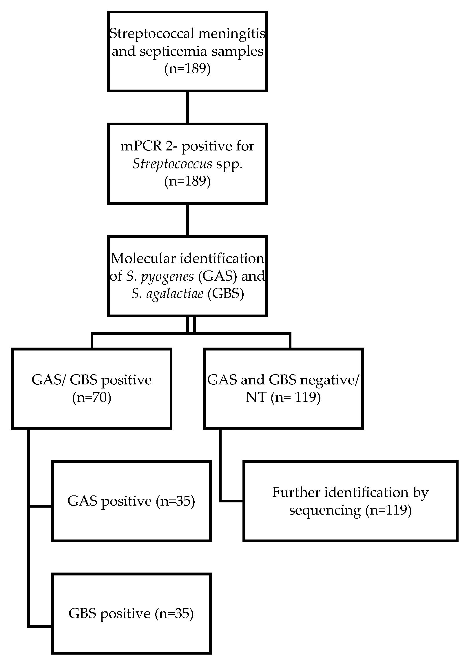
3.1. PCR Amplification

The amplification of the target genes formed products of 341 bp (GAS) and 259 bp (GBS), respectively (Figure 2). The GAS and GBS identification was positive for 70 of 189 samples (37.04%). Specifically, 35 of the 70 samples were identified as GAS and 35 of the samples were identified as GBS, respectively.
The PCR amplicons of the tuf gene PCR assay were 198 bp. Moreover, this technique provided positive results for most of the cases (116 (97.5%) out of 119 clinical samples) (Figure 3).

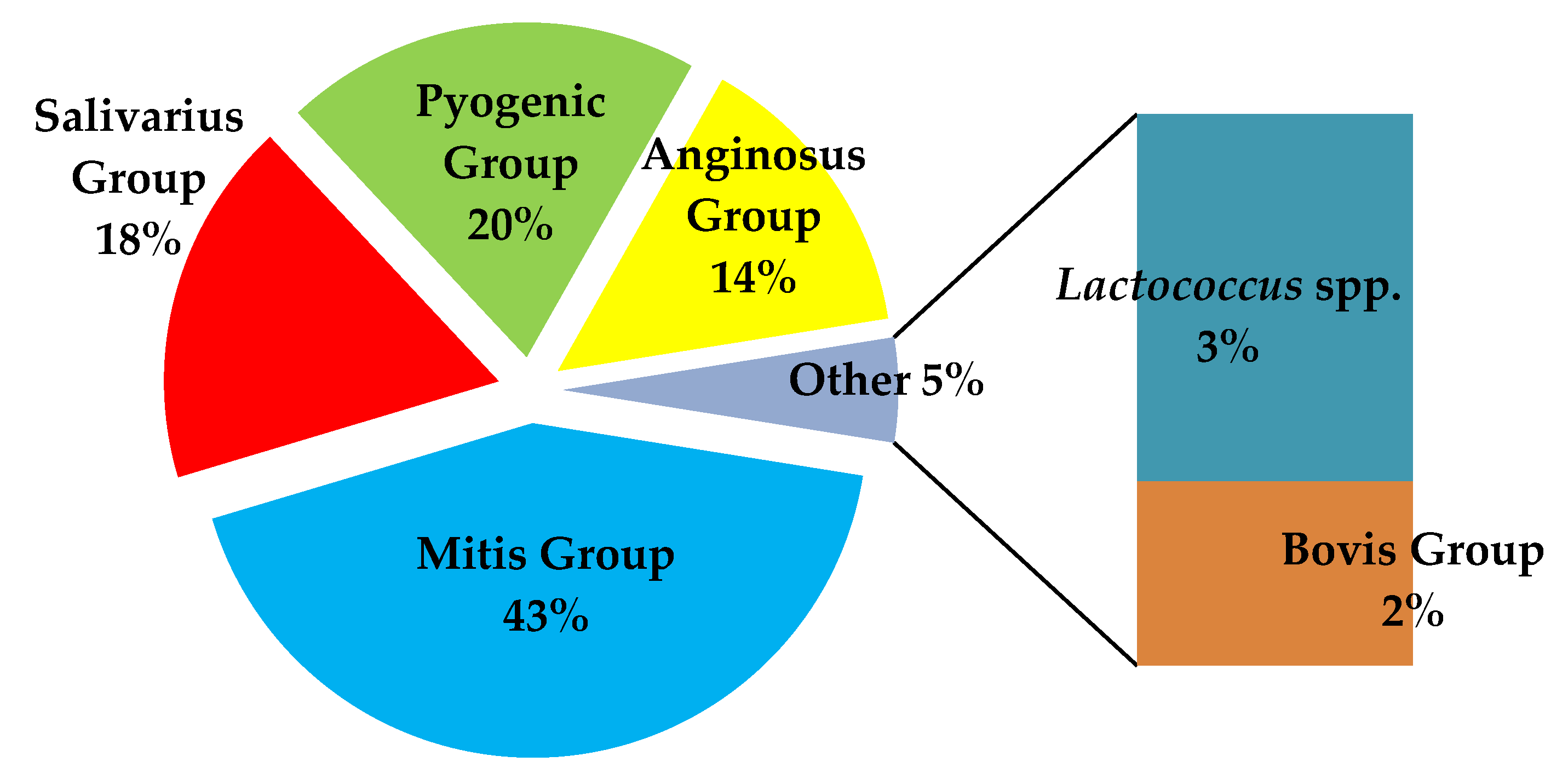
3.2. Sequencing Results

Sequencing analysis revealed that the majority of the genus Streptococcus spp. belonged to the Viridans Streptococci Group (VS), Bovis, Pyogenic Group and Lactococcus spp. According to the sequencing results, the most prevalent group was the mitis group (43%; 51/119). Among this group, S. mitis was successfully identified in 30 samples (59%; 30/51), followed by S. oralis in 21 samples (41%; 21/51). Furthermore, among the 17 streptococci belonging to the Anginosus group, S. anginosus and S. intermidius were identified in seven samples (41%; 7/17) each, respectively, while S. constellatus was identified in three samples (3/17; 18%).
Among the pyogenic group, S. dysgalactiae was identified in all 24 samples (20.1%; 24/119). In addition, among the salivarius group, S. salivarius was identified in all 21 samples (18%; 21/119). Finally, among the two positive samples belonging to Bovis Group, S. bovis was identified. Further, Lactococcus spp. was identified in all 3 samples (3%) (Figure 4).
Further analysis of the sequencing results over time revealed that the mitis group was predominant throughout the target years, followed by the salivarius group and the pyogenic group. Specifically, an increase in the mitis group, although observed during the pre-pandemic period (2017–2018), with the exception of the COVID-19 period (2020–2021), was also observed during the post-COVID-19 era (2022–2024), almost reaching pre-pandemic levels (Figure 5).
Regarding the number of meningitis and/or septicemia cases caused by the pyogenic group and, in particular, S. dysgalactiae, an increase was observed in 2022–2023, while a decrease was observed in 2024 (Figure 6).

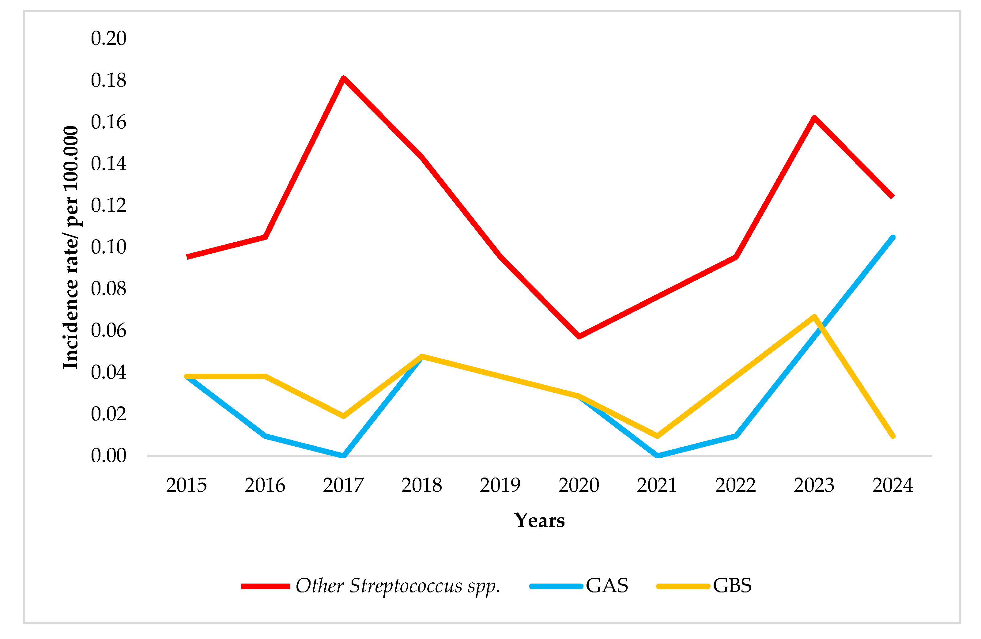
3.3. Contribution to Laboratory Surveillance

In general, the annual average incidence rate (IR) for meningitis and septicemia due to S. pyogenes (GAS) was 0.03/100,000. However, the incidence rate varied throughout the study period. Specifically, the pre-pandemic period was observed in 2018 (0.04/100,000). However, although during the pandemic period there were no GAS cases, a gradual increase was observed during the post-pandemic period (IRs of 0.02, 0.06 and 0.09 per 100,000 for 2022, 2023 and 2024, respectively) (Figure 5).
On the other hand, meningitis and septicemia caused by S. agalactiae (GBS) presented a different pattern in the annual incidence rate. Specifically, the incidence rate seemed to adhere to an average rate of 0.03/100,000 throughout the study years, while a decrease attributed to COVID-19 pandemic measures was observed in 2021. Although a peak was observed during 2021–2023 with an average rate of 0.02/100,000 population, the increasing trend was not observed for 2024 (Figure 5).
With regard to the remaining Streptococcus spp., identified neither as GAS nor GBS, an increase in the incidence rate was observed in 2017 (0.18/100,000) while a dramatic reduction was observed during the following years (2018–2020, average IR 0.11/100,000). However, an increase in the incidence rate was observed during the post-COVID-19 period (2021–2023)—with an average rate of 0.16/100,000—although a slight decline (0.12/100,000) was observed during 2024 (Figure 5).

3.4. Age Distribution

With regard to age, for most of the meningitis and septicemia cases due to Streptococcus spp., the highly affected age group was 0–4 years old (31.8%; 60/189) followed by the age groups > 60 years old (24.8%; 46/189) and 5–14 years old (23.8%; 45/189). In general, Streptococcus spp. other than GAS and GBS were responsible for the highest percentage of meningitis/septicemia cases in this study (Figure 7).
Analysis of the identified streptococci in relation to age revealed that the most predominant species was the mitis group streptococci, especially in children aged 0–4 years old (33%; 17/51) and 5–14 years old, as well as in adults >60 years of age (30%; 9/30) (Figure 8).
Salivarius group streptococci were found mainly in adults over 60 years of age (43%; 13/30) (Figure 8).
The pyogenic group of Streptococcus spp. was found in children 0–4 (21%; 11/51) and 5–14 years old (12%; 6/51), as well as in adults over 60 years old (13%; 4/30) (Figure 8).
Furthermore, Lactococcus spp. were found in children aged 0–4 (two cases) and 5–14 years old (two cases), respectively (Figure 8).

4. Discussion

The identification of Streptococcus spp. is important as it provides a great and vital amount of information on their biology and pathogenicity. Although conventional techniques are commonly used for the identification of bacteria (e.g., cultures and biochemical tests), the development and application of molecular assays resolves important issues in laboratory practice, especially when it comes to the surveillance of microorganisms [25].
In the case of Streptococcus spp., identification by culture combined with biochemical assays is not always possible in cases such as mitis group Streptococci (the main species of which are Streptococcus pneumoniae, Streptococcus mitis and Streptococcus oralis), for which some biochemical characteristics are presented in more than one species. For instance, while S. pneumoniae colonies are usually mucous, several cases lack this characteristic. Furthermore, as optochin testing was one of the main characteristics tests for S. pneumoniae, recent studies have indicated that streptococci belonging to the S. mitis species also present the same sensitivity to optochin [20]. Hence, molecular identification methods have an advantage in terms of further identification over the conventional ones as they are more specific and sensitive and less time-consuming [26].
Specifically, the application of the combined assays used in the present study successfully identified the majority of the genus Streptococcus spp. belonging to the Viridans Streptococci Group (VS), the mitis, salivarius and anginosus groups, as well as the bovis and pyogenic groups [18]. Further, the tuf gene sequencing assay also identified Lactococcus spp., which is in agreement with previous studies [17].
With regard to age, mitis group streptococci (S. mitis/pseudopneumoniae, S. oralis) were identified in patients 0–4 years old and in adults > 50 years old, which is in agreement with clinical case studies indicating that meningitis/septicemia from Streptococcus mitis/pseudopneumoniae and Streptococcus oralis occur mainly in children, as well as in older adults, due to underlying diseases and weak immune systems [6,27,28,29]. S. salivarius is one of the first microorganisms to colonize the mucosa, in the very first days after birth. Although it is not pathogenic, it can cause infections, leading to sepsis and meningitis [30], as well as in cases associated with surgery; e.g., epidural anesthesia or myelography [30,31]. According to our results, a significant percentage of S. salivarius was found in patients of >50 years of age, which is in agreement with previous studies [31,32], as well as in children 0–4 years of age.
The anginosus group (SAG) is usually found in the normal mucosal flora, colonizing both the urinary tract and the female genitals [33,34]. However, they could be potentially pathogenic in aseptic environments, such as CSF, and may cause severe infections. The aforementioned streptococcal group was identified in a low number of cases (14%) in all age groups, which is in agreement with previous studies indicating that infections caused by S. anginosus do not follow any specific age pattern [35,36].
Streptococcus dysgalactiae is a member of the pyogenic group; infections are associated with skin infections, which cause high rates of bacteremia, resulting in meningitis mainly in children [37]. This is in agreement with our findings as Streptococcus dysgalactiae was identified in most of the samples (20%) taken from children 0–15 years old.
Lactococcus spp. was identified in four samples (3%; 4/119) from the age groups 0–2 years old and 5–14 years old, respectively. This is an interesting observation indicating that the transmission of Lactococcus spp. might occur during breastfeeding as—according to previous studies—it has been found to be present in breast milk, among other bacteria [38]. Naturally, bovines are the natural reservoirs of Lactococcus spp., which can infect humans through the consumption of contaminated raw milk or fermented dairy products. Upon ingestion, Lactococcus lactis may colonize the human gastrointestinal tract and may potentially cause infections by invading sterile body sites [39].
The proposed methodology has advantages over Matrix-Assisted Laser Desorption/Ionization Time of Flight (MALDI-TOF), as MALDITOF is highly dependent on both the quality and thoroughness of the database [40]. Furthermore, as a low bacterial load in clinical samples such as CSF or blood often occurs, this may require additional steps such as sample purification, which is complex and time-consuming [41]. Furthermore, using MALDI-TOF MS to identify closely related microorganisms could be difficult for genetically related species such as Streptococcus spp. with similar protein profiles [42]. In such cases, the development of molecular methods, such as the PCR sequencing-based method, provides more reliable results.
PCR sequencing-based methods are fast, reliable, cost-effective, and suitable for small laboratories for monitoring streptococcal infections such as meningitis and septicemia.

5. Conclusions

In conclusion, no previous studies have focused on the molecular identification of meningitis and septicemia/bacteremia caused by Streptococcus spp. in the context of epidemiological monitoring. The existing literature primarily consists of individual case reports, which lack further specific identification and comprehensive data for epidemiological analysis. This study, therefore, presents a significant advance in the field, providing essential molecular insights into the prevalence, identification, and impact on public health of Streptococcus spp. related to severe infections.
The development of a protocol that can be applied directly to clinical samples initially contributes to the reduction in the turnaround time required for identification, without relying on culture-based methods. This advantage ensures the production of reliable and reproductive results, while minimizing the risk of contamination as the sample undergoes minimal processing before the assay’s application. Moreover, the application of this protocol revealed new data concerning meningitis and septicemia/bacteremia due to Streptococcus spp. other than S. pneumoniae, GAS and GBS. Finally, the implementation of the present molecular technique has significantly enhanced the laboratory-based surveillance of streptococcal meningitis.

Author Contributions

Conceptualization, C.K. and A.X.; methodology, C.K., A.X. and G.T.; resources, G.T.; data curation, C.K., A.X., S.S., M.D., A.P. and G.T.; writing—original draft preparation, C.K.; writing—review and editing, C.K. and G.T.; visualization, C.K.; supervision, A.X. and G.T.; project administration, C.K. and G.T. All authors have read and agreed to the published version of the manuscript.

Funding

This research received no external funding.

Institutional Review Board Statement

Ethical review and approval were waived in this study as the procedures being performed were part of the routine laboratory diagnosis and surveillance of bacterial meningitis. The study was conducted in accordance with the 1964 Helsinki Declaration and its later amendments or comparable ethical standards.

Informed Consent Statement

Patient consent was waived as depersonalized data from laboratory findings were used.

Data Availability Statement

The original contributions presented in this study are included in the article. Further inquiries can be directed to the corresponding author.

Conflicts of Interest

The authors declare no conflict of interest.

References

  1. Ku, L.C.; Boggess, K.A.; Cohen-Wolkowiez, M. Bacterial meningitis in infants. Clin. Perinatol. 2015, 42, 29–45. [Google Scholar] [CrossRef]
  2. McGill, F.; Heyderman, R.S.; Panagiotou, S.; Tunkel, A.R.; Solomon, T. Acute bacterial meningitis in adults. Lancet 2016, 388, 3036–3047. [Google Scholar] [CrossRef]
  3. Brouwer, M.C.; van de Beek, D. Epidemiology of community-acquired bacterial meningitis. Curr. Opin. Infect. Dis. 2018, 31, 78. [Google Scholar] [CrossRef]
  4. Davis, L.E. Acute Bacterial Meningitis. Contin. Lifelong Learn. Neurol. 2018, 24, 1264–1283. [Google Scholar] [CrossRef] [PubMed]
  5. Mańdziuk, J.; Kuchar, E.P. Streptococcal Meningitis. In StatPearls; StatPearls Publishing: Treasure Island, FL, USA, 2023. Available online: https://www.ncbi.nlm.nih.gov/books/NBK554448/ (accessed on 20 December 2024).
  6. Fukayama, H.; Shoji, K.; Yoshida, M.; Iijima, H.; Maekawa, T.; Ishiguro, A.; Miyairi, I. Bacterial meningitis due to the Streptococcus mitis group in children with cerebrospinal fluid leak. IDCases 2022, 27, e01406. [Google Scholar] [CrossRef] [PubMed]
  7. Spellerberg, B.; Brandt, C. Streptococcus. In Manual of Clinical Microbiology; John Wiley & Sons, Ltd.: Hoboken, NJ, USA, 2015; pp. 383–402. [Google Scholar] [CrossRef]
  8. Centers for Disease Control and Prevention. Bacterial Meningitis. 2023. Available online: https://www.cdc.gov/meningitis/about/bacterial-meningitis.html?CDC_AAref_Val=https://www.cdc.gov/meningitis/bacterial.html (accessed on 20 December 2024).
  9. Chandnani, H.K.; Jain, R.; Patamasucon, P. Group C Streptococcus Causing Rheumatic Heart Disease in a Child. J. Emerg. Med. 2015, 49, 12–14. [Google Scholar] [CrossRef]
  10. Ghazanfar, H.; Qureshi, Z.; Kalangi, H.; Ata, S.; Jyala, A.; Perez, E.A. Recurrent group G Streptococcus bacteremia: A case report and literature review. Clin. Case Rep. 2022, 10, e6162. [Google Scholar] [CrossRef] [PubMed]
  11. Malke, H. Genetics and Pathogenicity Factors of Group C and G Streptococci. Microbiol. Spectr. 2019, 7, 10–1128. [Google Scholar] [CrossRef]
  12. Lancefield, R.C. A serological differentiation of human and other groups of hemolytic streptococci. J. Exp. Med. 1933, 57, 571–595. [Google Scholar] [CrossRef]
  13. Sherman, J.M. The Streptococci. Bacteriol. Rev. 1937, 1, 3–97. [Google Scholar] [CrossRef]
  14. Facklam, R.F.; Martin, D.R.; Marguerite, L.; Dwight, R.J.; Efstratiou, A.; Thompson, T.A.; Gowan, S.; Kriz, P.; Tyrrell, G.J.; Kaplan, E.; et al. Extension of the Lancefield Classification for Group A Streptococci by Addition of 22 New M Protein Gene Sequence Types from Clinical Isolates: emm103 to emm124. Clin. Infect. Dis. 2002, 34, 28–38. [Google Scholar] [CrossRef] [PubMed]
  15. Kilpper-Bälz, R.; Schleifer, K.H. Nucleic acid hybridization and cell wall composition studies of pyogenic streptococci. FEMS Microbiol. Lett. 1984, 24, 355–364. [Google Scholar] [CrossRef]
  16. Ludwig, W.; Seewaldt, E.; Kilpper-Balz, R.; Heinz, K.; Magrum, L.; Woese, C.R.; Fox, G.E.; Stackebrandt, E. The Phylogenetic Position of Streptococcus and Enterococcus. Microbiology 1985, 131, 543–551. [Google Scholar] [CrossRef] [PubMed]
  17. Picard, F.J.; Ke, D.; Boudreau, D.K.; Boissinot, M.; Huletsky, A.; Richard, D.; Ouellette, M.; Roy, P.H.; Bergeron, M.G. Use of tuf Sequences for Genus-Specific PCR Detection and Phylogenetic Analysis of 28 Streptococcal Species. J. Clin. Microbiol. 2004, 42, 3686–3695. [Google Scholar] [CrossRef]
  18. Toit Mdu Huch, M.; Cho, G.-S.; Franz, C.M.A.P. The genus Streptococcus. In Lactic Acid Bacteria; John Wiley & Sons, Ltd.: Hoboken, NJ, USA, 2014; pp. 457–505. [Google Scholar] [CrossRef]
  19. Varghese, R.; Jayaraman, R.; Veeraraghavan, B. Current challenges in the accurate identification of Streptococcus pneumoniae and its serogroups/serotypes in the vaccine era. J. Microbiol. Methods 2017, 141, 48–54. [Google Scholar] [CrossRef]
  20. Sadowy, E.; Hryniewicz, W. Identification of Streptococcus pneumoniae and other Mitis streptococci: Importance of molecular methods. Eur. J. Clin. Microbiol. Infect. Dis. 2020, 39, 2247–2256. [Google Scholar] [CrossRef]
  21. Shahi, S.; Zununi Vahed, S.; Fathi, N.; Sharifi, S. Polymerase chain reaction (PCR)-based methods: Promising molecular tools in dentistry. Int. J. Biol. Macromol. 2018, 117, 983–992. [Google Scholar] [CrossRef]
  22. Xirogianni, A.; Tzanakaki, G.; Karagianni, E.; Markoulatos, P.; Kourea-Kremastinou, J. Development of a single-tube polymerase chain reaction assay for the simultaneous detection of Haemophilus influenzae, Pseudomonas aeruginosa, Staphylococcus aureus, and Streptococcus spp. directly in clinical samples. Diagn. Microbiol. Infect. Dis. 2009, 63, 121–126. [Google Scholar] [CrossRef]
  23. Liu, D.; Hollingshead, S.; Swiatlo, E.; Lawrence, M.L.; Austin, F.W. Rapid identification of Streptococcus pyogenes with PCR primers from a putative transcriptional regulator gene. Res. Microbiol. 2005, 156, 564–567. [Google Scholar] [CrossRef]
  24. Kong, F.; Ma, L.; Gilbert, G.L. Simultaneous detection and serotype identification of Streptococcus agalactiae using multiplex PCR and reverse line blot hybridization. J. Med. Microbiol. 2005, 54, 1133–1138. [Google Scholar] [CrossRef]
  25. Moore, M.S.; McCann, C.D.; Jordan, J.A. Molecular Detection of Culture-Confirmed Bacterial Bloodstream Infections with Limited Enrichment Time. J. Clin. Microbiol. 2020, 51, 3720–3725. [Google Scholar] [CrossRef]
  26. Váradi, L.; Luo, J.L.; Hibbs, D.E.; Perry, J.D.; Anderson, R.J.; Orenga, S.; Groundwater, P.W. Methods for the detection and identification of pathogenic bacteria: Past, present, and future. Chem. Soc. Rev. 2017, 46, 4818–4832. [Google Scholar] [CrossRef]
  27. Colomba, C.; Garbo, V.; Boncori, G.; Albano, C.; Bagarello, S.; Condemi, A.; Giordano, S.; Canduscio, L.A.; Gallo, C.; Parrinello, G.; et al. Streptococcus mitis as a New Emerging Pathogen in Pediatric Age: Case Report and Systematic Review. Antibiotics 2023, 12, 1222. [Google Scholar] [CrossRef] [PubMed]
  28. Poi, B.N.; Pasupulety Venkata, N.K.; Auckland, C.R.; Paul, S.P. Neonatal meningitis and maternal sepsis caused by Streptococcus oralis. J. Neonatal-Perinat. Med. 2018, 11, 331–334. [Google Scholar] [CrossRef] [PubMed]
  29. Wydall, S.; Durrant, F.; Scott, J.; Cheesman, K. Streptococcus oralis endocarditis leading to central nervous system infection in pregnancy. Anaesth. Rep. 2021, 9, e12133. [Google Scholar] [CrossRef] [PubMed]
  30. Delorme, C.; Abraham, A.-L.; Renault, P.; Guédon, E. Genomics of Streptococcus salivarius, a major human commensal. Infect. Genet. Evol. 2015, 33, 381–392. [Google Scholar] [CrossRef]
  31. Yanagida, M.; Hosoi, Y.; Kawano, T.; Otake, Y.; Yamanaka, Y.; Baba, T.; Ito, M. Noniatrogenic Meningitis Caused by Streptococcus salivarius Associated with Early Esophageal Cancer and Early Gastric Cancer. Intern. Med. Tokyo Jpn. 2024, 63, 457–460. [Google Scholar] [CrossRef]
  32. Jovanovic, U.; Freyer, M.; Heckmann, J.G. Streptococcus salivarius meningitis: A spontaneous case in a 74-year-old man. Acta Neurol. Belg. 2019, 119, 481–482. [Google Scholar] [CrossRef]
  33. Al Majid, F.; Aldrees, A.; Barry, M.; Binkhamis, K.; Allam, A.; Almohaya, A. Streptococcus anginosus group infections: Management and outcome at a tertiary care hospital. J. Infect. Public Health 2020, 13, 1749–1754. [Google Scholar] [CrossRef]
  34. Pilarczyk-Zurek, M.; Sitkiewicz, I.; Koziel, J. The Clinical View on Streptococcus anginosus Group—Opportunistic Pathogens Coming Out of Hiding. Front. Microbiol. 2022, 37, 210–217. [Google Scholar] [CrossRef]
  35. Băncescu, G.; Băncescu, A.; Constantinescu, M.V. Streptococcus anginosus group—Brief characterization and its contribution to the brain abscess pathogenesis. Stomatol. EDU J. 2015, 2, 153–161. [Google Scholar] [CrossRef]
  36. Madathil, S.; Matsumoto, S.; Mathews, K.D.; Glykys, J. Central Nervous System Infections Due to Streptococcus anginosus Group: A Single-Center Case Series. J. Child Neurol. 2022, 37, 210–217. [Google Scholar] [CrossRef] [PubMed]
  37. Baracco, G.J. Infections Caused by Group C and G Streptococcus (Streptococcus dysgalactiae subsp. equisimilis and Others): Epidemiological and Clinical Aspects. Microbiol. Spectr. 2019, 7, 10–1128. [Google Scholar] [CrossRef]
  38. Chathyushya, K.B.; Prakash, M.S.; Kodandapani, Y.Y.; Bheema, B.; Babu, J.J.G.; Rajkumar, R. Studies on the isolation and molecular characterization of Lactobacillus spp. from human breast milk and assessment of their probiotic potential. Environ. Exp. Biol. 2021, 19, 209–218. [Google Scholar] [CrossRef]
  39. Uchida, Y.; Morita, H.; Adachi, S.; Asano, T.; Taga, T.; Kondo, N. Bacterial meningitis and septicemia of neonate due to Lactococcus lactis. Pediatr. Int. 2011, 53, 119–120. [Google Scholar] [CrossRef] [PubMed]
  40. Alizadeh, M.; Yousefi, L.; Pakdel, F.; Ghotaslou, R.; Rezaee, M.A.; Khodadadi, E.; Oskouei, M.A.; Soroush Barhaghi, M.H.; Kafil, H.S. MALDI-TOF Mass Spectroscopy Applications in Clinical Microbiology. Adv. Pharmacol. Pharm Sci. 2021, 2021, 9928238. [Google Scholar] [CrossRef]
  41. Haider, A.; Ringer, M.; Kotroczó, Z.; Mohácsi-Farkas, C.; Kocsis, T. The Current Level of MALDI-TOF MS Applications in the Detection of Microorganisms: A Short Review of Benefits and Limitations. Microbiol. Res. 2023, 14, 80–90. [Google Scholar] [CrossRef]
  42. Pérez-Sancho, M.; Vela, A.I.; García-Seco, T.; Gottschalk, M.; Domínguez, L.; Fernández-Garayzábal, J.F. Assessment of MALDI-TOF MS as Alternative Tool for Streptococcus suis Identification. Front. Public Health 2015, 3, 202. [Google Scholar] [CrossRef]
Figure 1. Flowchart for sample identification included in the study.
Diagnostics 15 01632 g001
Figure 2. GAS and GBS molecular identification by PCR assay of lanes: (1) 100 bp ladder; (2,3) positive controls (5 ng of each DNA)—GAS (MW: 341 bp) and GBS (MW: 259 bp); (4,6–8) clinical samples; and (5,9) negative controls.
Diagnostics 15 01632 g002
Figure 3. Tuf gene amplification assay. Lanes: (1) 100 bp ladder; (2) positive control (MW: 198 bp); (8) negative control; and (3–7,9–14) clinical samples.
Diagnostics 15 01632 g003
Figure 4. Sequencing analysis of tuf gene directly in clinical samples.
Diagnostics 15 01632 g004
Figure 5. Incidence rate of streptococcal meningitis/septicemia in Greece (2015–2024).
Diagnostics 15 01632 g005
Figure 6. Annual cases of meningitis and septicemia from other streptococcal species (2015–2024).
Diagnostics 15 01632 g006
Figure 7. Age distribution of meningitis and septicemia due to streptococci in Greece (2015–2024).
Diagnostics 15 01632 g007
Figure 8. Age distribution in relation to Streptococcus spp. identification.
Diagnostics 15 01632 g008
Table 1. Oligonucleotide sequencing and PCR primers used for amplification of Spy1258 (GAS), cfb (GBS) and tuf (other Streptococcus spp.) genes.
PrimersEncoding GeneSequence 5′ → 3′ProductReference
SpyF
SpyR
spy1258ACTCTGGATGATTTGTACCG
TCAGTGGTTTCTTGATAGCC
314 bp[23]
CFBS
CFBA
cfbATGATGTATCTATCTGGAACTCT
CGCAAT GAAGTCTTTAATTTTTC
259 bp[24]
Str1
Str2
tufGTACAGTTGCTCAGGACGTATC
ACGTTCGATTTCATCACGTTG
198 bp[17]
Disclaimer/Publisher’s Note: The statements, opinions and data contained in all publications are solely those of the individual author(s) and contributor(s) and not of MDPI and/or the editor(s). MDPI and/or the editor(s) disclaim responsibility for any injury to people or property resulting from any ideas, methods, instructions or products referred to in the content.

