Article

Risk Factors Influencing Right and Left Ventricular Variables Assessed with Gated Cadmium–Zinc–Telluride Equilibrium Radionuclide Angiocardiography in Oncology Patients

by
Olav Monsson
1,2,3,*,
Marc Nielsen
1,
Thomas Kümler
4,5,6,
Christian Haarmark
1,6 and
Bo Zerahn
1
1
Department of Nuclear Medicine, Herlev og Gentofte Hospital, 2730 Herlev, Denmark
2
Department of Radiology, Odense University Hospital, 5000 Odense, Denmark
3
Research Unit of Radiology, Department of Clinical Research, University of Southern Denmark, 5000 Odense, Denmark
4
Complications Research, Steno Diabetes Center Copenhagen, 2730 Herlev, Denmark
5
Department of Cardiology, Herlev-Gentofte Hospital, 2730 Herlev, Denmark
6
Department of Clinical Medicine, Copenhagen University, 2200 Copenhagen, Denmark
*
Author to whom correspondence should be addressed.
Diagnostics 2025, 15(10), 1274; https://doi.org/10.3390/diagnostics15101274 (registering DOI)
Submission received: 10 April 2025 / Revised: 9 May 2025 / Accepted: 12 May 2025 / Published: 17 May 2025
(This article belongs to the Section Medical Imaging and Theranostics)

Abstract

:
Background: Left ventricular ejection fraction remains the primary focus in cardiac monitoring for oncology patients undergoing potentially cardiotoxic chemotherapy, while right ventricular function is seldom examined. This study evaluates how established risk factors for left ventricular dysfunction affect right ventricular function. Methods: This retrospective cohort study included 1770 patients undergoing cadmium–zinc–telluride equilibrium radionuclide angiocardiography before chemotherapy. Patients were categorized based on risk factors for left ventricular dysfunction—diabetes (DM), atrial fibrillation (AF), coronary heart disease (CHD), and previous oncological therapy—and compared to controls using independent t-tests. Results: Patients with previous oncological therapy exhibited a significantly lower right ventricular end-diastolic volume (RVEDV) (mean difference: −4.4 mL/m2, 95% CI: −6.1 to −2.7, p < 0.001), lower right ventricular end-systolic volume (RVESV) (−2.3 mL/m2, 95% CI: −3.4 to −1.2, p < 0.001), and lower right ventricular stroke volume (RVSV) (−2.1 mL/m2, 95% CI: −3 to −1.2, p < 0.001). In patients with CHD, there was a higher right ventricular ejection fraction (RVEF) (3.0 mL/m2, 95% CI: 0.8 to 5.2, p < 0.01), whereas patients with DM had lower RVEDV (−5.1 mL/m2, 95% CI: −9.2 to −1, p < 0.05) and RVESV (−3.0 mL/m2, 95% CI: −5.5 to −0.4, p < 0.05). No ventricular variables differed from the control group among patients with AF. Conclusions: Risk factors known to affect the left ventricle also impacted the right ventricle, with the exception of AF.

1. Introduction

Cardiotoxicity is a significant concern in the administration of antineoplastic drugs and underscores the complexity of balancing effective cancer treatment against the potential for adverse cardiac events [1,2,3]. According to guidelines [2], routine assessment is primarily centered on left ventricular function assessed by left ventricular ejection fraction (LVEF) along with other left ventricular variables such as diastolic function and strain, whereas the right ventricular function is somewhat overlooked in part due to the lack of available, practical, and standardized assessment methods, but also to an extent limited recognition of its clinical importance in many pathological conditions [4]. Nonetheless, growing evidence highlights the right ventricle’s importance in cardiac efficiency and hemodynamic stability, leading to increased focus on its function [5,6].
The most widely used technology to assess cardiac function is two-dimensional transthoracic echocardiography (2D-TTE) recommended in guidelines [2]. Due to its anatomy, assessment of the right ventricle using 2D-TTE has proven difficult [4]. Three-dimensional transthoracic echocardiography (3D-TTE) offers enhanced accuracy and detail along with diastolic function and strain. Despite its advancements, 3D-TTE remains challenging for accurate volumetric assessment of the right ventricle [7].
Emerging insights into cardiac physiology suggest that the right ventricle plays an equally vital role in the pathophysiology of various cardiac and systemic conditions, including coronary heart disease (CHD), diabetes (DM), atrial fibrillation (AF), and the aftermath of potential cardiotoxic oncological interventions [8,9,10,11,12]. This necessitates a broader understanding of cardiac health that encompasses both ventricles. Moreover, the advent of advanced imaging techniques, such as gated cadmium–zinc–telluride single-photon emission computed tomography equilibrium radionuclide angiocardiography (CZT-SPECT-ERNA), has provided new avenues for detailed cardiac assessment, enabling a more nuanced evaluation of ventricular function beyond conventional measures.
Equilibrium radionuclide angiography (ERNA) is a widely recognized and non-invasive imaging [13]. Notably, CZT-SPECT-ERNA stands out due to its practicality, operator independence, and high reproducibility [14,15,16]. Recent research has further optimized this technique by reducing the radiotracer dose while maintaining diagnostic value [17].
We hypothesize that while the right and left ventricles are interconnected but the extent to which common risk factors such as CHD, DM, AF, and previous oncological therapy affect right ventricular function remains insufficiently characterized. This study aims to quantify these effects and determine whether they differ from established left ventricular changes. By performing so, it seeks to elucidate the broader implications of these risk factors on cardiac function, with a particular emphasis on the right ventricle.
As a secondary objective, this study aims to assess whether the presence of these left ventricular risk factors significantly alters left ventricular function. By quantifying these differences, we seek to justify the exclusion of patients with these comorbidities in previously published normative data [18,19].

2. Materials and Methods

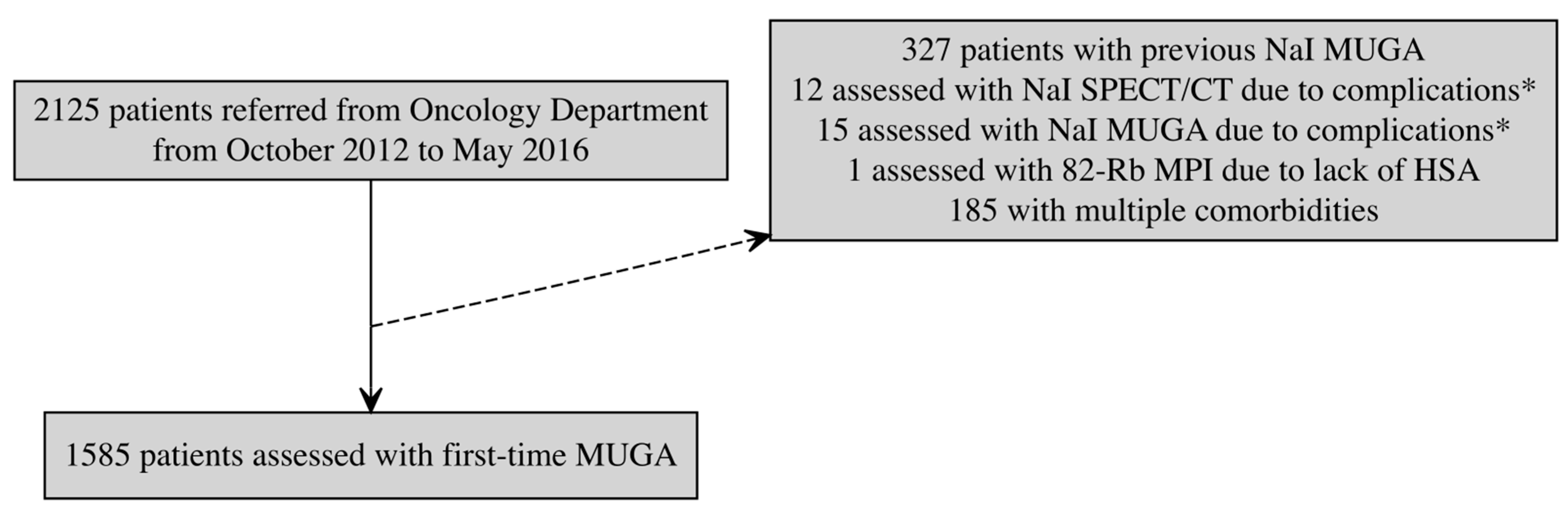
2.1. Study Design, Population, and Data Collection

This is a retrospective cohort study of patients referred from a tertiary oncological department at Herlev-Gentofte University Hospital to undergo gated CZT-SPECT-ERNA for baseline evaluation before initiating antineoplastic treatments with potential cardiotoxicity. The patient population is described in the publications of Haarmark et al. and Hansen et al. [18,19], and was originally collected between October 2012 and 30 September 2014.
Patients with CHD, DM, AF, and previous oncological therapy comprised four different test groups, while the control group had none of these conditions. Patients were excluded if they had pleural effusions, prior lung resection, or more than one of the risk factors (CHD, DM, AF or previous oncological treatment) in order to ensure more homogeneous comparison groups.
Screening was conducted by manual reading of the patient’s electronic hospital records (EPJ) and the Shared Medication Record (FMK). CHD was defined as a history of myocardial infarction, coronary artery bypass grafting, or coronary revascularization. DM was defined as current or previous elevation of hemoglobin A1c > 48 mmol/mol (HbA1c) or receiving antidiabetic treatment. AF was defined as having a medical history of prior or current atrial fibrillation. Previous oncological therapy was defined as any history of treatment with any chemotherapeutic agents and/or radiation therapy to the thoracic region.
Left and right ventricular ejection fractions (LVEF, RVEF), end-systolic volumes (LVESV, RVESV), end-diastolic volumes (LVEDV, RVEDV), and stroke volumes (LVSV, RVSV) were assessed using gated CZT-SPECT-ERNA with 99mTc-labeled human serum albumin (HSA) as the radiotracer.
To enhance statistical power, the inclusion period was extended to May 2016. Hence, patients were included if the first CZT-SPECT-ERNA scan was performed between October 2012 and May 2016, and excluded if they were assessed with alternative methods (see Figure 1).

2.2. Ethical Considerations

The study was approved by the Danish data protection Agency (R-24081714).
The corresponding author had full access to all the data in this study and takes responsibility for its integrity and the accuracy of the analysis.

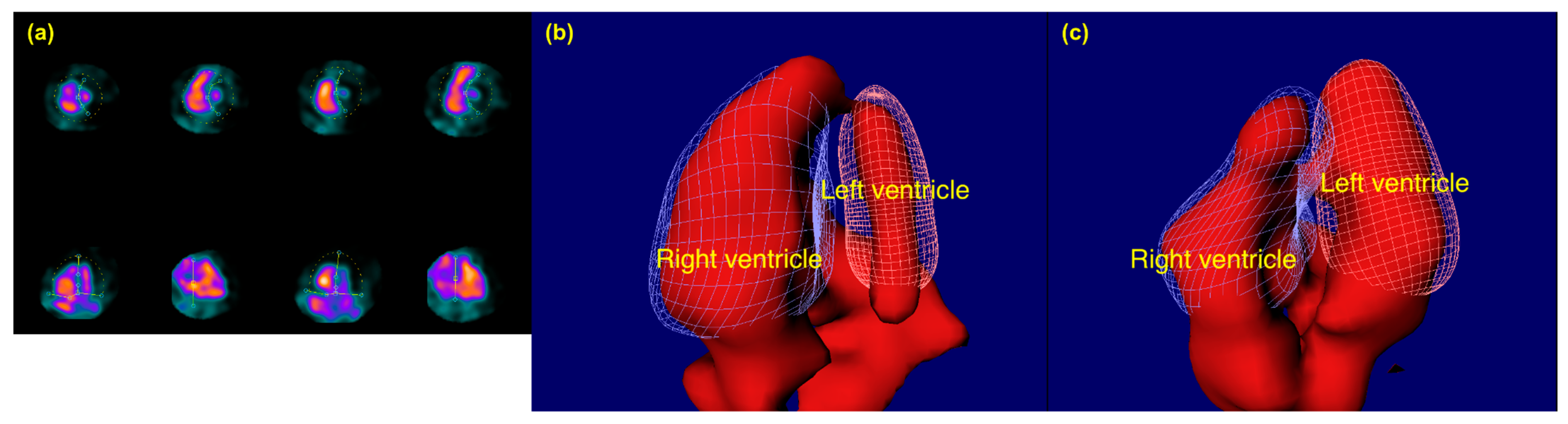
2.3. Gated CZT-SPECT-ERNA Procedure

Equilibrium radionuclide angiocardiography (ERNA) was performed using a cadmium zinc telluride (CZT) gamma camera system, GE Discovery 530c (GE Healthcare, Milwaukee, WI, USA). A dose of 550 MBq 99mTc-labeled HSA was administered intravenously. Patients were positioned supine with both arms extended above their heads. Gated data acquisition was obtained with 16 image frames (bins) per cardiac cycle, requesting 600 accepted beats, and a 20% energy window centered on 140 keV. Subsequently, the acquired data were processed and analyzed using Xeleris 3 Imaging workstation reorientation software (GE Healthcare, Milwaukee, WI, USA, version no. 3.0562) and Cedars-Sinai QBS processing software (Cedars-Sinai, Los Angeles, CA, USA, revision 2009.0) to generate spline-based time-activity curves for the ventricles, which were then used to calculate end-diastolic and -systolic volumes for both right and left ventricle. Figure 2 illustrates representative end-diastolic frames from patients with normal and dilated RVs.
Each acquisition was analyzed twice by two experienced technologists. If LVEF differed more than two percentage points between the two analyses, repeated processing was performed until sufficient agreement was obtained. In cases where RVEF differed by more than ten percentage points between two replicate analyses, a third analysis was performed, and the two most concordant values were used for reporting.

2.4. General Considerations

Right ventricular volumes are dependent on body surface [20] and were therefore corrected for this using Mostellar corrections.

2.5. Statistical Analysis

For continuous variables, independent t-tests were employed to assess statistical differences between the groups. For dichotomized variables, Chi-square tests were used.
Ad hoc univariate linear regression analyses were performed to identify candidate predictors of right ventricular volumes. Variables with p < 0.25 were considered for inclusion in multivariate models. In addition, CHD, AF, diabetes, and previous oncological therapy were included in all models regardless of univariate significance, due to their clinical relevance. Model performance was compared using adjusted R-squared values and ANOVA.
Adjusted β coefficients and 95% confidence intervals were visualized using forest plots. To further illustrate the independent effect of selected predictors (diabetes and previous oncological therapy), we generated adjusted effect plots based on model-derived predicted values, using the ggeffects package in R.
Extreme outliers were identified and excluded using the 3 × IQR rule.
All analyses were performed using RStudio 2022.07.1 + 554 “Spotted Wakerobin” Release and R version 4.3.1, and a p-value < 0.05 was considered statistically significant.

3. Results

The patient flow chart is presented in Figure 1. The study population detailed in Table 1, consists of 1770 patients of whom 811 were categorized as the control group and the remainder as having one of the risk factors.
Comparisons of the control group with AF, CHD, diabetes, and previous oncological therapy are presented in Table 2 and Figure 3.
Patients with previous oncological therapy had a lower LVEDV (p = 0.0051), leading to a lower LVEF (p < 0.001) and LVSV (p < 0.001). The same can be seen for RVEDV (p < 0.001). However, unlike the left ventricle, RVESV was also reduced (p < 0.001), leading to an unchanged RVEF.
For patients with DM, a statistically significant lower LVEDV (p = 0.027) and LVESV (p = 0.012) was noted; however, the lower LVESV was the most pronounced, leading to a higher LVEF (p = 0.015). Similar differences were seen in the right ventricle, except for an unchanged RVEF. RVEDV was lower in DM patients (p = 0.015) along with RVESV (p = 0.024).
For patients with CHD, the RVEF was significantly higher (p = 0.0082), despite RVESV and RVEDV remaining statistically unchanged. Differences can be seen in the left ventricle through a lower LVEF (p < 0.001), attributed to a higher value of LVESV (p < 0.001) compared to LEDV (p < 0.001).
In the AF group, no significant differences from the control group were observed for either left or right ventricular values.
To assess the contribution of CHD, AF, DM, and previous oncological therapy, we compared a core regression model including age, blood pressure, heart rate, gender, hypertension, hypercholesterolemia, and smoking with a full model that also included the above clinical variables. For Right EDV, the addition of CHD, AF, diabetes, and previous oncological therapy led to a statistically significant improvement in model fit (adjusted R2 = 0.198 vs. 0.194, p = 0.013). For Right ESV, the full model showed only a minimal and statistically non-significant increase in explanatory power (adjusted R2 = 0.207 vs. 0.206, p = 0.233) (see Supplementary Table S1).
After adjusting for age, systolic blood pressure, diastolic blood pressure, heart rate, gender, and other cardiovascular risk factors, patients with diabetes had a predicted RVEDV of 60.9 mL/m2 (95% CI: 57.4 to 64.5), compared to 65.7 mL/m2 (95% CI: 64.3 to 67.0) in patients without diabetes. Similarly, patients with previous oncological therapy had a predicted RVESV of 33.9 mL/m2 (95% CI: 32.9 to 34.8), compared to 34.9 mL/m2 (95% CI: 34.0 to 35.8) in those without previous oncological therapy (Figure 4).
Results from the corresponding univariate analyses are presented in Supplementary Table S2.

4. Discussion

This study evaluates how different comorbidities affecting the left ventricle—CHD, AF, DM, and previous oncological therapy—influence the same cardiac variables for the right ventricle, utilizing gated CZT-SPECT-ERNA.
Patients with previous oncological therapy exhibited lower RVEDV, RVESV, and RVSV. A systematic review by Kariyanna et al. [21] reported a reduced RVEF in the follow-up of breast cancer patients receiving anthracycline and/or trastuzumab. A study by Grover et al. [22] using MRI in the follow-up of anthracycline and/or trastuzumab treatment, reported a reduction in RVEF, though an increase in right ventricular ESV and EDV, indexed by body surface area, at 12 month follow-up. The discrepancy from this study could be the result of differences in imaging techniques or lack of discrimination between the different oncological treatments in regard to potential cardiotoxic effects. Few studies have examined the volumetric impact on the right ventricle, although reductions in other right ventricular function parameters have been reported [23,24].
For patients with previous oncological therapy, our study found a lower left EDV and a related lower SV and LVEF. This agrees with the current literature [25,26]. The aforementioned MRI study by Grover et al. [22], showed a higher left ventricular EDV and ESV, and a lower LVEF.
The regression analysis demonstrated that gender, age, and heart rate were the most consistent contributors to variability in right ventricular volumes, in line with physiological expectations [27,28]. In addition, smoking was negatively associated with both EDV and ESV. Importantly, diabetes was independently associated with a significantly lower right ventricular EDV, and previous oncological therapy showed a modest but statistically significant association with lower EDV and a borderline effect on ESV. These findings from the multivariable analysis reinforce the primary hypothesis of the study—that specific clinical risk factors traditionally associated with left ventricular dysfunction may also affect right ventricular remodeling, albeit in different ways.
Noteworthy for this study is its cross-sectional approach, as opposed to the aforementioned follow-up studies. This also might be relevant for the other risk factors, as this study evaluates patients in a relatively stable disease state.
For patients with CHD, the observed lower LVEF due to a higher LVESV and LVEDV is consistent with the existing literature [29,30]. Ischemia-induced myocyte loss and fibrosis likely contribute to impaired contractility, increased ventricular stiffness, and dilatation, culminating in reduced LVEF.
However, the right ventricle does not display the same features. The right ventricle exhibits a higher EF, though no statistically significant differences in ESV or EDV. This might reflect the right ventricle’s compensatory capacity and resilience towards ischemia [31,32]. A study by van Wolferen et al. [33] of patients with idiopathic pulmonary arterial hypertension found that right ventricular dilatation and impaired left ventricular filling were associated with worse outcomes over a one-year period, although this population differs from ours. Considering the primary factor for pulmonary hypertension being left ventricular failure, the finding by van Wolferen et al. could further support our findings. This finding may indicate the right ventricle’s adaptive response to ischemia, potentially compensating for left ventricular dysfunction via increased contractility or improved coupling with pulmonary circulation.
In diabetic patients, lower RVEDV and RVESV, with preserved RVEF, align with findings from MRI studies [34]. These findings suggest a parallel diastolic dysfunction in the right ventricle, highlighting the systemic nature of diabetes-related cardiomyopathy. In patients with cardiomyopathy, gated CZT-SPECT-ERNA has been shown to be accurate and reproducible when measuring right ventricular function and is considered a good alternative to gold-standard cardiac assessment [35].
A lower left ventricular EDV and ESV, accompanied by a higher LVEF in diabetic patients in our study, are consistent with early diastolic dysfunction features seen in diabetic cardiomyopathy [36]. Diabetes is well-documented to affect myocardial compliance, with microvascular damage, interstitial fibrosis, and glycation of myocardial proteins contributing to stiffened ventricles and impaired filling [37,38].
While prior studies have demonstrated lower LVEF and higher LVESV in AF patients [39,40], this study’s findings suggest these differences may not be universally detectable, potentially influenced by differences in AF chronicity, ventricular remodeling, or underlying pathophysiology.
For patients with AF, the lack of significant differences in ventricular function compared to controls might reflect limitations in the sensitivity of CZT-SPECT-ERNA imaging in this subgroup. It is well known that AF is associated with dynamic changes in stroke volume and ventricular filling due to irregular ventricular response and loss of atrial contribution to cardiac output [41]. However, the variability in ventricular filling across cardiac cycles in AF may complicate accurate assessment, particularly in methods dependent on consistent temporal data acquisition like CZT-SPECT-ERNA. The wide confidence intervals observed further underscore the variability and potential measurement uncertainty in this population.
In addition to the primary risk factors examined, smoking was independently associated with reduced RVEDV and RVESV in our multivariate analysis. Although some studies in patients with advanced pulmonary disease or long-term smoking exposure have reported right ventricular dilation and dysfunction [10], our findings may reflect an earlier or subclinical pattern of RV remodeling. This could involve fibrosis, microvascular injury, or impaired myocardial compliance, leading to lower filling volumes without affecting ejection fraction. Supporting this interpretation, a study by Chahal et al. using cardiac MRI in the MESA cohort found that former smokers with greater emphysema had smaller RV end-diastolic volumes after adjusting for left ventricular volume, suggesting that smoking-related myocardial remodeling may present as reduced RV volumes rather than dilation in certain contexts [42].

Limitations

Several limitations of this study warrant consideration. First, being a retrospective, single-center study narrows down the generalizability to other contexts. Second, we chose not to correct for the large number of variables analyzed, due to the nature of the explorative study. As such, there is an evident risk for type I errors. Third, since this study also focuses on right ventricular values, it would have been interesting also to evaluate chronic obstructive pulmonary disease (COPD), pulmonary embolism (PE), and pulmonary hypertension (PH) as potential risk factors for reduced cardiac function. Fourth, potential confounding factors, such as medication use, disease duration, and comorbidities such as valvular disease, were not fully accounted for, which may influence the observed differences. As an example, there is no differentiation between anthracyclines and other types of chemotherapy. Further, there are differences in baseline characteristics (age, height, and weight) which obscures the interpretation of the ventricular volumes. Additionally, laboratory data on renal function (e.g., eGFR) were not available, although renal impairment is a recognized cardiovascular risk factor and potential confounder in volumetric cardiac assessments.
Although this study aims to isolate the effects of CHD, DM, AF, and oncological treatment on cardiac function, patients often have other comorbidities that may independently or synergistically influence outcomes. Additionally, variability in malignancies and metastatic disease introduces potential selection bias.

5. Conclusions

This study found that risk factors influencing the left ventricle could also influence the right ventricle. Notably, previous oncological therapy and DM appear to impact the right ventricle to a similar extent as the left.
Future studies should examine specific risk factors for right ventricular dysfunction to further investigate volumetric changes and their clinical significance, including potential applications for treatment monitoring and outcome prediction in cancer therapy and the impact of medical treatment and multimorbidity on right ventricular function.

Supplementary Materials

The following supporting information can be downloaded at: https://www.mdpi.com/article/10.3390/diagnostics15101274/s1, Supplementary Table S1: Multivariate linear regression analysis; Supplementary Table S2: Univariate linear regression analysis.

Author Contributions

Conceptualization, O.M., M.N., T.K., C.H. and B.Z.; methodology, O.M., M.N., T.K., C.H. and B.Z.; software, O.M. and M.N.; validation, O.M., M.N., T.K., C.H. and B.Z.; formal analysis, O.M., M.N. and B.Z.; investigation, C.H. and B.Z.; resources, C.H. and B.Z.; data curation, O.M. and B.Z.; writing—original draft preparation, O.M.; writing—review and editing, O.M., M.N., T.K., C.H. and B.Z.; visualization, O.M.; supervision, B.Z.; project administration, O.M., C.H. and B.Z.; funding acquisition, C.H. and B.Z. All authors have read and agreed to the published version of the manuscript.

Funding

No external funding was received for this study. The publication costs were covered by the Department of Nuclear Medicine, Herlev Hospital. No grant number applies.

Institutional Review Board Statement

The study was conducted in accordance with the Declaration of Helsinki, and approved by the Danish data protection Agency (R-24081714) on 18 December 2024.

Informed Consent Statement

Patient consent was waived by the relevant authority due to ethical considerations. The project involves patients, some of whom are deceased, and others who are currently in treatment and considered particularly vulnerable in relation to the study’s purpose. The regional journal data team (“Team for Journaldata”) assessed that obtaining consent from these individuals would be inappropriate. Therefore, access to patient data were granted without informed consent in accordance with applicable regulations.

Data Availability Statement

The data presented in this study are not publicly available due to Danish data protection regulations. Individual-level patient data were accessed through approval from Team for Journaldata, Region Hovedstaden (Project ID: R-24081714), under Section 46(2) of the Danish Health Act. Data are considered sensitive health information and cannot be shared publicly or anonymized for open release. Researchers who meet the criteria for access to confidential data can contact Team for Journaldata at journaldata.center-forsundhed@regionh.dk to request data access under similar regulatory approval.

Conflicts of Interest

The authors declare no conflicts of interest.

Abbreviations

The following abbreviations are used in this manuscript:
2D-TTETwo-Dimensional Transthoracic Echocardiography
3D-TTEThree-Dimensional Transthoracic Echocardiography
AFAtrial Fibrillation
CHDCoronary Heart Disease
COPDChronic Obstructive Pulmonary Disease
CZTCadmium–Zinc–Telluride
CZT-SPECT-ERNACadmium–Zinc–Telluride Single-Photon Emission Computed Tomography Equilibrium Radionuclide Angiocardiography
DMDiabetes Mellitus
EPJElektronisk Patientjournal (Electronic Patient Record)
ERNAEquilibrium Radionuclide Angiocardiography
FMKFælles Medicinkort (Shared Medication Record)
HbA1cHemoglobin A1c
HSAHuman Serum Albumin
LVEDVLeft Ventricular End-Diastolic Volume
LVEFLeft Ventricular Ejection Fraction
LVESVLeft Ventricular End-Systolic Volume
LVSVLeft Ventricular Stroke Volume
PEPulmonary Embolism
PHPulmonary Hypertension
RIS/PACSRadiology Information System/Picture Archiving and Communication System
RVEDVRight Ventricular End-Diastolic Volume
RVEFRight Ventricular Ejection Fraction
RVESVRight Ventricular End-Systolic Volume
RVSVRight Ventricular Stroke Volume
SVStroke Volume

References

  1. Jones, N.R.; Hobbs, F.R.; Taylor, C.J. Prognosis following a diagnosis of heart failure and the role of primary care: A review of the literature. BJGP Open 2017, 1, bjgpopen17X101013. [Google Scholar] [CrossRef]
  2. Lyon, A.R.; López-Fernández, T.; Couch, L.S.; Asteggiano, R.; Aznar, M.C.; Bergler-Klein, J.; Boriani, G.; Cardinale, D.; Cordoba, R.; Cosyns, B.; et al. 2022 ESC Guidelines on cardio-oncology developed in collaboration with the European Hematology Association (EHA), the European Society for Therapeutic Radiology and Oncology (ESTRO) and the International Cardio-Oncology Society (IC-OS). Eur. Heart J. 2022, 43, 4229–4361. [Google Scholar] [CrossRef] [PubMed]
  3. Herrmann, J. Adverse cardiac effects of cancer therapies: Cardiotoxicity and arrhythmia. Nat. Rev. Cardiol. 2020, 17, 474–502. [Google Scholar] [CrossRef]
  4. Kossaify, A. Echocardiographic Assessment of the Right Ventricle, from the Conventional Approach to Speckle Tracking and Three-Dimensional Imaging, and Insights into the “Right Way” to Explore the Forgotten Chamber. Clin. Med. Insights Cardiol. 2015, 9, 65–75. [Google Scholar] [CrossRef]
  5. Meyer, P.; Filippatos, G.S.; Ahmed, M.I.; Iskandrian, A.E.; Bittner, V.; Perry, G.J.; White, M.; Aban, I.B.; Mujib, M.; Dell’Italia, L.J.; et al. Effects of Right Ventricular Ejection Fraction on Outcomes in Chronic Systolic Heart Failure. Circulation 2010, 121, 252–258. [Google Scholar] [CrossRef] [PubMed]
  6. Sanz, J.; Sánchez-Quintana, D.; Bossone, E.; Bogaard, H.J.; Naeije, R. Anatomy, Function, and Dysfunction of the Right Ventricle. J. Am. Coll. Cardiol. 2019, 73, 1463–1482. [Google Scholar] [CrossRef] [PubMed]
  7. Seo, Y.; Ishizu, T.; Ieda, M.; Ohte, N. Right ventricular three-dimensional echocardiography: The current status and future perspectives. J. Echocardiogr. 2020, 18, 149–159. [Google Scholar] [CrossRef]
  8. Kartas, A.; Samaras, A.; Akrivos, E.; Vrana, E.; Papazoglou, A.S.; Moysidis, D.V.; Papanastasiou, A.; Baroutidou, A.; Botis, M.; Liampas, E.; et al. Τhe association of heart failure across left ventricular ejection fraction with mortality in atrial fibrillation. ESC Heart Fail. 2021, 8, 3189–3197. [Google Scholar] [CrossRef]
  9. Yoon, G.J.; Telli, M.L.; Kao, D.P.; Matsuda, K.Y.; Carlson, R.W.; Witteles, R.M. Left Ventricular Dysfunction in Patients Receiving Cardiotoxic Cancer Therapies. J. Am. Coll. Cardiol. 2010, 56, 1644–1650. [Google Scholar] [CrossRef]
  10. Hendriks, T.; Van Dijk, R.; Alsabaan, N.A.; Van Der Harst, P. Active Tobacco Smoking Impairs Cardiac Systolic Function. Sci. Rep. 2020, 10, 6608. [Google Scholar] [CrossRef]
  11. Siddiqui, A.J.; Holzmann, M.J. Association between reduced left ventricular ejection fraction following non-ST-segment elevation myocardial infarction and long-term mortality in patients of advanced age. Int. J. Cardiol. 2019, 296, 15–20. [Google Scholar] [CrossRef]
  12. Ehl, N.F.; Kühne, M.; Brinkert, M.; Müller-Brand, J.; Zellweger, M.J. Diabetes reduces left ventricular ejection fraction-irrespective of presence and extent of coronary artery disease. Eur. J. Endocrinol. 2011, 165, 945–951. [Google Scholar] [CrossRef]
  13. Hesse, B.; Lindhardt, T.B.; Acampa, W.; Anagnostopoulos, C.; Ballinger, J.; Bax, J.J.; Edenbrandt, L.; Flotats, A.; Germano, G.; Stopar, T.G.; et al. EANM/ESC guidelines for radionuclide imaging of cardiac function. Eur. J. Nucl. Med. Mol. Imaging 2008, 35, 851–885. [Google Scholar] [CrossRef] [PubMed]
  14. Jensen, M.M.; Schmidt, U.; Huang, C.; Zerahn, B. Gated tomographic radionuclide angiography using cadmium-zinc-telluride detector gamma camera; comparison to traditional gamma cameras. J. Nucl. Cardiol. 2014, 21, 384–396. [Google Scholar] [CrossRef] [PubMed]
  15. Duvall, W.L.; Guma-Demers, K.A.; George, T.; Henzlova, M.J. Radiation reduction and faster acquisition times with SPECT gated blood pool scans using a high-efficiency cardiac SPECT camera. J. Nucl. Cardiol. 2016, 23, 1128–1138. [Google Scholar] [CrossRef] [PubMed]
  16. Jensen, M.M.; Haase, C.; Zerahn, B. Interstudy repeatability of left and right ventricular volume estimations by serial-gated tomographic radionuclide angiographies using a cadmium-zinc-telluride detector gamma camera. Clin. Physiol. Funct. Imaging 2015, 35, 418–424. [Google Scholar] [CrossRef]
  17. Hansen, M.N.; Haarmark, C.; Kristensen, B.; Zerahn, B. An Algorithm for Individual Dosage in Cadmium–Zinc–Telluride SPECT-Gated Radionuclide Angiography. Diagnostics 2021, 11, 2268. [Google Scholar] [CrossRef]
  18. Haarmark, C.; Haase, C.; Jensen, M.M.; Zerahn, B. Pre-chemotherapy values for left and right ventricular volumes and ejection fraction by gated tomographic radionuclide angiography using a cadmium-zinc-telluride detector gamma camera. J. Nucl. Cardiol. 2016, 23, 87–97. [Google Scholar] [CrossRef]
  19. Hansen, N.L.; Haarmark, C.; Zerahn, B. Ventricular peak emptying and filling rates measured by gated tomographic radionuclide angiography using a cadmium–zinc–telluride SPECT camera in chemotherapy-naïve cancer patients. J. Nucl. Cardiol. 2020, 27, 1193–1201. [Google Scholar] [CrossRef]
  20. Kawel-Boehm, N.; Hetzel, S.J.; Ambale-Venkatesh, B.; Captur, G.; Francois, C.J.; Jerosch-Herold, M.; Salerno, M.; Teague, S.D.; Valsangiacomo-Buechel, E.; van der Geest, R.J.; et al. Reference ranges (“normal values”) for cardiovascular magnetic resonance (CMR) in adults and children: 2020 update. J. Cardiovasc. Magn. Reson. 2020, 22, 87. [Google Scholar] [CrossRef]
  21. Theetha Kariyanna, P.; Kumar, A.; Jayarangaiah, A.; Shetty, M.; Chowdhury, Y.; Das, S.; Jayarangaiah, A. Chemotherapy induced right ventricular cardiomyopathy; a systematic review and meta-analysis. Front. Cardiovasc. Med. 2023, 10, 1103941. [Google Scholar] [CrossRef] [PubMed]
  22. Grover, S.; Leong, D.P.; Chakrabarty, A.; Joerg, L.; Kotasek, D.; Cheong, K.; Joshi, R.; Joseph, M.X.; DePasquale, C.; Koczwara, B.; et al. Left and right ventricular effects of anthracycline and trastuzumab chemotherapy: A prospective study using novel cardiac imaging and biochemical markers. Int. J. Cardiol. 2013, 168, 5465–5467. [Google Scholar] [CrossRef] [PubMed]
  23. Tadic, M.; Baudisch, A.; Haßfeld, S.; Heinzel, F.; Cuspidi, C.; Burkhardt, F.; Escher, F.; Attanasio, P.; Pieske, B.; Genger, M. Right ventricular function and mechanics in chemotherapy- and radiotherapy-naïve cancer patients. Int. J. Cardiovasc. Imaging 2018, 34, 1581–1587. [Google Scholar] [CrossRef] [PubMed]
  24. Tanindi, A.; Demirci, U.; Tacoy, G.; Buyukberber, S.; Alsancak, Y.; Coskun, U.; Yalcin, R.; Benekli, M. Assessment of right ventricular functions during cancer chemotherapy. Eur. J. Echocardiogr. 2011, 12, 834–840. [Google Scholar] [CrossRef]
  25. Piper, S.E.; McDonagh, T.A. Chemotherapy-related Cardiomyopathy. Eur. Cardiol. Rev. 2015, 10, 19–24. [Google Scholar] [CrossRef]
  26. Parmentier, S.; Melin, J.A.; Piret, L.; Beckers, C. Assessment of left ventricular diastolic function in patients receiving anthracycline therapy. Eur. J. Nucl. Med. 1988, 13, 563–567. [Google Scholar] [CrossRef]
  27. Kawut, S.M.; Lima, J.A.C.; Barr, R.G.; Chahal, H.; Jain, A.; Tandri, H.; Praestgaard, A.; Bagiella, E.; Kizer, J.R.; Johnson, W.C.; et al. Sex and Race Differences in Right Ventricular Structure and Function: The Multi-Ethnic Study of Atherosclerosis–Right Ventricle Study. Circulation 2011, 123, 2542–2551. [Google Scholar] [CrossRef]
  28. Walker, L.A.; Buttrick, P.M. The Right Ventricle: Biologic Insights and Response to Disease: Updated. Curr. Cardiol. Rev. 2013, 9, 73–81. [Google Scholar]
  29. Maggioni, A.P.; Dahlström, U.; Filippatos, G.; Chioncel, O.; Leiro, M.C.; Drozdz, J.; Fruhwald, F.; Gullestad, L.; Logeart, D.; Fabbri, G.; et al. EURObservational Research Programme: Regional differences and 1-year follow-up results of the Heart Failure Pilot Survey (ESC-HF Pilot). Eur. J. Heart Fail. 2013, 15, 808–817. [Google Scholar] [CrossRef]
  30. Murphy, S.P.; Ibrahim, N.E.; Januzzi, J.L. Heart Failure With Reduced Ejection Fraction: A Review. JAMA 2020, 324, 488–504. [Google Scholar] [CrossRef]
  31. Goldstein, J.A. Right heart ischemia: Pathophysiology, natural history, and clinical management. Prog. Cardiovasc. Dis. 1998, 40, 325–341. [Google Scholar] [CrossRef] [PubMed]
  32. Lahm, T.; Douglas, I.S.; Archer, S.L.; Bogaard, H.J.; Chesler, N.C.; Haddad, F.; Hemnes, A.R.; Kawut, S.M.; Kline, J.A.; Kolb, T.M.; et al. Assessment of Right Ventricular Function in the Research Setting: Knowledge Gaps and Pathways Forward. An Official American Thoracic Society Research Statement. Am. J. Respir. Crit. Care Med. 2018, 198, e15–e43. [Google Scholar] [CrossRef] [PubMed]
  33. Van Wolferen, S.A.; Marcus, J.T.; Boonstra, A.; Marques, K.M.J.; Bronzwaer, J.G.F.; Spreeuwenberg, M.D.; Postmus, P.E.; Vonk-Noordegraaf, A. Prognostic value of right ventricular mass, volume, and function in idiopathic pulmonary arterial hypertension. Eur. Heart J. 2007, 28, 1250–1257. [Google Scholar] [CrossRef] [PubMed]
  34. Widya, R.L.; Van Der Meer, R.W.; Smit, J.W.A.; Rijzewijk, L.J.; Diamant, M.; Bax, J.J.; de Roos, A.; Lamb, H.J. Right Ventricular Involvement in Diabetic Cardiomyopathy. Diabetes Care 2013, 36, 457–462. [Google Scholar] [CrossRef]
  35. Apert, A.; Canu, M.; Jankowski, A.; Riou, L.; Broisat, A.; Charlon, C.; Augier, C.; Boignard, A.; Leenhardt, J.; Salvat, M.; et al. Comparison of Cadmium Zinc Telluride ECG-gated SPECT equilibrium radionuclide angiocardiography to magnetic resonance imaging to measure right ventricular volumes and ejection fraction in patients with cardiomyopathy. J. Nucl. Cardiol. 2022, 29, 1647–1656. [Google Scholar] [CrossRef]
  36. Triposkiadis, F.; Xanthopoulos, A.; Bargiota, A.; Kitai, T.; Katsiki, N.; Farmakis, D.; Skoularigis, J.; Starling, R.C.; Iliodromitis, E. Diabetes Mellitus and Heart Failure. J. Clin. Med. 2021, 10, 3682. [Google Scholar] [CrossRef]
  37. Mgbemena, O.; Zhang, Y.; Velarde, G. Role of Diabetes Mellitus in Heart Failure With Preserved Ejection Fraction: A Review Article. Cureus 2021, 13, e19398. Available online: https://www.cureus.com/articles/75988-role-of-diabetes-mellitus-in-heart-failure-with-preserved-ejection-fraction-a-review-article (accessed on 5 June 2024). [CrossRef]
  38. Abudureyimu, M.; Luo, X.; Wang, X.; Sowers, J.R.; Wang, W.; Ge, J.; Ren, J.; Zhang, Y. Heart failure with preserved ejection fraction (HFpEF) in type 2 diabetes mellitus: From pathophysiology to therapeutics. J. Mol. Cell Biol. 2022, 14, mjac028. [Google Scholar] [CrossRef]
  39. Alpert, J.S.; Petersen, P.; Godtfredsen, J. Atrial Fibrillation: Natural History, Complications, and Management. Annu. Rev. Med. 1988, 39, 41–52. [Google Scholar] [CrossRef]
  40. Marcusohn, E.; Kobo, O.; Postnikov, M.; Epstein, D.; Agmon, Y.; Gepstein, L.; Hellman, Y.; Zukermann, R. Left Ventricular Systolic Dysfunction Due to Atrial Fibrillation: Clinical and Echocardiographic Predictors. Card. Fail. Rev. 2021, 7, e16. [Google Scholar] [CrossRef]
  41. Kerr, A.J.; Williams, M.J.A.; Stewart, R.A.H. Ventricular rate and beat-to-beat variation of stroke volume in atrial fibrillation. Am. J. Cardiol. 2001, 87, 1116–1119. [Google Scholar] [CrossRef] [PubMed]
  42. Grau, M.; Barr, R.G.; Lima, J.A.; Hoffman, E.A.; Bluemke, D.A.; Carr, J.J.; Chahal, H.; Enright, P.L.; Jain, A.; Prince, M.R.; et al. Percent Emphysema and Right Ventricular Structure and Function. Chest 2013, 144, 136–144. [Google Scholar] [CrossRef] [PubMed]
Figure 1. Flow chart of the study population. Solid arrow indicate inclusion in the main study cohort, while dashed arrow indicate exclusion. * Complications include inability to abduct the left shoulder (n = 12), a too large thoracic circumference (n = 6), gating and trigger issues (n = 2), claustrophobia (n = 2), camera malfunction (n = 2), sarcoma proximally to the heart (n = 1), heart malrotation (n = 1), and lack of staff (n = 1).
Diagnostics 15 01274 g001
Figure 2. Representative end-diastolic CZT-SPECT-ERNA frames showing (a) two-dimensional adjustment mode, (b) dilated right ventricle, and (c) normal right ventricle as seen an apical four-chamber-like view. Images are representative of the assessment for RV dimensions in this study. In panel (a), colors are based on standardized uptake values confined by the color template (thermal), dotted lines indicate the region of interest, while solid lines with circles or squares denote anatomical landmarks such as the interventricular septum, tricuspid valve, and mitral valve.
Diagnostics 15 01274 g002
Figure 3. Dot plot with error bars of patients with (a) AF, (b) CHD, (c) DM, and (d) previous oncological therapy and their influence on left and right ventricular variables.
Diagnostics 15 01274 g003
Figure 4. Association between clinical predictors and right ventricular volumes. (a) and (b) Forest plots showing adjusted β coefficients (with 95% confidence intervals) from multivariate linear regression models for RVEDV and RVESV, respectively. (c) Model-predicted RVEDV for patients with and without diabetes, adjusted for all other covariates. (d) Model-predicted RVESV for patients with and without previous oncological therapy, adjusted for all other covariates. All values are indexed to body surface area (mL/m2).
Diagnostics 15 01274 g004
Table 1. Patient characteristics.
VariableControl
(n = 811)
AF
(n = 54)
CHD
(n = 104)
DM
(n = 81)
Previous Oncological Therapy (n = 535)
Gender (men)367 (45%)30 (56%)66 (63%)58 (72%)103 (19%)
Age61 (±15)73 (±7) *69 (±11) *67 (±8) *62 (±11) *
Height (women) [cm]166 (±7)167 (±5)164 (±6)164 (±8)166 (±6)
Height (men) [cm]178 (±7)179 (±5)176 (±6) *177 (±7)180 (±7)
Weight (women) [kg]70 (±14)72 (±12)68 (±12)77 (±25)70 (±14)
Weight (men) [kg]82 (±14)81 (±9)80 (±12)87 (±15) *83 (±15)
Systolic BP [mmHg]129 (±19)128 (±20)129 (±20)127 (±16)125 (±18) *
Diastolic BP [mmHg]77 (±10)76 (±11)75 (±10)73 (±10) *76 (±10)
Heart rate [bpm]75 (±13)76 (±17)73 (±15)77 (±15)77 (±14) *
Arterial hypertension229 (28%)32 (59%)64 (62%)55 (68%)138 (26%)
Hypercholesterolemia95 (12%)21 (39%)56 (54%)38 (47%)56 (10%)
Smoking (prior or current)350 (43%)27 (50%)65 (63%)47 (58%)208 (39%)
* p-value < 0.05.
Table 2. T-test comparison for each risk factor.
VariableLeft VentricleRight Ventricle
AF
EDV2.4 (−8.2–13, p = 0.65)−1.3 (−5.9–3.3, p = 0.56)
ESV5.5 (−1.7–13, p = 0.13)−1.1 (−5.3–3, p = 0.58)
EF−3.6 (−7.9–0.64, p = 0.094)1.5 (−1.8–4.8, p = 0.37)
SV−3.1 (−9.4–3.2, p = 0.33)−0.2 (−2.9–2.5, p = 0.88)
CHD
EDV22 (13–32, p < 0.001)0.6 (−3.7–4.9, p = 0.78)
ESV21 (13–29, p < 0.001)−1.1 (−4.2–2, p = 0.48)
EF−9.2 (−13–(−6), p < 0.001)3 (0.79–5.2, p = 0.008)
SV0.9 (−3.2–5, p = 0.66)1.7 (−0.35–3.8, p = 0.1)
DM
EDV−6.8 (−13–(−0.8), p = 0.027)−5.1 (−9.2–(−1), p = 0.015)
ESV−4.5 (−8–(−1), p = 0.012)−3 (−5.5–(−0.4), p = 0.024)
EF3.5 (0.7–6.2, p = 0.015)0.2 (−2–2.4, p = 0.83)
SV−2.3 (−6.2–1.5, p = 0.23)−2.2 (−4.6–0.23, p = 0.076)
Previous oncological therapy
EDV−3.8 (−6.5–(−1.2), p = 0.005)−4.4 (−6.1–(−2.7), p < 0.001)
ESV0.9 (−0.68–2.5, p = 0.26)−2.3 (−3.4–(−1.2), p < 0.001)
EF−2.2 (−3.4–(−1), p < 0.001)−0.01 (−0.92–0.90, p = 0.98)
SV−4.8 (−6.4–(−3.1), p < 0.001)−2.1 (−3–(−1.2), p < 0.001)
Disclaimer/Publisher’s Note: The statements, opinions and data contained in all publications are solely those of the individual author(s) and contributor(s) and not of MDPI and/or the editor(s). MDPI and/or the editor(s) disclaim responsibility for any injury to people or property resulting from any ideas, methods, instructions or products referred to in the content.

