Brief Report

Post-COVID-19 Vaccination Myocarditis: A Histopathologic Study on a Monocentric Series of Six Cases

1
Division of Cardiology, Department of Internal Medicine, Uijeongbu St. Mary’s Hospital, The Catholic University of Korea, Seoul 06591, Republic of Korea
2
Catholic Research Institute for Intractable Cardiovascular Disease (CRID), College of Medicine, The Catholic University of Korea, Seoul 06591, Republic of Korea
3
Midwest International Research Institute, Midwest University, 851 Parr Rd., Wentzville, MO 63385, USA
4
Department of Hospital Pathology, College of Medicine, The Catholic University of Korea, Seoul 06591, Republic of Korea
*
Author to whom correspondence should be addressed.
Diagnostics 2024, 14(2), 219; https://doi.org/10.3390/diagnostics14020219
Submission received: 6 December 2023 / Revised: 15 January 2024 / Accepted: 16 January 2024 / Published: 19 January 2024
(This article belongs to the Special Issue Diagnosis, Prognosis, and Management of Cardiovascular Disease)

Abstract

:
Many reports on the development of myocarditis following coronavirus disease 2019 (COVID-19) vaccination (PCVM) have emerged. However, only a few case studies have investigated endomyocardial biopsy (EMB) results. This study describes the clinicopathologic features of PCVM. We surveyed all hospitalized patients in a single university hospital in Korea and identified six cases of PCVM. All six patients underwent EMB, five of whom were men aged 15–85 years. All patients developed cardiac dysfunction. Among these patients, two had mild disease without sequelae, whereas the other four had dilated cardiomyopathy with depressed cardiac function. All six cases demonstrated lymphohistiocytic myocarditis. Two of our cases fulfilled the criterion of CD3+ T lymphocytes > 7 cells/mm2 (Case nos. 3 and 6), while the remaining four cases did not fulfill the Dallas criteria. In conclusion, most PCVM cases showed mild degree inflammation histopathologically, and some cases could not fulfill the Dallas criteria and were classified as borderline myocarditis.

1. Introduction

Myocarditis has various infectious and noninfectious etiologies, including viruses; autoimmune diseases, such as sarcoidosis; immune stimulation, such as from vaccines or cancer therapies; and exposure to toxins and drugs, including endogenous biochemical compounds, as seen in amyloidosis and in thyrotoxicosis [1].
Myocarditis has been recognized as a rare complication of the coronavirus disease 2019 (COVID-19) mRNA vaccinations, particularly in male adolescents and young adults [2]. According to a review summarizing 61 case reports or series on post-COVID-19 vaccination myocarditis (PCVM), the mean age of patients was 26 years old (range: 14–56), and 98% were male [2,3]. However, most of these studies reported no histopathological data obtained by endomyocardial biopsy (EMB) [2,4,5,6], and only a few case studies have reported EMB results (Table 1) [7,8,9,10,11,12].
The histopathologic spectrum of myocarditis is very broad, and myocarditis can be categorized according to the prevalent histopathologic pattern including lymphocytic, lymphohistiocytic, eosinophilic, and neutrophilic myocarditis and giant-cell myocarditis, as well as myocarditis with granulomata [13]. Chow et al. described a case of lymphohistiocytic myocarditis in which the histopathological findings of the EMB specimen showed patchy endocardial and intra-myocardial lymphohistiocytic infiltrates with scattered eosinophils and focal myocyte injury [7]. Immunohistochemical CD3 and CD68 staining confirmed the lymphocytic and histiocytic nature of the infiltrate, respectively. An ill-defined granuloma was also observed. The authors argued that these findings were suggestive of a post-vaccination hypersensitivity reaction. Kiblboeck et al. recently reported a case series of three young male patients with PCVM following vaccination with the BNT162b2 mRNA COVID-19 vaccine from BioNTech/Pfizer, including detailed histopathologic features of EMB [8].
In this case series, we describe six additional cases of suspected PCVM with detailed EMB histopathologic findings.

2. Materials and Methods

Case Identification and Clinical Data

Following our index case (Case no. 1 in Table 1), we performed an archival search using a free-text search tool in our pathology database to identify cases diagnosed by EMB at our institution from August 2021 to March 2022. We first looked for patients who required hospitalization among patients who clinically showed symptoms of myocarditis, such as sudden chest pain, fever, and heart failure, at our institution from August 2021 to March 2022. Among these patients, we re-selected those who had received a coronavirus vaccination 3 weeks prior to recent hospitalization. Among these patients, we excluded cases with the possibility of other causes, such as viral or autoimmune diseases: exclusion criteria are positive serological marker tests for other viruses, such as adenovirus, coxsackievirus B1, and parvovirus, or any evidence of autologous immune disease. Among these, patients who underwent EMB were selected, and six cases were identified. Coronary computed tomographic angiography or coronary angiography showed normal coronary arteries with no stenosis or occlusion in these six cases. All available diagnostic slides were retrieved from the pathology department archives and reviewed by one pathologist (KJ Seo). Clinical information and follow-up data were obtained from electronic medical records.

3. Results

The clinicopathologic features of previous PCVM cases and our six cases are summarized in Table 1.

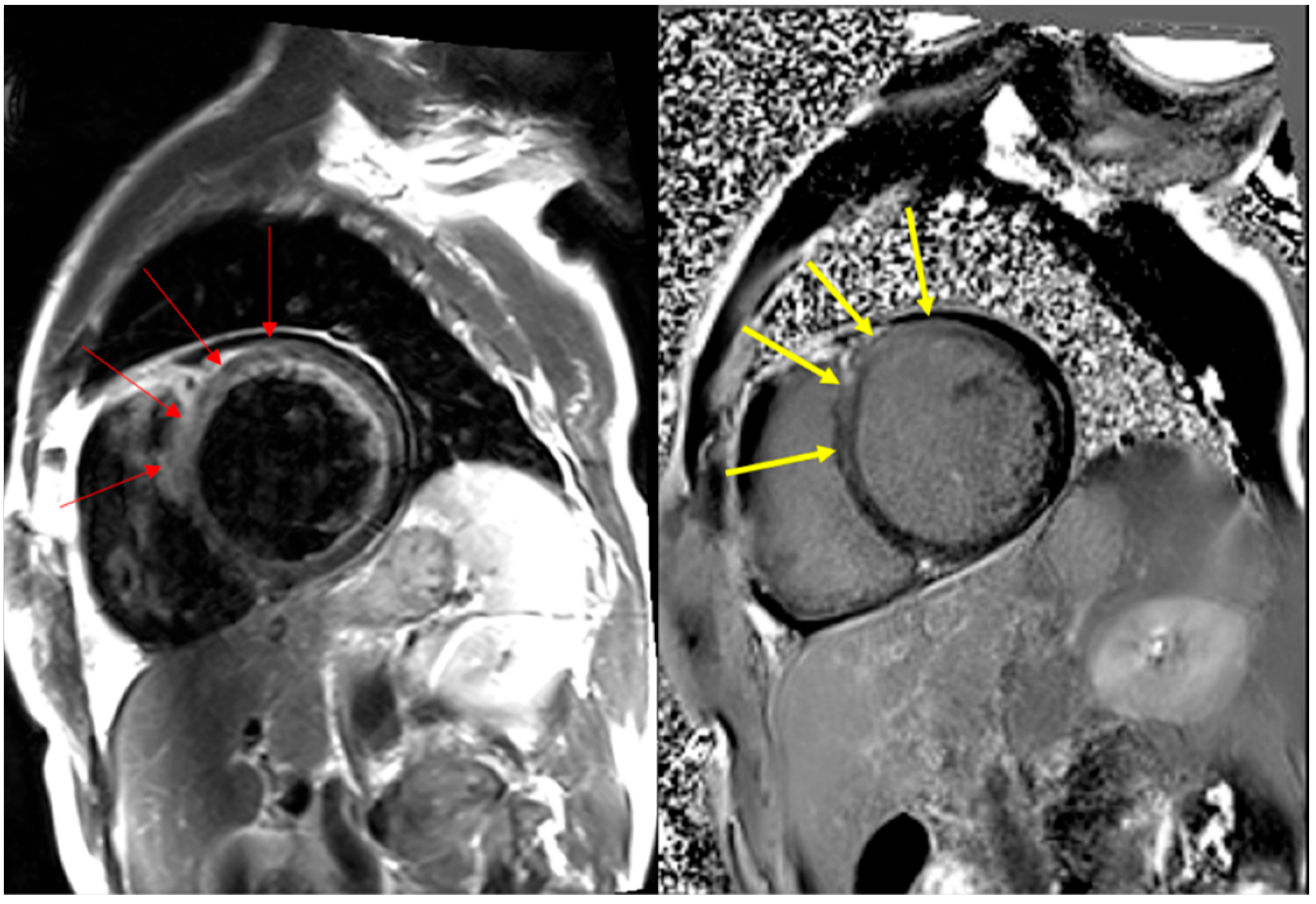
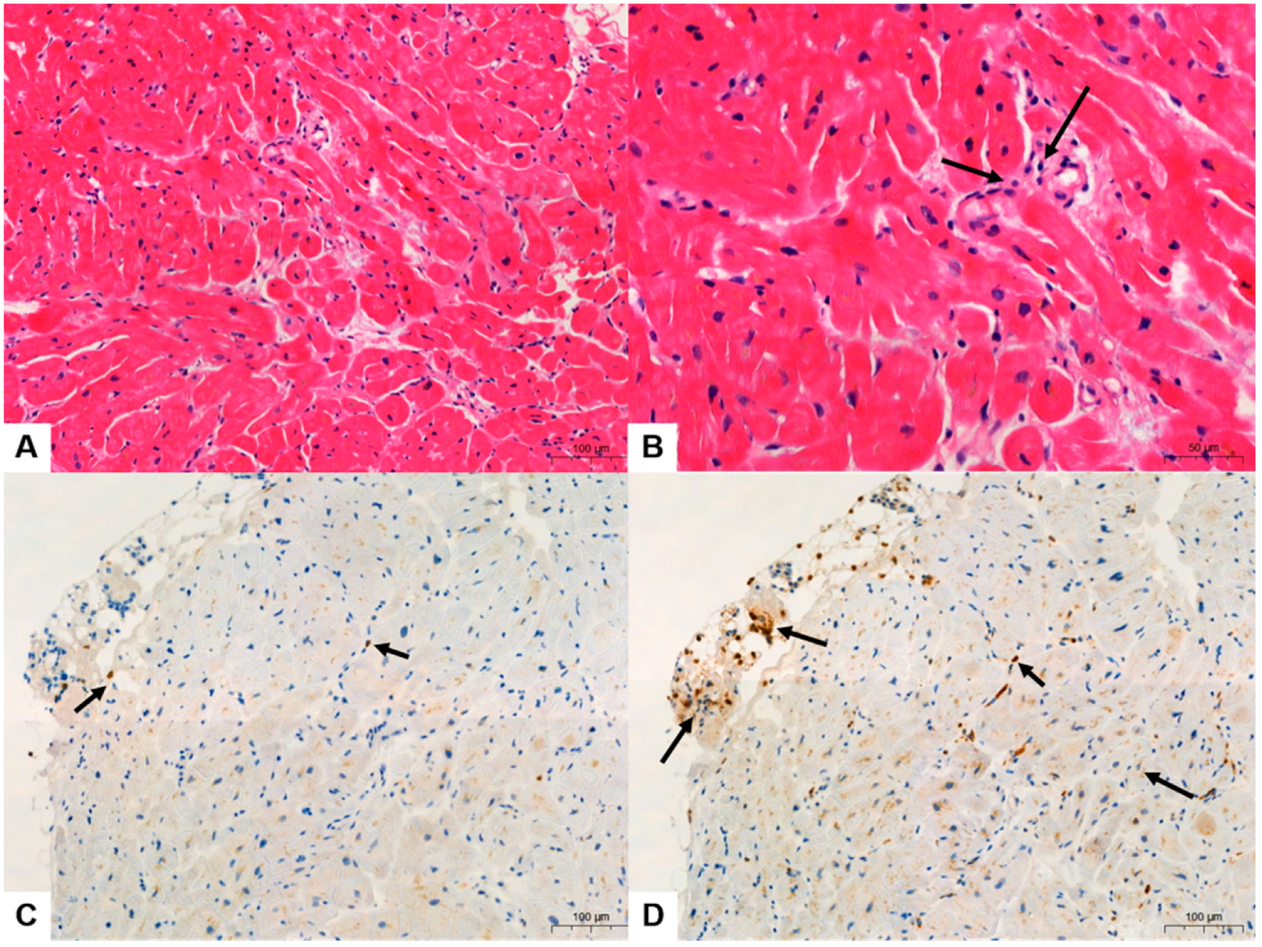
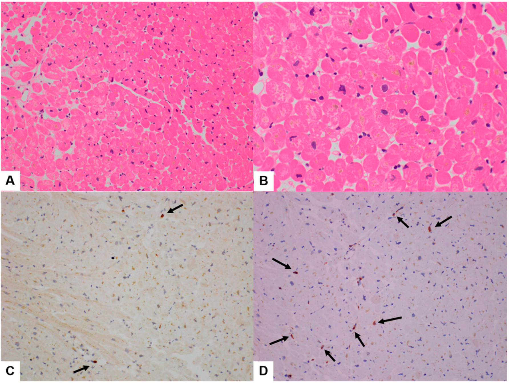
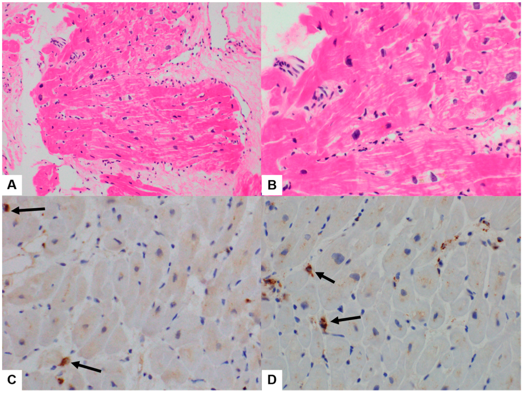
3.1. Clinicopathologic Features

Six EMB cases were identified and reviewed. A summary of the clinical features is presented in Table 1. Briefly, patients were aged 19–83 years old (median age 55.5 years). Four patients were vaccinated with the BioNTech/Pfizer BNT162b2 vaccine, whereas two were vaccinated with the Moderna mRNA-1273 vaccine before EMB. Cardiac symptoms developed 2–19 days after inoculation with the first, second, or third COVID-19 vaccine dose. The severity of cardiac dysfunction varied; left ventricular ejection fraction (LVEF) ranged from 18% to 65% (Table 1).
The symptoms of two patients were mild, with a normal range of LVEF, and had no sequelae (Case no. 3, LVEF 65%; Case no. 5, 60%; Table 1). However, these patients showed increased levels of myocardial enzymes, such as troponin-T and creatine kinase MB isoenzyme (CK-MB), and some delayed enhancement in the left ventricle on cardiac magnetic resonance imaging (MRI) (Figure 1). The T2-weighted black blood image (left) and delayed enhancement image (right) of Case no. 1 showed diffuse increased signal intensity (as compared with skeletal muscles) due to edema and abnormal subepicardial and mid-wall hyper-enhancement, particularly in the anterior and antero-septal portions of the left ventricle (in part and modified from Figure 1 in [14]). The other four patients (Case nos. 1, 2, 4, and 6) exhibited depressed LVEF with dilated cardiomyopathy (DCM). Furthermore, three of these four showed some delayed enhancement in the left ventricle on heart MRI. The assessment could not be performed on the fourth patient (Case no. 2) because of hypoxic brain damage caused by ventricular fibrillation (VF). A cardiac MRI could not be performed because breathing was not controlled, and myocardial enzymes (e.g., troponin-T and CK-MB) were elevated. Another patient (Case no. 4) underwent surgery for an implantable cardioverter-defibrillator for recurrent ventricular tachycardia. Overall, two patients (Case nos. 3 and 5) with mild symptoms received conservative therapy, whereas the remaining four (Case nos. 1, 2, 4, and 6) received treatment for heart failure. No immunosuppressive treatments were administered.
The quantitative analysis of infiltrating CD3+ lymphocytes and CD68+ histiocytes of reported PCVM cases and our six cases are summarized in Table 2.

3.2. Quantitative Analysis of Immunohistochemical Stains

All six cases showed various degrees of myocarditis with a few microfoci of myofiber eosinophilic changes and disrupted myocytes and inflammatory infiltrates (Figure 2, Figure 3 and Figure 4). Two cases met the >7 CD3-positive T lymphocytes/mm2 Dallas criterion (Case nos. 3 and 6), but the remaining four did not. However, three cases met the ≥14 leukocytes in the myocardium criterion, including up to 4 monocytes/mm2 (Case nos. 1, 2, 3, and 6), whereas the remaining three did not. These (Case nos. 2, 4, and 5) could be categorized as “borderline” myocarditis, and repeat biopsy may be indicated (Table 2).
In Cases 1–6, the histopathologic type of myocarditis was lymphohistiocytic myocarditis, showing 2–10 CD3-positive infiltrating lymphocytes/mm2 and 0–10 CD68-positive infiltrating histiocytes/mm2 (Figure 2, Figure 3 and Figure 4, Table 2). In Case 4, the histopathological type was lymphocytic myocarditis (LM), showing 1 CD3-positive lymphocytes/mm2 and 7 CD68-positive histiocytes/mm2 were identified and classified as borderline myocarditis (Figure 4, Table 2).

4. Discussion

Many heterogeneous etiologies can cause inflammatory cardiomyopathy, which is characterized by inflammatory cell infiltration into the myocardium and a high risk of deteriorating cardiac function [1]. Various factors, including viral, bacterial, protozoal, or fungal infections, as well as various toxic substances or drugs and systemic immune-mediated diseases, can cause inflammatory cardiomyopathy.
The clinical presentation of PCVM may include reduced LVEF, heart failure, advanced atrioventricular block, sustained ventricular arrhythmias, and, in severe cases, cardiogenic shock with an increased risk of death or need for heart transplantation. However, most cases of PCVM are mild [1]. According to one study that evaluated 238 patients with PCVM, most were male (87.1%), and the mean ± standard deviation age was 27.4 ± 16 (range 12–80) years [15]. The most common symptom was chest pain (93%). Approximately 30% of the patients had reduced LVEF, but more than half recovered on repeat imaging. Of the 238 patients, 11 developed cardiogenic shock, and 5 patients (1.7%) died. Chow et al. [7] pointed out some intriguing distinctions between the demographics and clinical history of one reported patient compared with previously documented cases of severe acute respiratory syndrome coronavirus 2 vaccination myocarditis in a case report.
In the present study, all six patients underwent EMB. The symptoms of the two patients were mild, with a normal range of left ventricular function, and had no sequelae. Two patients with mild symptoms received conservative therapy, whereas the remaining four received treatment for heart failure. No immunosuppressive treatments were administered.
Currently, most cardiac diseases can be diagnosed by non-invasive procedures such as echocardiography and cardiac MRI. Despite some limitations, such as inconsistencies in diagnosis, a group of pathological conditions that require biopsy for a conclusive diagnosis remains. These include myocarditis, amyloidosis, sarcoidosis, and giant-cell myocarditis [16]. Khan et al. [16] reported that in 29.2% of cases (73 out of 250), the results of EMB histology significantly influenced and altered patient management. Although the diagnostic utility of EMB for myocarditis is limited, the authors suggest that minimal lymphohistiocytic infiltration should raise suspicion for myocarditis. Histological diagnosis of myocarditis in EMB specimens is still based on the Dallas criteria. These criteria account for the distribution, extent, and cell types of inflammatory infiltrates [13,17,18]. To reduce inter-observer variability and establish a cut-off for defining abnormal lymphohistiocytic infiltration, the European Society of Cardiology introduced quantitative immunohistochemistry (IHC) criteria for diagnosing myocarditis: ≥14 leukocytes in the myocardium, including up to 4 monocytes/mm2, with the presence of >7 CD3-positive T lymphocytes/mm2. These IHC criteria may reduce inter-observer variations and thus enhance the diagnostic value of EMB for suspected myocarditis. In our study, three cases met the Dallas criteria, whereas the remaining three did not and were categorized as “borderline” myocarditis.
Of the eight cases described in the Larson et al. [5] case series, EMB was conducted with only one patient who did not demonstrate myocardial infiltrate before steroid initiation. In the report by Chow et al., EMB findings revealed lymphohistiocytic-type myocarditis. In a systematic review of the cumulative experience of 20,212 cases of PCVM that identified 238 positive patients, only 13 underwent an EMB or autopsy. Furthermore, 10/13 (77%) patients showed abnormal histopathology suggestive of myocarditis [15]. In our hospital’s experience, myocarditis due to other causes has a much more severe inflammatory response pathologically; however, the disease severity of PCVM is relatively mild. Although not pathologically severe, PCVM cases were clinically severe with conditions such as DCM, VF, and ventricular tachycardia. Therefore, histopathological confirmation of myocarditis is important in predicting prognosis.
In terms of the molecular pathogenesis of PCVM, we conducted single-cell RNA sequencing and single-cell T-cell receptor sequencing analyses of peripheral blood mononuclear cells (PBMCs) obtained from one of the patients (Case no. 1) and published the result implicating the immunologic background of PCVM: the greatest changes were observed in the transcriptomic profile of monocytes in terms of the number of differentially expressed genes [14].

5. Conclusions

Our report describes a series of clinically diagnosed PCVM cases and highlights the potential significance of EMB and histopathological findings in the diagnosis of PCVM. In our cases, the degree of inflammatory cell infiltration was mild, and 50% (3/6) did not fulfill the Dallas criteria. Therefore, these cases could be categorized as “borderline” myocarditis. Interestingly, inflammatory cell infiltration was not proportional to the severity of clinical symptoms.
Myocarditis is a rare complication of COVID-19 vaccinations, predominantly in male adolescents and young adults. The symptoms in most suspected cases of PCVM are mild; thus, EMB was often not performed. Most PCVM cases showed a mild degree of inflammation histopathologically, and some cases could not fulfill the Dallas criteria and were classified as borderline myocarditis.

Author Contributions

Conceptualization, H.-S.A. and K.-J.S.; data curation, K.-J.S.; formal analysis, K.-J.S.; funding acquisition, H.-S.A. and K.-J.S.; investigation, H.-S.A. and K.-J.S.; methodology, H.-S.A. and K.-J.S.; project administration, H.-S.A.; resources, H.-S.A.; supervision, K.-J.S.; validation, H.-S.A. and K.-J.S.; visualization, K.-J.S.; writing—original draft, H.-S.A. and K.-J.S.; writing—review and editing, Y.A., J.J., S.B., S.L., C.K., J.-M.L. and K.L. All authors have read and agreed to the published version of the manuscript.

Funding

This research was partly supported by the Catholic University of Korea, Uijeongbu St. Mary’s Hospital Clinical Research Laboratory Foundation, in the program year 2023 (5-2023-B0001-00253); Korean Heart Failure Registry III funded by the Korean Society of Heart Failure; and the Material Parts Technology Development Program funded by the Ministry of Trade, Industry & Energy (MOTIE), Korea (No. 20023097).

Institutional Review Board Statement

This study was approved by the Institutional Research Ethics Board of Uijeongbu St. Mary’s Hospital of the Catholic University of Korea (No. UC23RIS10041; approved 27 April 2023) and conducted in accordance with the tenets of the Declaration of Helsinki.

Informed Consent Statement

Not required due to the retrospective, observational nature of this study. For the purpose of the treatment, the patient’s consent was obtained prior to the study, and the all of the patients’ personal details have been de-identified.

Data Availability Statement

Data are available on reasonable request.

Conflicts of Interest

The authors declare no conflicts of interest.

References

  1. Basso, C. Myocarditis. N. Engl. J. Med. 2022, 387, 1488–1500. [Google Scholar] [CrossRef] [PubMed]
  2. Albert, E.; Aurigemma, G.; Saucedo, J.; Gerson, D.S. Myocarditis following COVID-19 vaccination. Radiol. Case Rep. 2021, 16, 2142–2145. [Google Scholar] [CrossRef] [PubMed]
  3. Bozkurt, B.; Kamat, I.; Hotez, P.J. Myocarditis with COVID-19 mRNA vaccines. Circulation 2021, 144, 471–484. [Google Scholar] [CrossRef] [PubMed]
  4. Abbate, A.; Gavin, J.; Madanchi, N.; Kim, C.; Shah, P.R.; Klein, K.; Boatman, J.; Roberts, C.; Patel, S.; Danielides, S. Fulminant myocarditis and systemic hyperinflammation temporally associated with BNT162b2 mRNA COVID-19 vaccination in two patients. Int. J. Cardiol. 2021, 340, 119–121. [Google Scholar] [CrossRef] [PubMed]
  5. Larson, K.F.; Ammirati, E.; Adler, E.D.; Cooper, L.T., Jr.; Hong, K.N.; Saponara, G.; Couri, D.; Cereda, A.; Procopio, A.; Cavalotti, C.; et al. Myocarditis After BNT162b2 and mRNA-1273 Vaccination. Circulation 2021, 144, 506–508. [Google Scholar] [CrossRef] [PubMed]
  6. Rosner, C.M.; Genovese, L.; Tehrani, B.N.; Atkins, M.; Bakhshi, H.; Chaudhri, S.; Damluji, A.A.; De Lemos, J.A.; Desai, S.S.; Emaminia, A. Myocarditis temporally associated with COVID-19 vaccination. Circulation 2021, 144, 502–505. [Google Scholar] [CrossRef] [PubMed]
  7. Chow, B.T.; Lai, C.K. Lymphohistiocytic Myocarditis Possibly Due to Moderna mRNA-1273 VaccineA Case Report. Am. J. Clin. Pathol. 2022, 158, 167–172. [Google Scholar] [CrossRef] [PubMed]
  8. Kiblboeck, D.; Klingel, K.; Genger, M.; Traxler, S.; Braunsteiner, N.; Steinwender, C.; Kellermair, J. Myocarditis following mRNA COVID-19 vaccination: Call for endomyocardial biopsy. ESC Heart Fail. 2021, 9, 1996–2002. [Google Scholar] [CrossRef] [PubMed]
  9. Sung, K.; McCain, J.; King, K.R.; Hong, K.; Aisagbonhi, O.; Adler, E.D.; Urey, M.A. Biopsy-Proven Giant Cell Myocarditis Following the COVID-19 Vaccine. Circ. Heart Fail. 2022, 15, e009321. [Google Scholar] [CrossRef] [PubMed]
  10. Ujueta, F.; Azimi, R.; Lozier, M.R.; Poppiti, R.; Ciment, A. Lymphohistocytic myocarditis after Ad26.COV2.S viral vector COVID-19 vaccination. Int. J. Cardiol. Heart Vasc. 2021, 36, 100869. [Google Scholar] [CrossRef] [PubMed]
  11. Verma, A.K.; Lavine, K.J.; Lin, C.-Y. Myocarditis after COVID-19 mRNA vaccination. N. Engl. J. Med. 2021, 385, 1332–1334. [Google Scholar] [CrossRef] [PubMed]
  12. Yamamoto, M.; Tajiri, K.; Ayuzawa, S.; Ieda, M. Pathological Findings of Clinically Suspected Myocarditis Temporally Associated with COVID-19 Vaccination. Eur. J. Heart Fail. 2022, 24, 1132–1138. [Google Scholar] [CrossRef] [PubMed]
  13. Leone, O.; Pieroni, M.; Rapezzi, C.; Olivotto, I. The spectrum of myocarditis: From pathology to the clinics. Virchows Arch. 2019, 475, 279–301. [Google Scholar] [CrossRef] [PubMed]
  14. Yoon, B.K.; Oh, T.G.; Bu, S.; Seo, K.J.; Kwon, S.H.; Lee, J.Y.; Kim, Y.; Kim, J.W.; Ahn, H.S.; Fang, S. The Peripheral Immune Landscape in a Patient with Myocarditis after the Administration of BNT162b2 mRNA Vaccine. Mol. Cells 2022, 45, 738–748. [Google Scholar] [CrossRef] [PubMed]
  15. Ilonze, O.J.; Guglin, M.E. Myocarditis following COVID-19 vaccination in adolescents and adults: A cumulative experience of 2021. Heart Fail. Rev. 2022, 27, 2033–2043. [Google Scholar] [CrossRef] [PubMed]
  16. Khan, T.; Selvakumar, D.; Trivedi, S.; Rao, K.; Harapoz, M.; Thiagalingam, A.; Denniss, A.R.; Varikatt, W. The value of endomyocardial biopsy in diagnosis and guiding therapy. Pathology 2017, 49, 750–756. [Google Scholar] [CrossRef] [PubMed]
  17. Leone, O.; Veinot, J.P.; Angelini, A.; Baandrup, U.T.; Basso, C.; Berry, G.; Bruneval, P.; Burke, M.; Butany, J.; Calabrese, F.; et al. 2011 Consensus statement on endomyocardial biopsy from the Association for European Cardiovascular Pathology and the Society for Cardiovascular Pathology. Cardiovasc. Pathol. 2012, 21, 245–274. [Google Scholar] [CrossRef] [PubMed]
  18. Thomas Aretz, H. Myocarditis: The Dallas criteria. Hum. Pathol. 1987, 18, 619–624. [Google Scholar] [CrossRef] [PubMed]
Figure 1. Cardiac magnetic resonance imaging findings of Case 1, post-COVID-19 vaccination myocarditis (PCVM). T2-weighted black blood image (left) and delayed enhancement image (right) show diffuse increased signal intensity (as compared with skeletal muscles) due to edema and abnormal subepicardial and mid-wall hyper-enhancement, particularly in the anterior and antero-septal portions of the left ventricle (red and yellow arrows). Reprinted in part with modification from Ref. [14].
Diagnostics 14 00219 g001
Figure 2. Case 1, post-COVID-19 vaccination myocarditis (PCVM). (A,B) Endomyocardial biopsy (EMB) showed active multifocal lymphocytic myocarditis (LM) with a few microfoci of myofiber eosinophilic changes and infiltrating lymphocytes (black arrows in (B)) (hematoxylin and eosin (H&E), 200× in (A) and 400× in (B)). (C) Infiltrating CD3-positive lymphocytes (black arrows) were 6/mm2. (D) Infiltrating CD68-positive histiocytes (black arrows) were 10/mm2 (IHC, 400× in (C,D)).
Diagnostics 14 00219 g002
Figure 3. Case 4 borderline post-COVID-19 vaccination myocarditis (PCVM). (A,B) EMB showed a few lymphocytic infiltrations with a few microfoci of myofiber eosinophilic changes and disrupted myocytes (H&E, 200× in (A) and 400× in (B)). (C) Infiltrating CD3-positive lymphocytes (black arrows) were 2/mm2. (D) Infiltrating CD68-positive histiocytes (black arrows) were 7/mm2 (IHC, 400× in (C,D)).
Diagnostics 14 00219 g003
Figure 4. Case 6 of post-COVID-19 vaccination myocarditis (PCVM). (A,B) EMB showed a few lymphocytic infiltrations with a few microfoci of myofiber eosinophilic changes and disrupted myocytes (H&E, 200× in (A) and 400× in (B)). (C) Infiltrating CD3-positive lymphocytes (black arrows) were 10/mm2. (D) Infiltrating CD68-positive histiocytes (black arrows) were 5/mm2 (IHC, 400× in (C,D)).
Diagnostics 14 00219 g004
Table 1. Clinicopathologic findings of previous cases with histopathologic examinations and our six cases of post-COVID vaccination myocarditis.
Case Series [Reference No.]Age (Yrs)SexCardiac Function LVEF (%)SymptomsMRI FindingsCK-MBCardiac Troponin * I or # TPathologic Myocarditis TypeSpecimen TypeVaccine Type (Dose)Symptoms Onset after Vaccination (Days)
Rosner et al. [6]28M51chest pain, no fever or coughingPatchy LGE, no EDU* 17.08 (ng/mL)UNDJ (1st)5
Rosner et al. [6]39M35–40chest pain, no fever or coughingPatchy LGE, no EDU* 11.01 (ng/mL)UNDP (2nd)3
Rosner et al. [6]39M60chest pain, fever, chills, and shortness of breathMultifocal LGE, no EDU* 13.01 (ng/mL)UNDP (2nd)4
Rosner et al. [6]24M53chest painMidmyocardial LGE, EDU* 0.37 (ng/mL)UNDP (1st)7
Rosner et al. [6]19M55chest painMultifocal LGE, EDU* 44.8 (ng/mL)UNDP (2nd)2
Rosner et al. [6]20M50–55chest painSubepicardial LGE, EDU* 8.36 (ng/mL)UNDP (2nd)3
Rosner et al. [6]23M58subjective fevers, chest pain, and myalgiaMidmyocardial LGE, no EDUUUNDP (2nd)3
Abbate et al. [4]27M20nausea and vomitingU252 (ng/ml)UUNDP (1st)2
Abbate et al. [4]34F15fever, cough, chest pain, nausea, and vomitingU42.4 (ng/ml)UUNDP (2nd)9
Larson et al. [5]22M50fever, chills, and myalgiaLGE U285 FULUNDM (2nd)3
Larson et al. [5]31M34fever, chills, and myalgiaLGE U46 FULUNDM (2nd)3
Larson et al. [5]40M47chest painED, LGE, PEU520 FULUNDP (2nd)2
Larson et al. [5]56M60chest painED, LGEU37 FULUNDP (2nd)3
Larson et al. [5]26M60cough, feverED, LGE, PEU100 FULUNDP (2nd)3
Larson et al. [5]35M50fever and chest painED, LGEU29 FULUNDP (2nd)2
Larson et al. [5]21M54fever and chest painED, LGE, PEU1164 FULUNDP (2nd)4
Larson et al. [5]22M53chest painED, LGEU1433 FULUNDM (2nd)2
Verma et al. [11]45F15–20dyspnea and dizzinessLGEU* 10.453 (ng/mL)LM admixed with eosinophils, B cells, and plasma cellsEMBP (1st)10
Verma et al. [11] 42M15dyspnea and chest painNot availableU* 44.30 (ng/mL)An inflammatory infiltrate admixed with macrophages, T cells, eosinophils, and B cellsAutopsyM (2nd)2wks
Ujueta et al. [10]62F29progressive body aches, weakness, and fatigueUU* 6.4 (ng/mL)LM with sparse eosinophilsAutopsyJ (1st)4
Sung et al. [9]63M35fever, fatigue, and coughUU# 5.816 (ng/mL)GCMEMBP (2nd)7
Kiblboek et al. [8]18M33chest pain, fever, and fatigueED, LGEU# 1.386 (ng/mL)LM with sparse eosinophilsEMBP (1st)3
Kiblboek et al. [8]22M40chest pain, no fever, and fatigueED, LGEU* 38.735 (ng/mL)LM with sparse eosinophilsEMBP (2nd)1
Kiblboek et al. [8]38M48chest pain, no fever, and fatigueED, LGEU# 1.104 (ng/mL)LM with sparse eosinophilsEMBP (1st)4
Yamamoto et al. [12] 41M15chest pain, myalgia, and feverU236 (U/L)# 15 (ng/mL)Severe LM predominantly composed of cytotoxic T cells (CD8+) and macrophages (CD68+) admixed with B cells (CD20+) and a few eosinophilsEMBM (2nd)19
Yamamoto et al. [12] 18M27fever, chest pain, and fatigueU32 (U/L)# 3.09 (ng/mL)Severe LM predominantly composed of cytotoxic T cells (CD8+) and macrophages (CD68+)EMBP (1st)9
Yamamoto et al. [12]18M46fever and chest painU72 (U/L)# 1.3 (ng/mL)Inflammatory cell infiltration is trivial, obvious cardiomyocyte damage (loss of nuclei and mild vacuolar degeneration) and perivascular and interstitial fibrosisEMBM (2nd)2
Yamamoto et al. [12]18M62fever and chest painU32 (U/L)# 0.515 (ng/mL)Same as the aboveEMBM (2nd)3
Chow et al. [7]45F40progressive decline of exercise capacity, palpitation, fatigue, and exertional dyspneaMultifocal LGEUULM and a focal histiocytic collection suggestive of an ill-defined granulomaEMBM (1st)7
Our case 159M24dyspnea, leg edemaPatchy midmyocardial LGE22.7 (ng/mL)# 0.019 (ng/mL)LMEMBP (1st)3
Our case 252M35cardiac arrest d/t ventricular fibrillationND31.3 (ng/mL)# 2.00 (ng/mL)LMEMBP (2nd)19
Our case 319M65chest painLGE11.7 (ng/mL)# 0.085 (ng/mL)LMEMBM (2nd) 3
Our case 483M35dyspnea, palpitation due to ventricular tachycardiaLGE18.7 (ng/mL)# 0.82 (ng/mL)LMEMBP (3rd)2
Our case 569F60dyspnea, chest discomfortPatchy subepicardial and midmyocardial LGE23.4 (ng/mL)# 0.667 (ng/mL)LMEMBM (3rd)2
Our case 6 38M18myalgia, dyspneaPatchy subepicardial and midmyocardial LGE and PE15.96 (ng/mL)# 0.233 (ng/mL)LMEMBP (3rd)12
LVEF, left ventricular ejection fraction; ND, testing not done; U, unknown; J, Ad26.COV2.S viral vector (Janssen) COVID-19 vaccine (Johnson & Johnson, New Brunswick, NJ, USA); P, BNT162b2 vaccine (Pfizer-BioNTech, New York, NY, USA); M, mRNA-1273 vaccine (Moderna, Cambridge, MA, USA); LM, lymphohistiocytic myocarditis; ED, edema; LGE, late gadolinium enhancement; PE, pericardial effusion; * Troponin I, # Troponin T; FUL: fold of the upper limit; GCM, giant-cell myocarditis; EMB, endomyocardial biopsy.
Table 2. Quantitative analysis of CD3+ lymphocytes and CD68+ histiocytes of post-COVID vaccination myocarditis.
Case [Reference No.]Age (Years)SexMyocarditis TypeCD3 (Cells/mm2)CD68 (Cells/mm2)
Kiblboeck et al. [8]18MLM with sparse eosinophils>50>100
Kiblboeck et al. [8]22MLM with sparse eosinophils514
Kiblboeck et al. [8]38MLM with sparse eosinophils3034
Our case 159MLM (* myocarditis)610
Our case 252MLM (* borderline myocarditis)55
Our case 319MLM (* myocarditis)105
Our case 483MLM (* borderline myocarditis)27
Our case 569FLM (* borderline myocarditis)30
Our case 638MLM (* myocarditis)105
LM, lymphohistiocytic myocarditis; * classified according to the Dallas criteria.
Disclaimer/Publisher’s Note: The statements, opinions and data contained in all publications are solely those of the individual author(s) and contributor(s) and not of MDPI and/or the editor(s). MDPI and/or the editor(s) disclaim responsibility for any injury to people or property resulting from any ideas, methods, instructions or products referred to in the content.

