Single-Site Experience with an Automated Artificial Intelligence Application for Left Ventricular Ejection Fraction Measurement in Echocardiography
Abstract
1. Introduction
2. Methods
2.1. Study Design
2.2. Echocardiographic Imaging and Analysis by Cardiologists Using MBS Method
2.3. Briefing and Self-Study of Participating Cardiologist
2.4. Analysis by Artificial Intelligence Application
2.5. Categorization for Subgroup Analyses
2.6. Repeatability and Reliability
2.7. Statistics
3. Results
3.1. Study Population
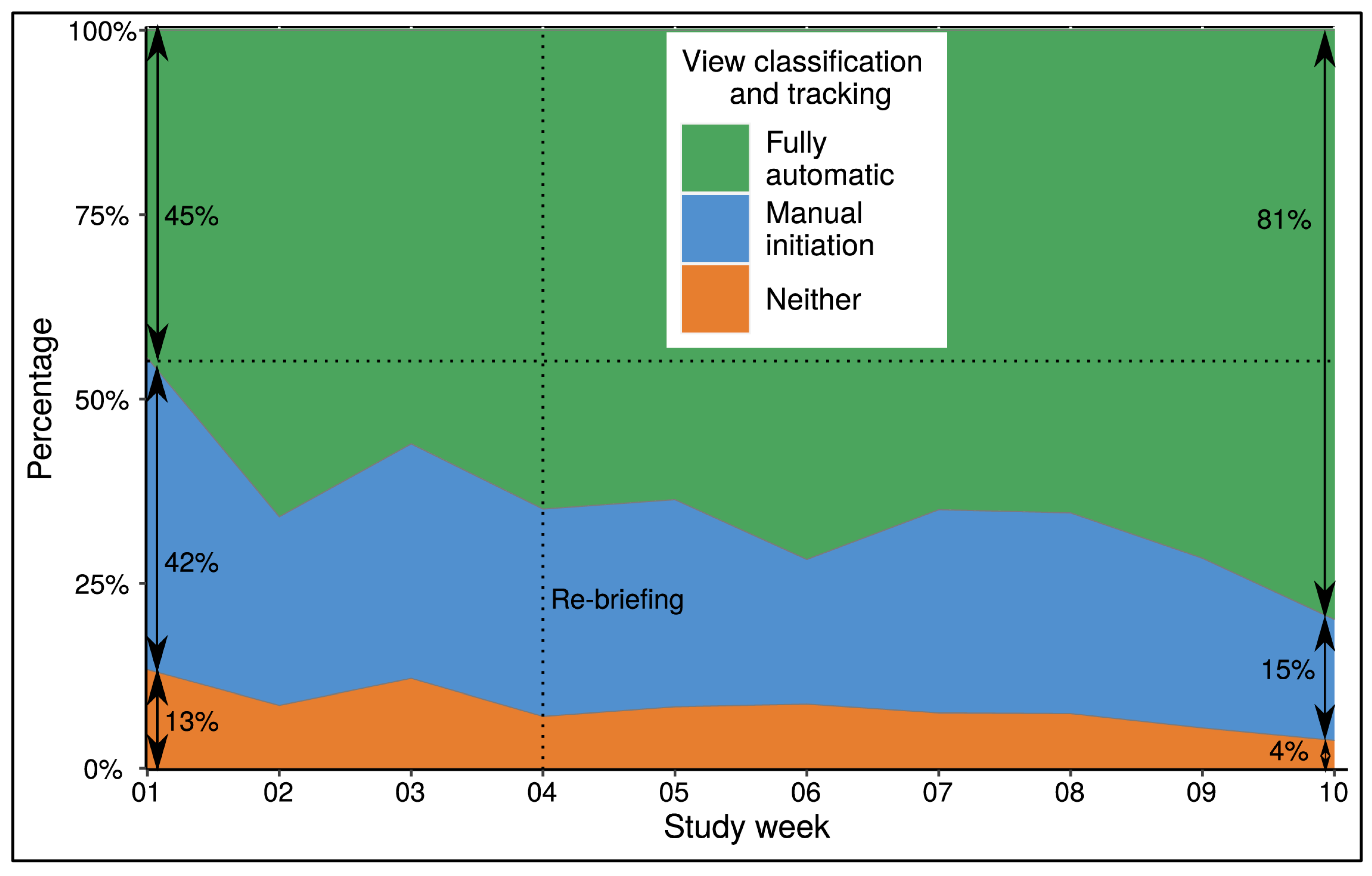
3.2. Feasibility
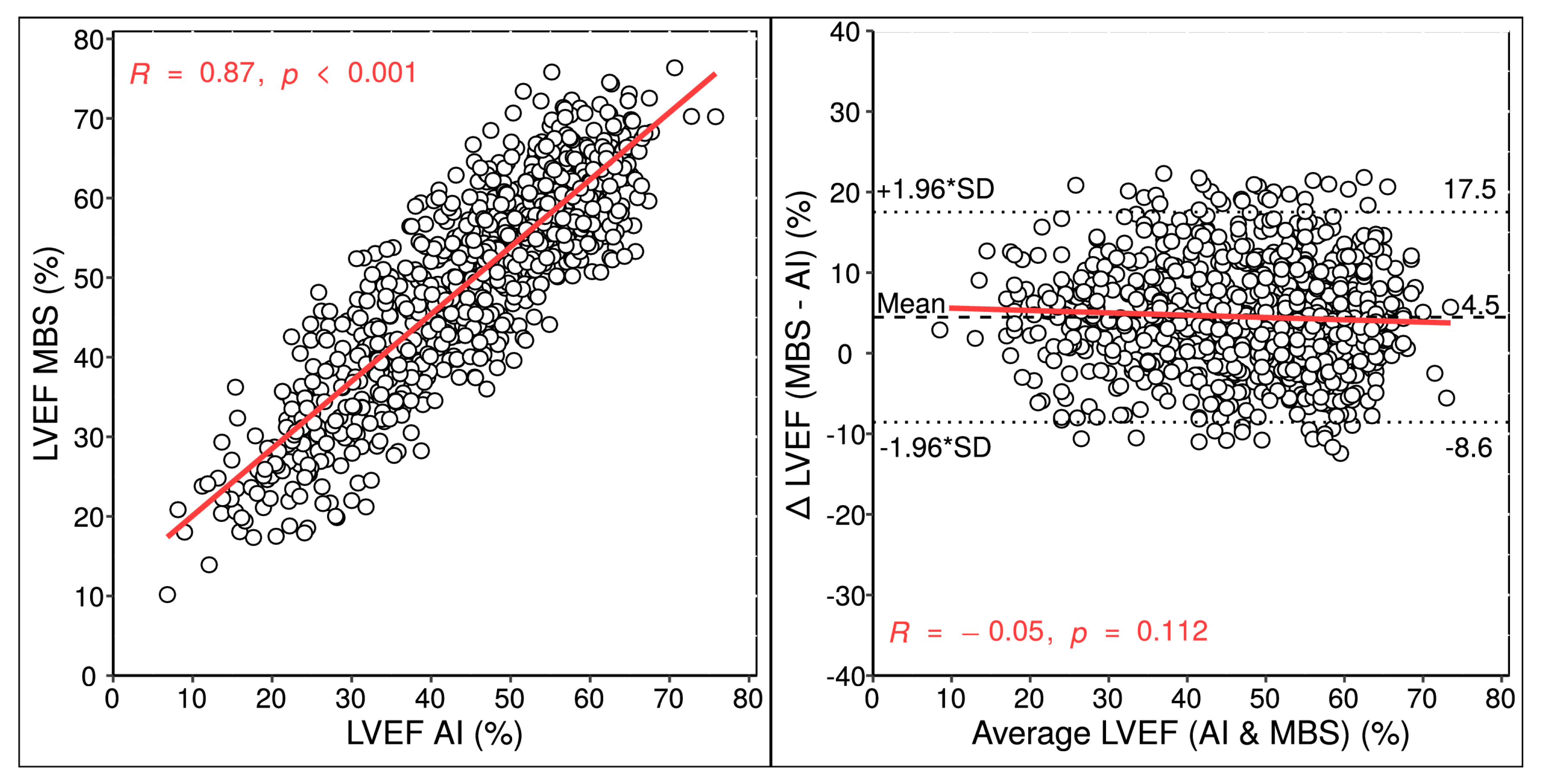
3.3. Measurement Comparison between Cardiologists and AI
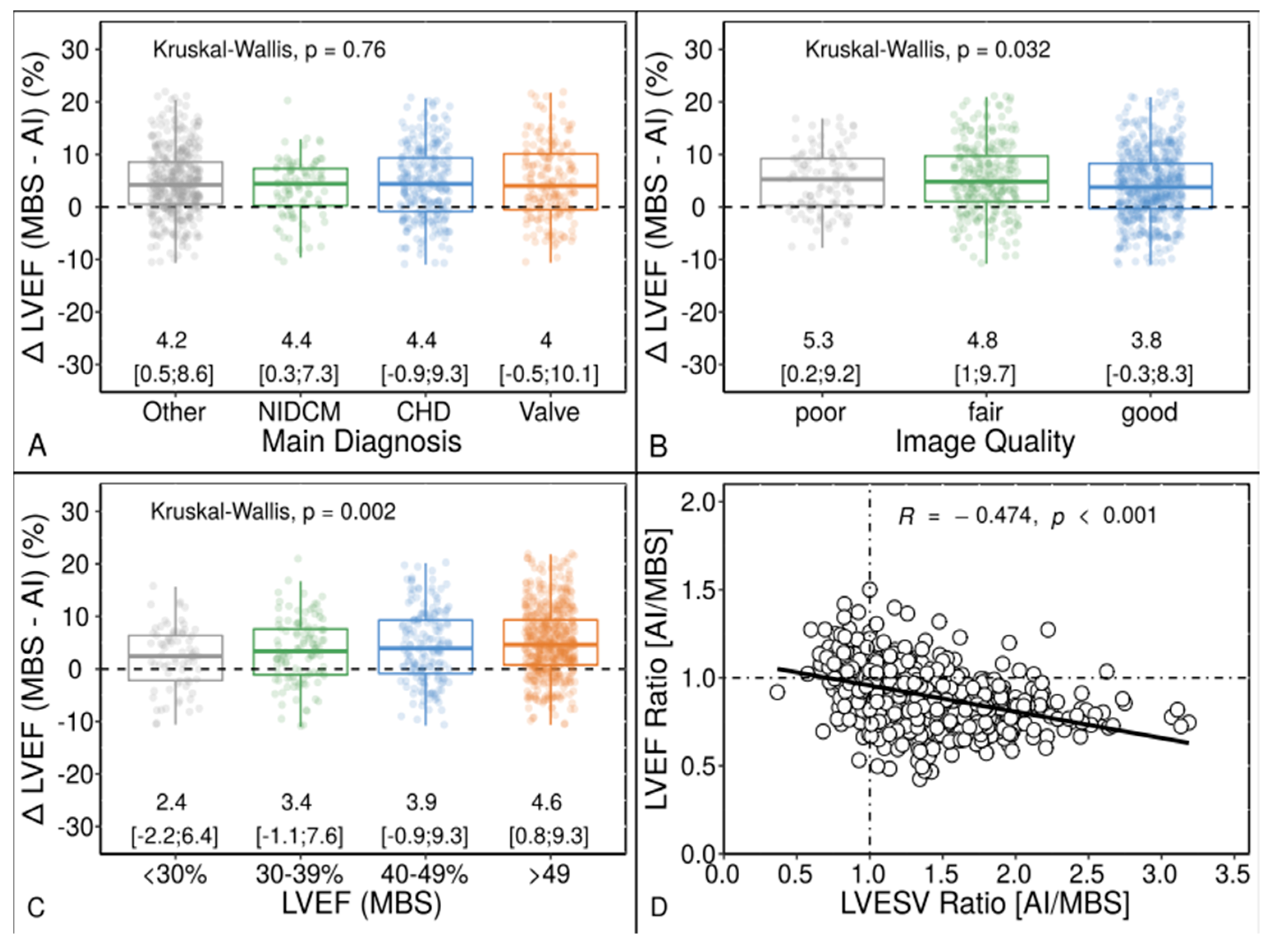
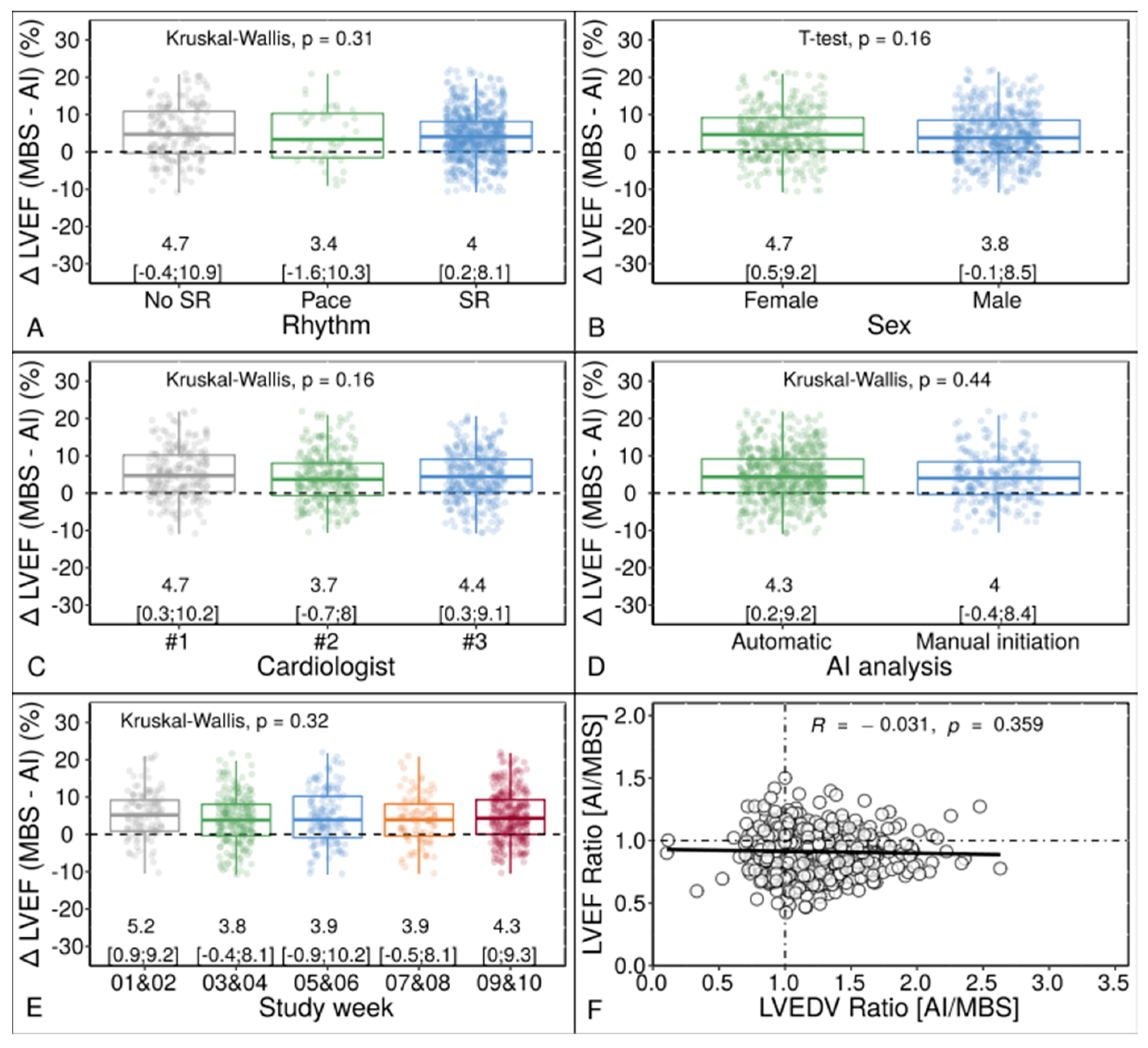
3.4. Agreement and Bias Categorized Using Clinical and Echocardiographic Characteristics
3.5. Comparison of Repeatability and Reliability between AI and MBS
4. Discussion
4.1. Feasibility
4.2. Comparison of LV Measurement Results
4.3. Repeatability, Reliability and Performance
4.4. Limitations
5. Conclusions
Supplementary Materials
Author Contributions
Funding
Institutional Review Board Statement
Informed Consent Statement
Data Availability Statement
Acknowledgments
Conflicts of Interest
References
- Lang, R.M.; Badano, L.P.; Mor-Avi, V.; Afilalo, J.; Armstrong, A.; Ernande, L.; Flachskampf, F.A.; Foster, E.; Goldstein, S.A.; Kuznetsova, T.; et al. Recommendations for cardiac chamber quantification by echocardiography in adults: An update from the American Society of Echocardiography and the European Association of Cardiovascular Imaging. J. Am. Soc. Echocardiogr. 2015, 28, 1–39.e14. [Google Scholar] [CrossRef]
- Aurich, M.; André, F.; Keller, M.; Greiner, S.; Hess, A.; Buss, S.J.; Katus, H.A.; Mereles, D. Assessment of left ventricular volumes with echocardiography and cardiac magnetic resonance imaging: Real-life evaluation of standard versus new semiautomatic methods. J. Am. Soc. Echocardiogr. 2014, 27, 1017–1024. [Google Scholar] [CrossRef]
- Hoffmann, R.; Barletta, G.; von Bardeleben, S.; Vanoverschelde, J.L.; Kasprzak, J.; Greis, C.; Becher, H. Analysis of left ventricular volumes and function: A multicenter comparison of cardiac magnetic resonance imaging, cine ventriculography, and unenhanced and contrast-enhanced two-dimensional and three-dimensional echocardiography. J. Am. Soc. Echocardiogr. 2014, 27, 292–301. [Google Scholar] [CrossRef]
- Knackstedt, C.; Bekkers, S.C.; Schummers, G.; Schreckenberg, M.; Muraru, D.; Badano, L.; Franke, A.; Bavishi, C.; Omar, A.M.S.; Sengupta, P.P. Fully Automated Versus Standard Tracking of Left Ventricular Ejection Fraction and Longitudinal Strain the FAST-EFs Multicenter Study. J. Am. Coll. Cardiol. 2015, 66, 1456–1466. [Google Scholar] [CrossRef]
- Thavendiranathan, P.; Grant, A.D.; Negishi, T.; Plana, J.C.; Popović, Z.B.; Marwick, T.H. Reproducibility of Echocardiographic Techniques for Sequential Assessment of Left Ventricular Ejection Fraction and Volumes. J. Am. Coll. Cardiol. 2013, 61, 77–84. [Google Scholar] [CrossRef]
- Asch, F.M.; Descamps, T.; Sarwar, R.; Karagodin, I.; Singulane, C.C.; Xie, M.; Tucay, E.S.; Rodrigues, A.C.T.; Vasquez-Ortiz, Z.Y.; Monaghan, M.J.; et al. Human versus Artificial Intelligence–Based Echocardiographic Analysis as a Predictor of Outcomes: An Analysis from the World Alliance Societies of Echocardiography COVID Study. J. Am. Soc. Echocardiogr. 2022, 35, 1226–1237.e7. [Google Scholar] [CrossRef]
- Tromp, J.; Seekings, P.J.; Hung, C.-L.; Iversen, M.B.; Frost, M.J.; Ouwerkerk, W.; Jiang, Z.; Eisenhaber, F.; Goh, R.S.M.; Zhao, H.; et al. Automated interpretation of systolic and diastolic function on the echocardiogram: A multicohort study. Lancet Digit. Health 2022, 4, e46–e54. [Google Scholar] [CrossRef]
- Zhang, J.; Gajjala, S.; Agrawal, P.; Tison, G.H.; Hallock, L.A.; Beussink-Nelson, L.; Lassen, M.H.; Fan, E.; Aras, M.A.; Jordan, C.; et al. Fully automated echocardiogram interpretation in clinical practice: Feasibility and diagnostic accuracy. Circulation 2018, 138, 1623–1635. [Google Scholar] [CrossRef]
- Samtani, R.; Bienstock, S.; Lai, A.C.; Liao, S.; Baber, U.; Croft, L.; Stern, E.; Beerkens, F.; Ting, P.; Goldman, M.E. Assessment and validation of a novel fast fully automated artificial intelligence left ventricular ejection fraction quantification software. Echocardiography 2022, 39, 473–482. [Google Scholar] [CrossRef]
- Akkus, Z.; Aly, Y.; Attia, I.; Lopez-Jimenez, F.; Arruda-Olson, A.; Pellikka, P.; Pislaru, S.; Kane, G.; Friedman, P.; Oh, J. Artificial intelligence (ai)-empowered echocardiography interpretation: A state-of-the-art review. J. Clin. Med. 2021, 10, 1391. [Google Scholar] [CrossRef]
- Bunting, K.V.; Steeds, R.P.; Slater, L.T.; Rogers, J.K.; Gkoutos, G.V.; Kotecha, D. A Practical Guide to Assess the Reproducibility of Echocardiographic Measurements. J. Am. Soc. Echocardiogr. 2019, 32, 1505–1515. [Google Scholar] [CrossRef] [PubMed]
- Quer, G.; Arnaout, R.; Henne, M.; Arnaout, R. Machine Learning and the Future of Cardiovascular Care: JACC State-of-the-Art Review. J. Am. Coll. Cardiol. 2021, 77, 300–313. [Google Scholar] [CrossRef] [PubMed]
- Litjens, G.; Ciompi, F.; Wolterink, J.M.; de Vos, B.D.; Leiner, T.; Teuwen, J.; Išgum, I. State-of-the-Art Deep Learning in Cardiovascular Image Analysis. JACC Cardiovasc. Imaging 2019, 12, 1549–1565. [Google Scholar] [CrossRef]
- Asch, F.M.; Poilvert, N.; Abraham, T.; Jankowski, M.; Cleve, J.; Adams, M.; Romano, N.; Hong, H.; Mor-Avi, V.; Martin, R.P.; et al. Automated Echocardiographic Quantification of Left Ventricular Ejection Fraction Without Volume Measurements Using a Machine Learning Algorithm Mimicking a Human Expert. Circ. Cardiovasc. Imaging 2019, 12, e009303. [Google Scholar] [CrossRef]
- Kusunose, K.; Haga, A.; Yamaguchi, N.; Abe, T.; Fukuda, D.; Yamada, H.; Harada, M.; Sata, M. Deep Learning for Assessment of Left Ventricular Ejection Fraction from Echocardiographic Images. J. Am. Soc. Echocardiogr. 2020, 33, 632–635.e1. [Google Scholar] [CrossRef]
- Bhuva, A.N.; Bai, W.; Lau, C.; Davies, R.H.; Ye, Y.; Bulluck, H.; McAlindon, E.; Culotta, V.; Swoboda, P.P.; Captur, G.; et al. A Multicenter, Scan-Rescan, Human and Machine Learning CMR Study to Test Generalizability and Precision in Imaging Biomarker Analysis. Circ. Cardiovasc. Imaging 2019, 12, e009214. [Google Scholar] [CrossRef]
- Yuan, N.; Jain, I.; Rattehalli, N.; He, B.; Pollick, C.; Liang, D.; Heidenreich, P.; Zou, J.; Cheng, S.; Ouyang, D. Systematic Quantification of Sources of Variation in Ejection Fraction Calculation Using Deep Learning. JACC Cardiovasc. Imaging 2021, 14, 2260–2262. [Google Scholar] [CrossRef]
- Ghorbani, A.; Ouyang, D.; Abid, A.; He, B.; Chen, J.H.; Harrington, R.A.; Liang, D.H.; Ashley, E.A.; Zou, J.Y. Deep learning interpretation of echocardiograms. Npj Digit. Med. 2020, 3, 10. [Google Scholar] [CrossRef]
- Playford, D.; Bordin, E.; Mohamad, R.; Stewart, S.; Strange, G. Enhanced Diagnosis of Severe Aortic Stenosis Using Artificial Intelligence: A Proof-of-Concept Study of 530,871 Echocardiograms. JACC Cardiovasc. Imaging 2020, 13, 1087–1090. [Google Scholar] [CrossRef]
- Pellikka, P.A.; Strom, J.B.; Pajares-Hurtado, G.M.; Keane, M.G.; Khazan, B.; Qamruddin, S.; Tutor, A.; Gul, F.; Peterson, E.; Thamman, R.; et al. Automated analysis of limited echocardiograms: Feasibility and relationship to outcomes in COVID-19. Front. Cardiovasc. Med. 2022, 9. [Google Scholar] [CrossRef]
- Salte, I.M.; Østvik, A.; Smistad, E.; Melichova, D.; Nguyen, T.M.; Karlsen, S.; Brunvand, H.; Haugaa, K.H.; Edvardsen, T.; Lovstakken, L.; et al. Artificial Intelligence for Automatic Measurement of Left Ventricular Strain in Echocardiography. JACC Cardiovasc. Imaging 2021, 14, 1918–1928. [Google Scholar] [CrossRef] [PubMed]
- Otani, K.; Nakazono, A.; Salgo, I.S.; Lang, R.M.; Takeuchi, M. Three-Dimensional Echocardiographic Assessment of Left Heart Chamber Size and Function with Fully Automated Quantification Software in Patients with Atrial Fibrillation. J. Am. Soc. Echocardiogr. 2016, 29, 955–965. [Google Scholar] [CrossRef] [PubMed]
- Zhang, J.; Chatham, J.C.; Young, M.E. Circadian Regulation of Cardiac Physiology: Rhythms That Keep the Heart Beating. Annu. Rev. Physiol. 2020, 82, 79–101. [Google Scholar] [CrossRef] [PubMed]
- Houard, L.; Militaru, S.; Tanaka, K.; Pasquet, A.; Vancraeynest, D.; Vanoverschelde, J.L.; Pouleur, A.C.; Gerber, B.L. Test-retest reliability of left and right ventricular systolic function by new and conventional echocardiographic and cardiac magnetic resonance parameters. Eur. Heart J. Cardiovasc. Imaging 2021, 22, 1157–1167. [Google Scholar] [CrossRef] [PubMed]
- Cannesson, M.; Tanabe, M.; Suffoletto, M.S.; McNamara, D.M.; Madan, S.; Lacomis, J.M.; Gorcsan, J. A Novel Two-Dimensional Echocardiographic Image Analysis System Using Artificial Intelligence-Learned Pattern Recognition for Rapid Automated Ejection Fraction. J. Am. Coll. Cardiol. 2007, 49, 217–226. [Google Scholar] [CrossRef]


| Demographics | |
| Age, years | 71 [59; 80] |
| Male sex | 542 (61) |
| Anthropomorphic characteristics | |
| Body mass index, kg/m² | 27 [24; 29] |
| Heart rate, beats/min | 70 [65; 76] |
| Systolic blood pressure, mm Hg | 132 [102; 145] |
| Sinus rhythm | 666 (75) |
| Main Clinical Diagnosis | |
| Valvular heart disease, severe | 181 (20) |
| Aortic valve stenosis | 73 (40) |
| Mitral valve regurgitation | 57 (31) |
| Tricuspid valve regurgitation | 41 (23) |
| Others | 10 (6) |
| Coronary heart disease | 236 (27) |
| Non-ischemic cardiomyopathy | 88 (10) |
| Screening and clinical workup for | 384 (43) |
| Suspected coronary artery disease | 198 (52) |
| Suspected or known myocarditis | 71 (18) |
| Others | 115 (30) |
| AI | MBS | p-Value | Bias | p-Value | LOA [Lower; Upper] | ICC | R | p-Value | |
|---|---|---|---|---|---|---|---|---|---|
| LVEF, % | 48.7 [36.8; 56.2] | 53.1 [42.2; 59.5] | <0.001 | +4.5 | <0.001 | −8.6; 17.5 | 0.90 | 0.87 | <0.001 |
| LV EDV, mL | 122 [87; 149] | 98 [73; 130] | <0.001 | −12 | <0.001 | −59; 34 | 0.90 | 0.89 | <0.001 |
| LV ESV, mL | 56 [40; 86] | 45 [31; 71] | <0.001 | −11 | <0.001 | −41; 20 | 0.89 | 0.93 | <0.001 |
| Other N = 384 | NICM N = 88 | CHD N = 236 | VHD N = 181 | p-Value | |
|---|---|---|---|---|---|
| Age, years | 67 [54; 77] | 64 [56; 72] | 72 [64; 80] | 78 [69; 83] | <0.001 |
| Male sex | 209 (54) | 60 (68) | 162 (69) | 111 (61) | <0.001 |
| Sinus rhythm | 336 (88) | 77 (88) | 144 (61) | 109 (60) | <0.001 |
| Poor image quality | 41 (11) | 11 (10) | 29 (12) | 18 (10) | 0.982 |
| LVEF, % | 59 [55; 63] | 34 [26; 45] | 44 [37; 48] | 50 [36; 58] | <0.001 |
| LV EDV, mL | 79 [64; 99] | 187 [150; 211] | 99 [74; 123] | 115 [86; 156] | <0.001 |
| LV ESV, mL | 33 [25; 43] | 121 [96; 149] | 53 [37; 70] | 56 [39; 94] | <0.001 |
| Categorization of Left Ventricular Ejection Fraction by MBS Method | |
| >50% | 534 (60) |
| 40–49% | 169 (19) |
| 30–39% | 111 (12) |
| <30% | 75 (8) |
| Echocardiographic image quality | |
| Poor | 99 (11) |
| Fair | 258 (29) |
| Good | 532 (60) |
| AI | MBS | AI vs. MBS | AI vs. MBS | |||||
|---|---|---|---|---|---|---|---|---|
| 1st vs. 2nd Echo | 1st vs. 2nd Echo | 1st Echo | 2nd Echo | |||||
| ICC | COV | ICC | COV | ICC | Bias (unit) | ICC | Bias (unit) | |
| LVEF | 0.98 | 3.2% | 0.89 | 5.9% | 0.93 | +4.5% | 0.92 | +4.7% |
| LV EDV | 0.92 | 5.6% | 0.89 | 7.1% | 0.91 | −13 mL | 0.91 | −15 mL |
| LV ESV | 0.97 | 6.3% | 0.94 | 11.5% | 0.95 | −9 mL | 0.95 | −13 mL |
Disclaimer/Publisher’s Note: The statements, opinions and data contained in all publications are solely those of the individual author(s) and contributor(s) and not of MDPI and/or the editor(s). MDPI and/or the editor(s) disclaim responsibility for any injury to people or property resulting from any ideas, methods, instructions or products referred to in the content. |
© 2023 by the authors. Licensee MDPI, Basel, Switzerland. This article is an open access article distributed under the terms and conditions of the Creative Commons Attribution (CC BY) license (https://creativecommons.org/licenses/by/4.0/).
Share and Cite
Sveric, K.M.; Botan, R.; Dindane, Z.; Winkler, A.; Nowack, T.; Heitmann, C.; Schleußner, L.; Linke, A. Single-Site Experience with an Automated Artificial Intelligence Application for Left Ventricular Ejection Fraction Measurement in Echocardiography. Diagnostics 2023, 13, 1298. https://doi.org/10.3390/diagnostics13071298
Sveric KM, Botan R, Dindane Z, Winkler A, Nowack T, Heitmann C, Schleußner L, Linke A. Single-Site Experience with an Automated Artificial Intelligence Application for Left Ventricular Ejection Fraction Measurement in Echocardiography. Diagnostics. 2023; 13(7):1298. https://doi.org/10.3390/diagnostics13071298
Chicago/Turabian StyleSveric, Krunoslav Michael, Roxana Botan, Zouhir Dindane, Anna Winkler, Thomas Nowack, Christoph Heitmann, Leonhard Schleußner, and Axel Linke. 2023. "Single-Site Experience with an Automated Artificial Intelligence Application for Left Ventricular Ejection Fraction Measurement in Echocardiography" Diagnostics 13, no. 7: 1298. https://doi.org/10.3390/diagnostics13071298
APA StyleSveric, K. M., Botan, R., Dindane, Z., Winkler, A., Nowack, T., Heitmann, C., Schleußner, L., & Linke, A. (2023). Single-Site Experience with an Automated Artificial Intelligence Application for Left Ventricular Ejection Fraction Measurement in Echocardiography. Diagnostics, 13(7), 1298. https://doi.org/10.3390/diagnostics13071298


