Artificial Intelligence-Aided Diagnosis Solution by Enhancing the Edge Features of Medical Images
Abstract
1. Introduction
2. Related Work
3. Methodology
3.1. Data Pre-Processing
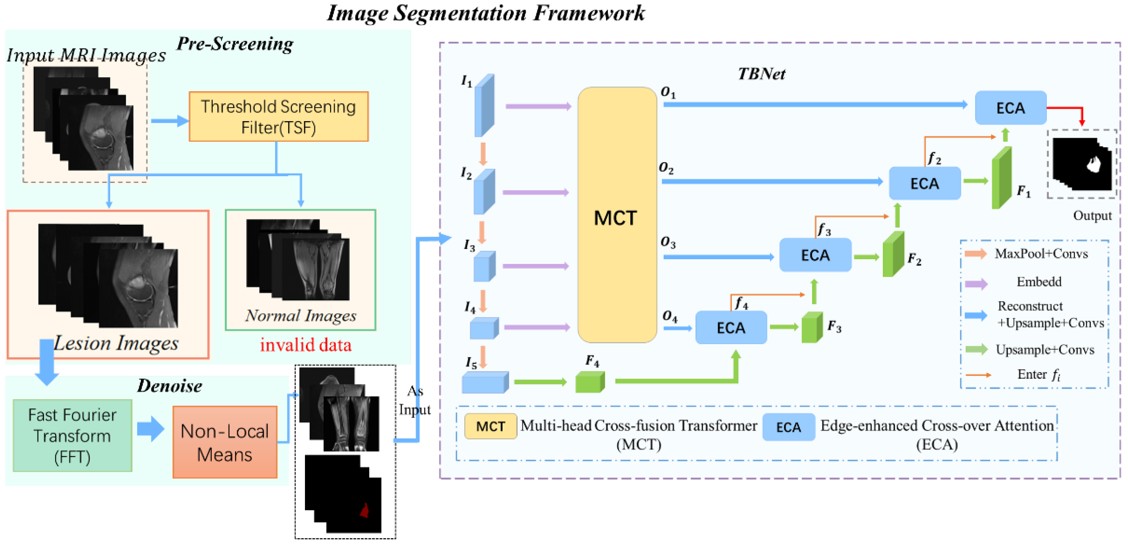
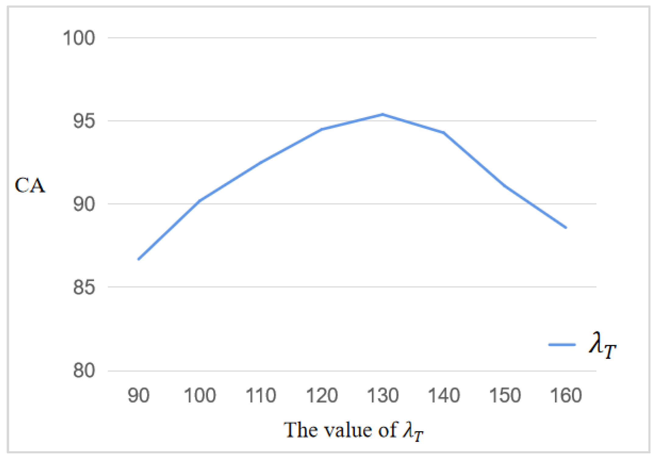
3.1.1. MRI Image Pre-Screening Based on Threshold Screening Filter (TSF)
3.1.2. Noise Reduction Based on Fast NLM Algorithm
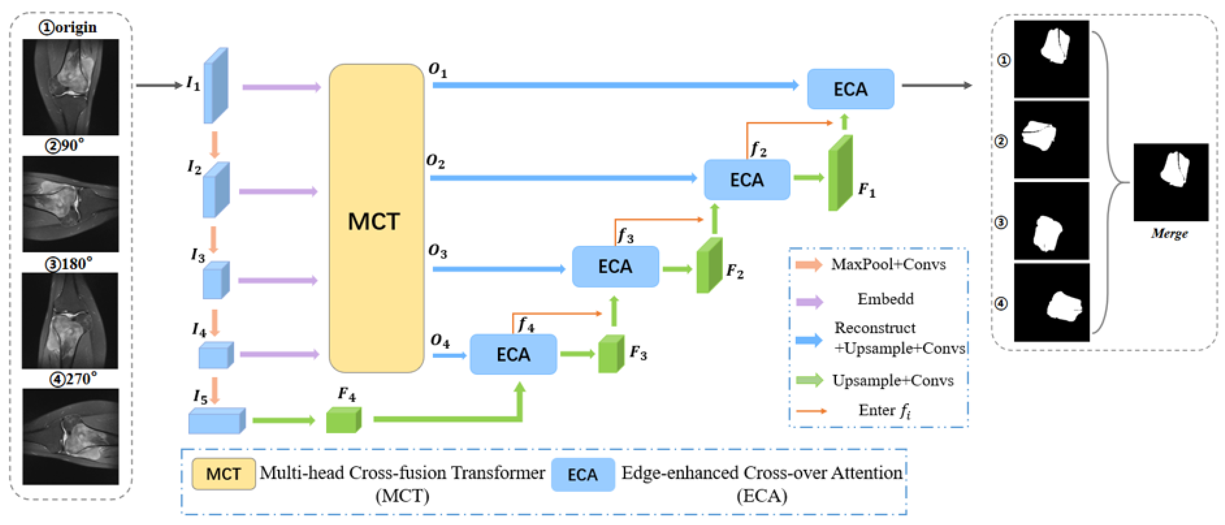
3.2. Tumor Localization
- (1)
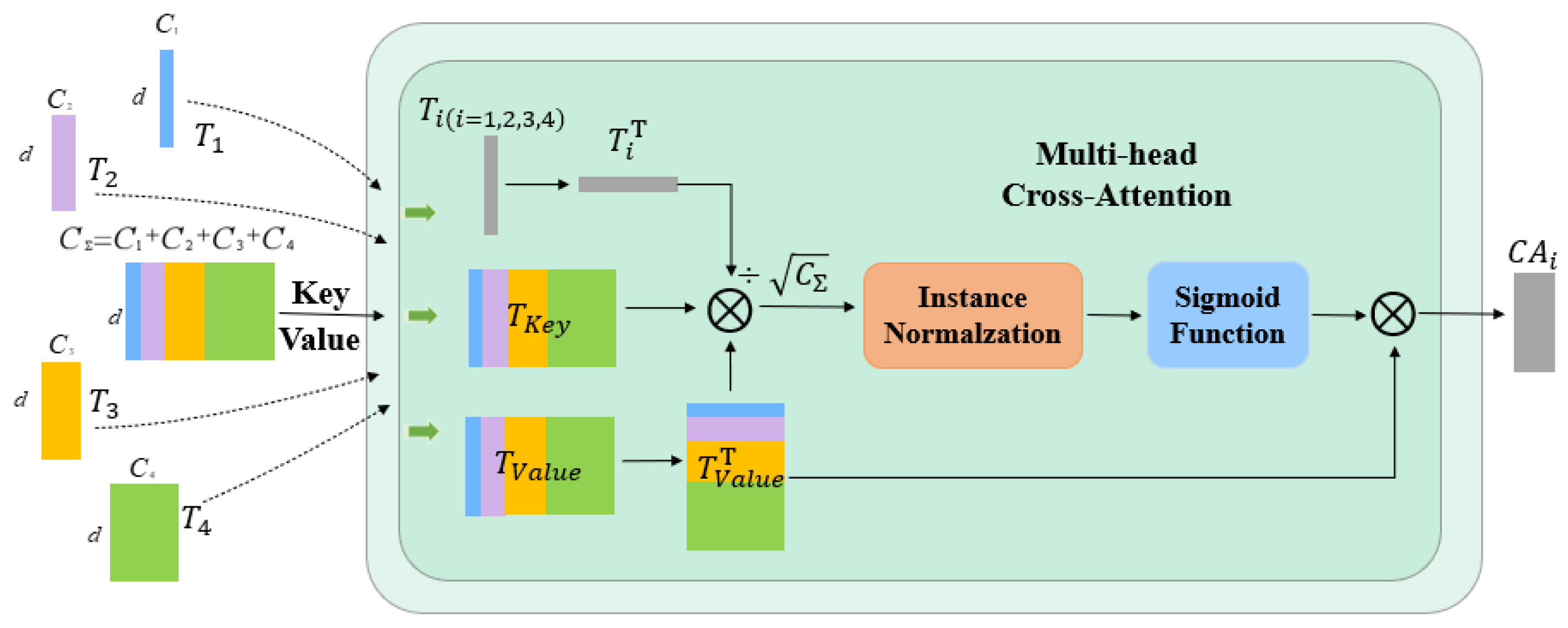
- Multi-head cross-fusion transformer (MCT) for encoder feature transformation
- (2)
- Edge-Enhanced Cross Attention (ECA) module for decoder feature fusion
4. Experimental Analysis
4.1. Dataset
4.2. Evaluation Metrics
4.3. Algorithm Comparison
- (1)
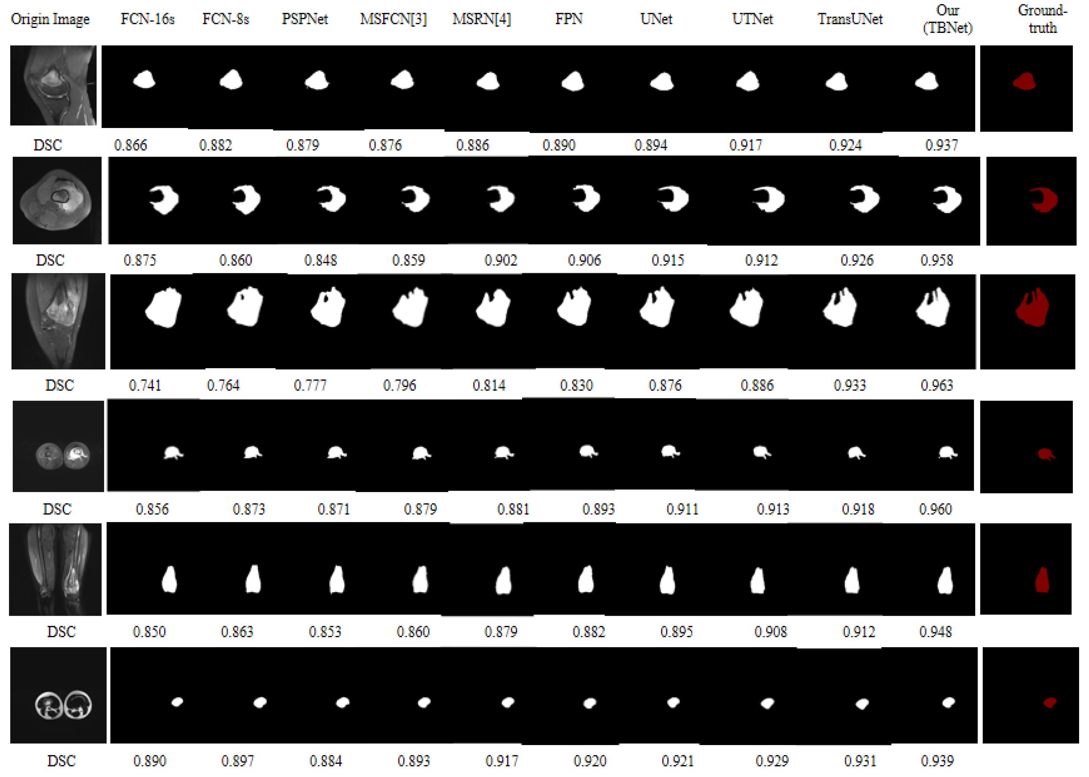
- A fully convolutional network (FCN) is a pixel-level classification of images, using skip structures to achieve fine segmentation [57]. In this paper, 2 networks with 8 and 16 up-sampling are used FCN-8s and FCN-16s.
- (2)
- The PSPNet focuses on the pyramid pool module as a technique of extracting global contextual information, collecting and fusing contextual information at various scales, thus being particularly effective in acquiring global information [58].
- (3)
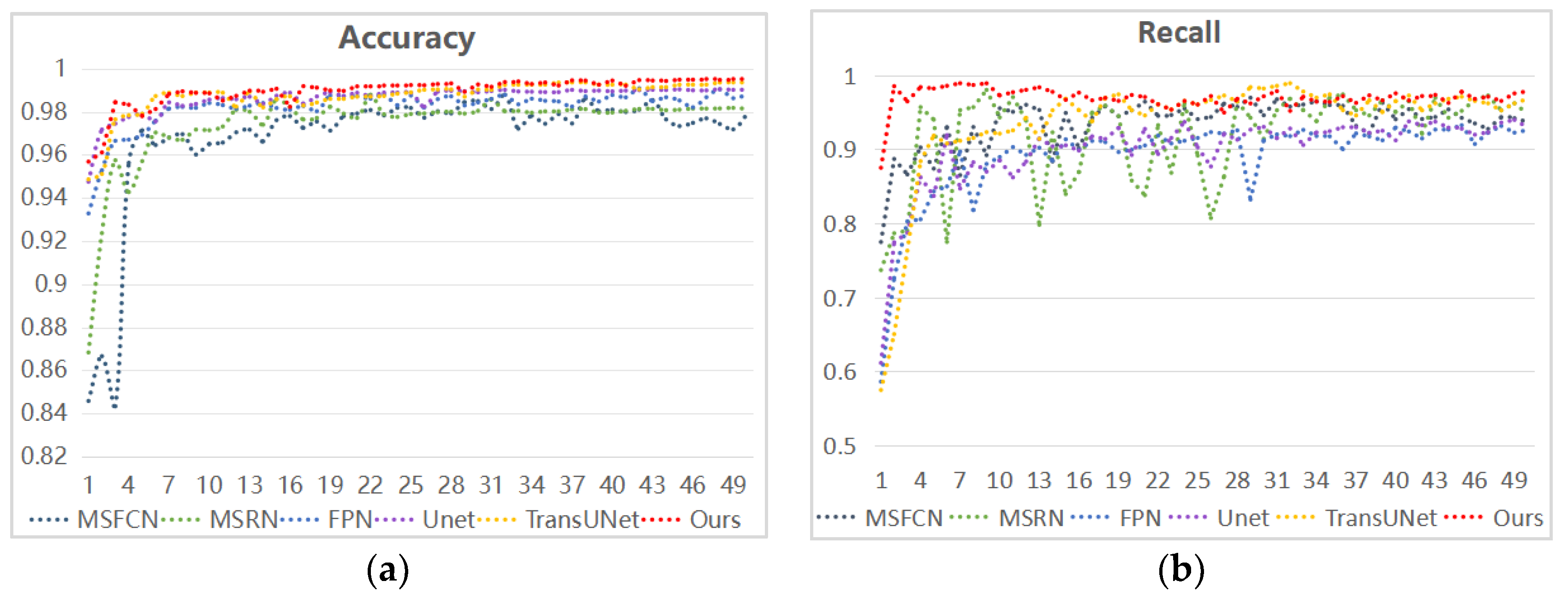
- The MSFCN is a fully convolutional network with many supervised lateral output layers for automatic volume segmentation [59]. To enable the effective learning of local and global visual features, a supervised lateral layer is added to the three layers of the convolutional network to provide a system-level structure to guide multidimensional feature learning.
- (4)
- Multi-scale residual networks (MSRN) [60] can adaptively identify image features at different scales and make them interact to produce effective high-resolution picture information. The generation of structural multiscale residual block MSRBs is achieved by combining convolution kernels of different sizes on the basis of the creation of residual blocks.
- (5)
- U-Net is a symmetric U-shaped structure that enables image-semantic-level segmentation [61]. The left side is a convolutional layer (systolic path) responsible for feature extraction and the right side is an up-sampling layer (extended path) responsible for feature reduction. It can use very little data to obtain the best results.
- (6)
- Feature pyramid networks (FPN) [62] use low-level feature semantic information with accurate target locations and information-rich high-level feature semantic information to make predictions independently at different feature layers using multi-scale feature fusion.
- (7)
- The UTNet network [24] hybrid transformer design incorporates self-attention into convolutional neural networks. The self-attention module is used in both the encoder and decoder in UTNet, and it is combined with relative position coding to considerably minimize the complexity of the self-attention process. In addition, a self-attentive decoder is presented to recover fine-grained features from the encoder’s skipped connections, addressing the problem that the transformer requires a considerable quantity of data to acquire the visual sensing bias.
- (8)
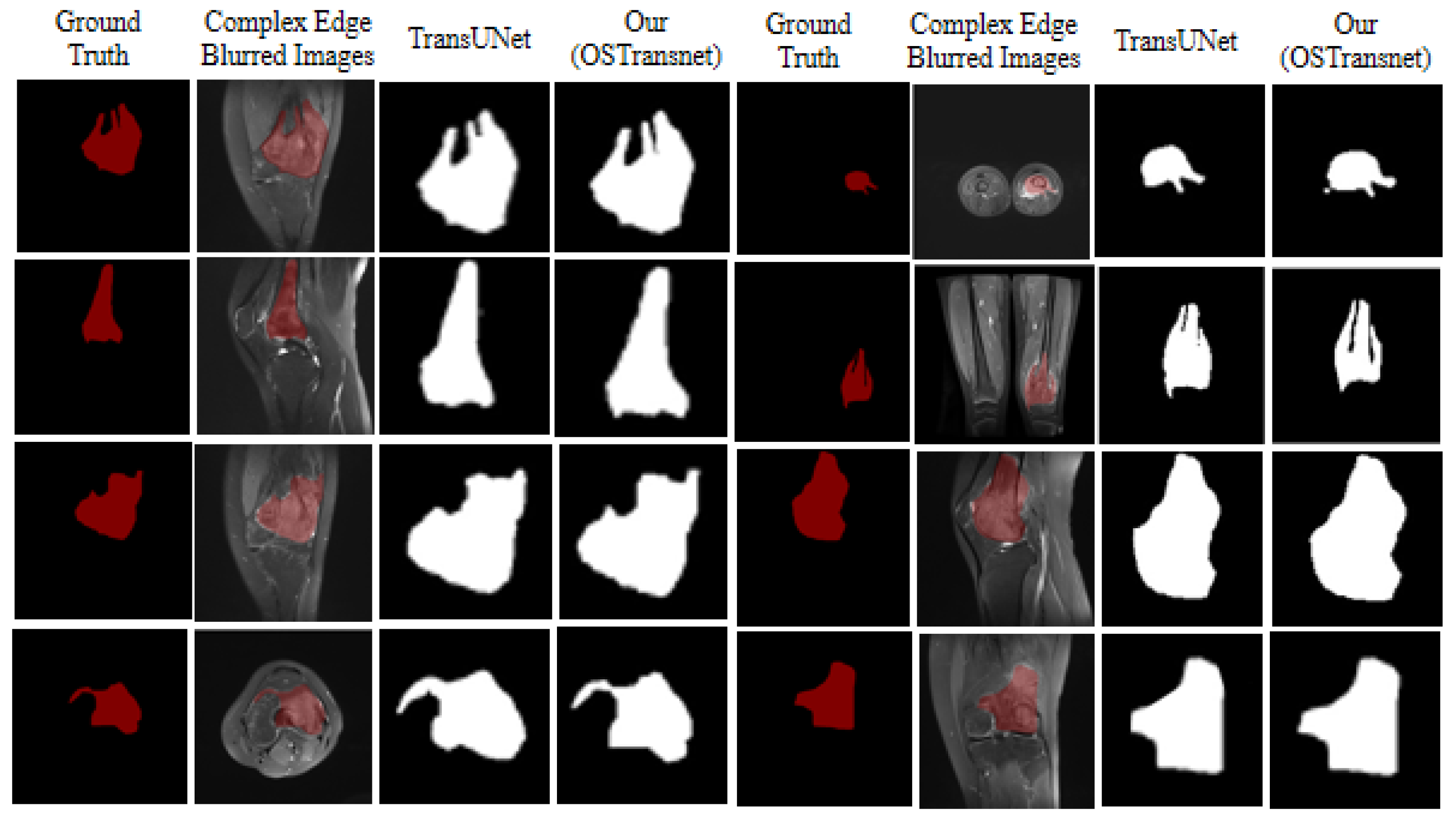
- The TransUNet network [21] combines the advantages of a transformer and U-Net in a hybrid CNN transformer design. The global context extraction encodes the tokenized picture blocks in the CNN feature map as input sequences by the transformer. The decoder performs up-sampling to achieve accurate localization. This operation is performed before combining the encoded features with the high-resolution CNN feature map.
4.4. Influence of Super Parameters
4.5. Results
4.6. Discussion
5. Conclusions
Author Contributions
Funding
Institutional Review Board Statement
Informed Consent Statement
Data Availability Statement
Conflicts of Interest
References
- Corre, I.; Verrecchia, F.; Crenn, V.; Redini, F.; Trichet, V. The Osteosarcoma Microenvironment: A Complex but Targetable Ecosystem. Cells 2020, 9, 976. [Google Scholar] [CrossRef] [PubMed]
- Wu, J.; Xiao, P.; Huang, H.; Gou, F.; Zhou, Z.; Dai, Z. An Artificial Intelligence Multiprocessing Scheme for the Diagnosis of Osteosarcoma MRI Images. IEEE J. Biomed. Health Inform. 2022, 26, 4656–4667. [Google Scholar] [CrossRef] [PubMed]
- Gao, S.-S.; Wang, Y.-J.; Zhang, G.-X.; Zhang, W.-T. Potential diagnostic value of miRNAs in peripheral blood for osteosarcoma: A meta-analysis. J. Bone Oncol. 2020, 23, 100307. [Google Scholar] [CrossRef]
- Wu, J.; Guo, Y.; Gou, F.; Dai, Z. A medical assistant segmentation method for MRI images of osteosarcoma based on DecoupleSegNet. Int. J. Intell. Syst. 2022, 37, 8436–8461. [Google Scholar] [CrossRef]
- Wang, L.; Yu, L.; Zhu, J.; Tang, H.; Gou, F.; Wu, J. Auxiliary Segmentation Method of Osteosarcoma in MRI Images Based on Denoising and Local Enhancement. Healthcare 2022, 10, 1468. [Google Scholar] [CrossRef] [PubMed]
- Chouhan, S.S.; Kaul, A.; Singh, U.P. Image Segmentation Using Computational Intelligence Techniques: Review. Arch. Comput. Methods Eng. 2019, 26, 533–596. [Google Scholar] [CrossRef]
- Heo, Y.-C.; Kim, K.; Lee, Y. Image Denoising Using Non-Local Means (NLM) Approach in Magnetic Resonance (MR) Imaging: A Systematic Review. Appl. Sci. 2020, 10, 7028. [Google Scholar] [CrossRef]
- Tang, H.; Huang, H.; Liu, J.; Zhu, J.; Gou, F.; Wu, J. AI-Assisted Diagnosis and Decision-Making Method in Developing Countries for Osteosarcoma. Healthcare 2022, 10, 2313. [Google Scholar] [CrossRef]
- Chang, L.; Wu, J.; Moustafa, N.; Bashir, A.K.; Yu, K. AI-Driven Synthetic Biology for Non-Small Cell Lung Cancer Drug Effectiveness-Cost Analysis in Intelligent Assisted Medical Systems. IEEE J. Biomed. Health Inform. 2021, 26, 5055–5066. [Google Scholar] [CrossRef]
- Gou, F.; Wu, J. Data Transmission Strategy Based on Node Motion Prediction IoT System in Opportunistic Social Networks. Wirel. Pers. Commun. 2022, 126, 1751–1768. [Google Scholar] [CrossRef]
- Ling, Z.; Yang, S.; Gou, F.; Dai, Z.; Wu, J. Intelligent Assistant Diagnosis System of Osteosarcoma MRI Image Based on Transformer and Convolution in Developing Countries. IEEE J. Biomed. Health Inform. 2022, 26, 5563–5574. [Google Scholar] [CrossRef] [PubMed]
- Zhan, X.; Liu, J.; Long, H.; Zhu, J.; Tang, H.; Gou, F.; Wu, J. An Intelligent Auxiliary Framework for Bone Malignant Tumor Lesion Segmentation in Medical Image Analysis. Diagnostics 2023, 13, 223. [Google Scholar] [CrossRef] [PubMed]
- Shen, Y.; Gou, F.; Dai, Z. Osteosarcoma MRI Image-Assisted Segmentation System Base on Guided Aggregated Bilateral Network. Mathematics 2022, 10, 1090. [Google Scholar] [CrossRef]
- Gou, F.; Wu, J. An Attention-based AI-assisted Segmentation System for Osteosarcoma MRI Images. In Proceedings of the 2022 IEEE International Conference on Bioinformatics and Biomedicine (BIBM), Las Vegas, NV, USA, 6–8 December 2022; pp. 1539–1543. [Google Scholar] [CrossRef]
- Wu, J.; Liu, Z.; Gou, F.; Zhu, J.; Tang, H.; Zhou, X.; Xiong, W. BA-GCA Net: Boundary-Aware Grid Contextual Attention Net in Osteosarcoma MRI Image Segmentation. Comput. Intell. Neurosci. 2022, 2022, 3881833. [Google Scholar] [CrossRef] [PubMed]
- Zhuang, Q.; Dai, Z.; Wu, J. Deep Active Learning Framework for Lymph Node Metastasis Prediction in Medical Support System. Comput. Intell. Neurosci. 2022, 2022, 4601696. [Google Scholar] [CrossRef] [PubMed]
- Jiao, Y.; Qi, H.; Wu, J. Capsule network assisted electrocardiogram classification model for smart healthcare. Biocybern. Biomed. Eng. 2022, 42, 543–555. [Google Scholar] [CrossRef]
- Gou, F.; Liu, J.; Zhu, J.; Wu, J. A Multimodal Auxiliary Classification System for Osteosarcoma Histopathological Images Based on Deep Active Learning. Healthcare 2022, 10, 2189. [Google Scholar] [CrossRef]
- Lv, B.; Liu, F.; Gou, F.; Wu, J. Multi-Scale Tumor Localization Based on Priori Guidance-Based Segmentation Method for Osteosarcoma MRI Images. Mathematics 2022, 10, 2099. [Google Scholar] [CrossRef]
- Wu, J.; Zhou, L.; Gou, F.; Tan, Y. A Residual Fusion Network for Osteosarcoma MRI Image Segmentation in Developing Countries. Comput. Intell. Neurosci. 2022, 2022, 7285600. [Google Scholar] [CrossRef]
- Chen, J.; Lu, Y.; Yu, Q.; Luo, X.; Adeli, E.; Wang, Y.; Lu, L.; Yuille, A.L.; Zhou, Y. TransUNet: Transformers Make Strong Encoders for Medical Image Segmentation. arXiv 2021, arXiv:2102.04306. [Google Scholar]
- Liu, Z.; Lin, Y.; Cao, Y.; Hu, H.; Wei, Y.; Zhang, Z.; Lin, S.; Guo, B. Swin Transformer: Hierarchical Vision Transformer Using Shifted Windows. arXiv 2021, arXiv:2103.14030. [Google Scholar]
- Valanarasu, J.M.J.; Oza, P.; Hacihaliloglu, I.; Patel, V.M. Medical transformer: Gated axial-attention for medical image segmentation. In Medical Image Computing and Computer Assisted Intervention–MICCAI 2021: 24th International Conference, Strasbourg, France, 27 September –1 October 2021, Proceedings, Part I 24; Springer International Publishing: New York, NY, USA, 2021; pp. 36–46. [Google Scholar]
- Gao, Y.; Zhou, M.; Metaxas, D. UTNet: A Hybrid Transformer Architecture for Medical Image Segmentation. arXiv 2021, arXiv:2107.00781. [Google Scholar]
- Ji, Y.; Zhang, R.; Wang, H.; Li, Z.; Wu, L.; Zhang, S.; Luo, P. Multi-compound transformer for accurate biomedical image segmentation. In Medical Image Computing and Computer Assisted Intervention–MICCAI 2021: 24th International Con-ference, Strasbourg, France, September 27–October 1 2021, Proceedings, Part I 24; Springer International Publishing: New York, NY, USA, 2021; pp. 326–336. [Google Scholar]
- Petit, O.; Thome, N.; Rambour, C.; Soler, L. U-net transformer: Self and cross attention for medical image segmentation. arXiv 2021, arXiv:2103.06104. [Google Scholar]
- Zhang, Y.; Liu, H.; Hu, Q. TransFuse: Fusing Transformers and CNNs for Medical Image Segmentation. In Medical Image Computing and Computer Assisted Intervention–MICCAI 2021: 24th International Conference, Strasbourg, France, 27 September–1 October 2021, Proceedings, Part I 24; Springer International Publishing: New York, NY, USA, 2021; pp. 14–24. [Google Scholar] [CrossRef]
- Heidari, M.; Kazerouni, A.; Soltany, M.; Azad, R.; Aghdam, E.K.; Cohen-Adad, J.; Merhof, D. HiFormer: Hierarchical Multi-scale Representations Using Transformers for Medical Image Segmentation. In Proceedings of the IEEE/CVF Winter Conference on Applications of Computer Vision, Waikoloa, HI, USA, 2–7 January 2023; pp. 6191–6201. [Google Scholar] [CrossRef]
- Wei, C.; Ren, S.; Guo, K.; Hu, H.; Liang, J. High-Resolution Swin Transformer for Automatic Medical Image Segmentation. arXiv 2022, arXiv:2207.11553. [Google Scholar]
- Sun, R.; Pang, Y.; Li, W. Efficient Lung Cancer Image Classification and Segmentation Algorithm Based on an Improved Swin Transformer. Electronics 2023, 12, 1024. [Google Scholar] [CrossRef]
- Valanarasu, J.M.J.; Patel, V.M. UNeXt: MLP-Based Rapid Medical Image Segmentation Network. arXiv 2022, arXiv:2203.04967. [Google Scholar]
- Tran, M.; Vo-Ho, V.; Quinn, K.; Nguyen, H.V.; Luu, K.; Le, N.T. CapsNet for Medical Image Segmentation. arXiv 2022, arXiv:2203.08948. [Google Scholar]
- Anisuzzaman, D.; Barzekar, H.; Tong, L.; Luo, J.; Yu, Z. A deep learning study on osteosarcoma detection from histological images. Biomed. Signal Process. Control. 2021, 69, 102931. [Google Scholar] [CrossRef]
- Fu, Y. Deep Model with Siamese Network for Viability and Necrosis Tumor Assessment in Osteosarcoma. Med. Phys. 2019, 47, 4895–4905. [Google Scholar] [CrossRef]
- Barzekar, H.; Yu, Z. C-Net: A Reliable Convolutional Neural Network for Biomedical Image Classification. arXiv 2020. [Google Scholar] [CrossRef]
- Nabid, R.; Rahman, M.; Hossain, M.F. Classification of Osteosarcoma Tumor from Histological Image Using Sequential RCNN. In Proceedings of the 2020 11th International Conference on Electrical and Computer Engineering (ICECE), Dhaka, Bangladesh, 17–19 December 2020. [Google Scholar]
- D’Acunto, M.; Martinelli, M.; Moroni, D. From human mesenchymal stromal cells to osteosarcoma cells classification by deep learning. J. Intell. Fuzzy Syst. 2019, 37, 7199–7206. [Google Scholar] [CrossRef]
- Parlak, Ş.; Ergen, F.B.; Yüksel, G.Y.; Karakaya, J.; Aydın, G.B.; Kösemehmetoğlu, K.; Aydıngöz, Ü. Diffusion-weighted imaging for the differentiation of Ewing sarcoma from osteosarcoma. Skelet. Radiol. 2021, 50, 2023–2030. [Google Scholar] [CrossRef] [PubMed]
- Ho, D.J.; Agaram, N.P.; Schüffler, P.J.; Vanderbilt, C.M.; Jean, M.-H.; Hameed, M.R.; Fuchs, T.J. Deep Interactive Learning: An Efficient Labeling Approach for Deep Learning-Based Osteosarcoma Treatment Response Assessment. In International Conference on Medical Image Computing and Computer-Assisted Intervention; Springer: Cham, Switzerland, 2020; pp. 540–549. [Google Scholar]
- Im, H.-J.; Solaiyappan, M.; Lee, I.; Bradshaw, T.; Daw, N.C.; Navid, F.; Shulkin, B.L.; Cho, S.Y. Multi-level otsu method to define metabolic tumor volume in positron emission tomography. Am. J. Nucl. Med. Mol. Imaging 2018, 8, 373–386. [Google Scholar] [PubMed]
- Shuai, L.; Gao, X.; Wang, J. Wnet ++: A Nested W-shaped Network with Multiscale Input and Adaptive Deep Supervision for Osteosarcoma Segmentation. In Proceedings of the 2021 IEEE 4th International Conference on Electronic Information and Communication Technology (ICEICT), Xi’an, China, 8–20 August 2021; pp. 93–99. [Google Scholar] [CrossRef]
- Huang, W.-B.; Wen, D.; Yan, Y.; Yuan, M.; Wang, K. Multi-target osteosarcoma MRI recognition with texture context features based on CRF. In Proceedings of the 2016 International Joint Conference on Neural Networks (IJCNN), Vancouver, BC, Canada, 24–29 July 2016; pp. 3978–3983. [Google Scholar] [CrossRef]
- Gou, F.; Wu, J. Novel data transmission technology based on complex IoT system in opportunistic social networks. Peer-to-Peer Netw. Appl. 2022, 185, 1–18. [Google Scholar] [CrossRef]
- Xiong, W.; Chen, H.; Jiao, Y.; Yang, M.; Zhou, X. A user cache management and cooperative transmission mechanism based on edge community computing in opportunistic social networks. IET Commun. 2022, 16, 2045–2058. [Google Scholar] [CrossRef]
- Zhou, Z.; Gou, F.; Tan, Y.; Wu, J. A Cascaded Multi-Stage Framework for Automatic Detection and Segmentation of Pulmonary Nodules in Developing Countries. IEEE J. Biomed. Health Inform. 2022, 26, 5619–5630. [Google Scholar] [CrossRef]
- Qin, Y.; Li, X.; Wu, J.; Yu, K. A management method of chronic diseases in the elderly based on IoT security environment. Comput. Electr. Eng. 2022, 102, 108188. [Google Scholar] [CrossRef]
- Gou, F.; Wu, J. Triad link prediction method based on the evolutionary analysis with IoT in opportunistic social networks. Comput. Commun. 2021, 181, 143–155. [Google Scholar] [CrossRef]
- Han, K.; Hhan, M.; Cheon, J.H. Improved Homomorphic Discrete Fourier Transforms and FHE Bootstrapping. IEEE Access 2019, 7, 57361–57370. [Google Scholar] [CrossRef]
- Deng, Y.; Gou, F.; Wu, J. Hybrid data transmission scheme based on source node centrality and community reconstruction in opportunistic social networks. Peer-to-Peer Netw. Appl. 2021, 14, 3460–3472. [Google Scholar] [CrossRef]
- Wu, J.; Yu, L.; Gou, F. Data transmission scheme based on node model training and time division multiple access with IoT in opportunistic social networks. Peer-to-Peer Netw. Appl. 2022, 15, 2719–2743. [Google Scholar] [CrossRef]
- Liu, F.; Zhu, J.; Lv, B.; Yang, L.; Sun, W.; Dai, Z.; Gou, F.; Wu, J. Auxiliary Segmentation Method of Osteosarcoma MRI Image Based on Transformer and U-Net. Comput. Intell. Neurosci. 2022, 2022, 9990092. [Google Scholar] [CrossRef]
- Yu, G.; Wu, J. Efficacy prediction based on attribute and multi-source data collaborative for auxiliary medical system in developing countries. Neural Comput. Appl. 2022, 34, 5497–5512. [Google Scholar] [CrossRef]
- Suriyan, K.; Ramaingam, N.; Rajagopal, S.; Sakkarai, J.; Asokan, B.; Alagarsamy, M. Performance analysis of peak sig-nal-to-noise ratio and multipath source routing using different denoising method. Bull. Electr. Eng. Infor-Matics 2022, 11, 286–292. [Google Scholar] [CrossRef]
- Wu, J.; Yang, S.; Gou, F.; Zhou, Z.; Xie, P.; Xu, N.; Dai, Z. Intelligent Segmentation Medical Assistance System for MRI Images of Osteosarcoma in Developing Countries. Comput. Math. Methods Med. 2022, 2022, 7703583. [Google Scholar] [CrossRef] [PubMed]
- Ouyang, T.; Yang, S.; Gou, F.; Dai, Z.; Wu, J. Rethinking U-Net from an Attention Perspective with Transformers for Osteosarcoma MRI Image Segmentation. Comput. Intell. Neurosci. 2022, 2022, 7973404. [Google Scholar] [CrossRef]
- Liu, F.; Gou, F.; Wu, J. An Attention-Preserving Network-Based Method for Assisted Segmentation of Osteosarcoma MRI Images. Mathematics 2022, 10, 1665. [Google Scholar] [CrossRef]
- Long, J.; Shelhamer, E.; Darrell, T. Fully convolutional networks for semantic segmentation. In Proceedings of the IEEE Conference on Computer Vision and Pattern Recognition, Boston, MA, USA, 7–12 June 2015; pp. 3431–3440. [Google Scholar]
- Zhao, H.; Shi, J.; Qi, X.; Wang, X.; Jia, J. Pyramid scene parsing network. In Proceedings of the IEEE Conference on Computer Vision and Pattern Recognition, Honolulu, HI, USA, 21–26 July 2017; pp. 2881–2890. [Google Scholar]
- Huang, L.; Xia, W.; Zhang, B.; Qiu, B.; Gao, X. MSFCNmultiple supervised fully convolutional networks for the osteosar-coma segmentation of CT images. Comput. Methods Programs Biomed. 2017, 143, 67–74. [Google Scholar] [CrossRef]
- Zhang, R.; Huang, L.; Xia, W.; Zhang, B.; Qiu, B.; Gao, X. Multiple supervised residual network for osteosarcoma segmentation in CT images. Comput. Med. Imaging Graph. 2018, 63, 1–8. [Google Scholar] [CrossRef]
- Ronneberger, O.; Fischer, P.; Brox, T. U-Net: Convolutional networks for biomedical image segmentation. In Medical Image Computing and Computer-Assisted Intervention 2015; Navab, N., Hornegger, J., Wells, W.M., Frangi, A.F., Eds.; Springer International Publishing: Cham, Switzerland, 2015; pp. 234–241. [Google Scholar] [CrossRef]
- Lin, T.-Y.; Dollár, P.; Girshick, R.; He, K.; Hariharan, B.; Belongie, S. Feature pyramid networks for object detection. In Proceedings of the IEEE Conference on Computer Vision and Pattern Recognition, Honolulu, HI, USA, 21–26 July 2017; pp. 2117–2125. [Google Scholar]



| Notation | Meaning |
|---|---|
| The osteosarcoma MRI image domain | |
| initialized noise reduction | |
| The hyperparameter of the TSF | |
| p | The Euclidean distance |
| The fixed threshold parameter | |
| X | MR noise data |
| S | Original MR images and Phases |
| k(a) | Noise component intensity of the i-th pixel |
| The modified Bessel function | |
| Real and virtual channel impact | |
| k(a) | The activation function of TSF |
| ) | The function of weighted similarity |
| The DCT transform operator (FFT) and its inverse | |
| Z(i) | The normalization constant |
| The boundary level set | |
| Search window side half-length | |
| The similarity matrix and Mask edge diagram | |
| The instance normalization | |
| The ReLU operator | |
| Supplementary layer feature map for layer i − 1 | |
| The probabilistic output of the network |
| True | Lesion Image | Normal Image | |
|---|---|---|---|
| Predicted | |||
| Lesion image | 49 | 4 | |
| Normal image | 3 | 44 | |
| Model | ACC | IOU | DSC | Pre | Re | F1 |
|---|---|---|---|---|---|---|
| TBNet | 0.991 | 0.904 | 0.931 | 0.927 | 0.974 | 0.949 |
| TBNet+Denoise | 0.993 | 0.915 | 0.943 | 0.932 | 0.968 | 0.951 |
| TBNet+Denoise+Pre-Screening | 0.997 | 0.915 | 0.949 | 0.941 | 0.969 | 0.954 |
| Model | ACC | IOU | DSC | Pre | Re | F1 | FLOPS | Params |
|---|---|---|---|---|---|---|---|---|
| FCN-16s | 0.989 | 0.824 | 0.859 | 0.922 | 0.882 | 0.900 | 187.35 G | 122.4 M |
| FCN-8s | 0.993 | 0.830 | 0.876 | 0.941 | 0.873 | 0.901 | 187.18 G | 122.4 M |
| PSPNet | 0.975 | 0.772 | 0.870 | 0.856 | 0.888 | 0.872 | 103.55 G | 47.70 M |
| MSFCN | 0.991 | 0.841 | 0.874 | 0.881 | 0.936 | 0.906 | 1642.43 G | 24.53 M |
| MSRN | 0.988 | 0.853 | 0.887 | 0.893 | 0.945 | 0.918 | 1346.12 G | 12.42 M |
| FPN | 0.989 | 0.852 | 0.888 | 0.914 | 0.924 | 0.919 | 134.14 G | 47.82 M |
| UNet | 0.990 | 0.867 | 0.892 | 0.922 | 0.924 | 0.923 | 160.16 G | 17.26 M |
| UTNet | 0.990 | 0.879 | 0.919 | 0.924 | 0.934 | 0.936 | 264.27 G | 52.53 M |
| TransUNet | 0.993 | 0.898 | 0.923 | 0.919 | 0.959 | 0.948 | 240.26 G | 40.38 M |
| Our(TBNet)+Denoise+Pre-Screening | 0.997 | 0.915 | 0.949 | 0.941 | 0.969 | 0.954 | 235.26 G | 36.51 M |
Disclaimer/Publisher’s Note: The statements, opinions and data contained in all publications are solely those of the individual author(s) and contributor(s) and not of MDPI and/or the editor(s). MDPI and/or the editor(s) disclaim responsibility for any injury to people or property resulting from any ideas, methods, instructions or products referred to in the content. |
© 2023 by the authors. Licensee MDPI, Basel, Switzerland. This article is an open access article distributed under the terms and conditions of the Creative Commons Attribution (CC BY) license (https://creativecommons.org/licenses/by/4.0/).
Share and Cite
Lv, B.; Liu, F.; Li, Y.; Nie, J.; Gou, F.; Wu, J. Artificial Intelligence-Aided Diagnosis Solution by Enhancing the Edge Features of Medical Images. Diagnostics 2023, 13, 1063. https://doi.org/10.3390/diagnostics13061063
Lv B, Liu F, Li Y, Nie J, Gou F, Wu J. Artificial Intelligence-Aided Diagnosis Solution by Enhancing the Edge Features of Medical Images. Diagnostics. 2023; 13(6):1063. https://doi.org/10.3390/diagnostics13061063
Chicago/Turabian StyleLv, Baolong, Feng Liu, Yulin Li, Jianhua Nie, Fangfang Gou, and Jia Wu. 2023. "Artificial Intelligence-Aided Diagnosis Solution by Enhancing the Edge Features of Medical Images" Diagnostics 13, no. 6: 1063. https://doi.org/10.3390/diagnostics13061063
APA StyleLv, B., Liu, F., Li, Y., Nie, J., Gou, F., & Wu, J. (2023). Artificial Intelligence-Aided Diagnosis Solution by Enhancing the Edge Features of Medical Images. Diagnostics, 13(6), 1063. https://doi.org/10.3390/diagnostics13061063


