Article

Histological Changes in Portal Cavernoma Cholangiopathy

Departments of Pathology Radiodiagnosis Hepatology, Institute of Liver and Biliary Sciences, D1, Vasant Kunj, New Delhi 110070, India
*
Author to whom correspondence should be addressed.
These authors contributed equally to this work.
Diagnostics 2023, 13(3), 436; https://doi.org/10.3390/diagnostics13030436

Submission received: 26 December 2022 / Revised: 19 January 2023 / Accepted: 20 January 2023 / Published: 25 January 2023
(This article belongs to the Section Pathology and Molecular Diagnostics)

Abstract

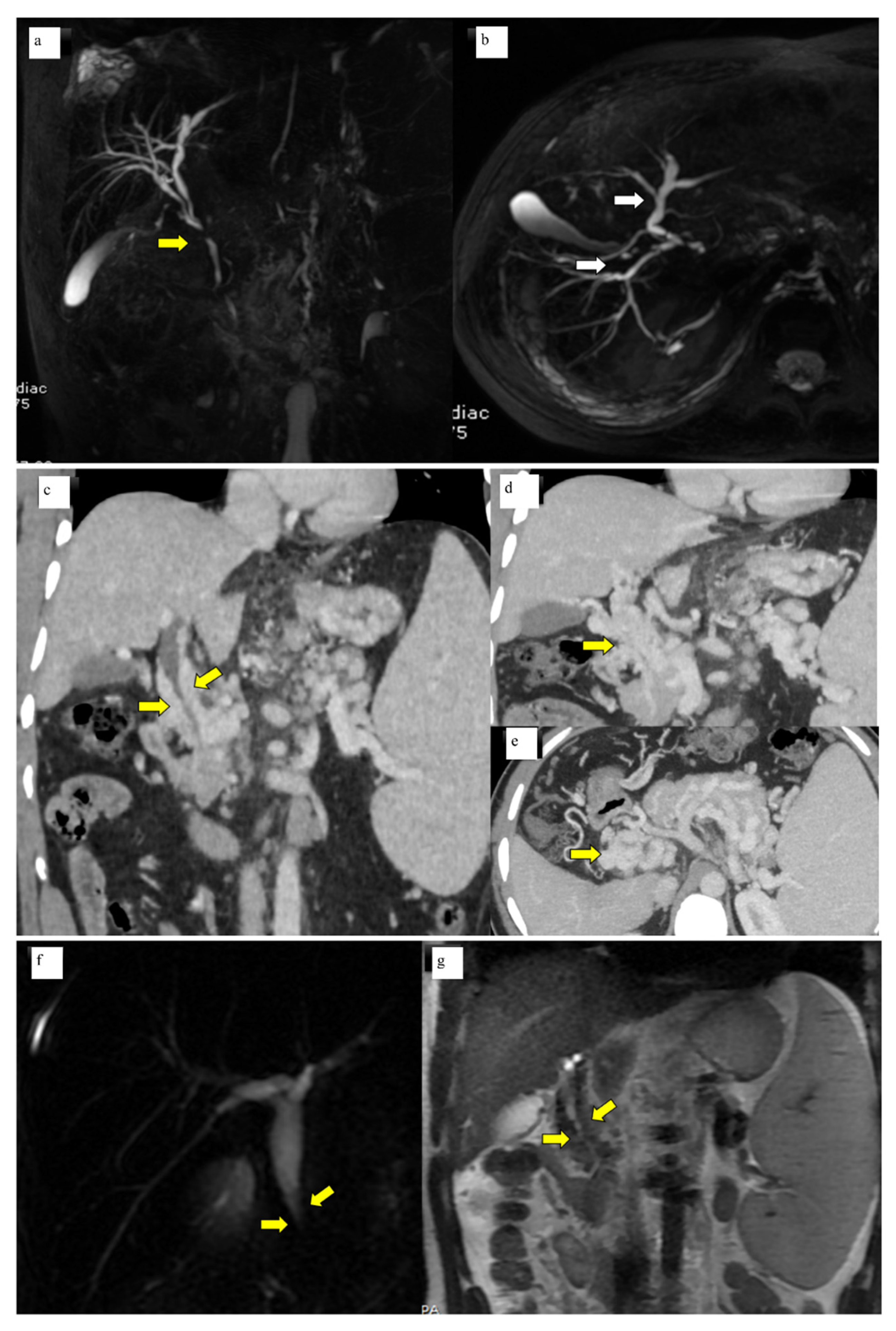
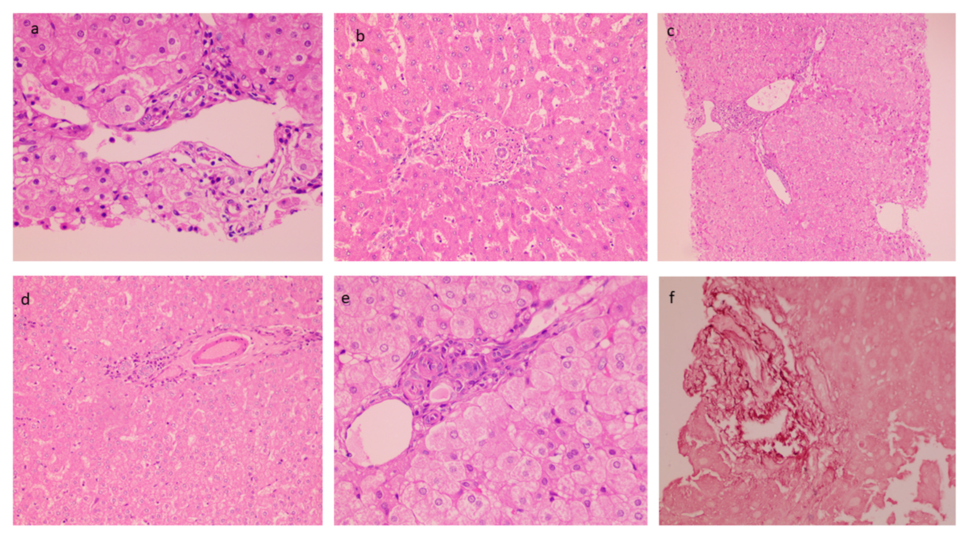
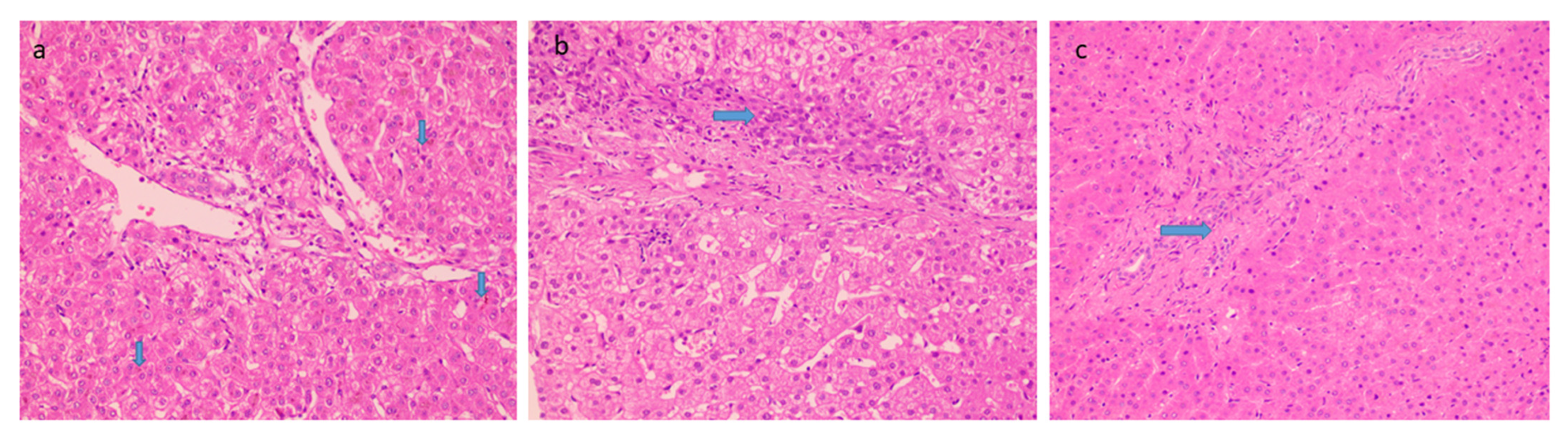
Introduction: Portal cavernoma cholangiopathy (PCC)’ refers to abnormalities of the extrahepatic and intrahepatic bile ducts in patients with portal hypertension. Although there is data on clinical and imaging aspects of PCC, the description of liver pathology has been strikingly deficient. The purpose of this study was to examine the histopathological characteristics of PCC. Patients and Methods: A retrospective study of patients clinically diagnosed with extrahepatic portal vein obstruction (EHPVO) with portal cavernoma cholangiopathy, was conducted. Vascular anatomy was characterized by computerized tomographic angiography, and endoscopic retrograde cholangiography (ERC) and magnetic resonance cholangiography (MRC) were used to characterize the biliary anatomy. Histological features were analyzed by two hepatopathologists in a blinded manner, with mutual discussion to resolve any discrepancies. Results: A total of 50 patients with portal cavernoma cholangiopathy were included in the study. The mean age of the patients was 26.2 ± 11.6 years. Radiologically, bilobar intrahepatic biliary dilatation was seen in 98% with common bile duct abnormality in 100% of patients, along with extrinsic ductal impressions in 77 % of cases. Liver tests were deranged total bilirubin 1.5 mg/dL (IQR 0.8–2.4) and alkaline phosphatase 109.5 IU/L (IQR 70–193). Histologically; dilated multiple portal venous channels (72%), hepatic artery thickening (70%). The presence of aberrant vascular channels around portal tracts (54%), elastosis of portal veins (50%), and bile ductular reaction in (44%) were the other prominent findings. A 12% of cases show focal thin bridges. Advanced fibrosis was not seen in any of the cases. One-fourth of the cases showed concomitant minimal to mild hepatocyte steatosis. Conclusions: Histologically, intrahepatic portal vein and portal tract abnormalities were noted in cases with portal cavernoma cholangiopathy, associated with mild derangement of liver tests.
Keywords: portal biliopathy; portal cavernoma; liver biopsy; portal vein; bile ducts portal biliopathy; portal cavernoma; liver biopsy; portal vein; bile ducts