Review

The Role of the Spleen and the Place of Splenectomy in Autoimmune Hemolytic Anemia—A Review of Current Knowledge

by
Zorica Cvetković
1,2,*,
Nikola Pantić
3,
Mirjana Cvetković
3,
Marijana Virijević
2,3,
Nikica Sabljić
3,
Gligorije Marinković
1,
Vladimir Milosavljević
4,
Zlatko Pravdić
3,
Nada Suvajdžić-Vuković
2,3 and
Mirjana Mitrović
2,3
1
Department of Hematology, University Hospital Medical Center Zemun, 11080 Belgrade, Serbia
2
Medical Faculty, University of Belgrade, 11000 Belgrade, Serbia
3
Clinic for Hematology, University Clinical Center of Serbia, 11000 Belgrade, Serbia
4
Department for HPB Surgery, University Hospital Medical Center Bežanijska Kosa, 11070 Belgrade, Serbia
*
Author to whom correspondence should be addressed.
Diagnostics 2023, 13(18), 2891; https://doi.org/10.3390/diagnostics13182891
Submission received: 31 July 2023 / Revised: 4 September 2023 / Accepted: 6 September 2023 / Published: 9 September 2023
(This article belongs to the Special Issue Diagnosis of Spleen Disorders)

Abstract

:
Autoimmune hemolytic anemia (AIHA) is a rare, very heterogeneous, and sometimes life-threatening acquired hematologic disease characterized by increased red blood cell (RBC) destruction by autoantibodies (autoAbs), either with or without complement involvement. Recent studies have shown that the involvement of T- and B-cell dysregulation and an imbalance of T-helper 2 (Th2) and Th17 phenotypes play major roles in the pathogenesis of AIHA. AIHA can be primary (idiopathic) but is more often secondary, triggered by infections or drug use or as a part of other diseases. As the location of origin of autoAbs and the location of autoAb-mediated RBC clearance, as well as the location of extramedullary hematopoiesis, the spleen is crucially involved in all the steps of AIHA pathobiology. Splenectomy, which was the established second-line therapeutic option in corticosteroid-resistant AIHA patients for decades, has become less common due to increasing knowledge of immunopathogenesis and the introduction of targeted therapy. This article provides a comprehensive overview of current knowledge regarding the place of the spleen in the immunological background of AIHA and the rapidly growing spectrum of novel therapeutic approaches. Furthermore, this review emphasizes the still-existing expediency of laparoscopic splenectomy with appropriate perioperative thromboprophylaxis and the prevention of infection as a safe and reliable therapeutic option in the context of the limited availability of rituximab and other novel therapies.

1. Introduction

Autoimmune hemolytic anemia (AIHA) is an acquired autoimmune disease characterized by the increased destruction of autologous red blood cells (RBCs) due to the presence of immunoglobulin (Ig)G, IgM, IgA, or complements (usually C3d) bound to RBC membrane antigens [1,2,3]. Although this rare, very heterogeneous, and sometimes life-threatening condition occurs across all age groups, with a reported annual incidence of 1–3 per 100,000 persons, the incidence of chronic and relapsing cases increases with age (i.e., over the age of 40) [2].
Shortened RBC survival due to hemolysis clinically manifests in symptoms which include weakness, dyspnea, jaundice, acrocyanosis or Raynaud phenomena, and splenomegaly, and laboratory tests reveal normocytic/macrocytic anemia, reticulocytosis, and elevated indirect (unconjugated) bilirubin and lactate dehydrogenase with reduced or fully consumed haptoglobin. The spherocytosis, agglutination, and polychromasia of RBCs can be seen in a peripheral blood smear. Hemoglobinuria and the presence of urinary hemosiderin indicate the onset of extravascular hemolysis [3].
The severity of AIHA is determined by the type and affinity of autoantibodies (autoAbs), which reduce RBC lifespan, and by the capacity of bone marrow to compensate for hemolysis. A diagnosis of hemolytic anemia is issued when RBC survival drops below 100 days [4]. The spleen plays a major role in the pathobiology of AIHA, both in the synthesis of autoAbs and in the immune destruction of RBCs.

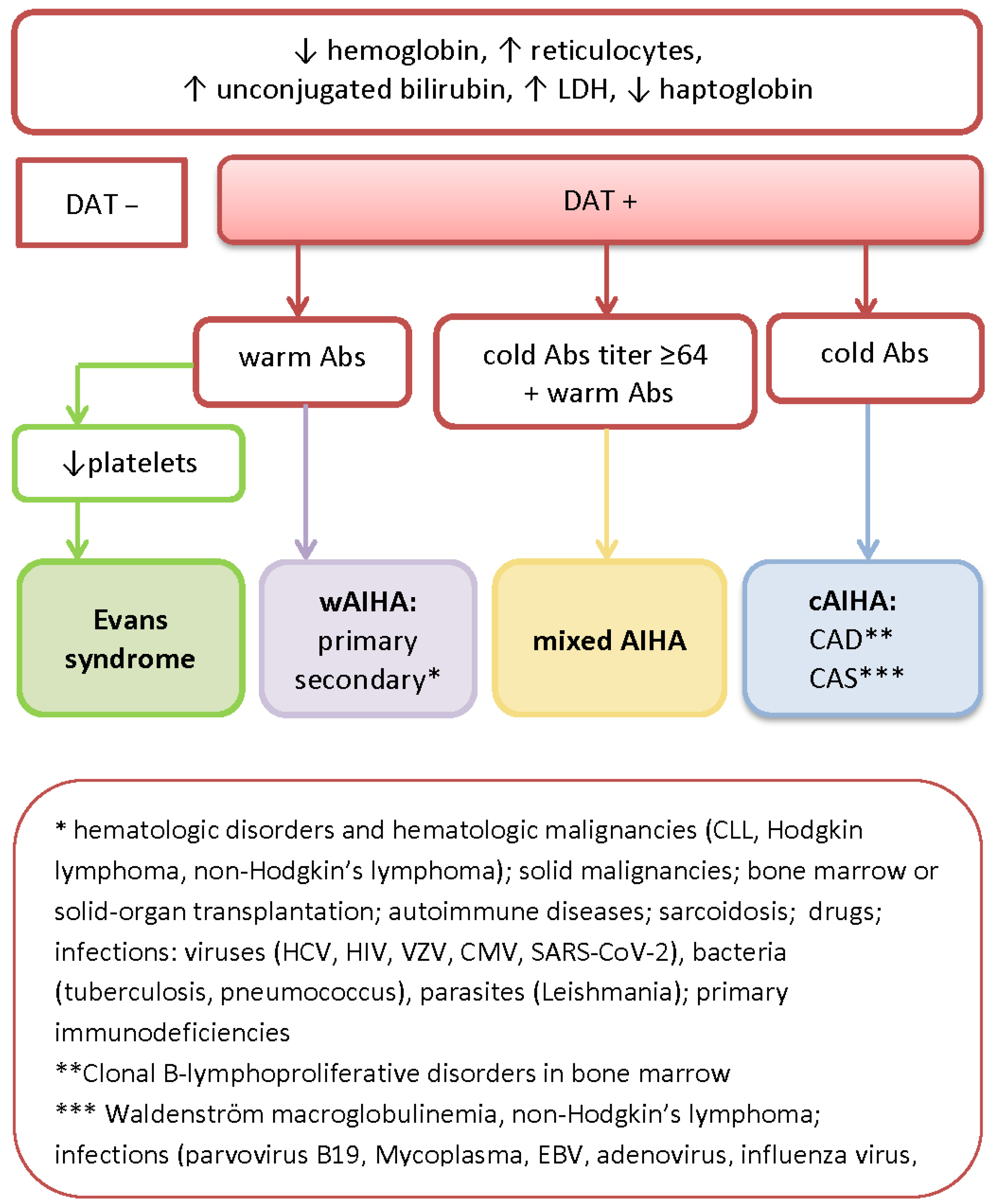
2. Diagnostic Workup, Serological Features, and the Classification of AIHA

The diagnostic hallmark of AIHA is a positive direct Coombs test (antiglobulin test—DAT), and the classification of AIHA into warm AIHA (wAIHA) and cold AIHA (cAIHA) is based on the serological and thermal characteristics of the detected autoAbs. The most common type of AIHA is wAIHA, which accounts for 60–70% cases and is mediated by the autoreactive polyclonal IgG that binds to RBC antigens maximally at 37 °C. The most frequently occurring type of cAIHA, and the second most frequent type of AIHA (20% of all AIHA cases), is cold agglutinin disease (CAD). CAD is mediated in 90% of patients by oligoclonal or clonal IgM autoAbs that maximally react at 4 °C and strongly activate complement due to their high density on RBC surfaces and the structure of IgM [5]. A rare subtype of cAIHA is paroxismal cold hemoglobinuria (PCH). This represents only 1% of all AIHA cases, is usually seen in childhood, and is characterized by transient hemolysis triggered by an upper respiratory tract infection. PCH is defined by DAT positivity only for the complement and the presence of biphasic Donath–Landsteiner Abs, which adsorb and fix the early components of the complement system on the RBC membrane at low temperatures and afterwards, with warming, dissociate from the RBC, leading to the activation of downstream complement cascade effects, the formation of a lytic complex (MAC–membrane attack complex), and the onset of intravascular hemolysis [6].
In 5–10% of patients with the laboratory characteristics of both wAIHA and CAD, the diagnosis of mixed-type AIHA is established [7]. Furthermore, wAIHA can concomitantly or sequentially be associated with other autoimmune cytopenias (thrombocytopenia and, rarely neutropenia), the conditions usually assigned as Evans syndrome [8].
Simplified laboratory algorithms for AIHA diagnosis and the classification of AIHA are shown in Figure 1 and Table 1 according to the autoAb istotype and the temperature at which the autoAbs show maximal binding.
It is important to notice that DATs may be negative in nearly 10% of patients with AIHA, predominantly those with wAIHA. This usually happens if the affinity and density of the autoAbs on the RBC surface is low (i.e., less than 500 autoAbs), or because of the presence of IgA [9]. On the other hand, false-positive Coombs test results are seen in the presence of antiphosholipid Abs (lupus anticoagulant and anti-cardiolipin Abs) due to cytophilic or non-specifically adsorbed IgG being on the surface of RBCs, and the eluates of RBCs can determine whether the Abs were immunologically or nonspecifically attached to these cells [10,11]. False-positive Coombs test results are also seen if the IgG in the serum is elevated after the transfusion of RBCs; after the administration of intravenous Ig (IVIG), monoclonal antiCD38Ab (daratumumab), and some other therapies; and even in healthy blood donors [5,12,13]. In patients successfully treated for AIHA, DAT may remain persistently positive [5]. DAT false positives and DAT false negatives in atypical AIHA highlight the necessity and the importance of careful clinical and serological correlation, especially given that evidence of hemolysis is necessary for AIHA diagnosis.
AIHA is idiopathic (primary) in less than 50% of cases. In the remaining patients, the underlying diseases are identified, the most common being hematological malignancies, solid tumors, autoimmune and inflammatory diseases, and immunodeficiencies. A high incidence of AIHA is also observed in transplant recipients. AIHA can be triggered by acute and chronic bacterial and viral infections, including SARS-CoV-2, and numerous drugs [5,14]. Of particular interest is the distinction between primary and secondary CAD. Primary CAD is defined as the absence of underlying diseases, excluding clonal B-lymphoproliferative disorders of the bone marrow in the absence of clinical and radiological evidence of active disease, while the presence of cold agglutinins in the course of apparent malignancy or infection is defined as cold agglutinin syndrome (CAS) [3]. Severe AIHA implies a drop in hemoglobin (Hb) to a level below 80 g/L and the need for transfusions at intervals of ≤7 days [5].

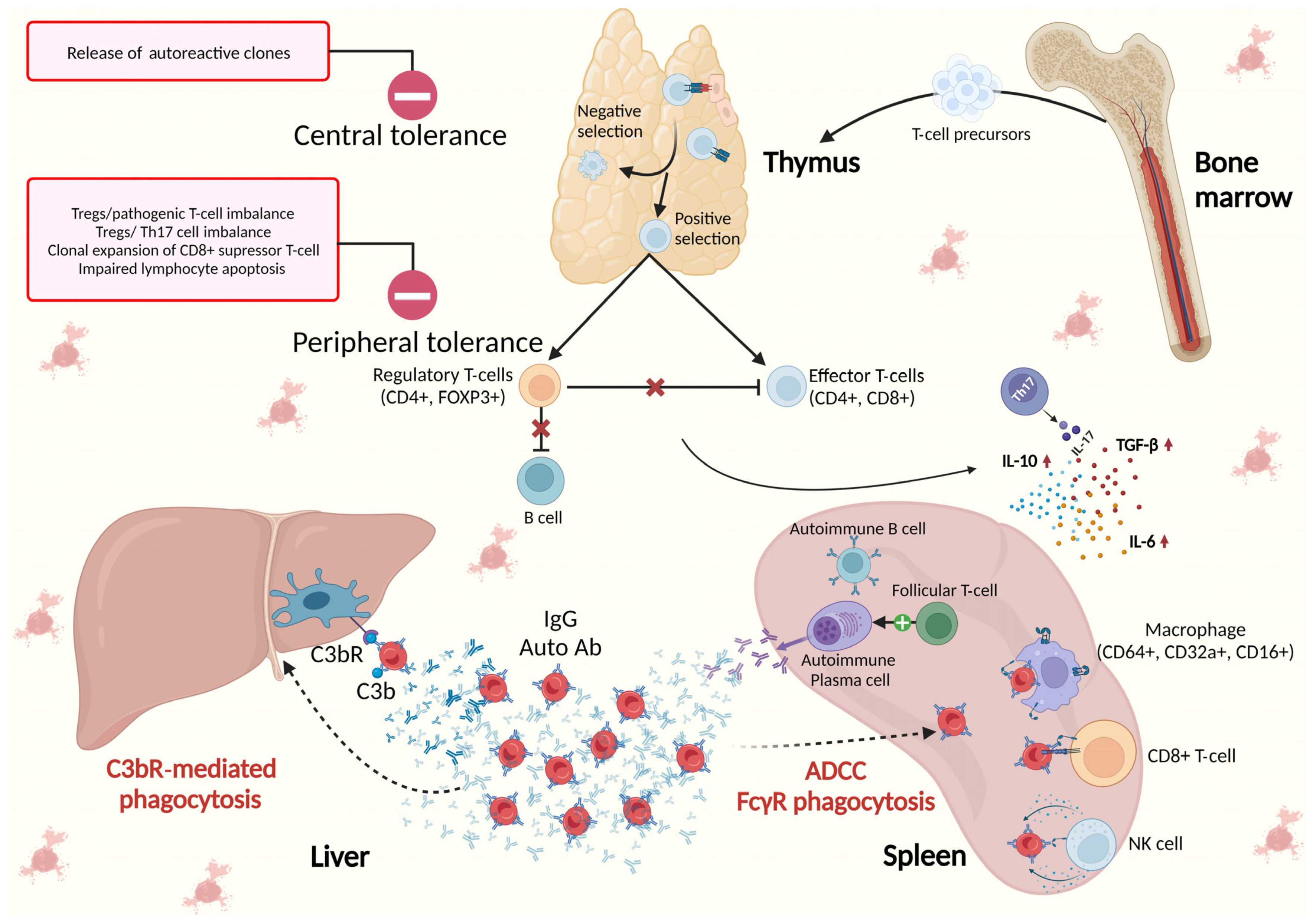
3. The Role of the Spleen and Bone Marrow in the Immunological Background of AIHA

3.1. Spleen as the Place of Origin of Autoantibodies in AIHA

Hitherto, the pathogenesis of AIHA has not been fully understood. Today, it is believed that, as in autoimmune and lymphoproliferative diseases, the breakdown of immunological central and peripheral tolerance, T- and B-cell dysregulation, and a shift towards T-helper 2 (Th2) and Th17 phenotype play major roles in the generation of autoAbs directed against RBCs.
The spleen is the largest secondary lymphoid organ and plays an important role in host defense but also in autoimmunity. The spleen is divided by function and structure into red and white pulp, with a perifollicular (marginal) zone in between. The primary immunological region of the spleen comprises white pulp, which occupies less than a quarter of splenic tissue. Unlike the lymph nodes, the spleen lacks afferent lymphatic vessels, and therefore, all cells and antigens enter the spleen via the blood [15]. The secretion of Abs by lymphoid follicle germinal center (GC) plasma cells (PCs) that have lifespans of 2–3 days is tightly regulated by negative feedback interactions with follicular Th cells (Tfh). Tfh cells produce IL-21, a protein critical for the processes of affinity maturation, GC longevity and function, and B-lymphocyte terminal differentiation [16]. Conventional PCs are designated to participate in bone marrow homing. Embedded in bone marrow niches, PCs may survive as long-lived PCs (LLPCs), persisting for up to the lifetime of the hosts in the absence of repeated antigen stimulation, together with memory CD4+ and memory CD8+ T cells [17,18]. It has been shown that, unlike conventional PCs, autoimmune LLPCs that secrete high-affinity IgG autoAbs abnormally accumulate in the spleen and show positive feedback with Tfh cells [19,20,21,22]. In the recently published work of Zeng et al., splenic white pulp fibroblastic reticular cells (FRCs) are recognized as a key factor that control autoreactive B-cell responses through CD36-mediated lipid uptake and the consequently enhanced mitochondrial oxidative phosphorylation in B cells [22].
The association between autoimmune disorders and neoplastic diseases, especially lymphoid neoplasms of B-cell origin (chronic lymphocytic leukemia(CLL), B-cell non-Hodgkin’s lymphoma, and Hodgkin’s lymphoma), was noted a long time ago. Autoimmune diseases, predominantly rheumatological (systemic lupus erythematosus, rheumatoid arthritis, and ankylosing spondylitis), endocrinological (type-1 insulin-dependent diabetes, Hashimoto thyroiditis, and Graves’ disease), neurological (multiple sclerosis and myasthenia gravis), and dermatological (pemphigus vulgaris) ones, have been linked with various autoimmune cytopenias (anemia, thrombocytopenia, neutropenia, pure red cell aplasia) and also with lymphoproliferative diseases and solid cancer. They share a common underlying etiology and are caused by the impaired self-tolerance of the immune system [14,23,24,25,26]. AIHA may occur synchronously with lymphoma, may precede or follow its diagnosis for several years, and may even be related to antilymphoma therapy [24]. In addition, AIHA that is resistant to therapy is emerging in recipients who have solid organs or are recipients of hematopoietic stem cell (HCT) transplants from an unrelated HLA donor [27,28].
The relationship between AIHA and congenital conditions such as common variable immunodeficiency/hyper IgM syndrome/autoimmune lymphoproliferative syndrome/Kabuki syndrome suggest that genetic background is an important factor regarding AIHA onset. Recurrent somatic mutations of KMT2D and mono-allelic CARD11 were demonstrated in patients with CAD. The loss of KMT2D function is related to the stimulation of the auto-reactive IGHV4-34-encoded immunoglobulin receptor, disturbed class switch recombination, and enhanced B-cell proliferation and survival. Mono-allelic CARD11 mutations result in B-cell proliferation and auto-antibody production. Similarly, oncogenic bi-allelic CARD11 mutations that result in constitutive nuclear factor (NF)-κB activation were demonstrated in a subset of patients with diffuse large B-cell lymphoma [16].
Conversely, the origins of autoAbs in drug-induced AIHA (DIHA) are well established. More than 160 drugs are suspected of inducing AIHA, the most commonly reported being antibiotics (penicillins, cephalosporins, and cotrimoxazole), antimycotics (fluconazole and amphotericin B), diclofenac, ibuprofen and other non-steroidal anti-inflammatory drugs, immunosuppressive (azathioprine) and antineoplastic drugs (both conventional ones like fludarabine, chlorambucil, and bendamustine, and novel options, such as immune checkpoint inhibitors), cardiovascular drugs (methyldopa, furosemide, and enalapril), and many others, including corticosteroids [2,29,30]. It is worth mentioning that some vaccines, including the mRNA-COVID 19 vaccine, have also been implicated as causes of the new onset or relapse of AIHA and Evans syndrome [31]. Although rare, DIHA encompasses about 10% of all AIHA cases [32]. There are two main subtypes of DIHA. In drug-dependent DIHA, Abs are directed to drugs adsorbed onto the RBC membrane, or to immune complexes of drug and cell membrane compounds (neoantigen). In drug-independent DIHA, IgG warm autoAbs can be detected in the absence of drugs [1]. Several drugs can modify the RBC membrane so that Ig (IgM or IgG), complement, albumin, and other plasma proteins adsorb nonimmunologically (non-immunologic protein adsorption(NIPA)), leading to a positive DAT result accompanied by slow and slight hemolysis [33].

3.2. Immune Clearance of Autoantibodies by Spleen Macrophages

Although an adaptive immune response to foreign antigens is initiated in the white pulp, immune effector function takes place and expands in spleen red pulp where neutrophils, monocytes, dendritic cells (DCs), gamma delta (γδ) T cells, and macrophages reside [17]. The most abundant Ig isotype in human serum and the predominant anti-RBC autoAb in wAIHA is IgG. IgG is composed of four subtypes whose constant regions differ in terms of the hinges and CH2 domains that are involved in binding to IgG-Fc receptors (FcγR) and C1q. The binding of IgG to FcγR on effector cells (macrophages, CD8+ T cells, and natural killer (NK) cells) triggers phagocytosis and antibody-dependent cell-mediated cytotoxicity (ADCC). In physiological conditions, IgG1, which is the most abundant IgG subclass, and IgG3 interact efficiently with most FcγR, while IgG2 and IgG4 show reduced affinity to a number of FcγR [34]. As a consequence, IgG1 and IgG3 shorten the RBC half-life more efficiently via ADCC than IgG2 and IgG4. In the case of high density (i.e., when two IgG molecules are in close proximity on the RBC surface), IgG1 and IgG3 can also fix C1q and activate the classical complement route, but usually not beyond C3b. The CD3b-opsonized RBCs undergo extravascular destruction caused by liver macrophages that carry receptors for C3b fragments. The downstream activation of complement cascade in wAIHA is very rare due to the monomeric IgG structure and the low Rh antigen density on RBCs. The capacity to perform terminal complement activation, the cleavage of C5, and the sequential formation of MAC (C5b-9) on the RBC surface is a feature of pentameric IgM and results in RBC agglutination and their direct and dramatic intravascular osmotic lysis, with a reported mortality rate of about 20% if the thermal activity of IgM is close to physiological temperatures [1,2,5,23,35]. The calculated speed of RBC destruction via intravascular hemolysis is 200 mL of RBCs per hour, whereas the extent of extravascular hemolysis is tenfold lesser (0.25 mL RBCs/kg/h, i.e., a patient with an average weight of 70 kg will experience a loss of 420 mL RBCs per day) [23].
Extravascular FcγR-mediated hemolysis takes place in the spleen and lymphoid organs. Spleen macrophages regulate RBC turnover in physiological and pathological circumstances. During a normal lifespan, RBCs encounter detrimental changes in plasma cell membrane and become less deformable. Senescent RBCs are too rigid to pass through the inter-endothelial slits of the spleen red pulp, and those trapped RBCs are phagocytized by macrophages located in the cords of the red pulp. Every day, approximately 1% of aged and irreversibly damaged RBCs are removed from circulation [36]. Besides the changes in deformability, over time, RBCs accumulate so-called “eat me” signals on their membrane that facilitate their clearance by macrophages. The exposure of phosphatidylserine on the outer leaflet is one of these “eat me” signals and is part of the programmed cell death process. Spleen macrophages possess receptors that recognize exposed phosphatidylserine on various cells and phagocyte them. As RBCs are non-nucleated, the term eryptocysis is used instead of apoptosis [37].
The most abundant integral membrane protein in RBCs is Band 3 (SLC4A1), with more than a million copies per cell. Band 3 is associated with a number of other membrane proteins including the Rh complex, glycophorins, and CD47. At the inner RBC membrane, Band 3 is attached to the cytoskeleton through interaction with ankyrin, while its carboxyl terminus is associated with carbonic anhydrase. Consequently, Band 3 has numerous fundamental functions in maintaining RBC integrity—from controlling RBC shape and deformability, through regulating CO2 transport, to mediating phagocytosis in the spleen. Namely, Band 3 exposes neoantigens that are recognized by naturally occurring Abs (Nabs) and cleared by macrophages. Conversely, CD47 represents one of the “do not eat me” signals, and binding to its receptor, signal-regulatory protein alpha (SIRPα), on macrophages suppresses phagocytosis by inhibiting the inside-out activation of integrin signaling [35]. In wAIHA, as well as in immune thrombocytopenia patients, CD47 is expressed at normal levels [38]. Spleen macrophages are not just responsible for clearing aged and damaged RBCs but also for repairing RBCs, as seen in the removal of inclusion bodies from circulating RBCs via spleen-facilitated vesiculation [39].
Splenic macrophages possess three types of FCγR receptors for the IgG heavy chain that activates phagocytosis: FcγRI (CD64), FcγRIIA (CD32a), and FcγRIII (CD16). Phagocytosed RBCs are targeted to phagolysosomes. FcγRI has the highest affinity for IgG molecules, and its activation induces signaling pathways. This includes phosphatidylinositol 3-kinase (PI3K) and mitogen-activated protein kinase (MAP), which are responsible for efficient erythrophagocytosis and a minimal release of harmful free hemoglobin into circulation. It is of clinical interest that spleen macrophages also express an inhibitory FcγRIIB and that its activation may explain the inefficiency of intravenous Ig (IVIG) in wAIHA as their therapeutic activity is mediated through binding to FcγRIIB [35].
Besides phagocytosis, spleen macrophages can only remove the IgG-coated portion of RBC membranes, resulting in a change in shape and RBCs becoming deformable. The formed rigid microspherocytes are retained in spleen red pulp sinusoids and cleared, as previously explained. Spleen CD8+ T cells and NK cells that also express FcγR are responsible for the ADCC destruction of opsonized RBCs [1,2,5,23,35].
The pivotal role of the spleen in the immunological background of AIHA is illustrated in Figure 2.

3.3. Autoimmune Reaction against Bone Marrow and Extramedullary Hematopoiesis in the Spleen

The presence of reticulocytopenia instead of reticulocytosis is not uncommon as a sign of insufficient erythropoiesis and poor prognosis. Indeed, this is observed in 20–40% of AIHA cases [41]. It has been suggested that in the pathogenesis of AIHA, as in ITP, an autoimmune reaction against bone marrow precursor plays an important role [42]. Recently reported data from a multicenter international study on the efficacy and safety of erythropoietin (EPO) treatment in AIHA revealed that the majority of the 51 included patients had inadequate reticulocytosis and reduced endogenous EPO levels considering the degree of anemia displayed. The overall response upon receiving EPO therapy (i.e., Hb increase greater than 20 g/L) was more than 70% and was observed both in primary and secondary wAIHA and CAD/CAS, as well in treatment-naïve patients and in relapsed/refractory patients previously treated with several treatment strategies. Several possible underlying mechanisms for reduced EPO levels in AIHA have been proposed—the inability of bone marrow to promptly respond to abrupt and massive RBC destruction, the negative feedback of hypoxia to the EPO kidney production, or a state of bone marrow corresponding to that seen in sepsis [15].
It has been estimated that adult humans make more than 2.5 × 10⁶ RBCs per second. The production of RBCs is in tight balance with the turnover of senescent RBCs by red pulp macrophages in the spleen [37,43]. Paulson et al. proposed a hypothesis that, as opposed to inhibiting steady-state erythropoiesis, inflammation induces stress erythropoiesis [44]. In anemia of critical illness (ACI), steady-state erythropoiesis is impaired due to HSC exhaustion and myelosuppression. The stress erythropoiesis seen in ACI is currently being intensively investigated. It is well documented that toll-like receptor 4 (TLR4) plays a crucial role via MYD88 (myeloid differentiation factor 88) or TRIF (Toll/interleukin-1 receptor (TIR)-domain-containing adapter-inducing interferon -β) in host response to bacterial infection, mediating the innate immune response and activating signaling pathways to promote the inflammatory cascade. These effects lead to increased cytokine secretion, such as of IL-1β, IL-6β, TNFα, and NF-κB, as well as to increased neutrophil infiltration and increased apoptosis [45,46]. TLR4 is also a key regulator of chronic inflammation [47]. However, on the other hand, TLR contributes to bone marrow failure—during sepsis, MYD88 activation leads to myelosuppression, while TRIF activation compromises HSC self-renewal. Furthermore, the downregulation of Spi1 and CebpA, two key transcription factors involved in the regulation of HSC and the myeloid transcriptional program, was observed [48]. The recently published results of Noel et al. indicate that IL-1/MyD88-dependent granulocyte colony stimulating factor (G-CSF) secretion plays a key role in impairing medullary erythropoiesis [49].
Besides the removal of aged, dead, or opsonized RBCs from circulation, spleen red pulp serves as a reservoir of RBCs, with the capacity to store 15–25% of total RBC volume that can be ejected as a physiological response to hypoxia (i.e., apnea, high altitude, exercise) [50]. Furthermore, the spleen is the main organ where stress erythropoiesis occurs. In humans, the spleen is one of the major sites of hematopoiesis during late embryonic life and at birth. In adults, HCTs in the spleen at steady state are less numerous than in bone marrow, but splenic HCTs are less quiescent and have faster reconstitution ability compared to bone marrow steady-state HCTs. It has demonstrated in mice that most splenic HSC are pre-activated, i.e., in the G1 phase that facilitates their fast cell cycle entry in emergency conditions [51]. Wang et al. analyzed splenic global gene expression patterns during stress erythropoiesis and found higher levels of expression of transcriptional factors that play the primary role in RBC differentiation (Gata1, Tal1, and Klf1). Conversely, genes involved in the immune response were inhibited, and NK cells decreased during stress erythropoiesis [52]. Extramedullary hematopoiesis contributes to the enlargement of the spleen in AIHA.

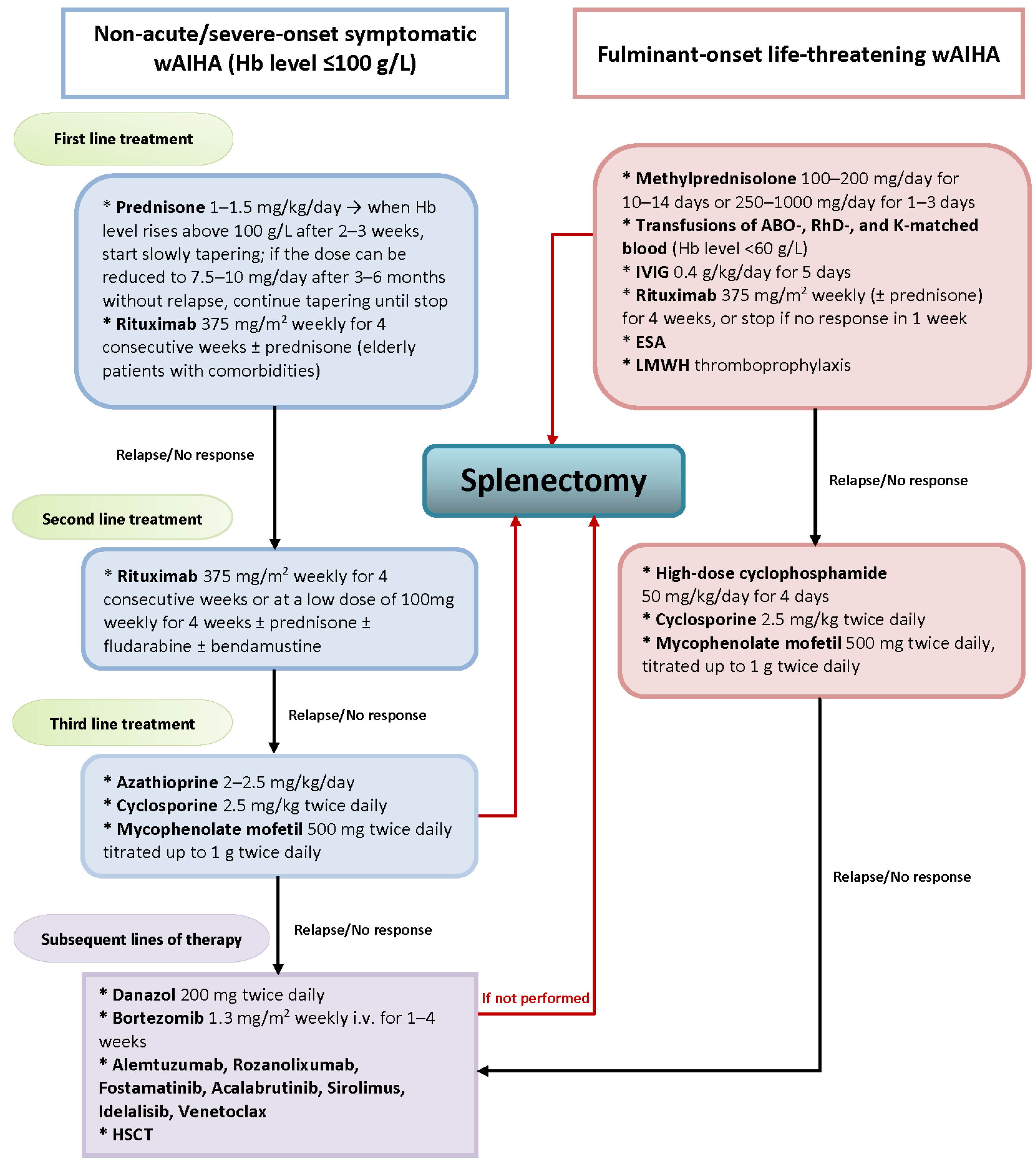
4. Novel Treatment Strategies for AIHA

Current guidelines recommend glucocorticoids and rituximab for first-line therapy [5]. Transfusions of ABO-, RhD-, and K-matched blood are only warranted for patients with life-threatening wAHA (Hb level ≤ 60 g/L), as they may further promote hemolysis. Due to the thermal characteristics of cold autoAbs, transfusions to cAIHA patients should be given using an in-line blood warmer. All active AIHA patients should receive folic acid (1–5 mg/d), and vitamin B12 if deficient, as well as ensuring thromboprophylaxis with low-molecular-weight heparin [5,14,32,53]. The exacerbation of hemolysis in cAIHA can simply be prevented if patients avoid cold temperatures and warm acral areas (nose, ears, digits) [53].
Specific first-line treatment strategies for AIHA are based on the inhibition of autoAb production. Patients with newly diagnosed symptomatic wAIHA (Hb level ≤ 100 g/L) are recommended to take oral prednisone 1 mg/kg daily. In steroid-responsive patients, i.e., those whose Hb rises above 100 g/L, the taper can begin after 2–3 weeks. If the dose can be reduced to 7.5 to 10 mg per day after 3 to 6 months without the relapse of clinically significant anemia, a further tapering should be attempted until the drug is discontinued. In the case of long-term corticosteroid treatment, patients should also receive treatment to prevent osteoporosis and fragile fractures (vitamin D, calcium, bisphosphonates) [1,2,5,14,32,54,55]. The tapering of corticosteroids is gradual and cautious as only 30–40% of patients who exhibit an initial response remain in remission after one year [56]. Corticosteroids are recommended in wAIHA due to the initial response in about 80% of patients, although they are not effective in cAIHA [5,53].
In patients with wAIHA who do not respond to corticosteroids, in those who relapse during corticosteroid tapering or after discontinuation, and in those requiring unacceptably high doses (>10–15 mg daily), the introduction of rituximab is recommended. It is important to emphasize that a lack of response to corticosteroids is suggestive of secondary wAIHA and indicates further investigation. Rituximab is a chimeric human IgG1-κ mAb directed against the extracellular domain of protein CD20, which is expressed on B-cells. Initially approved for the treatment of B-cell non-Hodgkin’s lymphoma, Rituximab has proven to be safe and effective therapy for the treatment of primary and secondary wAIHA [14,57,58,59,60], and as a result, other immune suppressants (azathioprine, cyclosporine, mycophenolate, etc.) have fallen out of use, most often in wAIHA associated with systemic lupus erythematosus. Rituximab may be given in a conventional form (375 mg/m2 weekly for 4 weeks) or at a low dose (100 mg weekly for 4 week) and combined with corticosteroids [5,55,61]. The use of rituximab in conventional doses introduces therapeutic options for cAIHA patients requiring treatment but is less effective against cAIHA than wAIHA. As complete responses are very rare, rituximab is used in combination with cytotoxic agents such as bendamustine or fludarabine [1,53].
Better insights into the pathobiology of AIHA have encouraged the investigation of numerous agents used to inhibit immune responses at various stages. Most of them are already approved for other indications. For example, rozanolixumab, the inhibitor of neonatal Fc receptor (FcRn), shortens IgG half-life from a normal 28 days to about 10% and is currently approved for the treatment of myasthenia gravis; fostamatinib [62], the splenic tyrosine kinase inhibitor, inhibits cellular mediators of phagocytosis and reduces the clearance of RBCs and platelets and is currently approved for the treatment of ITP; bruton tyrosine kinase inhibitors ibrutinib [63,64,65,66,67,68] and acalabrutinib are currently approved for treatment of CLL, and rizabrutinib is approved for the treatment of ITP; and sirolimus [69], the inhibitor of the mechanistic target of rapamycin kinase (mTOR), is approved for the treatment of malignant perivascular epithelioid cell tumors and is also effective in the prevention of graft rejection following organ transplant [14,55,62,69,70,71,72]. Idelalisib, which is a PI3K inhibitor, and venetoclax, a selective inhibitor of the anti-apoptotic protein B-cell lymphoma 2 (Bcl-2), are emerging as promising agents for the treatment of AIHA associated with CLL [63,73].
Drugs that target LLPCs include bortezomib, a proteasome inhibitor that is already included in recommendations for the treatment of cAIHA [53] and in refractory wAIHA either alone or combined with corticosteroids and dexamethason [74,75,76,77]. Daratumumab, a mAb targeting CD38 on LLPCs, is reported to be effective in treating wAIHA following alloHSCT and severe refractory AIHA cases [78,79,80], while a study on the safety, pharmacokinetics, and efficacy of ixatusimab, another antiCD38 mAb, was recently terminated based on strategic sponsor decisions [81]. Alemtuzumab, an mAb targeting CD52, may be useful in secondary wAIHA in the context of lymphoproliferative disorders [82,83,84,85,86,87,88,89]. The results of reports of targeted therapy in patients with primary and secondary wAIHA with sufficient clinical data are summarized in Table 2 [62,63,64,65,66,67,68,69,73,75,76,77,78,79,80,82,83,84,85,86,87,88,89,90,91,92]. Molecules that target the complement cascade, such as the C3 inhibitor pegcetacoplan and the C1q inhibitor cinryze, are reported to be effective in severe complement-mediated AIHA [93,94].

5. The Place of Splenectomy in the Era of Novel AIHA Treatment

The enlargement of spleen in AIHA is the consequence of Ab production, the immune destruction of RBC, and extramedullary hematopoiesis, crucial factors in IHA pathobiology. The surgical removal of the spleen was recognized and widely accepted as an effective therapeutic option in corticosteroid-resistant AIHA cases a long time ago. In a study by Allgoold and Chaplin on 44 AIHA patients published in 1967, an early response was obtained after splenectomy in 68% of corticosteroid-resistant patients, and 44% were able to maintain remission without supplementary steroid therapy after more than one year post-splenectomy. On the other side, the long-range mortality rate in this cohort was 40%, and the most common cause of death was pulmonary embolism [95]. Excellent efficacy was reported two decades later in a study on 52 splenectomized AIHA patients [96], with a reported sustained complete response rate of 64% after a mean follow-up period of 33 months, minimal morbidity, and an absence of mortality. Splenectomy was confirmed to be less efficient in secondary AIHA. A retrospective review of 30 patients with AIHA who underwent splenectomy revealed significantly inferior outcomes for AIHA patients with associated diseases (the most prevalent were B-cell malignancies and rheumatologic disorders), with an overall response rate of 56% and complete remission achieved in only three out of sixteen (19%) cases, while all patients with idiopathic AIHA demonstrated a response to splenectomy that was complete in nine out of eleven (82%) instances. Of interest is the fact that the reported median time from AIHA diagnosis to splenectomy in both groups was very short, at only 3.2 months for patients with secondary AIHA, and that seven patients within this group had associated thrombocytopenia, indicating Evans syndrome, a condition that may partly explain their worse outcome [97]. The irradiation of the spleen was successfully performed in several patients with associated lymphoproliferative diseases and high perioperative risk. As splenic irradiation in this setting can be considered experimental, a total irradiation dose is not established for performing these procedures and in published cases varies from 8 Gy to 20 Gy. It is assumed that a higher radiation dose may induce a “functional” splenectomy [98].
In the years that followed, although still prior to the introduction of novel therapy in the treatment of AIHA and underling diseases, the reported median time from AIHA diagnosis to splenectomy extended and the laparoscopic procedure prevailed over the open-surgery technique [99,100]. Due to the rarity of AIHA, patients with secondary AIHA were included in most published studies. In a case series of nine patients with symptomatic and uncontrolled AIHA diagnosed concurrently with CLL, splenectomy was performed after the failure of both CLL-targeted treatment with alkylating agents and/or purine analogues and AIHA targeted treatment with IVIG and/or corticosteroids. A median time to splenectomy of 62 months was reported, with patients’ average age at the time of splenectomy being 62 years. Seven of the nine patients (78%) achieved early complete response, and six (67%) retained remission from AIHA within a mean follow-up of 24 months after splenectomy. All patients underwent splenectomy via the laparoscopic technique and seven of the nine patients (78%) experienced no immediate postoperative complications [100]. Higher response rates were achieved in patients with primary AIHA. Long-term postoperative follow-up evaluations were obtained in three studies on the efficiency of laparascopic splenectomy performed in the context of malignant and non-malignant hematologic conditions, including primary AIHA [101,102,103]. In a retrospective study of 45 ITP and 15 AIHA patients who had undergone splenectomy from 1996 through to 2010, it was observed that the complete response rate was 93%, and that all patients survived the three-year post-splenectomy period [103]. Rosen et al. reported that uncontrolled AIHA was an indication for splenectomy in nine out of one hundred and forty-seven splenectomized patients and that, after a mean follow-up period of 20 +/− 14 months’, the response rate was 89% amongst the AIHA cohort [101]. Balagué et al. reported a response rate of 70% (seven out of ten patients) over a mean post-splenectomy period of 40 ± 18 months [102]. It is also noteworthy that, when analyzing the age at which splenectomy took place, the median age of patients with AIHA was significantly higher compared to the other indications given in these three previously mentioned studies. A comprehensive review of these studies on AIHA patients with a mean age above 55 years revealed that 94% of subjects (45 out of 48) underwent laparoscopic splenectomy with a complete response of 81% at 35.6 months, while the operative mortality was assessed at 0%, with no immediate or late thrombotic events [104]. Because of the high short-term efficacy and evidence of good long-term response, splenectomy was recommended as the most effective and best-evaluated second-line therapy for all steroid-refractory or steroid-dependent AIHA patients [105].
The introduction of rituximab was a turning point in the treatment of AIHA and gave impetus to the use of a wide range of innovative drugs in the treatment of AIHA, relegating splenectomy to the third and subsequent therapeutic lines [5,32,54,106,107], as shown on Figure 3. In a Spanish multicenter retrospective study of 93 patients diagnosed with AIHA between 1987 and 2017, the third line of therapy, including splenectomy, was needed in only 27% of patients, and a decrease in the rate of splenectomy was observed after the successful introduction of rituximab, which was applied in 34 patients with an overall response rate of close to 100% [60].
In addition to the abovementioned study conducted by Serbian authors, which compared the safety of laparsacopic and open techniques [99], by searching the Medline database, we managed to identify three studies published in English after 2012 that related to splenectomy in AIHA adult humans, and one study related to splenectomy in adults with Evans syndrome. Maskal et al. reported a short-term complete response of 61% and a complete response of 43% after a median follow up period of 33 months in splenectomized patients with primary and secondary AIHA [108]. Barron et al. analyzed the efficacy of splenectomy in autoimmune hematological disorders in the context of systemic lupus erythematosus and found that complete remission (defined as Hb ≥90 g/L) was achieved in eleven of the sixteen patients who had secondary AIHA or Evans syndrome [109]. Of interest is a study by Ogbue et al. that reviewed a cohort of 339 adult patients with immune cytopenias who had undergone a splenectomy, including 68 patients with AIHA. Aside from the high response rate (overall response was 74%, of which 86% were complete responses) and very low rate of postoperative complications with no mortality, the finding that up to 20% of the cases had a postoperative diagnosis is of importance, as this was discordant with the original indication for splenectomy, including benign conditions, such as Felty’s syndrome, and malignant diseases, predominantly mature B-, T-, and NK-cell lymphoma [110]. This emphasizes the role of splenectomy in securing a diagnosis and a subsequent optimal treatment level. In a study by Sulpizio et al. on seven patients who had undergone splenectomy to treat Evans syndrome (two of them having been associated with hematological malignancy), an early response was achieved in 85.7% of patients, and no major surgical complications were reported, albeit with a 42.8% relapse rate and a one-year response rate of 42.8% [111]. In all four of the abovementioned retrospective studies, AIHA patients who underwent splenectomy before the introduction of rituximab were also included. We could not find any published data addressing splenectomies performed only in the rituximab era.
According to the current guidelines, splenectomy or partial splenic embolization are still recommended in the emergency management of transfusion-dependent life-threatening wAIHA that is unresponsive to prednisolone. This should be combined with other supportive therapies (erythropoietin stimulating agents (ESA) and/or plasma exchange and/or IVIG) and active immunosuppression [5,55].
The loss of crucial functions of splenic macrophages in eliminating encapsulated bacteria and prolonged antecedent or concomitant immunosuppression renders splenectomized AIHA patients highly susceptible to bacterial infections, with a reported rate of 6–7% [112]. Among the 4756 AIHA patients identified using the California Discharge Dataset 1991–2014, the cumulative incidence of sepsis was 4.3% in those who had never undergone a splenectomy, compared to rate of 6.7% who had undergone a splenectomy. For the latter group, the rate of sepsis only increased in the late postoperative period [113]. In addition to postoperative antibiotic prophylaxis, vaccination with polysaccharide and conjugate vaccines against Streptococcus pneumoniae, Haemophilus influenzae, and Neisseria meningitidis should be performed at least 14 days before a scheduled splenectomy, or after the 14th postoperative day after an urgent splenectomy [114,115]. The use of live or live, attenuated vaccines in patients receiving >10 mg/day of prednisone or a cumulative dose of >700 mg over three months should be avoided, and the Food and Drug Agency recommended deferring procedures until at least one month after steroid discontinuation [116]. This is a need that cannot always be met in the case of refractory AIHA patients.
Thromboprophylaxis is also indicated in splenectomized AIHA patients in order to minimize the risk of venous thrombosis. It has long been recognized that AIHA has a prothrombotic state, per se [117], and thromboprophylaxis is advisable and recommended for all hospitalized patients admitted for wAIHA, as well as for outpatients with marked hemolysis [5,32,54]. The risk of venous thrombosis is further increased in splenectomized AIHA patients, leading to long-term morbidity and disability [113,118,119].
Splenectomy is not recommended to treat complement-mediated intravascular hemolysis, nor is it recommended for complement-mediated extravascular hemolysis where CD3b-opsonized RBCs are destroyed via liver macrophages that carry receptors for C3b fragments [51]. This is despite the fact that several case reports demonstrating the successful management of refractory CAD with splenectomy have been published, especially in recent years [120].

6. Conclusions and Future Direction

The spleen is the location of origin of autoAb and the location of antibody-mediated RBC phagocytosis in AIHA, as well as the location of extramedullary hematopoiesis. Splenectomy was the therapeutic option in corticosteroid-resistant AIHA patients for decades. In the era of modern therapy, splenectomy is scarcely performed, as it is either employed as a third or subsequent line of treatment or is recommended as a rescue management technique for transfusion-dependent, life-threatening wAIHA. The reported rate of complications following splenectomy has gradually decreased over time due to less invasive laparascopic techniques, vaccination against encapsulated bacteria, and thromboprophylaxis. Today, splenectomy is not associated with increased mortality. When taking into account this and the real-world evidence of the limited wide availability of rituximab and other costly targeted therapies inspired by clinical trials in most and not only developing countries, laparascopic splenectomy holds its place as a safe and good therapeutic option in the selected pools of AIHA patients.

Author Contributions

Conceptualization, Z.C. and M.M.; writing—original draft preparation, Z.C., N.P., M.C., M.V., N.S., G.M., V.M., Z.P. and M.M.; writing—review and editing, Z.C., N.S.-V. and M.M.; supervision, Z.C. and M.M. All authors have read and agreed to the published version of the manuscript.

Funding

This research received no external funding.

Institutional Review Board Statement

Not applicable.

Informed Consent Statement

Not applicable.

Data Availability Statement

Not applicable.

Conflicts of Interest

The authors declare no conflict of interest.

References

  1. Berentsen, S.; Barcellini, W. Autoimmune Hemolytic Anemias. N. Engl. J. Med. 2021, 385, 1407–1419. [Google Scholar] [CrossRef]
  2. Barcellini, W.; Zaninoni, A.; Giannotta, J.A.; Fattizzo, B. New Insights in Autoimmune Hemolytic Anemia: From Pathogenesis to Therapy Stage 1. J. Clin. Med. 2020, 9, 3859. [Google Scholar] [CrossRef] [PubMed]
  3. Hill, Q.A.; Stamps, R.; Massey, E.; Grainger, J.D.; Provan, D.; Hill, A.; British Society for Haematology. The diagnosis and management of primary autoimmune haemolyticanaemia. Br. J. Haematol. 2017, 176, 395–411. [Google Scholar] [CrossRef]
  4. Kuter, D.J. Warm autoimmune hemolytic anemia and the best treatment strategies. Hematology 2022, 2022, 105–113. [Google Scholar] [CrossRef] [PubMed]
  5. Jäger, U.; Barcellini, W.; Broome, C.M.; Gertz, M.A.; Hill, A.; Hill, Q.A.; Jilma, B.; Kuter, D.J.; Michel, M.; Montillo, M.; et al. Diagnosis and treatment of autoimmune hemolytic anemia in adults: Recommendations from the First International Consensus Meeting. Blood Rev. 2020, 41, 100648. [Google Scholar] [CrossRef]
  6. Jacobs, J.W.; Figueroa Villalba, C.A.; Booth, G.S.; Woo, J.S.; Stephens, L.D.; Adkins, B.D. Clinical and epidemiological features of paroxysmal cold hemoglobinuria: A systematic review. Blood Adv. 2023, 13, 2520–2527. [Google Scholar] [CrossRef] [PubMed]
  7. Hill, Q.A.; Hill, A.; Berentsen, S. Defining autoimmune hemolytic anemia: A systematic review of the terminology used for diagnosis and treatment. Blood Adv. 2019, 3, 1897–1906. [Google Scholar] [CrossRef]
  8. Audia, S.; Grienay, N.; Mounier, M.; Michel, M.; Bonnotte, B. Evans’ Syndrome: From Diagnosis to Treatment. J. Clin. Med. 2020, 9, 3851. [Google Scholar] [CrossRef]
  9. Barcellini, W. Pitfalls in the diagnosis of autoimmune haemolytic anaemia. Blood Transfus. 2015, 13, 3–5. [Google Scholar]
  10. Zarandona, J.M.; Yazer, M.H. The role of the Coombs test in evaluating hemolysis in adults. CMAJ 2006, 174, 305–307. [Google Scholar] [CrossRef]
  11. Ames, P.R.J.; Merashli, M.; Bucci, T.; Pastori, D.; Pignatelli, P.; Arcaro, A.; Gentile, F. Antiphospholipid Antibodies and Autoimmune Haemolytic Anaemia: A Systematic Review and Meta-Analysis. Int. J. Mol. Sci. 2020, 21, 4120. [Google Scholar] [CrossRef] [PubMed]
  12. Parker, V.; Tormey, C.A. The Direct Antiglobulin Test: Indications, Interpretation, and Pitfalls. Arch. Pathol. Lab. Med. 2017, 141, 305–310. [Google Scholar] [CrossRef] [PubMed]
  13. Ribkoff, J.; Deloughery, T.; Olson, S.R. Incidence of False-Positive Direct Anti Globulin Tests (DAT): A Single-Center Retrospective Analysis. Blood 2020, 136, 6. [Google Scholar] [CrossRef]
  14. Michalak, S.S.; Olewicz-Gawlik, A.; Rupa-Matysek, J.; Wolny-Rokicka, E.; Nowakowska, E.; Gil, L. Autoimmune hemolytic anemia: Current knowledge and perspectives. Immun. Ageing 2020, 17, 38. [Google Scholar] [CrossRef]
  15. Fattizzo, B.; Michel, M.; Zaninoni, A.; Giannotta, J.; Guillet, S.; Frederiksen, H.; Vos, J.M.I.; Mauro, F.R.; Jilma, B.; Patriarca, A.; et al. Efficacy of recombinant erythropoietin in autoimmune hemolytic anemia: A multicenter international study. Haematologica 2021, 106, 622–625. [Google Scholar] [CrossRef]
  16. Małecka, A.; Trøen, G.; Tierens, A.; Østlie, I.; Małecki, J.; Randen, U.; Wang, J.; Berentsen, S.; Tjønnfjord, G.E.; Delabie, J.M.A. Frequent somatic mutations of KMT2D (MLL2) and CARD11 genes in primary cold agglutinin disease. Br. J. Haematol. 2018, 183, 838–842. [Google Scholar] [CrossRef]
  17. Lewis, S.M.; Williams, A.; Eisenbarth, S.C. Structure and function of the immune system in the spleen. Sci. Immunol. 2019, 4, eaau6085. [Google Scholar] [CrossRef]
  18. Pelletier, N.; McHeyzer-Williams, L.J.; Wong, K.A.; Urich, E.; Fazilleau, N.; McHeyzer-Williams, M.G. Plasma cells negatively regulate the follicular helper T cell program. Nat. Immunol. 2010, 11, 1110–1118. [Google Scholar] [CrossRef] [PubMed]
  19. Slamanig, S.A.; Nolte, M.A. The Bone Marrow as Sanctuary for Plasma Cells and Memory T-Cells: Implications for Adaptive Immunity and Vaccinology. Cells 2021, 10, 1508. [Google Scholar] [CrossRef]
  20. Liu, X.; Yao, J.; Zhao, Y.; Wang, J.; Qi, H. Heterogeneous plasma cells and long-lived subsets in response to immunization, autoantigen and microbiota. Nat. Immunol. 2022, 23, 1564–1576. [Google Scholar] [CrossRef]
  21. Jang, E.; Cho, S.; Pyo, S.; Nam, J.W.; Youn, J. An Inflammatory Loop Between Spleen-Derived Myeloid Cells and CD4+ T Cells Leads to Accumulation of Long-Lived Plasma Cells That Exacerbates Lupus Autoimmunity. Front. Immunol. 2021, 12, 631472. [Google Scholar] [CrossRef]
  22. Hoyer, B.F.; Moser, K.; Hauser, A.E.; Peddinghaus, A.; Voigt, C.; Eilat, D.; Radbruch, A.; Hiepe, F.; Manz, R.A. Short-lived plasmablasts and long-lived plasma cells contribute to chronic humoral autoimmunity in NZB/W mice. J. Exp. Med. 2004, 199, 1577–1584. [Google Scholar] [CrossRef] [PubMed]
  23. Barcellini, W. New Insights in the Pathogenesis of Autoimmune Hemolytic Anemia. Transfus. Med. Hemotherapy 2015, 42, 287–293. [Google Scholar] [CrossRef]
  24. Porpaczy, E.; Jäger, U. How I manage autoimmune cytopenias in patients with lymphoid cancer. Blood 2022, 139, 1479–1488. [Google Scholar] [CrossRef] [PubMed]
  25. Khanmohammadi, S.; Shabani, M.; Tabary, M.; Rayzan, E.; Rezaei, N. Lymphoma in the setting of autoimmune diseases: A review of association and mechanisms. Crit. Rev. Oncol Hematol. 2020, 150, 102945. [Google Scholar] [CrossRef]
  26. Marques, H.S.; de Brito, B.B.; da Silva, F.A.F.; Santos, M.L.C.; de Souza, J.C.B.; Correia, T.M.L.; Lopes, L.W.; Neres, N.S.M.; Dórea, R.S.D.M.; Dantas, A.C.S.; et al. Relationship between Th17 immune response and cancer. World J. Clin. Oncol 2021, 12, 845–867. [Google Scholar] [CrossRef]
  27. Kanellopoulou, T. Autoimmune hemolytic anemia in solid organ transplantation-The role of immunosuppression. Clin. Transpl. 2017, 31, e13031. [Google Scholar] [CrossRef]
  28. Kennedy, C.; Jackson, D.E. The effect of HLA matching and donor relatedness on the risk of autoimmune haemolytic anaemia in haematopoietic stem cell transplant recipients: A systematic review and meta-analysis. EJHaem 2022, 3, 609–618. [Google Scholar] [CrossRef] [PubMed]
  29. Pereira-Maia, K.; Leguelinel-Blache, G.; Eiden, C.; Bernard, J.; Donnadieu-Rigole, H.; Peyrière, H. Drug-Induced Autoimmune Hemolytic Anemia: Detection of New Signals in the World Pharmacovigilance Database and Risk Assessment in a Nationwide Cohort Study in France. Blood 2022, 140, 8176–8177. [Google Scholar]
  30. Leaf, R.K.; Ferreri, C.; Rangachari, D.; Mier, J.; Witteles, W.; Ansstas, G.; Anagnostou, T.; Zubiri, L.; Piotrowska, Z.; Oo, T.H.; et al. Clinical and laboratory features of autoimmune hemolytic anemia associated with immune checkpoint inhibitors. Am. J. Hematol. 2019, 94, 563–574. [Google Scholar] [CrossRef]
  31. Cvetković, M.; Pantić, N.; Virijević, M.; Pravdić, Z.; Sabljić, N.; Mitrović, M.; Suvajdžić-Vuković, N. Relapse of Evans syndrome following BNT162b2 (Pfizer-BioNTech) COVID-19 vaccine: Case report and literature review. J. Infect. Dev. Ctries 2023, 17, 800–804. [Google Scholar] [CrossRef] [PubMed]
  32. Hill, Q.A.; Stamps, R.; Massey, E.; Grainger, J.D.; Provan, D.; Hill, A. Guidelines on the management of drug-induced immune and secondary autoimmune, haemolytic anaemia. Br. J. Haematol. 2017, 177, 208–220. [Google Scholar] [CrossRef] [PubMed]
  33. Wu, Y.; Wu, Y.; Guo, G.; Zeng, J.; Liu, Y.; Wu, Y. Piperacillin-tazobactam induced immune hemolytic anemia led to increased renal impairment and eventual death from multiple organ failure in a patient with hypertensive nephropathy: Case report and literature review. BMC Nephrol. 2023, 24, 173. [Google Scholar] [CrossRef] [PubMed]
  34. Vidarsson, G.; Dekkers, G.; Rispens, T. IgG subclasses and allotypes: From structure to effector functions. Front. Immunol. 2014, 5, 520. [Google Scholar] [CrossRef]
  35. Thiagarajan, P.; Parker, C.J.; Prchal, J.T. How Do Red Blood Cells Die? Front. Physiol. 2021, 12, 655393. [Google Scholar] [CrossRef]
  36. Kaufman, R. Red Blood Cell Life Span, Senescence, and destruction. In Anemia: Pathophysiology, Diagnosis and Management; Benz, E.J., Berliner, N., Schiffman, F.J., Eds.; Cambridge University Press: Cambridge, UK, 2017; pp. 19–22. [Google Scholar]
  37. Klei, T.R.; Meinderts, S.M.; van den Berg, T.K.; van Bruggen, R. From the Cradle to the Grave: The Role of Macrophages in Erythropoiesis and Erythrophagocytosis. Front. Immunol. 2017, 8, 73. [Google Scholar] [CrossRef]
  38. Ahrens, N.; Pagenkopf, C.; Kiesewetter, H.; Salama, A. CD47 is expressed at normal levels in patients with autoimmune haemolytic anaemia and/or immune thrombocytopenia. Transfus. Med. 2006, 16, 397–402. [Google Scholar] [CrossRef]
  39. Willekens, F.L.; Roerdinkholder-Stoelwinder, B.; Groenen-Döpp, Y.A.; Bos, H.J.; Bosman, G.J.; van den Bos, A.G.; Verkleij, A.J.; Were, J.M. Hemoglobin loss from erythrocytes in vivo results from spleen-facilitated vesiculation. Blood 2003, 101, 747–751. [Google Scholar] [CrossRef]
  40. Ohmes, J.; Comdühr, S.; Akbarzadeh, R.; Riemekasten, G.; Humrich, J.Y. Dysregulation and chronicity of pathogenic T cell responses in the pre-diseased stage of lupus. Front. Immunol. 2022, 13, 1007078. [Google Scholar] [CrossRef]
  41. Aladjidi, N.; Leverger, G.; Leblanc, T.; Picat, M.Q.; Michel, G.; Bertrand, Y.; Bader-Meunier, B.; Robert, A.; Nelken, B.; Gandemer, V.; et al. New insights into childhood autoimmune hemolytic anemia: A French national observational study of 265 children. Haematologica 2011, 96, 655–663. [Google Scholar] [CrossRef]
  42. Arbach, O.; Funck, R.; Seibt, F.; Salama, A. Erythropoietin May Improve Anemia in Patients with Autoimmune Hemolytic Anemia Associated with Reticulocytopenia. Transfus. Med. Hemotherapy 2012, 39, 221–223. [Google Scholar] [CrossRef]
  43. Palis, J. Primitive and definitive erythropoiesis in mammals. Front. Physiol. 2014, 5, 3. [Google Scholar] [CrossRef] [PubMed]
  44. Paulson, R.F.; Ruan, B.; Hao, S.; Chen, Y. Stress Erythropoiesis is a Key Inflammatory Response. Cells 2020, 9, 634. [Google Scholar] [CrossRef] [PubMed]
  45. Zhou, S.; Wang, G.; Zhang, W. Effect of TLR4/MyD88 signaling pathway on sepsis-associated acute respiratory distress syndrome in rats, via regulation of macrophage activation and inflammatory response. Exp. Ther. Med. 2018, 15, 3376–3384. [Google Scholar] [CrossRef]
  46. Cuenca, A.G.; Joiner, D.N.; Gentile, L.F.; Cuenca, A.L.; Wynn, J.L.; Kelly-Scumpia, K.M.; Scumpia, P.O.; Behrns, K.E.; Efron, P.A.; Nacionales, D.; et al. TRIF-dependent innate immune activation is critical for survival to neonatal gram-negative sepsis. J. Immunol. 2015, 194, 1169–1177. [Google Scholar] [CrossRef] [PubMed]
  47. Kiripolsky, J.; Romano, R.A.; Kasperek, E.M.; Yu, G.; Kramer, J.M. Activation of Myd88-Dependent TLRs Mediates Local and Systemic Inflammation in a Mouse Model of Primary Sjögren’s Syndrome. Front. Immunol. 2020, 10, 2963. [Google Scholar] [CrossRef]
  48. Zhang, H.; Rodriguez, S.; Wang, L.; Wang, S.; Serezani, H.; Kapur, R.; Cardoso, A.A.; Carlesso, N. Sepsis Induces Hematopoietic Stem Cell Exhaustion and Myelosuppression through Distinct Contributions of TRIF and MYD88. Stem Cell Rep. 2016, 6, 940–956. [Google Scholar] [CrossRef]
  49. Noel, J.G.; Ramser, S.W.; Pitstick, L.; Goetzman, H.S.; Dale, E.L.; Potter, A.; Adam, M.; Potter, S.S.; Gardner, J.C. IL-1/MyD88-Dependent G-CSF and IL-6 Secretion Mediates Postburn Anemia. J. Immunol. 2023, 210, 972–980. [Google Scholar] [CrossRef]
  50. Lauda, E.; Haam, E. Importance of the spleen as a reservoir for red blood cells. Exp Biol Med. 1931, 29, 260–262. [Google Scholar] [CrossRef]
  51. Coppin, E.; Florentin, J.; Vasamsetti, S.B.; Arunkumar, A.; Sembrat, J.; Rojas, M.; Dutta, P. Splenic hematopoietic stem cells display a pre-activated phenotype. Immunol. Cell Biol. 2018, 96, 772–784. [Google Scholar] [CrossRef] [PubMed]
  52. Wang, H.; Liu, D.; Song, P.; Jiang, F.; Chi, X.; Zhang, T. Exposure to hypoxia causes stress erythropoiesis and downregulates immune response genes in spleen of mice. BMC Genom. 2021, 22, 413. [Google Scholar] [CrossRef] [PubMed]
  53. Despotovic, J.M.; Kim, T.O. Cold AIHA and the best treatment strategies. Hematology 2022, 2022, 90–95. [Google Scholar] [CrossRef] [PubMed]
  54. Brodsky, R.A. Warm Autoimmune Hemolytic Anemia. N. Engl. J. Med. 2019, 381, 647–654. [Google Scholar] [CrossRef]
  55. Berentsen, S.; Fattizzo, B.; Barcellini, W. The choice of new treatments in autoimmune hemolytic anemia: How to pick from the basket? Front. Immunol. 2023, 14, 1180509. [Google Scholar] [CrossRef] [PubMed]
  56. Roumier, M.; Loustau, V.; Guillaud, C.; Languille, L.; Mahevas, M.; Khellaf, M.; Limal, N.; Noizat-Pirenne, F.; Godeau, B.; Michel, M. Characteristics and outcome of warm autoimmune hemolytic anemia in adults: New insights based on a single-center experience with 60 patients. Am. J. Hematol. 2014, 89, E150–E155. [Google Scholar] [CrossRef]
  57. Reynaud, Q.; Durieu, I.; Dutertre, M.; Ledochowski, S.; Durupt, S.; Michallet, A.S.; Vital-Durand, D.; Lega, J.C. Efficacy and safety of rituximab in auto-immune hemolytic anemia: A meta-analysis of 21 studies. Autoimmun. Rev. 2015, 14, 304–313. [Google Scholar] [CrossRef] [PubMed]
  58. Barcellini, W.; Fattizzo, B.; Zaninoni, A.; Radice, T.; Nichele, I.; Di Bona, E.; Lunghi, M.; Tassinari, C.; Alfinito, F.; Ferrari, A.; et al. Clinical heterogeneity and predictors of outcome in primary autoimmune hemolytic anemia: A GIMEMA study of 308 patients. Blood 2014, 124, 2930–2936. [Google Scholar] [CrossRef]
  59. Dierickx, D.; Kentos, A.; Delannoy, A. The role of rituximab in adults with warm antibody autoimmune hemolytic anemia. Blood 2015, 125, 3223–3229. [Google Scholar] [CrossRef]
  60. Jomarrón, I.G.; Rubio, M.L.; Arias, M.M.; Arrizabalaga, B.; de la Iglesia, S.; Beneitez, D.; Sáez, M.I.; Cervera, A.; Recasens, V.; Herrera, A.; et al. Autoimmune haemolytic anaemias: A retrospective study of 93 patients. Med. Clin. 2020, 154, 331–337. [Google Scholar] [CrossRef]
  61. Barcellini, W.; Zaja, F.; Zaninoni, A.; Imperiali, F.G.; Di Bona, E.; Fattizzo, B.; Consonni, D.; Cortelezzi, A.; Zanella, A. Sustained response to low-dose rituximab in idiopathic autoimmune hemolytic anemia. Eur. J. Haematol. 2013, 91, 546–551. [Google Scholar] [CrossRef]
  62. Kuter, D.J.; Rogers, K.A.; Boxer, M.A.; Choi, M.; Agajanian, R.; Arnold, D.M.; Broome, C.M.; Field, J.J.; Murakhovskaya, I.; Numerof, R.; et al. Fostamatinib for the treatment of warm antibody autoimmune hemolytic anemia: Phase 2, multicenter, open-label study. Am. J. Hematol. 2022, 97, 691–699. [Google Scholar] [CrossRef] [PubMed]
  63. Quinquenel, A.; Godet, S.; Dartigeas, C.; Ysebaert, L.; Dupuis, J.; Ohanyan, H.; Collignon, A.; Gilardin, L.; Lepretre, S.; Dilhuydy, M.S.; et al. Ibrutinib and idelalisib in the management of CLL-associated autoimmune cytopenias: A study from the FILO group. Am. J. Hematol. 2019, 94, E183–E185. [Google Scholar] [CrossRef] [PubMed]
  64. Vitale, C.; Ahn, I.E.; Sivina, M.; Ferrajoli, A.; Wierda, W.G.; Estrov, Z.; Konoplev, S.N.; Jain, N.; O’Brien, S.; Farooqui, M.; et al. Autoimmune cytopenias in patients with chronic lymphocytic leukemia treated with ibrutinib. Haematologica 2016, 101, e254–e258. [Google Scholar] [CrossRef]
  65. Cavazzini, F.; Lista, E.; Quaglia, F.M.; Formigaro, L.; Cavallari, M.; Martinelli, S.; Rigolin, G.M.; Foà, R.; Cuneo, A. Response to ibrutinib of refractory life-threatening autoimmune hemolytic anemia occurring in a relapsed chronic lymphocytic leukemia patient with 17p deletion. Leuk. Lymphoma 2016, 57, 2685–2688. [Google Scholar] [CrossRef]
  66. Garcia-Horton, A.; Bernard, R.S.; Lazo-Langner, A.; Xenocostas, A.; Mangel, J.; Howson-Jan, K.; Lam, S.; Hsia, C.C. Safe Start of Ibrutinib in Patients with Chronic Lymphocytic Leukemia and Uncontrolled Autoimmune Hemolytic Anemia. Blood 2018, 132, 5560. [Google Scholar] [CrossRef]
  67. Montillo, M.; O’Brien, S.; Tedeschi, A.; Byrd, J.C.; Dearden, C.; Gill, D.; Brown, J.R.; Barrientos, J.C.; Mulligan, S.P.; Furman, R.R.; et al. Ibrutinib in previously treated chronic lymphocytic leukemia patients with autoimmune cytopenias in the resonate study. Blood Cancer J. 2017, 3, e524. [Google Scholar] [CrossRef]
  68. Galinier, A.; Delwail, V.; Puyade, M. Ibrutinib Is Effective in the Treatment of Autoimmune Haemolytic Anaemia in Mantle Cell Lymphoma. Case Rep. Oncol 2017, 10, 127–129. [Google Scholar] [CrossRef] [PubMed]
  69. Li, H.; Ji, J.; Du, Y.; Huang, Y.; Gu, H.; Chen, M.; Wu, R.; Han, B. Sirolimus is effectivefor primary relapsed/refractory autoimmune cytopenia: A multicenter study. Exp. Hematol. 2020, 89, 87–95. [Google Scholar] [CrossRef]
  70. Barcellini, W.; Fattizzo, B. How I treat warm autoimmune hemolytic anemia. Blood 2021, 137, 1283–1294. [Google Scholar] [CrossRef]
  71. Fattizzo, B.; Barcellini, W. New Therapies for the Treatment of Warm Autoimmune Hemolytic Anemia. Transfus. Med. Rev. 2022, 36, 175–180. [Google Scholar] [CrossRef]
  72. Xiao, Z.; Murakhovskaya, I. Development of New Drugs for Autoimmune Hemolytic Anemia. Pharmaceutics 2022, 14, 1035. [Google Scholar] [CrossRef] [PubMed]
  73. Galindo-Navarro, P.; Delgado-García, A.; Rodríguez-Gil, M.A.; Puerta-Puerta, J.M. Venetoclax for treating refractory autoimmune hemolytic anemia in chronic lymphocytic leukemia: Report of two cases in Spain. Haematologica 2023, 108, 2261–2264. [Google Scholar] [CrossRef]
  74. Yao, M.; Zhang, J.; Li, Y.; Lv, L.; Jia, L.; Yang, C.; Huang, Y.; Liu, H.; Wang, J.; Chen, M.; et al. Combination of low-dose rituximab, bortezomib and dexamethasone for the treatment of autoimmune hemolytic anemia. Medicine 2022, 101, e28679. [Google Scholar] [CrossRef] [PubMed]
  75. Ames, P.R.J.; Jeffrey, S. Bortezomib and rituximab in multiply relapsed primary warm autoimmune hemolytic anemia. Ann. Hematol. 2021, 100, 2415–2416. [Google Scholar] [CrossRef] [PubMed]
  76. Chen, M.; Zhuang, J.; Yang, C.; Zhang, L.; Wang, W.; Cai, H.; Yu, Y.; Li, J.; Zhou, D.; Han, B. Rapid response to a single-dose rituximab combined with bortezomib in refractory and relapsed warm autoimmune hemolytic anemia. Ann. Hematol. 2020, 99, 1141–1143. [Google Scholar] [CrossRef]
  77. Chineke, I.; Kagbo-Kue, S.; Aniekwena, J.; Rose, M. Successful treatment of severe idiopathic mixed autoimmune hemolytic anemia with bortezomib and intravenous immunoglobulin. Int. J. Blood Transfus. Immunohematol. 2019, 9, 100046Z02IC2019. [Google Scholar] [CrossRef]
  78. Jain, A.; Gupta, D.K. Daratumumab for refractory warm autoimmune hemolytic anemia. Ann. Hematol. 2021, 100, 1351–1353. [Google Scholar] [CrossRef]
  79. Rieger, M.J.; Stolz, S.M.; Ludwig, S.; Benoit, T.M.; Bissig, M.; Widmer, C.C.; Schwotzer, R.; Müller, A.M.; Nair, G.; Hegemann, I.; et al. Daratumumab in rituximab-refractory autoimmune haemolytic anaemia. Br. J. Haematol. 2021, 194, 931–934. [Google Scholar] [CrossRef] [PubMed]
  80. McGlothlin, J.; Abeykoon, J.; Al-Hattab, E.; Ashrani, A.A.; Elliott, M.; Hook, C.C.; Pardanani, A.; Pruthi, R.; Sridharan, M.; Wolanskyj, A.; et al. Bortezomib and daratumumab in refractory autoimmune hemolytic anemia. Am. J. Hematol. 2023, 7. [Google Scholar] [CrossRef] [PubMed]
  81. U.S. National Library of Medicine. Safety, Pharmacokinetics, and Efficacy of Subcutaneous Isatuximab in Adults with Warm Autoimmune Hemolytic Anemia (wAIHA). National Library of Medicine: Bethesda, MD, USA, 2023. [Google Scholar]
  82. McAlister, R.K.; Talbott, M.S.; Reddy, N.M. Durable responses in refractory autoimmune hemolytic anemia with alemtuzumab. J. Oncol Pharm. Pract. 2019, 25, 706–709. [Google Scholar] [CrossRef]
  83. Willis, F.; Marsh, J.C.; Bevan, D.H.; Killick, S.B.; Lucas, G.; Griffiths, R.; Ouwehand, W.; Hale, G.; Waldmann, H.; Gordon-Smith, E.C. The effect of treatment with Campath-1H in patients with autoimmune cytopenias. Br. J. Haematol. 2001, 114, 891–898. [Google Scholar] [CrossRef] [PubMed]
  84. Rodon, P.; Breton, P.; Courouble, G. Treatment of pure red cell aplasia and autoimmune haemolytic anaemia in chronic lymphocytic leukaemia with Campath-1H. Eur. J. Haematol. 2003, 70, 319–321. [Google Scholar] [CrossRef]
  85. Cheung, W.W.; Hwang, G.Y.; Tse, E.; Kwong, Y.L. Alemtuzumab induced complete remission of autoimmune hemolytic anemia refractory to corticosteroids, splenectomy and rituximab. Haematologica 2006, 91, ECR13. [Google Scholar]
  86. Lundin, J.; Karlsson, C.; Celsing, F. Alemtuzumab therapy for severe autoimmune hemolysis in a patient with B cell chronic lymphocytic leukemia. Med. Oncol. 2006, 23, 137–139. [Google Scholar] [CrossRef]
  87. Karlsson, C.; Hansson, L.; Celsing, F.; Lundin, J. Treatment of severe refractory autoimmune hemolytic anemia in B-cell chronic lymphocytic leukemia with alemtuzumab (humanized CD52 monoclonal antibody). Leukemia 2007, 21, 511–514. [Google Scholar] [CrossRef]
  88. Royer, B.; Vaida, I.; Etienne, A.; Garidi, R.; Damaj, G.; Marolleau, J.P. Treatment of severe autoimmune hemolytic anemia in B-cell chronic lymphocytic leukemia with alemtuzumab. Leukemia 2007, 21, 1841–1842. [Google Scholar] [CrossRef]
  89. Laurenti, L.; Tarnani, M.; Efremov, D.G.; Chiusolo, P.; De Padua, L.; Sica, S.; Leone, G. Efficacy and safety of low-dose alemtuzumab as treatment of autoimmune hemolytic anemia in pretreated B-cell chronic lymphocytic leukemia. Leukemia 2007, 21, 1819–1821. [Google Scholar] [CrossRef]
  90. Barcellini, W.; Murakhovskaya, I.; Terriou, L.; Pane, F.; Patriarca, A.; Butler, K.; Moran, S.; Wei, S.; Jäger, U. Long-term efficacy and safety results from an ongoing open-label phase 2 study of parsaclisib for the treatment of autoimmune hemolytic anemia (Aiha). HemaSphere 2022, 6, 186–187. [Google Scholar] [CrossRef]
  91. Lacerda, M.P.; Guedes, N.R.; Yamakawa, P.E.; Pereira, A.D.; Fonseca, A.R.B.M.D.; Chauffaille, M.L.L.F.; Goncalves, M.V.; Yamamoto, M.; Rodrigues, C.A. Treatment of refractory autoimmune hemolytic anemia with venetoclax in relapsed chronic lymphocytic leukemia with del(17p). Ann. Hematol. 2017, 96, 1577–1578. [Google Scholar] [CrossRef] [PubMed]
  92. Gordon, M.J.; Maldonado, E.; Danilov, A.V. Refractory Autoimmune Cytopenias Treated with Venetoclax. Hemasphere 2019, 4, e202. [Google Scholar] [CrossRef]
  93. Grossi, F.; Shum, M.K.; Gertz, M.A.; Roman, E. Inhibition of C3 with APL-2 Controls Hemolysis and Increases Hemoglobin Levels in Subjects with Autoimmune Hemolytic Anemia (AIHA). Blood 2018, 132, 3623. [Google Scholar] [CrossRef]
  94. de Boer, E.C.W.; Jalink, M.; Delvasto-Nuñez, L.; Meulenbroek, E.M.; Baas, I.; Janssen, S.R.; Folman, C.C.; Gelderman, K.A.; Wouters, D.; Engel, M.D.; et al. C1-inhibitor treatment in patients with severe complement-mediated autoimmune hemolytic anemia. Blood Adv. 2023, 7, 3128–3139. [Google Scholar] [CrossRef] [PubMed]
  95. Allgood, J.W.; Chaplin, H., Jr. Idiopathic acquired autoimmune hemolytic anemia. Areview of forty-seven cases treated from 1955 through 1965. Am. J. Med. 1967, 43, 254–273. [Google Scholar] [CrossRef] [PubMed]
  96. Coon, W.W. Splenectomy in the treatment of hemolytic anemia. Arch. Surg. 1985, 120, 625–628. [Google Scholar] [CrossRef]
  97. Akpek, G.; McAneny, D.; Weintraub, L. Comparative response to splenectomy in Coombs-positive autoimmune hemolytic anemia with or without associated disease. Am. J. Hematol. 1999, 61, 98–102. [Google Scholar] [CrossRef]
  98. Weinmann, M.; Becker, G.; Einsele, H.; Bamberg, M. Clinical indications and biological mechanisms of splenic irradiation in autoimmune diseases. Strahlenther. Onkol 2001, 177, 105–111. [Google Scholar] [CrossRef] [PubMed]
  99. Milosavljevic, V.; Tadic, B.; Grubor, N.; Eric, D.; Reljic, M.; Matic, S. Analysis of the surgical treatment of the patients operated on by using laparoscopic and classic splenectomy due to benign disorders of the spleen. Turk. J. Surg. 2019, 35, 111–116. [Google Scholar] [CrossRef] [PubMed]
  100. Hill, J.; Walsh, R.M.; McHam, S.; Brody, F.; Kalaycio, M. Laparoscopic splenectomy for autoimmune hemolytic anemia in patients with chronic lymphocytic leukemia: A case series and review of the literature. Am. J. Hematol. 2004, 75, 134–138. [Google Scholar] [CrossRef]
  101. Rosen, M.; Brody, F.; Walsh, R.M.; Tarnoff, M.; Malm, J.; Ponsky, J. Outcome of laparoscopic splenectomy based on hematologic indication. Surg. Endosc. 2002, 16, 272–279. [Google Scholar] [CrossRef]
  102. Balagué, C.; Targarona, E.M.; Cerdán, G.; Novell, J.; Montero, O.; Bendahan, G.; García, A.; Pey, A.; Vela, S.; Diaz, M.; et al. Long-term outcome after laparoscopic splenectomy related to hematologic diagnosis. Surg. Endosc. 2004, 18, 1283–1287. [Google Scholar] [CrossRef]
  103. Patel, N.Y.; Chilsen, A.M.; Mathiason, M.A.; Kallies, K.J.; Bottner, W.A. Outcomes and complications after splenectomy for hematologic disorders. Am. J. Surg. 2012, 204, 1014–1019, discussion 1019–1020. [Google Scholar] [CrossRef]
  104. Giudice, V.; Rosamilio, R.; Ferrara, I.; Seneca, E.; Serio, B.; Selleri, C. Efficacy and safety of splenectomy in adult autoimmune hemolytic anemia. Open Med. 2016, 11, 374–380. [Google Scholar] [CrossRef]
  105. Lechner, K.; Jäger, U. How I treat autoimmune hemolytic anemias in adults. Blood 2010, 116, 1831–1838. [Google Scholar] [CrossRef] [PubMed]
  106. Bonnet, S.; Guédon, A.; Ribeil, J.A.; Suarez, F.; Tamburini, J.; Gaujoux, S. Indications and outcome of splenectomy in hematologic disease. J. Visc. Surg. 2017, 154, 421–429. [Google Scholar] [CrossRef]
  107. Sys, J.; Provan, D.; Schauwvlieghe, A.; Vanderschueren, S.; Dierickx, D. The role of splenectomy in autoimmune hematological disorders: Outdated or still worth considering? Blood Rev. 2017, 31, 159–172. [Google Scholar] [CrossRef] [PubMed]
  108. Maskal, S.; Al Marzooqi, R.; Fafaj, A.; Zolin, S.; Naples, R.; Iyer, A.; Petro, C.; Krpata, D.; Prabhu, A.; Rosen, M.; et al. Clinical and surgical outcomes of splenectomy for autoimmune hemolytic anemia. Surg. Endosc. 2022, 36, 5863–5872. [Google Scholar] [CrossRef]
  109. Barron, N.; Arenas-Osuna, J.; Medina, G.; Cruz-Dominguez, M.P.; González-Romero, F.; Velásques-García, J.A.; Ayala-López, E.A.; Jara, L.J. Splenectomy in systemic lupus erythematosus and autoimmune hematologic disease: A comparative analysis. Clin. Rheumatol. 2018, 37, 943–948. [Google Scholar] [CrossRef]
  110. Ogbue, O.; Bahaj, W.; Kewan, T.; Ahmed, R.; Ullah, F.; Visconte, V.; Maskal, S.; Gurnari, C.; Rosenblatt, S.; Maciejewski, J.P. Splenectomy for Immune Cytopenias: Treatment Outcomes and Predictors of Response. Blood 2022, 140, 1226–1227. [Google Scholar] [CrossRef]
  111. Sulpizio, E.D.; Raghunathan, V.; Shatzel, J.J.; Zilberman-Rudenko, J.; Worrest, T.; Sheppard, B.C.; DeLoughery, T.G. Long-term remission rates after splenectomy in adults with Evans syndrome compared to immune thrombocytopenia: A single-center retrospective study. Eur. J. Haematol. 2020, 104, 55–58. [Google Scholar] [CrossRef]
  112. Giannotta, J.A.; Fattizzo, B.; Cavallaro, F.; Barcellini, W. Infectious Complications in Autoimmune Hemolytic Anemia. J. Clin. Med. 2021, 10, 164. [Google Scholar] [CrossRef] [PubMed]
  113. Ho, G.; Brunson, A.; Keegan, T.H.M.; Wun, T. Splenectomy and the incidence of venous thromboembolism and sepsis in patients with autoimmune hemolytic anemia. Blood Cells Mol. Dis. 2020, 81, 102388. [Google Scholar] [CrossRef]
  114. Leone, G.; Pizzigallo, E. Bacterial Infections Following Splenectomy for Malignant and Nonmalignant Hematologic Diseases. Mediterr. J. Hematol. Infect. Dis. 2015, 7, e2015057. [Google Scholar] [CrossRef] [PubMed]
  115. Webb, C.W.; Crowell, K.; Cravens, D. Clinical inquiries. Which vaccinations are indicated after splenectomy? J. Fam. Pract. 2006, 55, 711–712. [Google Scholar] [PubMed]
  116. Lee, G.; Malpica Castillo, L.E.; Walter, J.E. Infection Risk, Immunization Recommendations, and Antimicrobial Prophylaxis Needs When Treating Non-Malignant Hematologic Disorders—Wash Your Hands and What Else? Education Program. In Proceedings of the 62nd ASH Annual Meeting and Exposition, San Diego, CA, USA, 5–8 December 2020. [Google Scholar]
  117. Lecouffe-Desprets, M.; Néel, A.; Graveleau, J.; Leux, C.; Perrin, F.; Visomblain, B.; Artifoni, M.; Masseau, A.; Connault, J.; Pottier, P.; et al. Venous thromboembolism related to warm autoimmune hemolytic anemia: A case-control study. Autoimmun. Rev. 2015, 14, 1023–1028. [Google Scholar] [CrossRef] [PubMed]
  118. Petit, P.; Bret, P.M.; Atri, M.; Hreno, A.; Casola, G.; Gianfelice, D. Splenic vein thrombosis after splenectomy: Frequency and role of imaging. Radiology 1994, 190, 65–68. [Google Scholar] [CrossRef]
  119. Hassn, A.M.; Al-Fallouji, M.A.; Ouf, T.I.; Saad, R. Portal vein thrombosis following splenectomy. Br. J. Surg. 2000, 87, 362–373. [Google Scholar] [CrossRef]
  120. Okamoto, S.; Urade, T.; Yakushijin, K.; Kido, M.; Kuramitsu, K.; Komatsu, S.; Gon, H.; Yamashita, H.; Shirakawa, S.; Tsugawa, D.; et al. Successful Management of Refractory Autoimmune Hemolytic Anemia with Cold Agglutinin Disease with Splenectomy: A Case Report with Review of Literature. Kobe J. Med. Sci. 2023, 68, E30–E34. [Google Scholar]
Figure 1. Simplified algorithm for the diagnosis of AIHA. Abbreviations: LDH—lactate dehydrogenase; DAT—direct antiglobulin test; Abs—antibodies; TMA—thrombotic microangiopathy; TTP—thrombotic thrombocytopenic purpura; HUS—hemolytic uremic syndrome; DIC—disseminated intravascular coagulation; wAIHA—warm autoimmune hemolytic anemia; cAIHA—cold autoimmune hemolytic anemia; CAD—cold agglutinin disease; CAS—cold agglutinin syndrome; CLL—chronic lymphocytic leukemia; HCV—hepatitis C; HIV—human immunodeficiency virus; CMV—cytomegalovirus; EBV—Epstein–Barr virus.
Diagnostics 13 02891 g001
Figure 2. The spleen in the immunological background of AIHA. Central tolerance takes place in primary lymphoid organs (bone marrow for B-cells and thymus for T-cells) and is responsible for identifying immature self-reactive lymphocytes, i.e., T- or B-cell clones that possess receptors, to identify self-antigens with high affinity. These autoreactive clones, upon binding with self-antigens, undergo deletion or apoptosis (negative selection) or may, in the case of B-cells, change their specificity or, in the case of T cells, develop regulatory tolerance (Treg). Peripheral tolerance takes place in the germinal centers (GCs) of lymphoid follicles in secondary lymphoid organs (the spleen, lymph nodes, and mucosal lymphoid tissues) where foreign antigens are normally encountered. The importance of peripheral tolerance in preventing autoimmunity is in maintaining unresponsiveness to self-antigens that are expressed in peripheral tissues and not in primary lymphoid organs, and in preserving tolerance to self-antigens that are expressed in adult life after the production of mature lymphocytes. Peripheral tolerance is driven by specific CD4+FOXP3+ Tregs, and the imbalance between Tregs and pathogenic effector/memory CD+ T cells, as well as Tregs and Th17, together with increased clonal expansion of CD8+ suppressor T cells and impaired lymphocyte apoptosis, lead to the loss of control over T-cell activation and autoimmunity [14,23,40]. Th17 cells play crucial roles in the immune response against microorganisms and in autoimmunity and facilitate the development and progression of various cancers. They are responsible for the secretion of proinflammatory cytokine interleukin-17 (IL-17), which promotes and characterizes humoral autoimmune response, together with observed increased levels of IL-6, IL-10, and transforming growth factor (TGF)-β and reduced tumor necrosis factor (TNF)-α [15,26,40]. In contrast to normal plasma cells (PCs), autoimmune plasma cells accumulate in the spleen, establishing positive feedback with follicular T-helper cells (Tfh). Opsonized erythrocytes undergo elimination in the spleen through FcγR-mediated phagocytosis and antibody-dependent cellular cytotoxicity (ADCC), as well as in the liver via C3bR phagocytosis.
Diagnostics 13 02891 g002
Figure 3. The role of splenectomy in the current treatment landscape of wAIHA. Abbreviations: wAIHA—warm autoimmune hemolytic anemia; Hb—hemoglobin; IVIG—intravenous immunoglobulin; ESA—erythropoietin stimulating agent; HSCT—hematopoietic stem cell transplantation.
Diagnostics 13 02891 g003
Table 1. Serological and thermal characteristics of autoantibodies in AIHA.
AIHA
Subtype
AutoantibodiesHemolysis
Antibody
Specificity
Thermal
Amplitude
SpecificityDAT
wAIHAIgG
(+ possible C fixation)
37 °C (0–40)
98.6 °F (32–104)
Rh system
glycophorins A–D
IgG or
IgG + C3d
Extravascular
(mainly spleen)
cAIHACADIgM, rare IgG (common C fixation)4 °C (4–34)
39.2 °F (39.2–93.2)
I/i system, rarely
Pr or IH antigen
C3d
CA titer ≥64
Extravascular (liver)
Intravascular
PCHIgG, rare IgM (common C fixation)react at 4 °C (39.2 °F)
hemolyze at
37 °C (98.6 °F)
P antigenC3d
(Donath–Landsteiner test)
Intravascular
Mixed AIHAwarm IgG and
cold IgM
4 °C (39.2 °F) and
37 °C (98.6 °F)
IgG + high-titer cold IgMIntra- and extravascular
Abbreviations: wAIHA—warm autoimmune hemolytic anemia; cAIHA—cold autoimmune hemolytic anemia; CAD—cold agglutinin disease; PCH—paroxysmal cold hemoglobinuria; IgG—immunoglobulin G; IgM—immunoglobulin M; DAT—direct antiglobulin test; C—complement; CA—cold agglutinin.
Table 2. Real-world evidence on the effectiveness of novel treatment strategies in patients with primary and secondary wAIHA.
Drug/Mechanism of ActionDose ScheduleNumber of PatientsResponseTime to
Response
Duration of
Responses
Reference
Primary wAIHA
Fostamatinib/
syk inhibitor
100–150 p.o. mg bid197/192–4 weeks24 weeksKuter et al. 2022 [62]
Parsaclisib/
PI3Kδ inhibitor
1 mg p.o. qd
2.5 mg p.o. qd
1675%
(PR 25%)
2 weeks12 weeksBarcellini et al. 2022 [90]
Bortezomib/Proteasome inhibitor1.3 mg/s.c. qw for 4 weeks + Rituximab11/1NA9 monthsAmes et al. 2010 [75]
1.3 mg/s.c. biw
for 2 weeks + Rituximab
44/41–3 weeks3–5 monthsChen et al. 2020 [76]
2.7 mg every 72 h
total of four doses
11/11 weeks7 monthsChineke et al. 2019 [77]
Daratumumab/
antiDC38 mAb
16 mg/kg qw for 6 weeks11/1NA5 monthsRieger et al. 2021 [79]
16 mg/kg qw for 4 weeks11/112 weeks2 monthsJain et al. 2020 [78]
16 mg/kg qw for 2 months11/14 weeksNAMcGlothlin et al. 2022 [80]
Sirolimus/
mTOR inhibitor
1–3 mg/d 6–18 months
1412/14 (4 PR)12–24 weeks16 monthsLi et al. 2020 [69]
Secondary wAIHA
Alemtuzumab/
antiCD52 mAb
wAIHA+CLL
10 mg qd for 1.5 weeks22/2 (1 PR)8 weeks16 monthsWillis et al. 2001 [83]
30 mg d5, then 30 mg tiw for 3 weeks11/13 weeks10 monthsRondon et al. 2003 [84]
3 mg d1, 10 mg d3, and 30 mg tiw for 8 weeks11/1NA16 monthsCheung et al. 2006 [85]
10 mg s.c. d1, then 30 mg tiw for 8 weeks11/18 weeks15 monthsLundin et al. 2007 [86]
30 mg s.c. tiw 3–12 w for 8 weeks55/55 weeks
(4–7)
15 monthsKarlsson et al. 2007 [87]
30 mg tiw:
8 weeks i.v.—episode 1
11 weeks s.c.—episode 2
11/1eeks—episode 1
NA—episode 2
17 months (episode 1)
3 months (episode 2)
Royer et al. 2007 [88]
3 mg d1-d3, then 10 mg tiw for
10–13 weeks
33/35–8 weeks9–26 monthsLeurenti et al. 2007 [89]
after dose escalation,
30 mg tiw 8–16 weeks
33/3NA7–68 monthsMc Alister et al. 2019 [82]
Idelalisib/
PI3Kδ inhibitor
150 mg p.o.bid
+ Rituximab
1211/12
(8 PR)
NAmedian PFS not reached after 2 yearsQuinquenel et al. 2019 [63]
Ibrutinib/
BTK inhibitor
wAIHA+ CLL
420 mg p.o. qd
+/− Rituximab
1615/16
(8 PR)
NAmedian PFS 19 monthsQuinquenel et al. 2019 [63]
420 mg p.o.
+/− Rituximab
86/8NA17.6 monthsVitale et al. 2016 [64]
420 mg p.o. qd
+ Prednisone 12.5 mg p.o. qd for 4 weeks
11/15 weeks12 monthsCavazzini et al. 2016 [65]
420 mg p.o.
+ oral corticosteroids 6–17 weeks
55/56–10 weeks15–150 weeksGarcia-Horton A et al. 2016 [66]
420 mg
+ Prednisone ≤ 20 mg qd *
21NANA17.5 monthsMontilo et al. 2017 [67]
Ibrutinib/
BTK inhibitor
wAIHA+ MCL
560 mg p.o. qd11/17 weeks6 monthsGalinier et al. 2017 [68]
Venetoclax/
Bcl-2 inhibitor
wAIHA+CLL
after standard ramp up,
400 mg p.o. qd
11/13 months10 monthsLacerda et al. 2017 [91]
after standard ramp up,
400 mg p.o. qd
11/14 weeks6 monthsGordon et al. 2019 [92]
after standard ramp up,
400 mg p.o. qd
+ Rituximab
22/23 months13 mo and
29 months
Galindo-Navarro et al. 2023 [73]
Abbreviations: wAIHA—warm autoimmune hemolytic anemia; FcRn—neonatal Fc receptor; mAb—monoclonal antibody; syk—splenic tyrosine kinase; BTK—bruton tyrosine kinase; PI3Kδ -phosphatidylinositol 3-kinase δ inhibitor; mTOR—mammalian target of rapamycin; Bcl-2—B-cell lymphoma 2; CLL—chronic lymphocytic leukemia; MCL—mantle cell lymphoma; PR—partial response; i.v.—intravenous: s.c.—subcutaneous; p.o.—orally (per os); qd—once daily; bid—twice daily; qw—once weekly; biw—twice weekly; tiw—three times weekly; NA—not applicable * patients with uncontrolled AIHA requiring >20 mg prednisone daily were excluded from the RESONATE study.
Disclaimer/Publisher’s Note: The statements, opinions and data contained in all publications are solely those of the individual author(s) and contributor(s) and not of MDPI and/or the editor(s). MDPI and/or the editor(s) disclaim responsibility for any injury to people or property resulting from any ideas, methods, instructions or products referred to in the content.

