The Differential Diagnosis of Congenital Developmental Midline Nasal Masses: Histopathological, Clinical, and Radiological Aspects
Abstract
1. Introduction
2. Clinical Manifestations
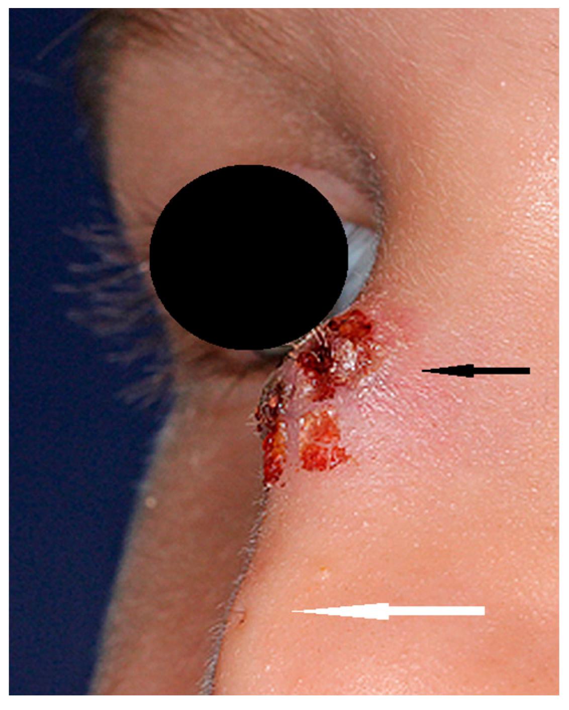
2.1. Nasal Dermoids
2.2. Congenital Encephaloceles
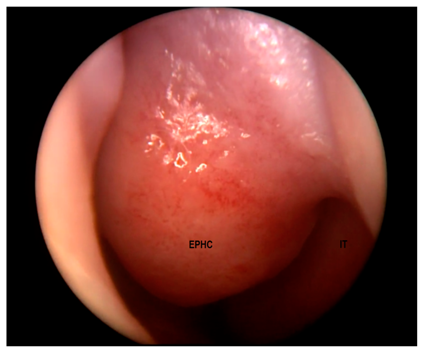
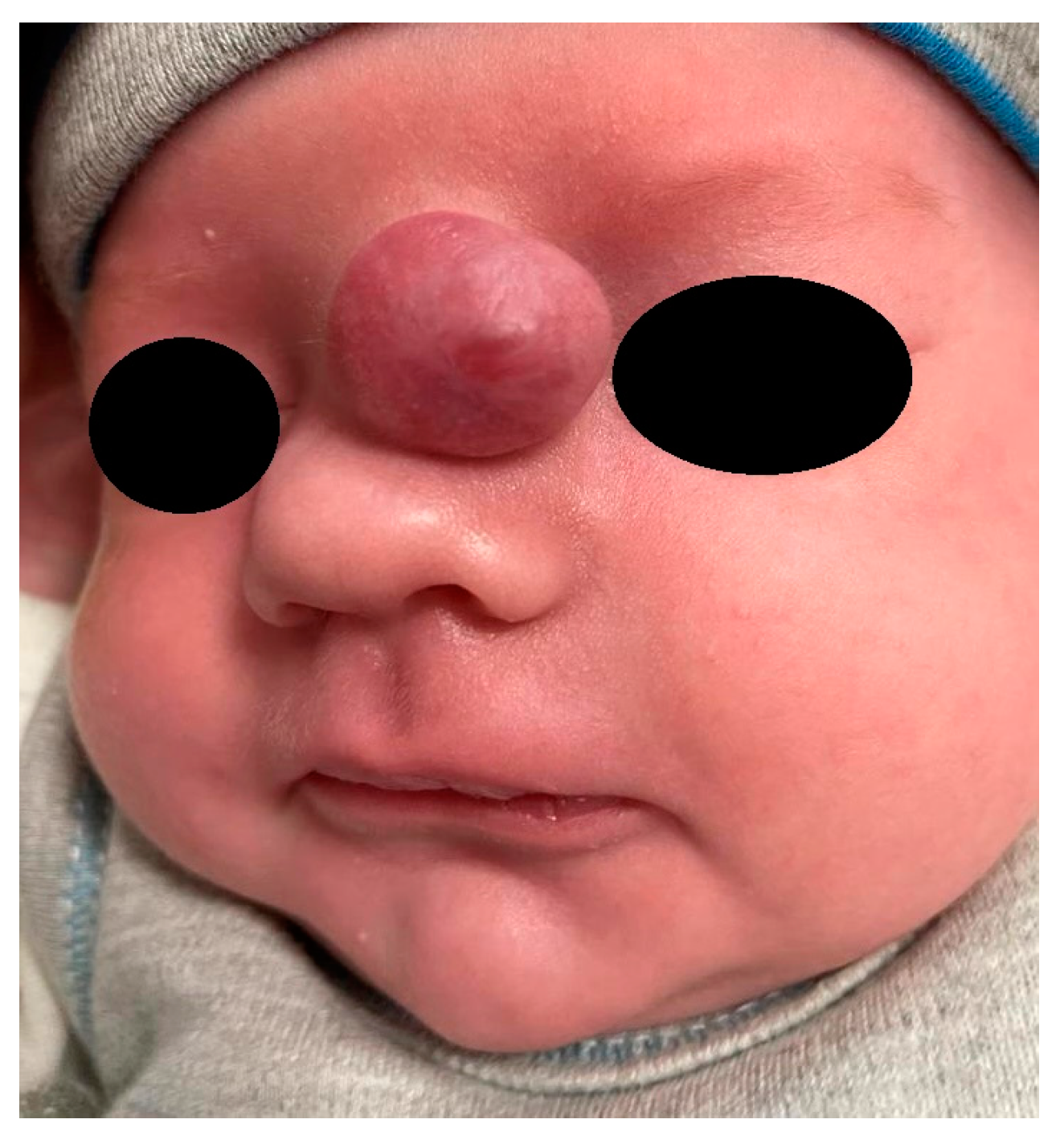
2.3. Nasal Glial Heterotopias
3. Histopathological Findings
3.1. Epidermoid/Dermoid Lesions
3.2. Nasal Glial Heterotopias
3.3. Encephaloceles
4. Imaging Studies
4.1. General Considerations
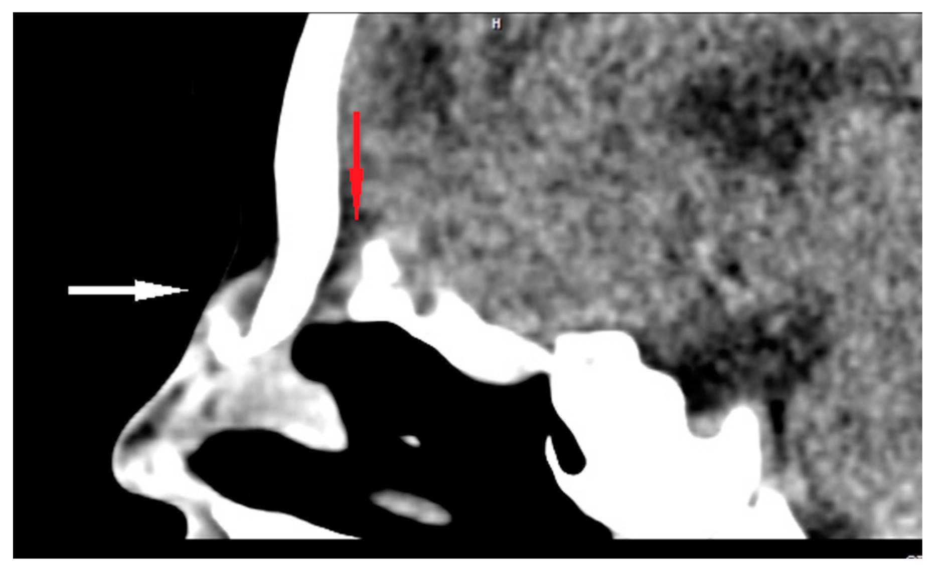
4.2. Nasal Dermoids
4.3. Encephaloceles
4.4. Nasal Glial Heterotopias
4.5. Role of Ultrasonography
5. Future Directions of Research
Funding
Institutional Review Board Statement
Informed Consent Statement
Data Availability Statement
Conflicts of Interest
References
- Hedlund, G. Congenital frontonasal masses: Developmental anatomy, malformations, and MR imaging. Pediatr. Radiol. 2006, 36, 647–662; quiz 726–727. [Google Scholar] [CrossRef]
- Saettele, M.; Alexander, A.; Markovich, B.; Morelli, J.; Lowe, L.H. Congenital midline nasofrontal masses. Pediatr. Radiol. 2012, 42, 1119–1125. [Google Scholar] [CrossRef]
- Kotowski, M.; Szydlowski, J. Congenital midline upper lip sinuses with intracranial extension—A variant of nasal dermoid? An embryology-based concept. Int. J. Pediatr. Otorhinolaryngol. 2023, 164, 111394. [Google Scholar] [CrossRef]
- Hoving, E.W. Nasal encephaloceles. Child’s Nerv. Syst. 2000, 16, 702–706. [Google Scholar] [CrossRef]
- Adil, E.; Huntley, C.; Choudhary, A.; Carr, M. Congenital nasal obstruction: Clinical and radiologic review. Eur. J. Pediatr. 2012, 171, 641–650. [Google Scholar] [CrossRef]
- Ziade, G.; Hamdan, A.L.; Homsi, M.T.; Kazan, I.; Hadi, U. Spontaneous Transethmoidal Meningoceles in Adults: Case Series with Emphasis on Surgical Management. Sci. World J. 2016, 2016, 3238297. [Google Scholar] [CrossRef]
- Macfarlane, R.; Rutka, J.T.; Armstrong, D.; Phillips, J.; Posnick, J.; Forte, V.; Humphreys, R.P.; Drake, J.; Hoffman, H.J. Encephaloceles of the anterior cranial fossa. Pediatr. Neurosurg. 1995, 23, 148–158. [Google Scholar] [CrossRef]
- Barkovich, A.J. Congenital malformations of the brain and skull. In Pediatric Neuroimaging, 4th ed.; Lippincott Williams & Wilkins: Philadelphia, PA, USA, 2005; pp. 308–313. [Google Scholar]
- Harnsberger, R. Nose and sinus. In Diagnostic Imaging: Head and Neck, 1st ed.; Amirsys: Salt Lake City, UT, USA, 2004; pp. 14–25. [Google Scholar]
- Hoving, E.W.; Vermeij-Keers, C. Frontoethmoidal encephaloceles, a study of their pathogenesis. Pediatr. Neurosurg. 1997, 27, 246–256. [Google Scholar] [CrossRef]
- Arifin, M.; Suryaningtyas, W.; Bajamal, A.H. Frontoethmoidal encephalocele: Clinical presentation, diagnosis, treatment, and complications in 400 cases. Child’s Nerv. Syst. 2018, 34, 1161–1168. [Google Scholar] [CrossRef]
- Suwanwela, C.; Suwanwela, N. A morphological classification of sincipital encephalomeningoceles. J. Neurosurg. 1972, 36, 201–211. [Google Scholar] [CrossRef]
- Cunha, B.; Kuroedov, D.; Conceição, C. Imaging of pediatric nasal masses: A review. J. Neuroimaging 2022, 32, 230–244. [Google Scholar] [CrossRef] [PubMed]
- Mahatumarat, C.; Rojvachiranonda, N.; Taecholarn, C. Frontoethmoidal encephalomeningocele: Surgical correction by the Chula technique. Plast. Reconstr. Surg. 2003, 111, 556–567. [Google Scholar] [CrossRef] [PubMed]
- Yeoh, G.P.; Bale, P.M.; de Silva, M. Nasal cerebral heterotopia: The so-called nasal glioma or sequestered encephalocele and its variants. Pediatr. Pathol. 1989, 9, 531–549. [Google Scholar] [CrossRef] [PubMed]
- Penner, C.R.; Thompson, L. Nasal glial heterotopia: A clinicopathologic and immunophenotypic analysis of 10 cases with a review of the literature. Ann. Diagn. Pathol. 2003, 7, 354–359. [Google Scholar] [CrossRef]
- Chan, J.K.; Lau, W.H. Nasal astrocytoma or nasal glial heterotopia? Arch. Pathol. Lab. Med. 1989, 113, 943–945. [Google Scholar]
- Diop, M.; Balakrishnan, K. Neonatal Nasal Obstruction. Curr. Otorhinolaryngol. Rep. 2021, 9, 389–398. [Google Scholar] [CrossRef]
- Tashiro, Y.; Sueishi, K.; Nakao, K. Nasal glioma: An immunohistochemical and ultrastructural study. Pathol. Int. 1995, 45, 393–398. [Google Scholar] [CrossRef]
- Wang, C.S.; Brown, C.; Mitchell, R.B.; Shah, G. Diagnosis and Management of Pediatric Nasal CSF Leaks and Encephaloceles. Curr. Treat. Options Allerg. 2020, 7, 326–334. [Google Scholar] [CrossRef]
- Wardinsky, T.D.; Pagon, R.A.; Kropp, R.J.; Hayden, P.W.; Clarren, S.K. Nasal dermoid sinus cysts: Association with intracranial extension and multiple malformations. Cleft Palate Craniofac. J. 1991, 28, 87–95. [Google Scholar] [CrossRef]
- Yavuzer, R.; Bier, U.; Jackson, I.T. Be careful: It might be a nasal dermoid cyst. Plast. Reconstr. Surg. 1999, 103, 2082–2083. [Google Scholar] [CrossRef]
- Denoyelle, F.; Ducroz, V.; Roger, G.; Garabedian, E.N. Nasal dermoid sinus cysts in children. Laryngoscope 1997, 107, 795–800. [Google Scholar] [CrossRef] [PubMed]
- Sessions, R.B. Nasal dermal sinuses--new concepts and explanations. Laryngoscope 1982, 92 Pt 2 (Suppl. S29), 1–28. [Google Scholar] [CrossRef] [PubMed]
- Pensler, J.M.; Bauer, B.S.; Naidich, T.P. Craniofacial dermoids. Plast. Reconstr. Surg. 1988, 82, 953–958. [Google Scholar] [CrossRef] [PubMed]
- Rohrich, R.J.; Lowe, J.B.; Schwartz, M.R. The role of open rhinoplasty in the management of nasal dermoid cysts. Plast. Reconstr. Surg. 1999, 104, 2163–2170; quiz 2171. [Google Scholar] [CrossRef]
- Rahbar, R.; Shah, P.; Mulliken, J.B.; Robson, C.D.; Perez-Atayde, A.R.; Proctor, M.R.; Kenna, M.A.; Scott, M.R.; McGill, T.J.; Healy, G.B. The presentation and management of nasal dermoid: A 30-year experience. Arch. Otolaryngol. Head Neck Surg. 2003, 129, 464–471. [Google Scholar] [CrossRef]
- Posnick, J.C.; Bortoluzzi, P.; Armstrong, D.C.; Drake, J.M. Intracranial nasal dermoid sinus cysts: Computed tomographic scan findings and surgical results. Plast. Reconstr. Surg. 1994, 93, 745–756. [Google Scholar] [CrossRef]
- Owusu-Ayim, M.; Locke, R.; Clement, W.A.; Kubba, H. Quantifying the annual risk of infection in congenital midline nasal dermoid cysts in children. Clin. Otolaryngol. 2023, 48, 254–258. [Google Scholar] [CrossRef]
- Kotowski, M.; Adamczyk, P.; Szydlowski, J. Nasal Dermoids in Children: Factors Influencing the Distant Result. Indian J. Otolaryngol. Head Neck Surg. 2022, 74 (Suppl. S2), 1412–1419. [Google Scholar] [CrossRef]
- Hartley, B.E.; Eze, N.; Trozzi, M.; Toma, S.; Hewitt, R.; Jephson, C.; Cochrane, L.; Wyatt, M.; Albert, D. Nasal dermoids in children: A proposal for a new classification based on 103 cases at Great Ormond Street Hospital. Int. J. Pediatr. Otorhinolaryngol. 2015, 79, 18–22. [Google Scholar] [CrossRef]
- El-Fattah, A.M.; Naguib, A.; El-Sisi, H.; Kamal, E.; Tawfik, A. Midline nasofrontal dermoids in children: A review of 29 cases managed at Mansoura University Hospitals. Int. J. Pediatr. Otorhinolaryngol. 2016, 83, 88–92. [Google Scholar] [CrossRef]
- Zapata, S.; Kearns, D.B. Nasal dermoids. Curr. Opin. Otolaryngol. Head Neck Surg. 2006, 14, 406–411. [Google Scholar] [CrossRef] [PubMed]
- Gnagi, S.H.; Schraff, S.A. Nasal obstruction in newborns. Pediatr. Clin. N. Am. 2013, 60, 903–922. [Google Scholar] [CrossRef] [PubMed]
- Bloom, D.C.; Carvalho, D.S.; Dory, C.; Brewster, D.F.; Wickersham, J.K.; Kearns, D.B. Imaging and surgical approach of nasal dermoids. Int. J. Pediatr. Otorhinolaryngol. 2002, 62, 111–122. [Google Scholar] [CrossRef] [PubMed]
- Hanikeri, M.; Waterhouse, N.; Kirkpatrick, N.; Peterson, D.; Macleod, I. The management of midline transcranial nasal dermoid sinus cysts. Br. J. Plast. Surg. 2005, 58, 1043–1050. [Google Scholar] [CrossRef] [PubMed]
- Castelnuovo, P.; Bignami, M.; Pistochini, A.; Battaglia, P.; Locatelli, D.; Dallan, I. Endoscopic endonasal management of encephaloceles in children: An eight-year experience. Int. J. Pediatr. Otorhinolaryngol. 2009, 73, 1132–1136. [Google Scholar] [CrossRef] [PubMed]
- Blumenfeld, R.; Skolnik, E.M. Intranasal encephaloceles. Arch. Otolaryngol. 1965, 82, 527–531. [Google Scholar] [CrossRef]
- Xue, L.; Gehong, D.; Ying, W.; Jianhua, T.; Hong, Z.; Honggang, L. Nasal meningoencephalocele: A retrospective study of clinicopathological features and diagnosis of 16 patients. Ann. Diagn. Pathol. 2020, 49, 151594. [Google Scholar] [CrossRef]
- Suurna, M.V.; Maurrasse, S.E. Congenital nasal anomalies. In Current Diagnosis Treatment Otolaryngology—Head and Neck Surgery, 4th ed.; Lalwani, A.K., Ed.; McGraw-Hill: New York, NY, USA, 2021. [Google Scholar]
- Tirumandas, M.; Sharma, A.; Gbenimacho, I.; Shoja, M.M.; Tubbs, R.S.; Oakes, W.J.; Loukas, M. Nasal encephaloceles: A review of etiology, pathophysiology, clinical presentations, diagnosis, treatment, and complications. Child’s Nerv. Syst. 2013, 29, 739–744. [Google Scholar] [CrossRef]
- Oddone, M.; Granata, C.; Dalmonte, P.; Biscaldi, E.; Rossi, U.; Toma, P. Nasal glioma in an infant. Pediatr. Radiol. 2002, 32, 104–105. [Google Scholar] [CrossRef]
- Mills, S.E.; Gaffey, M.J.; Frierson, H.F. Neural, Neuroendocrine, and Neuroectodermal Neoplasia: Tumors of the Upper Aerodigestive Tract and Ear; Armed Forces Institute of Pathology: Washington, DC, USA, 1997; pp. 119–200.
- Turgut, M. Glial heterotopias of the nose. Child’s Nerv. Syst. 1997, 13, 569. [Google Scholar] [CrossRef]
- Alvo, A.; Villarroel, G.; Sedano, C. Neonatal nasal obstruction. Eur. Arch. Otorhinolaryngol. 2021, 278, 3605–3611. [Google Scholar] [CrossRef]
- Hoeger, P.H.; Schaefer, H.; Ussmueller, J.; Helmke, K. Nasal glioma presenting as capillary haemangioma. Eur. J. Pediatr. 2001, 160, 84–87. [Google Scholar] [CrossRef]
- Batsakis, J.G. Non-odontogenic and fissural cysts. ORL 1971, 33, 19. [Google Scholar]
- Batsakis, J.G. Tumours of the Head and Neck: Clinical and Pathological Considerations, 2nd ed.; Williams and Wilkins: Baltimore, ML, USA, 1979; pp. 226–231. [Google Scholar]
- Balasundaram, P.; Garg, A.; Prabhakar, A.; Joseph Devarajan, L.S.; Gaikwad, S.B.; Khanna, G. Evolution of epidermoid cyst into dermoid cyst: Embryological explanation and radiological-pathological correlation. Neuroradiol. J. 2019, 32, 92–97. [Google Scholar] [CrossRef] [PubMed]
- Smith, A.S. Myth of the mesoderm. AJNR Am. J. Neuroradiol. 1989, 10, 449. [Google Scholar] [PubMed]
- Abdel-Rahman, N.; Al-Awadi, Y.; Ali Ramadan, A.; Choudhury, A.R. Cerebral heterotopia of the temporofacial region. Case report. J. Neurosurg. 1999, 90, 770–772. [Google Scholar] [CrossRef] [PubMed]
- Cerdá-Nicolás, M.; Sanchez Fernandez de Sevilla, C.; Lopez-Ginés, C.; Peydro-Olaya, A.; Llombart-Bosch, A. Nasal glioma or nasal glial heterotopia? Morphological, immunohistochemical and ultrastructural study of two cases. Clin. Neuropathol. 2002, 21, 66–71. [Google Scholar]
- Bozoky, B.; Stiller, D.; Ormos, J. Immunohistochemical demonstration of glial fibrillary acidic protein (GFAP) in nasal gliomas. Acta Histochem. 1987, 81, 117–123. [Google Scholar] [CrossRef]
- Dini, M.; Lo Russo, G.; Colafranceschi, M. So-called nasal glioma: Case report with immunohistochemical study. Tumori 1998, 84, 398–402. [Google Scholar] [CrossRef]
- Kindblom, L.G.; Angervall, L.; Haglid, K. An immunohistochemical analysis of S-100 protein and glial fibrillary acidic protein in nasal glioma. Acta Pathol. Microbiol. Immunol. Scand. A 1984, 92, 387–389. [Google Scholar] [CrossRef]
- Barnes, L. (Ed.) Surgical Pathology of the Head and Neck, 2nd ed.; Marcel Dekker: New York, NY, USA, 2001; Volume 3. [Google Scholar]
- Mahapatra, A.K.; Suri, A. Anterior encephaloceles: A study of 92 cases. Pediatr. Neurosurg. 2002, 36, 113–118. [Google Scholar] [CrossRef] [PubMed]
- Formica, F.; Iannelli, A.; Paludetti, G.; Di Rocco, C. Transsphenoidal meningoencephalocele. Child’s Nerv. Syst. 2002, 18, 295–298. [Google Scholar] [CrossRef] [PubMed]
- Lavezzi, A.M.; Corna, M.F.; Matturri, L. Neuronal nuclear antigen (NeuN): A useful marker of neuronal immaturity in sudden unexplained perinatal death. J. Neurol. Sci. 2013, 329, 45–50. [Google Scholar] [CrossRef] [PubMed]
- Rahbar, R.; Resto, V.A.; Robson, C.D.; Perez-Atayde, A.R.; Goumnerova, L.C.; McGill, T.J.; Healy, G.B. Nasal glioma and encephalocele: Diagnosis and management. Laryngoscope 2003, 113, 2069–2077. [Google Scholar] [CrossRef]
- Warf, B.C.; Stagno, V.; Mugamba, J. Encephalocele in Uganda: Ethnic distinctions in lesion location, endoscopic management of hydrocephalus, and survival in 110 consecutive children. J. Neurosurg. Pediatr. 2011, 7, 88–93. [Google Scholar] [CrossRef]
- Kotowski, M.; Szydlowski, J. Radiological diagnostics in nasal dermoids: Pitfalls, predictive values and literature analysis. Int. J. Pediatr. Otorhinolaryngol. 2021, 149, 110842. [Google Scholar] [CrossRef]
- Barkovich, A.J.; Vandermarck, P.; Edwards, M.S.; Cogen, P.H. Congenital nasal masses: CT and MR imaging features in 16 cases. AJNR Am. J. Neuroradiol. 1991, 12, 105–116. [Google Scholar]
- Herrington, H.; Adil, E.; Moritz, E.; Robson, C.; Perez-Atayde, A.; Proctor, M.; Rahbar, R. Update on current evaluation and management of pediatric nasal dermoid. Laryngoscope 2016, 126, 2151–2160. [Google Scholar] [CrossRef]
- Huisman, T.A.; Schneider, J.F.; Kellenberger, C.J.; Martin-Fiori, E.; Willi, U.V.; Holzmann, D. Developmental nasal midline masses in children: Neuroradiological evaluation. Eur. Radiol. 2004, 14, 243–249. [Google Scholar] [CrossRef][Green Version]
- Lindbichler, F.; Braun, H.; Raith, J.; Ranner, G.; Kugler, C.; Uggowitzer, M. Nasal dermoid cyst with a sinus tract extending to the frontal dura mater: MRI. Neuroradiology 1997, 39, 529–531. [Google Scholar] [CrossRef]
- Adil, E.; Rahbar, R. Paediatric nasal dermoid: Evaluation and management. Curr. Opin. Otolaryngol. Head Neck Surg. 2021, 29, 487–491. [Google Scholar] [CrossRef]
- Rodriguez, D.P.; Orscheln, E.S.; Koch, B.L. Masses of the Nose, Nasal Cavity, and Nasopharynx in Children. Radiographics 2017, 37, 1704–1730. [Google Scholar] [CrossRef]
- Barkovich, A.J.; Raybaud, C. Congenital malformations of the brain and skull. In Pediatric Neuroimaging, 6th ed.; Barkovich, A.J., Raybaud, C., Eds.; Lippincott Williams & Wilkins: Philadelphia, PA, USA, 2018; pp. 363–657. [Google Scholar]
- Valencia, M.P.; Castillo, M. Congenital and acquired lesions of the nasal septum: A practical guide for differential diagnosis. Radiographics 2008, 28, 205–224; quiz 326. [Google Scholar] [CrossRef] [PubMed]
- Franco, D.; Alonso, N.; Ruas, R.; da Silva Freitas, R.; Franco, T. Transsphenoidal meningoencephalocele associated with cleft lip and palate: Challenges for diagnosis and surgical treatment. Child’s Nerv. Syst. 2009, 25, 1455–1458. [Google Scholar] [CrossRef]
- Steven, R.A.; Rothera, M.P.; Tang, V.; Bruce, I.A. An unusual cause of nasal airway obstruction in a neonate: Trans-sellar, trans-sphenoidal cephalocoele. J. Laryngol. Otol. 2011, 125, 1075–1078. [Google Scholar] [CrossRef] [PubMed]
- Choudhury, A.R.; Bandey, S.A.; Haleem, A.; Sharif, H. Glial heterotopias of the nose: A report of two cases. Child’s Nerv. Syst. 1996, 12, 43–47. [Google Scholar] [CrossRef]
- Shafqat, A.; Shafquat, S.; Magableh, H.M.F.; Shaik, A.; Elshaer, A.N.; Alfehaid, W.K.; Hatatah, N.; Islam, S.S. Radiologic diagnosis of nasal gliomas with an illustrative case report. Indian J. Otolaryngol. Head Neck Surg. 2022, 74, 4623–4627. [Google Scholar] [CrossRef] [PubMed]
- Karunakaran, P.; Duraikannu, C.; Pulupula, V.N.K. An unusual presentation of neuroglial heterotopia: Case report. BJR Case Rep. 2020, 6, 20190116. [Google Scholar] [CrossRef]
- Ajose-Popoola, O.; Lin, H.W.; Silvera, V.M.; Teot, L.A.; Madsen, J.R.; Meara, J.G.; Rahbar, R. Nasal glioma: Prenatal diagnosis and multidisciplinary surgical approach. Skull Base Rep. 2011, 1, 83–88. [Google Scholar] [CrossRef]
- Yan, Y.Y.; Zhou, Z.Y.; Bi, J.; Fu, Y. Nasal glial heterotopia in children: Two case reports and literature review. Int. J. Pediatr. Otorhinolaryngol. 2020, 129, 109728. [Google Scholar] [CrossRef]
- He, F.; Soriano, P. A critical role for PDGFRα signaling in medial nasal process development. PLoS Genet. 2013, 9, e1003851. [Google Scholar] [CrossRef] [PubMed]
- Som, P.M.; Streit, A.; Naidich, T.P. Illustrated review of the embryology and development of the facial region, part 3: An overview of the molecular interactions responsible for facial development. AJNR Am. J. Neuroradiol. 2014, 35, 223–229. [Google Scholar] [CrossRef]
- Marcucio, R.; Hallgrimsson, B.; Young, N.M. Facial Morphogenesis: Physical and Molecular Interactions Between the Brain and the Face. Curr. Top. Dev. Biol. 2015, 115, 299–320. [Google Scholar] [CrossRef] [PubMed]
- Adameyko, I.; Fried, K. The Nervous System Orchestrates and Integrates Craniofacial Development: A Review. Front. Physiol. 2016, 7, 49. [Google Scholar] [CrossRef] [PubMed]
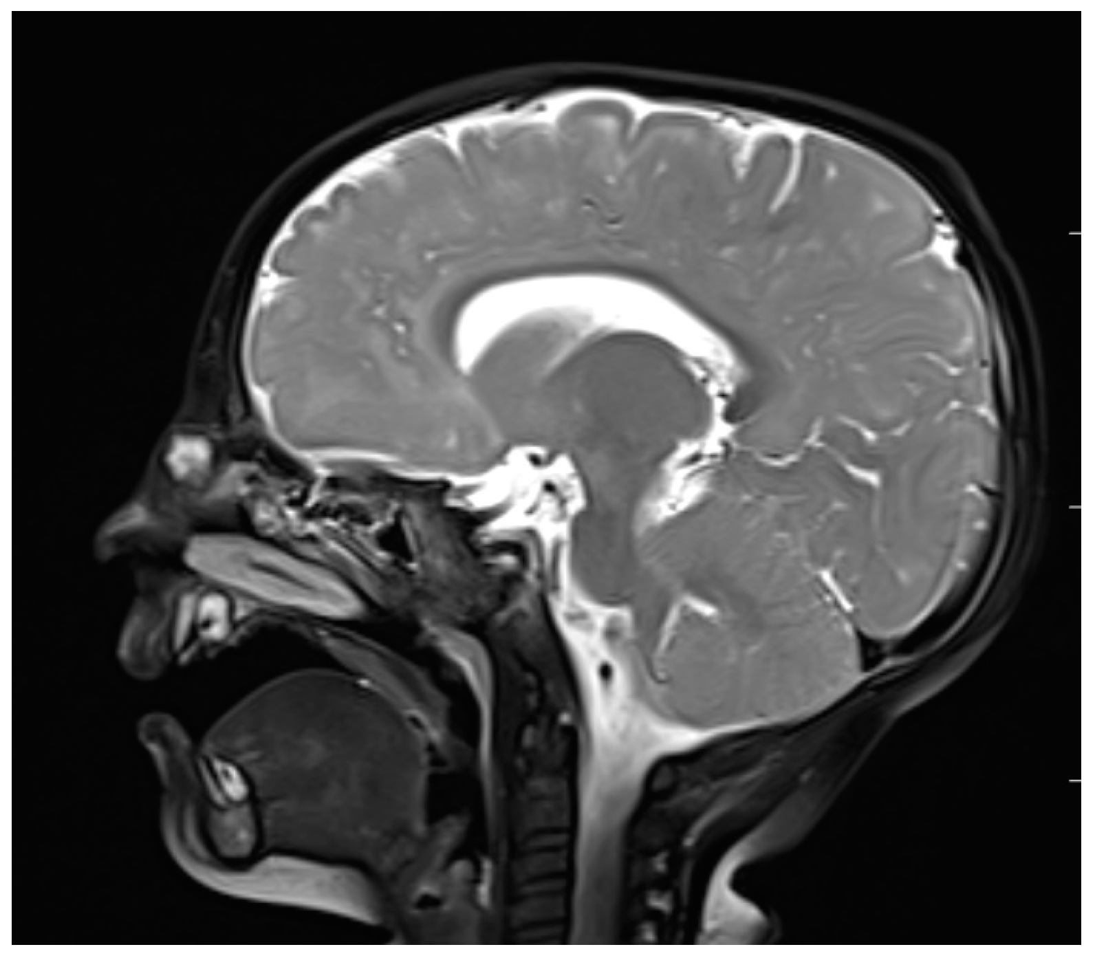
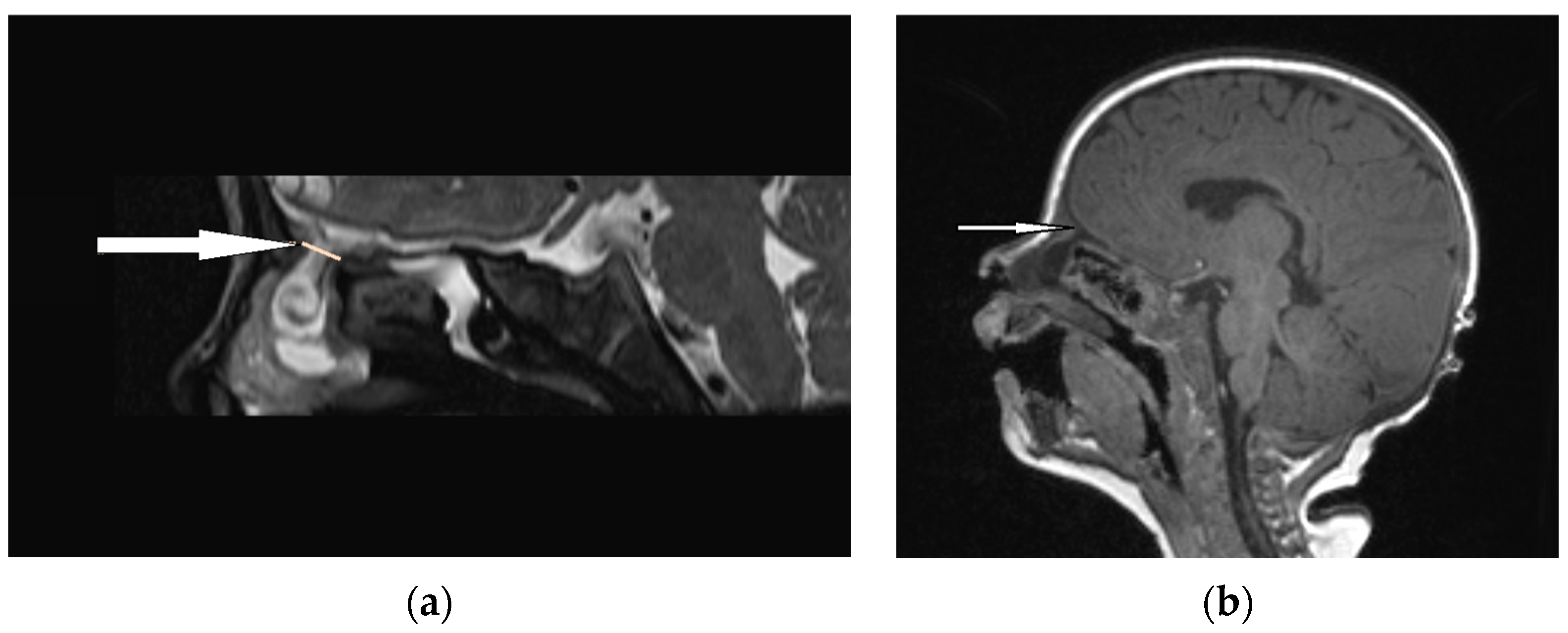
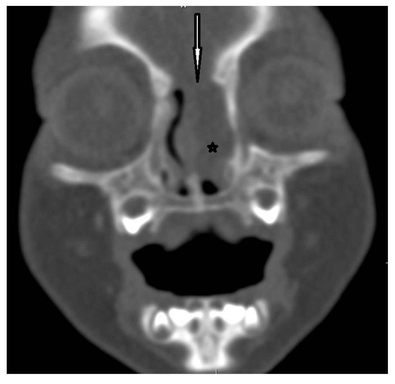
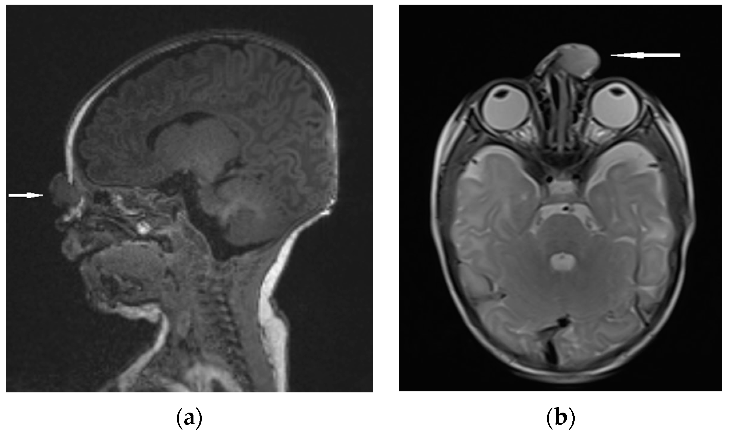
| Type of Pathology | ||||
|---|---|---|---|---|
| ND | NGH | EPHC | ||
| Clinical manifestation | Location | Anywhere from the philtrum of the upper lip to the glabellar region | Usually glabella, nasal dorsum, or intranasal | Transethmoidal, sphenoethmoidal, transsphenoidal, sphenoorbital, nasofrontal, nasoethmoidal, or nasoorbital |
| Form | Cyst, sinus, or fistula; possible intracranial extension | Extranasal, intranasal, or combined mass | Intra- or extranasal mass | |
| Characteristics | Hair visible in the skin ostium; non-expansile, non-pulsatile, non-compressible, and non-transilluminating mass | Tends to grow along with the patient; firm and non-compressible mass, non-transilluminating; intranasal—pale, “polypoid”; extranasal—smooth, well-circumscribed, reddish or bluish, often with telangiectasis on its surface | Smooth “polypoid” mass, pink or bluish, covered with mucous membrane, usually pulsatile, transilluminating, soft, and compressible | |
| Furstengerg’s sign | Negative | Negative | Positive | |
| Radiological characteristics | CT | No correlation between the particular location of the sinus ostium or cyst and the presence of intracranial extension; bifid crista galli and widening of foramen caecum (suggestive of intracranial extension); dermoid cyst—density of fat; epidermoid cyst—density of water | Bony defect may be revelaed | Developmental bony defect of the skull base |
| MRI | Variable signal intensity depending on the protein content; fat-suppressed T1-weighted images—differentiation between skull base defects and enhancing non-ossified cartilage of anterior cranial fossa; DWI—typically high-signal-intensity lesion with corresponding low signal intensity on ADC maps | Discontinuity with the brain parenchyma; variable visualization of a fibrous stalk connection to CNS; well-circumscribed, rounded, or polypoid mass—isointense or rarely hypointense to gray matter on T1-weighted imaging; neural tissue—more hyperintense on T2-weighted images to normal brain parenchyma in most cases; dysplastic tissue usually corresponds with no enhancement or moderate enhancement; noticeable enhancement at the lesion periphery | Herniation of intracranial tissue and its continuity with the brain | |
| Histopathological findings | Lined by ectodermal-derived stratified squamous epithelium; consists of a characteristic oily substance including fat and hair | Varying proportions of neurons and glia; dysplastic neuroglial components including mainly astrocytes, but occasionally also gemistocytic astrocytes, neurons, or ependymal cells; usually no significant leptomeningeal, ependymal, or choroid plexus structures; reactive gliosis—routinely observed; degree of fibrosis/sclerosis increasing with age; external overlying epithelium represents different characteristics (depending on NGH clinical form and location) | Histologically similar to NGHs; covered with columnar or cuboidal epithelium and dense fibrous dura-like band; layer of leptomeninges surrounding neural structures; brain tissue—usually a mature neuroglial tissue composed of neurons and large ovoid or spindle astrocytes | |
| Staining/IHC | Hematoxylin-eosin staining; Masson trichrome staining; Nissl staining or neuron-specific enolase (neural structures in unclear cases); immunohistochemistry— S-100, GFAP (astrocytes marker); laminin and collagen type IV—for reactive fibrosis | Hematoxylin-eosin staining; immunohistochemistry—S100, vimentin, GFAP; glial tissue—positive to NeuN; EMA to identify the presence of meningeal components | ||
Disclaimer/Publisher’s Note: The statements, opinions and data contained in all publications are solely those of the individual author(s) and contributor(s) and not of MDPI and/or the editor(s). MDPI and/or the editor(s) disclaim responsibility for any injury to people or property resulting from any ideas, methods, instructions or products referred to in the content. |
© 2023 by the author. Licensee MDPI, Basel, Switzerland. This article is an open access article distributed under the terms and conditions of the Creative Commons Attribution (CC BY) license (https://creativecommons.org/licenses/by/4.0/).
Share and Cite
Kotowski, M. The Differential Diagnosis of Congenital Developmental Midline Nasal Masses: Histopathological, Clinical, and Radiological Aspects. Diagnostics 2023, 13, 2796. https://doi.org/10.3390/diagnostics13172796
Kotowski M. The Differential Diagnosis of Congenital Developmental Midline Nasal Masses: Histopathological, Clinical, and Radiological Aspects. Diagnostics. 2023; 13(17):2796. https://doi.org/10.3390/diagnostics13172796
Chicago/Turabian StyleKotowski, Michal. 2023. "The Differential Diagnosis of Congenital Developmental Midline Nasal Masses: Histopathological, Clinical, and Radiological Aspects" Diagnostics 13, no. 17: 2796. https://doi.org/10.3390/diagnostics13172796
APA StyleKotowski, M. (2023). The Differential Diagnosis of Congenital Developmental Midline Nasal Masses: Histopathological, Clinical, and Radiological Aspects. Diagnostics, 13(17), 2796. https://doi.org/10.3390/diagnostics13172796


