Article

The Impact of Liquid Biopsies Positive for EGFR Mutations on Overall Survival in Non-Small Cell Lung Cancer Patients

by
Jonnathan Roldan Ruiz
1,
Marta Gracia Fuentes Gago
2,
Luis Miguel Chinchilla Tabora
3,
Idalia Gonzalez Morais
3,
José María Sayagués
3,
Mar Abad Hernández
3,
Maria Rosa Cordovilla Pérez
4,
Maria Dolores Ludeña de la Cruz
3,
Edel del Barco Morillo
1 and
Marta Rodriguez Gonzalez
3,*
1
Department of Clinical Oncology, Biomedical Research Institute of Salamanca (IBSAL), University Hospital of Salamanca, University of Salamanca, 37007 Salamanca, Spain
2
Department of Thoracic Surgery, University Hospital of Salamanca, 37007 Salamanca, Spain
3
Department of Pathology, Biomedical Research Institute of Salamanca (IBSAL), University Hospital of Salamanca, 37007 Salamanca, Spain
4
Department of Pulmonology, University Hospital of Salamanca, 37007 Salamanca, Spain
*
Author to whom correspondence should be addressed.
Diagnostics 2023, 13(14), 2347; https://doi.org/10.3390/diagnostics13142347
Submission received: 31 May 2023 / Revised: 6 July 2023 / Accepted: 10 July 2023 / Published: 12 July 2023
(This article belongs to the Special Issue Diagnosis and Management of Lung Cancer)

Abstract

:
In recent years, non-small cell lung cancer treatment has been revolutionized. EGFR tyrosine kinase inhibitors and our improved understanding of its alterations have driven new diagnostic strategies. Liquid biopsies have emerged as a useful tool in these contexts, showing potential utility in early diagnosis combined with low-dose CT scans, as well as potential in monitoring treatment response and predicting the development of patients. We studied the circulating tumor DNA (ctDNA) of 38 EGFR-mutated non-small cell lung cancer patients at diagnosis in different moments of their disease by liquid biopsy techniques. Our results show that mean overall survival was significantly lower when a liquid biopsy was positive for the detection of EGFR mutations compared with wild-type patients in their liquid biopsy in both univariate (29 ± 4 vs. 104 ± 19 months; p = 0.004) and multivariate analysis (p = 0.008). Taking this into consideration, liquid biopsies could be key to improving the control of this disease.

1. Introduction

Lung cancer is the leading cause of death from cancer worldwide, representing 11.4% of new diagnoses and accounting for 18% of all cancer deaths [1,2,3]. There are two main types of lung cancer: small cell lung cancer (SCLC) and non-small cell lung cancer (NSCLC). About 85% of lung cancers are NSCLCs, which encompasses different histological subtypes such as adenocarcinomas, squamous cell carcinomas, and large cell carcinomas [4]. Up to 80–90% of patients with NSCLC are symptomatic at the time of diagnosis, showing frequent respiratory symptoms related to primary tumors. Patients may also present symptoms associated with metastasis, particularly neurological symptoms because of the involvement of the central nervous system, and bone pain [4]. The main risk factor for the development of NSCLC continues to be tobacco. Although, over the last decades, tobacco consumption rates have fallen among the global population, in Spain, tobacco consumption remains at a high level among men aged 15–64 years [5,6]. Furthermore, an increase in NSCLC cases among non-smoking women has been observed, meaning that the underlying causes are unclear (e.g., they may be related to hormonal exposure, previous radiotherapy for the treatment of other malignancies such as breast cancer, home environmental factors, etc.) [7,8].
The treatment of patients with lung cancer depends on the morphology, tumor stage, molecular characteristics, and assessment of a patient’s overall medical (OS) condition. Surgery is the most effective treatment, but it is reserved for patients with stage I, II, or IIIA NSCLC (representing only 35% of total cases) [4]. Since most NSCLC patients present an advanced-stage disease at diagnosis, systemic treatment with cytotoxic agents is often used as the standard treatment, showing a modest survival benefit [9].
A better understanding of the underlying molecular mechanisms that encourage tumorigenesis in NSCLC has led to the development of many target tools aimed at specific genetic abnormalities. The discovery of activating mutations of the Epidermal Growth Factor Receptor (EGFR) gene, as well as the advancement of EGFR tyrosine kinase inhibitors (TKI), has had a significant impact on adaptive treatment strategies and, consequently, on survival among patients with advanced NSCLC [10]. In addition to these TKI acting against EGFR mutations, in recent years, there has been a genuine revolution in the treatment of lung cancer through the development of drugs that act against ALK, ROS1, and RET rearrangements [11,12,13], BRAF [14,15,16], and KRAS mutations [17], as well as the standardization immunotherapy based on the immunohistochemical expression of PD-L1 [18,19]. Therefore, NSCLC has brought personalized medicine to the fore globally, and the most recently published guidelines propose that advanced non-squamous NSCLC and selected patients with squamous cell carcinomas (SCCs) should undergo genetic testing [20,21].
Despite these advances on the onset of the disease, monitoring patients for early relapse, and detecting resistance to new targeted therapies, challenges continue to persist. Diagnoses and histological typification are carried out by examining a small piece of tissue sample or even through cytology, with a likely lack of sufficient material for a proper molecular diagnosis or without the possibility to obtain a new tumor sample [22,23,24,25,26].
A liquid biopsy (LB) is a minimally invasive alternative to surgical biopsies that is based on analyzing circulating tumor cells (CTCs), cell-free DNA in plasma (cfDNA), circulating miRNA, exosomes, and tumor-educated platelets (TEP) in a biological fluid (mainly blood) [27]. These methods have emerged as a powerful tool to identify molecular alterations in patients with cancer. In addition to the practically non-invasive nature of this procedure, other advantages of liquid biopsies include their rapid processing and optimal reflections of tumor heterogeneity. They are also highly useful both after ablative treatment and in monitoring the response to targeted therapy. The major constraint is the very low amount of ctDNA. At present, there are various strategies and platforms available; however, thus far, there is no clear consensus on when or when not to perform a liquid biopsy [26,28,29].
Over the past few years at our hospital, a referral hospital in the mid-west of Spain, we have performed LBs and examined EGFR mutational status in accordance with the Medical Oncology Service’s guidelines; in most cases, patients have an evolution that is worse than expected. The objectives of the present study were to analyze our hospital’s clinical management of EGFR-mutated NSCLC patients, with a specific emphasis on how we used LB techniques, and assess the impact of LB techniques on tumor evolution and OS among patients.

2. Materials and Methods

2.1. Study Design

This is a retrospective observational study based on the first 79 liquid biopsies of patients treated at the Pathological Anatomy Service of the Complejo Asistencial Universitario de Salamanca between October 2016 and December 2022. Patients without EGFR alterations in the biopsy were excluded; consequently, this meant that our cohort consisted of 38 patients. All of them had non-squamous NSCLC diagnosed via small biopsy specimen or fine needle aspiration (FNA). Clinical (age, sex, symptoms, manifestations, type of sample for diagnosis, stage, surgery), histological (diagnosis, TTF1 and PDL1, staining), and evolutionary (treatment, recurrence, death) variables were collected. The biomarkers studied in the clinical treatment of NSCLC (ALK, ROS1, and BRAF genes) did not show relevance in the present study (Supplementary Table S1).

2.2. Tissue Biopsy Procedure

Tissue samples were obtained from surgical or biopsy specimens and were formalin-fixed paraffin-embedded (FFPE) tissue blocks. A 3 μm section stained with hematoxylin-eosin was made from each of the samples. Immunohistochemistry techniques (TTF1 (Leica, Microsystem Ltd., Milton Keynes, UK)) and p40 staining (A. Menarini Diagnostics, San Diego, CA, USA, a 1:200 dilution) were used for all the samples via the use of an automated immunostainer (Bond Polymer Refine Detection, Leica Microsystem Ltd., Milton Keynes, UK) and following the manufacturer’s instructions. Nuclear staining was considered positive in >10% of tumor cells. Furthermore, in 4 of these cases, histochemical techniques, such as periodic acid–Schiff (PAS) and periodic acid–Schiff–diastase (PAS diastase) for glycogen detection with the Alcian Blue/Periodic Acid-Schiff Stain kit on Agilent ArtisanLink platform from DAKO (Stevens Creek Blvd. Santa Clara, CA, USA), were also performed. A real-time PCR test was performed to examine the most common mutations in exons 18, 19, 20, and 21 of EGFR gene (Cobas® 4800 EGFR mutation test v2) on purified DNA samples that had been isolated from the tissue samples derived from biopsies.

2.3. Liquid Biopsy Procedure

When it was not possible to perform a re-biopsy and in cases where the oncologist needed updated patient information, data on at least one prior liquid biopsy were obtained per patient during the course of their disease (Supplementary Table S2). Peripheral blood samples (usually around 10 mL) were collected in Vacutainer EDTA tubes (BD, Plymouth, UK) for each liquid biopsy. For the first hour, the plasma was subjected to high-speed centrifugation (10,000 rpm for 10 min at 4 °C), and immediately after that, a second centrifugation (16,000 rpm for 10 min at 4 °C) was performed to pellet cell debris. After centrifugation, at least 2 mL of the supernatant was aliquoted into 2 mL cryotubes and stored at −80 °C. The commercially available Cobas® cfDNA sample preparation kit was then used to extract ctDNA. DNA quantification was carried out by using a NanoDrop® ND-1000 spectrophotometer (ND-1000, NanoDrop Technologies, Wilmington, DE, USA). We used the FDA-approved LB test for detecting EGFR mutations (Cobas® EGFR Mutation Test v2 for blood/from plasma samples) (CE-IVD) (Roche Molecular Diagnostics, Branchburg, NJ, USA).

2.4. Statistical Analysis
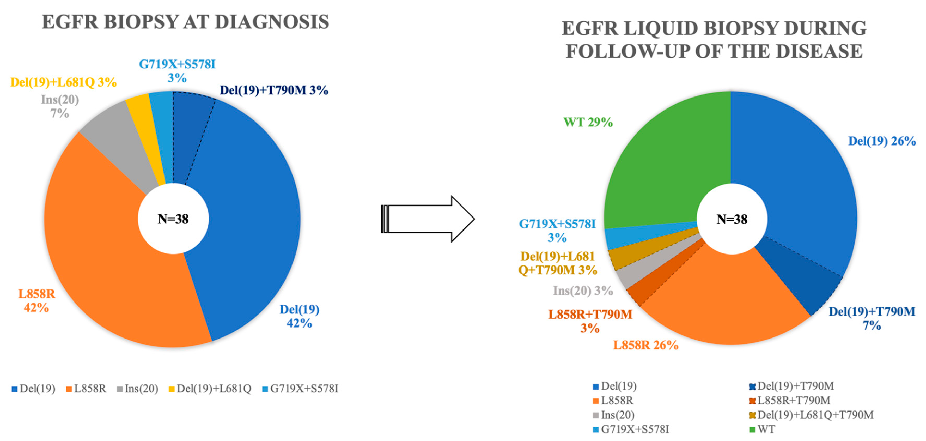
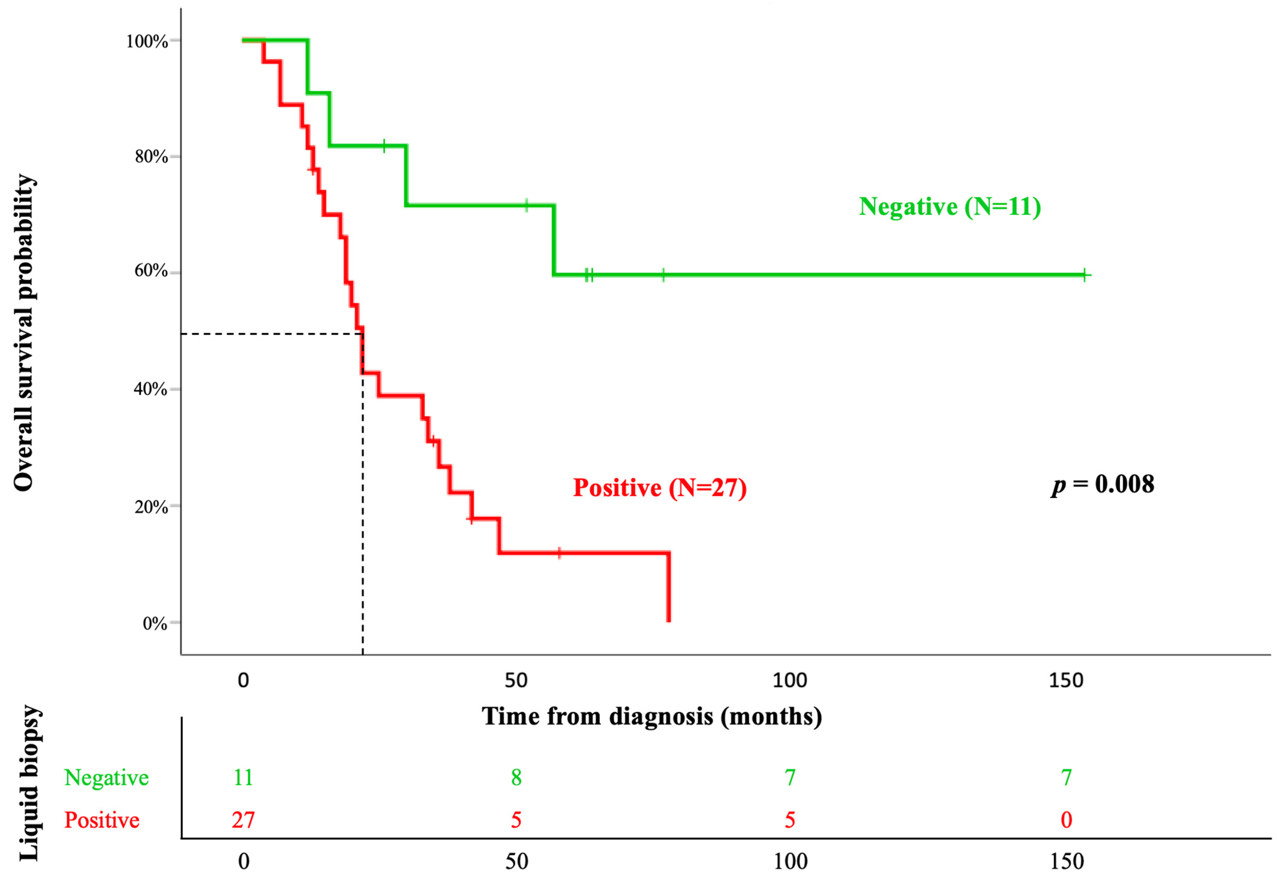
All the clinical/biological, morphological, genetic, and evolutionary variables were collected in a database, and the statistical program SPSS v.21 (IBM Corp., Armonk, NY, USA) was used to calculate the statistical significance of the different variables, as well as OS. For normally distributed continuous variables, we used Student’s t-test, and for non-normally distributed ones, the Mann–Whitney U test was used. OS curves were determined according to the Kaplan–Meier test. Multivariate analyses of prognostic factors influencing OS were performed using Cox regression, only taking into a count the variables that showed a significant association with OS in the univariate analysis; a p value of <0.05 (or Pearson-corrected p, if applicable) indicated statistical significance.

3. Results

Of the 38 patients included in the present study, 17 were men (45%) and 21 women (55%); the median age of 68 years (43–85 years). The majority (25; 66%) were non-smokers (Table 1). Functional status was assessed at two levels. Although it is true that the 38 patients presented good general status, 24 of them were classified as 0 (66%) according to the Eastern Cooperative Oncology Group’s (ECOG) guidelines, and the remaining 14 were classified as ECOG 1 (34%). Notably, 18% of patients (seven cases) were completely asymptomatic, and their diagnosis was incidental. This observation was made before complementary control or monitoring tests were carried out for the other non-tumor pathologies. A total of 22 patients (58%) presented symptoms associated with the primary tumor, the majority of which were manifested via coughing, expectoration, hemoptysis, dyspnea, or chest pain; 9 (24%) patients showed symptoms related to the location of the metastases, highlighting neurological manifestations, and pain at different levels of the spine. Nearly three quarters of the patients had stage IV diseases (28 cases, 74%); five patients had a locally advanced stage III level of disease (13%), and a small minority presented early-stage diseases (three with stage I, accounting for 8% of patients, and two with stage II, accounting for of patients 5%). Most of the patients did not undergo surgery (33; 87%); therefore, their histological diagnosis was obtained, in most cases, via a small biopsy (transbronchial biopsy, core-needle biopsy (CNB) of the lung, fine needle aspiration (FNA), or a cytological examination based on pleural fluid analysis).
Among the samples received (36 cases), 95% were diagnosed as adenocarcinomas, while two (5%) were deemed undifferentiated NSCLC (Table 2). On the one hand, p40 was entirely negative in all cases. On the other hand, 91% (35 patients) were TTF1 positive, and we performed PAS and PAS diastase in two of the three negative cases (9%), with positive results being obtained for both samples.
In the tissue samples, del(19) and L858R mutations were found to be the most frequently observed mutations (n = 16 cases; 42%), followed by Ins(20) (n = 3; 8%) and the combined mutations del(19) + L861Q (n = 1; 3%), del(19) + T790M (n = 1; 3%), and G719X + S578I (n = 1; 3%) (Supplementary Table S2 and Figure S1). Of the 38 patients, 32 (84%) started first-line treatment with first-generation tyrosine kinases inhibitors (TKIs), 20 with Erlonitib, and 10 with Gefitinib; 2 patients were assigned chemotherapy as their standard for of treatment to manage mutational mutation, specifically Ins(20) resistance. In addition, four patients (10%) received chemotherapy after post-surgical recurrence.
At least one liquid biopsy was performed to study EGFR mutations in all patients in the cohort (Supplementary Table S2), and the median follow-up was 24 months (4–154 months). All LBs were performed at the request of the Medical Oncology Service of our hospital. As shown in Figure 1, in 71% of the patients (n = 27), the presence of EGFR mutations was detected in at least one LB, with del(19) and L858R being the most frequent mutations (10 cases; 26%), followed by the combined mutations del(19) + T790M (4 cases; 10%). During patient monitoring, 13 patients developed T790M mutation resistance, leading to a change in their therapeutic approach to incorporate Osimertinib, a third-generation TKI with sensitivity to this mutation. Only one patient presented this alteration at diagnosis (associated with del(19)). In these 13 patients, the median onset of the T790M mutation from diagnosis was 34 months (7–78 months).
As expected, positivity for TTF1 was observed in most cases (n = 36; 95%), while expression in tumor cells was detected in half of the cases studied, with expression not being associated with LB positivity (Table 2).
Finally, the mean OS among patients was 48 ± 10 months. During patient monitoring, 31 of the 38 patients died (82%), showing statistically significant differences between patients with positive and negative LBs (27 cases (71%) vs. 11 cases (29%), respectively, p = 0.004). From a prognostic point of view, the variables that showed a significant influence on OS in the univariate analysis were treatment, ECOG, and LB (p < 0.05) (Table 3). However, in the multivariate analysis, only LB maintained significance (HR = 4.7; p = 0.008), with a mean OS of 29 ± 4 vs. 104 ± 20 months in positive and negative LBs, respectively (p 0.004) (Figure 2).

4. Discussion

EGFR mutations have become a very important therapeutic target in patients with NSCLC, especially in advanced stages. They have a significant impact on both OS and quality of life [30]. Therefore, improving our knowledge of these alterations, as well as the role that LBs play in detecting them and in the monitoring of these patients, is essential for improving therapeutic strategies and adapting treatment regimes so that align with patient’s stage of disease. The prevalence of lung cancer has led to recent developments in personalized medicine and revolutionized patient management by forcing clinical institutions to integrate clinical data, pathological findings, and molecular profiling.
The results obtained in our study are consistent with the literature regarding the profile of the patients studied. Overall, 60% of our patients were women, and 67% declared themselves to be non-smokers [31,32]. This confirms the increasing trend of NSCLC cases among women, especially non-smokers [7,8]. Even though tobacco use rates have generally decreased over time among men, they continue to be high [5,33,34]. In fact, half of the male patients (n = 8) in our study were smokers or ex-smokers, and we only found this habit in 23% of the female patients (6 out of 26), a trend that is seemingly consistent with what is published in the literature [3,34,35,36,37]. Likewise, it is estimated that 53% of lung cancer cases are diagnosed between the ages of 55 and 74 [34], with the median age of diagnosis in our study being 68 years. It should be noted that the median age of diagnosis in women is approximately 8 years older than in men (65 ± 13 years vs. 72 ± 10 years), probably due to decreased direct exposure to tobacco, although the etiological factors of lung cancer not associated with smoking are yet to be defined (e.g., hormonal exposure, previous radiotherapy due to other neoplasms such as breast cancer or environmental factors) [7,36,38,39,40] and are not well-characterized.
Even though surgery is the best form of treatment for patients with early-stage NSCLC, approximately 35–55% of patients relapse during the first 5 years after surgery [41,42,43,44]. The causes of this circumstance have not been adequately elucidated, but some authors have pointed to the percentage of solid components in the lesion, the histological subtype, the micropapillary growth pattern, necrosis, airborne spread, etc. [45,46,47]. In our series, 5 of the 38 patients were considered to be in the early stage of their disease (I–II), and after surgery, all of them presented local or distant relapses throughout the course of the disease. Nevertheless, up to 80% of patients with lung cancer are diagnosed in advanced stages, where the main diagnostic technique is usually a small biopsy or FNA [48,49,50]. This frequency was slightly higher in our study as almost 90% of the patients were in a locally advanced stage (n = 6; 14%) or metastatic (n = 32; 74%), confirming the fact that lung cancer is often diagnosed in the advanced stages, when therapeutic options are limited. Because of this reason, detection systems that allow for early stages diagnoses and therefore increase the possibility of curative treatment strategies are being extensively researched. In this regard, studies have been carried out to demonstrate the benefit of low-dose computed tomography (LDCT) in patients with risk factors [51]. However, this approximation method has some limitations, examples of which include false positives (with consequent overdiagnosis) and the radiation dose administered to patients who are healthy a priori [28]. In order to solve those problems, the value of the LB as an auxiliary tool to LDCT is being explored both as a screening method and as a diagnostic method in the early stages of the disease through molecular identification in peripheral blood [42,52]. Indeed, TRACERx has shown that ctDNA can be detected between 6 and 12 months before the presence of radiologically detectable lesions, which, in a selected population, could complement LDCT, decreasing the number of diagnostic interventions [53].
Previous reports based on the study of the mutational status of the EGFR gene, both in tissue and fluid biopsies, show that the mutations del(19) and L858R, followed by ins(20), are the most frequent mutations observed in NSCLC patients [54]. In line with other studies that used similar methodological approaches to that of this study, our results showed the presence of del(19) and L858R in almost half of the NSCLC cases studied. We only observed a complex mutation in two patients (G719X + S578I, and del(19) + L681Q). Complex EGFR mutations comprise a heterogenous group of mutations with a prevalence between 5 and 15% of all EGFR mutations [54]. In alignment with these observations, in our study, complex mutations were observed in the 6% of cases. The presence of the T790M mutation in the diagnosis of the disease is infrequent. Indeed, it has been suggested that it is a germline mutation of the EGFR gene [55] and has been associated with a poor prognosis [54,56,57,58]. In our study, only one patient presented a mutation associated with del(19) at diagnosis, and no significantly lower median survival was observed compared to other patients with different mutations (19 vs. 20 months).
Even though routine LB tests are not recommended in the diagnosis and follow-up of NSCLC [59], the results obtained in our study, although small, are strikingly significant. We observed that, in patients in whom the EGFR mutation was not detected in LB during follow-up, overall survival was four times higher than patients with at least one EGFR mutation detected during their LB (mean OS of 29 ± 4 vs. 104 ± 19 months; p 0.001, respectively). These data suggest that LB could have a powerful predictive value in the outcome of patients with NSCLC. Most of the cases with positive LB were detected between the first and the second determination, which implies that two ordinary blood extractions could infer the evolution of the patient. In our study, we used quantitative PCR (Cobas EGFR mutation test V2); however, there are currently more sensitive techniques and technologies available, such as microfluidic technology (digital PCR [60] and BEAMing [61]) or massive parallel sequencing (NGS) [62], which could further increase the predictive value of the mutational status of the EGFR gene. In the FASTACT-2 study [63], a baseline LB was performed, and on the first day of the third cycle, they observed the presence of EGFR mutations in the second LB, associating it with a negative impact on overall and disease-free survival. Similarly, a LUNGCA-1 study observed that the detection of EGFR mutations through the LBs of operated patients can predict relapses [64]. Although it is true that there is no consensus on when to perform a LB, aside from when searching for resistant mutations (T790M to first- or second-generation EGFR-TKI; C797S to third generation EGFR-TKI) or after an observed decreased in response to treatment, our own findings reinforce the idea of incorporating it into the routine of oncological control [65]. In this way, a LB could be performed at baseline even before starting treatment and/or in the postoperative period and every 3 months during the first year, when resistant mutations usually appear [10,63,66,67,68].
In 2019, the addition of Osimertinib to the first-line treatment of advanced NSCLC [69] decreased interest in searching for the T790M resistant mutation in newly diagnosed patients. In our study, LBs detected this mutation in 13 cases (34%) throughout the follow-up period; thus, it is possible that LBs could enable shifts to more effective treatment strategies, positively impacting quality of life and survival [63]. At present, the mutations demonstrating resistance to Osimertinib mechanisms are already known, such as the loss of the T790M mutation (early event) or C797S mutation, which is present in 7% of cases [54], and more information on these mechanisms will be made available over time thanks to new NGS techniques

5. Conclusions

In summary, our results show that the presence of mutations in the EGFR gene detected using LB techniques in patients with EGFR-mutated NSCLC is an independent prognostic factor for OS. Liquid biopsies make it possible to monitor the mutations of a tumor in a relatively simple and quick way, which, together with other diagnostic tools, could be useful in our daily clinical practice, improve prognostic assessments, and guide clinical decision making regarding the treatment of NSCLC patients. Additional prospective studies with larger cohorts are required to validate the utility of LB techniques in the diagnosis and follow-up of NSCLC patients.

Supplementary Materials

The following supporting information can be downloaded at: https://www.mdpi.com/2075-4418/13/14/2347/s1, Table S1: Correlation between the mutational status of the EGFR gene detected by liquid biopsy techniques and alterations in the ALK, ROS1 and BRAF genes; Table S2: Type of mutations detected in the EGFR gene in plasma samples from EGFR mutated non-small cell lung cancer (NSCLC) patients (n = 38) during disease follow-up.

Author Contributions

Conceptualization, M.R.G. and M.A.H.; methodology, J.R.R.; validation, M.R.G. and I.G.M.; formal analysis, J.M.S. and M.G.F.G.; interpretation of the results of the PCR, FISH and IHC techniques, M.A.H. and L.M.C.T.; investigation, J.R.R. and M.R.C.P.; writing—original draft preparation, M.R.G.; writing—review and editing, M.D.L.d.l.C. and E.d.B.M.; visualization, L.M.C.T.; supervision, M.R.G. All authors have read and agreed to the published version of the manuscript.

Funding

This research received no external funding.

Institutional Review Board Statement

The study was conducted in accordance with the Declaration of Helsinki and approved by the Local Ethics Committee of University Hospital of Salamanca, Salamanca, Spain (IP 2019/02/189).

Informed Consent Statement

Informed consent was obtained from all subjects involved in the study.

Data Availability Statement

All study data can be viewed in the manuscript.

Acknowledgments

We are grateful to Carmen Rodriguez, Saray Muñoz, and Belen Rivas (Pathology Department, University Hospital of Salamanca) for their technical assistance. We are also thankful to Javier Garcia de la Iglesia for providing graphical support.

Conflicts of Interest

The authors declare no potential conflict of interest with respect to the research, authorship, or publication of this article.

References

  1. Barta, J.A.; Powell, C.A.; Wisnivesky, J.P. Global Epidemiology of Lung Cancer. Ann. Glob. Health 2019, 85, 8. [Google Scholar] [CrossRef] [PubMed]
  2. Sung, H.; Ferlay, J.; Siegel, R.L.; Laversanne, M.; Soerjomataram, I.; Jemal, A.; Bray, F. Global Cancer Statistics 2020: GLOBOCAN Estimates of Incidence and Mortality Worldwide for 36 Cancers in 185 Countries. CA. Cancer J. Clin. 2021, 71, 209–249. [Google Scholar] [CrossRef]
  3. Siegel, R.L.; Miller, K.D.; Fuchs, H.E.; DVM, A.J. Cancer Statistics, 2022—Siegel—2022—CA: A Cancer Journal for Clinicians—Wiley Online Library. Available online: https://acsjournals.onlinelibrary.wiley.com/doi/full/10.3322/caac.21708?utm_source=newsletter&utm_medium=email&utm_campaign=work-in-progress&utm_content=20230117 (accessed on 21 March 2023).
  4. WHO Classification of Tumours Editorial Board. Thoracic Tumours; WHO: Geneva, Switzerland, 2021; ISBN 978-92-832-4506-3. [Google Scholar]
  5. Rábade Castedo, C.; de Granda-Orive, J.I.; González-Barcala, F.J. Incremento de la prevalencia del tabaquismo: ¿causas y actuación? Arch. Bronconeumol. 2019, 55, 557–558. [Google Scholar] [CrossRef]
  6. Lidón-Moyano, C.; Fu, M.; Ballbè, M.; Martín-Sánchez, J.C.; Matilla-Santander, N.; Martínez, C.; Fernández, E.; Martínez-Sánchez, J.M. Impact of the Spanish smoking laws on tobacco consumption and secondhand smoke exposure: A longitudinal population study. Addict. Behav. 2017, 75, 30–35. [Google Scholar] [CrossRef]
  7. MacRosty, C.R.; Rivera, M.P. Lung Cancer in Women. Clin. Chest Med. 2020, 41, 53–65. [Google Scholar] [CrossRef]
  8. Stapelfeld, C.; Dammann, C.; Maser, E. Sex-specificity in lung cancer risk. Int. J. Cancer 2020, 146, 2376–2382. [Google Scholar] [CrossRef] [PubMed]
  9. Lu, T.; Yang, X.; Huang, Y.; Zhao, M.; Li, M.; Ma, K.; Yin, J.; Zhan, C.; Wang, Q. Trends in the incidence, treatment, and survival of patients with lung cancer in the last four decades. Cancer Manag. Res. 2019, 11, 943–953. [Google Scholar] [CrossRef] [PubMed]
  10. Rosell, R.; Moran, T.; Queralt, C.; Porta, R.; Cardenal, F.; Camps, C.; Majem, M.; Lopez-Vivanco, G.; Isla, D.; Provencio, M.; et al. Screening for Epidermal Growth Factor Receptor Mutations in Lung Cancer. N. Engl. J. Med. 2009, 361, 958–967. [Google Scholar] [CrossRef]
  11. Tan, A.C.; Tan, D.S.W. Targeted Therapies for Lung Cancer Patients with Oncogenic Driver Molecular Alterations. J. Clin. Oncol. Off. J. Am. Soc. Clin. Oncol. 2022, 40, 611–625. [Google Scholar] [CrossRef]
  12. Gendarme, S.; Bylicki, O.; Chouaid, C.; Guisier, F. ROS-1 Fusions in Non-Small-Cell Lung Cancer: Evidence to Date. Curr. Oncol. Tor. Ont 2022, 29, 641–658. [Google Scholar] [CrossRef]
  13. Cascetta, P.; Sforza, V.; Manzo, A.; Carillio, G.; Palumbo, G.; Esposito, G.; Montanino, A.; Costanzo, R.; Sandomenico, C.; De Cecio, R.; et al. RET Inhibitors in Non-Small-Cell Lung Cancer. Cancers 2021, 13, 4415. [Google Scholar] [CrossRef] [PubMed]
  14. Hwang, I.; Choi, Y.-L.; Lee, H.; Hwang, S.; Lee, B.; Yang, H.; Chelakkot, C.; Han, J. Selection Strategies and Practical Application of BRAF V600E-Mutated Non-Small Cell Lung Carcinoma. Cancer Res. Treat. 2022, 54, 782–792. [Google Scholar] [CrossRef] [PubMed]
  15. Imyanitov, E.N.; Iyevleva, A.G.; Levchenko, E.V. Molecular testing and targeted therapy for non-small cell lung cancer: Current status and perspectives. Crit. Rev. Oncol. Hematol. 2021, 157, 103194. [Google Scholar] [CrossRef] [PubMed]
  16. Alexander, M.; Kim, S.Y.; Cheng, H. Update 2020: Management of Non-Small Cell Lung Cancer. Lung 2020, 198, 897–907. [Google Scholar] [CrossRef]
  17. Reck, M.; Carbone, D.P.; Garassino, M.; Barlesi, F. Targeting KRAS in non-small-cell lung cancer: Recent progress and new approaches. Ann. Oncol. Off. J. Eur. Soc. Med. Oncol. 2021, 32, 1101–1110. [Google Scholar] [CrossRef]
  18. Patel, S.A.; Weiss, J. Advances in the Treatment of Non-Small Cell Lung Cancer: Immunotherapy. Clin. Chest Med. 2020, 41, 237–247. [Google Scholar] [CrossRef]
  19. Isla, D.; Lozano, M.D.; Paz-Ares, L.; Salas, C.; de Castro, J.; Conde, E.; Felip, E.; Gómez-Román, J.; Garrido, P.; Enguita, A.B. New update to the guidelines on testing predictive biomarkers in non-small-cell lung cancer: A National Consensus of the Spanish Society of Pathology and the Spanish Society of Medical Oncology. Clin. Transl. Oncol. Off. Publ. Fed. Span. Oncol. Soc. Natl. Cancer Inst. Mex. 2022, 25, 1252–1267. [Google Scholar] [CrossRef]
  20. Rolfo, C.; Mack, P.C.; Scagliotti, G.V.; Baas, P.; Barlesi, F.; Bivona, T.G.; Herbst, R.S.; Mok, T.S.; Peled, N.; Pirker, R.; et al. Liquid Biopsy for Advanced Non-Small Cell Lung Cancer (NSCLC): A Statement Paper from the IASLC. J. Thorac. Oncol. 2018, 13, 1248–1268. [Google Scholar] [CrossRef]
  21. Mosele, F.; Remon, J.; Mateo, J.; Westphalen, C.B.; Barlesi, F.; Lolkema, M.P.; Normanno, N.; Scarpa, A.; Robson, M.; Meric-Bernstam, F.; et al. Recommendations for the use of next-generation sequencing (NGS) for patients with metastatic cancers: A report from the ESMO Precision Medicine Working Group. Ann. Oncol. 2020, 31, 1491–1505. [Google Scholar] [CrossRef]
  22. Mlika, M.; Hofman, P.; Dziri, C.; Mezni, F. La biopsie liquide dans le cancer du poumon Liquid biopsy in lung cancer. Tunis. Med. 2017, 95, 965–971. [Google Scholar]
  23. Bernabé, R.; Hickson, N.; Wallace, A.; Blackhall, F.H. What do we need to make circulating tumour DNA (ctDNA) a routine diagnostic test in lung cancer? Eur. J. Cancer 2017, 81, 66–73. [Google Scholar] [CrossRef] [PubMed]
  24. Bonanno, L.; Dal Maso, A.; Pavan, A.; Zulato, E.; Calvetti, L.; Pasello, G.; Guarneri, V.; Conte, P.; Indraccolo, S. Liquid biopsy and non-small cell lung cancer: Are we looking at the tip of the iceberg? Br. J. Cancer 2022, 127, 383–393. [Google Scholar] [CrossRef]
  25. Vicidomini, G.; Cascone, R.; Carlucci, A.; Fiorelli, A.; Di Domenico, M.; Santini, M. Diagnostic and prognostic role of liquid biopsy in non-small cell lung cancer: Evaluation of circulating biomarkers. Explor. Target. Anti-Tumor Ther. 2020, 1, 343–354. [Google Scholar] [CrossRef] [PubMed]
  26. Rijavec, E.; Coco, S.; Genova, C.; Rossi, G.; Longo, L.; Grossi, F. Liquid Biopsy in Non-Small Cell Lung Cancer: Highlights and Challenges. Cancers 2019, 12, 17. [Google Scholar] [CrossRef] [PubMed]
  27. Heitzer, E.; Haque, I.S.; Roberts, C.E.S.; Speicher, M.R. Current and future perspectives of liquid biopsies in genomics-driven oncology. Nat. Rev. Genet. 2019, 20, 71–88. [Google Scholar] [CrossRef]
  28. Li, W.; Liu, J.-B.; Hou, L.-K.; Yu, F.; Zhang, J.; Wu, W.; Tang, X.-M.; Sun, F.; Lu, H.-M.; Deng, J.; et al. Liquid biopsy in lung cancer: Significance in diagnostics, prediction, and treatment monitoring. Mol. Cancer 2022, 21, 25. [Google Scholar] [CrossRef]
  29. Qvick, A.; Stenmark, B.; Carlsson, J.; Isaksson, J.; Karlsson, C.; Helenius, G. Liquid biopsy as an option for predictive testing and prognosis in patients with lung cancer. Mol. Med. 2021, 27, 68. [Google Scholar] [CrossRef]
  30. Arbour, K.C.; Riely, G.J. Systemic Therapy for Locally Advanced and Metastatic Non–Small Cell Lung Cancer: A Review. JAMA 2019, 322, 764. [Google Scholar] [CrossRef]
  31. Dubin, S.; Griffin, D. Lung Cancer in Non-Smokers. Mo Med. 2020, 117, 375–379. [Google Scholar]
  32. Santoro, I.L.; Ramos, R.P.; Franceschini, J.; Jamnik, S.; Fernandes, A.L.G. Non-small cell lung cancer in never smokers: A clinical entity to be identified. Clinics 2011, 66, 1873–1877. [Google Scholar] [CrossRef] [PubMed]
  33. González-Marrón, A.; Martín-Sánchez, J.C.; Miró, Q.; Matilla-Santander, N.; Cartanyà-Hueso, À.; Mucci, L.; Martínez-Sánchez, J.M. Relation between tobacco control policies and population at high risk of lung cancer in the European Union. Environ. Res. 2019, 179, 108594. [Google Scholar] [CrossRef]
  34. Sun, P.-L.; Seol, H.; Lee, H.J.; Yoo, S.B.; Kim, H.; Xu, X.; Jheon, S.; Lee, C.-T.; Lee, J.-S.; Chung, J.-H. High Incidence of EGFR Mutations in Korean Men Smokers with No Intratumoral Heterogeneity of Lung Adenocarcinomas: Correlation with Histologic Subtypes, EGFR/TTF-1 Expressions, and Clinical Features. J. Thorac. Oncol. 2012, 7, 323–330. [Google Scholar] [CrossRef]
  35. Banks, K.C.; Sumner, E.T.; Alabaster, A.; Hsu, D.S.; Quesenberry Jr, C.P.; Sakoda, L.C.; Velotta, J.B. Sociodemographic and clinical characteristics associated with never-smoking status in patients with lung cancer: Findings from a large integrated health system. Transl. Cancer Res. 2022, 11, 3522–3534. [Google Scholar] [CrossRef]
  36. Mederos, N.; Friedlaender, A.; Peters, S.; Addeo, A. Gender-specific aspects of epidemiology, molecular genetics and outcome: Lung cancer. ESMO Open 2020, 5, e000796. [Google Scholar] [CrossRef]
  37. Tang, A.; Ahmad, U.; Toth, A.J.; Bourdakos, N.; Raja, S.; Raymond, D.P.; Blackstone, E.H.; Murthy, S.C. Non-small cell lung cancer in never- and ever-smokers: Is it the same disease? J. Thorac. Cardiovasc. Surg. 2021, 161, 1903–1917.e9. [Google Scholar] [CrossRef]
  38. Isla, D.; Majem, M.; Viñolas, N.; Artal, A.; Blasco, A.; Felip, E.; Garrido, P.; Remón, J.; Baquedano, M.; Borrás, J.M.; et al. A consensus statement on the gender perspective in lung cancer. Clin. Transl. Oncol. 2017, 19, 527–535. [Google Scholar] [CrossRef]
  39. Alberg, A.J.; Wallace, K.; Silvestri, G.A.; Brock, M.V. Invited Commentary: The Etiology of Lung Cancer in Men Compared With Women. Am. J. Epidemiol. 2013, 177, 613–616. [Google Scholar] [CrossRef] [PubMed]
  40. Vavalà, T.; Catino, A.; Pizzutilo, P.; Longo, V.; Galetta, D. Gender Differences and Immunotherapy Outcome in Advanced Lung Cancer. Int. J. Mol. Sci. 2021, 22, 11942. [Google Scholar] [CrossRef] [PubMed]
  41. Uramoto, H.; Tanaka, F. Recurrence after surgery in patients with NSCLC. Transl. Lung Cancer Res. 2014, 3, 242–249. [Google Scholar] [CrossRef]
  42. Revelo, A.E.; Martin, A.; Velasquez, R.; Kulandaisamy, P.C.; Bustamante, J.; Keshishyan, S.; Otterson, G. Liquid biopsy for lung cancers: An update on recent developments. Ann. Transl. Med. 2019, 7, 349. [Google Scholar] [CrossRef] [PubMed]
  43. Varlotto, J.; Fakiris, A.; Flickinger, J.; Medford-Davis, L.; Liss, A.; Shelkey, J.; Belani, C.; DeLuca, J.; Recht, A.; Maheshwari, N.; et al. Matched-pair and propensity score comparisons of outcomes of patients with clinical stage I non-small cell lung cancer treated with resection or stereotactic radiosurgery: Comparison of SBRT to Surgery in NSCLC. Cancer 2013, 119, 2683–2691. [Google Scholar] [CrossRef]
  44. Varlotto, J.M.; Recht, A.; Flickinger, J.C.; Medford-Davis, L.N.; Dyer, A.-M.; DeCamp, M.M. Varying recurrence rates and risk factors associated with different definitions of local recurrence in patients with surgically resected, stage I nonsmall cell lung cancer. Cancer 2010, 116, 2390–2400. [Google Scholar] [CrossRef]
  45. Kadota, K.; Nitadori, J.; Sima, C.S.; Ujiie, H.; Rizk, N.P.; Jones, D.R.; Adusumilli, P.S.; Travis, W.D. Tumor Spread through Air Spaces is an Important Pattern of Invasion and Impacts the Frequency and Location of Recurrences after Limited Resection for Small Stage I Lung Adenocarcinomas. J. Thorac. Oncol. 2015, 10, 806–814. [Google Scholar] [CrossRef]
  46. Bains, S.; Eguchi, T.; Warth, A.; Yeh, Y.-C.; Nitadori, J.-I.; Woo, K.M.; Chou, T.-Y.; Dienemann, H.; Muley, T.; Nakajima, J.; et al. Procedure-Specific Risk Prediction for Recurrence in Patients Undergoing Lobectomy or Sublobar Resection for Small (≤2 cm) Lung Adenocarcinoma: An International Cohort Analysis. J. Thorac. Oncol. Off. Publ. Int. Assoc. Study Lung Cancer 2019, 14, 72–86. [Google Scholar] [CrossRef]
  47. Eguchi, T.; Kadota, K.; Park, B.J.; Travis, W.D.; Jones, D.R.; Adusumilli, P.S. The New IASLC/ATS/ERS Lung Adenocarcinoma Classification: What the surgeon should know. Semin. Thorac. Cardiovasc. Surg. 2014, 26, 210–222. [Google Scholar] [CrossRef]
  48. Keyhanian, K.; Sekhon, H.S. Do fine needle aspirate cytomorphological features correlate with positron emission tomography findings of metastatic non-small cell lung carcinoma in lymph nodes? Cancer Med. 2023, 12, 8218–8227. [Google Scholar] [CrossRef]
  49. Travis, W.D.; Brambilla, E.; Noguchi, M.; Nicholson, A.G.; Geisinger, K.R.; Yatabe, Y.; Beer, D.G.; Powell, C.A.; Riely, G.J.; Van Schil, P.E.; et al. International association for the study of lung cancer/american thoracic society/european respiratory society international multidisciplinary classification of lung adenocarcinoma. J. Thorac. Oncol. Off. Publ. Int. Assoc. Study Lung Cancer 2011, 6, 244–285. [Google Scholar] [CrossRef]
  50. Righi, L.; Graziano, P.; Fornari, A.; Rossi, G.; Barbareschi, M.; Cavazza, A.; Pelosi, G.; Scagliotti, G.V.; Papotti, M. Immunohistochemical subtyping of nonsmall cell lung cancer not otherwise specified in fine-needle aspiration cytology. Cancer 2011, 117, 3416–3423. [Google Scholar] [CrossRef] [PubMed]
  51. de Koning, H.J.; van der Aalst, C.M.; de Jong, P.A.; Scholten, E.T.; Nackaerts, K.; Heuvelmans, M.A.; Lammers, J.-W.J.; Weenink, C.; Yousaf-Khan, U.; Horeweg, N.; et al. Reduced Lung-Cancer Mortality with Volume CT Screening in a Randomized Trial. N. Engl. J. Med. 2020, 382, 503–513. [Google Scholar] [CrossRef] [PubMed]
  52. Horgan, D.; Čufer, T.; Gatto, F.; Lugowska, I.; Verbanac, D.; Carvalho, Â.; Lal, J.A.; Kozaric, M.; Toomey, S.; Ivanov, H.Y.; et al. Accelerating the Development and Validation of Liquid Biopsy for Early Cancer Screening and Treatment Tailoring. Healthc. Basel Switz. 2022, 10, 1714. [Google Scholar] [CrossRef] [PubMed]
  53. Jamal-Hanjani, M.; Wilson, G.A.; McGranahan, N.; Birkbak, N.J.; Watkins, T.B.K.; Veeriah, S.; Shafi, S.; Johnson, D.H.; Mitter, R.; Rosenthal, R.; et al. Tracking the Evolution of Non-Small-Cell Lung Cancer. N. Engl. J. Med. 2017, 376, 2109–2121. [Google Scholar] [CrossRef] [PubMed]
  54. Reita, D.; Pabst, L.; Pencreach, E.; Guérin, E.; Dano, L.; Rimelen, V.; Voegeli, A.-C.; Vallat, L.; Mascaux, C.; Beau-Faller, M. Molecular Mechanism of EGFR-TKI Resistance in EGFR-Mutated Non-Small Cell Lung Cancer: Application to Biological Diagnostic and Monitoring. Cancers 2021, 13, 4926. [Google Scholar] [CrossRef] [PubMed]
  55. Pao, W.; Girard, N. New driver mutations in non-small-cell lung cancer. Lancet Oncol. 2011, 12, 175–180. [Google Scholar] [CrossRef] [PubMed]
  56. Attili, I.; Karachaliou, N.; Conte, P.; Bonanno, L.; Rosell, R. Therapeutic approaches for T790M mutation positive non-small-cell lung cancer. Expert Rev. Anticancer Ther. 2018, 18, 1021–1030. [Google Scholar] [CrossRef]
  57. Attili, I.; Passaro, A.; Pisapia, P.; Malapelle, U.; de Marinis, F. Uncommon EGFR Compound Mutations in Non-Small Cell Lung Cancer (NSCLC): A Systematic Review of Available Evidence. Curr. Oncol. 2022, 29, 255–266. [Google Scholar] [CrossRef]
  58. Cheng, G.; Song, Z.; Chen, D. Clinical efficacy of first-generation EGFR-TKIs in patients with advanced non-small-cell lung cancer harboring EGFR exon 20 mutations. OncoTargets Ther. 2016, 9, 4181–4186. [Google Scholar] [CrossRef]
  59. Pascual, J.; Attard, G.; Bidard, F.-C.; Curigliano, G.; De Mattos-Arruda, L.; Diehn, M.; Italiano, A.; Lindberg, J.; Merker, J.D.; Montagut, C.; et al. ESMO recommendations on the use of circulating tumour DNA assays for patients with cancer: A report from the ESMO Precision Medicine Working Group. Ann. Oncol. 2022, 33, 750–768. [Google Scholar] [CrossRef]
  60. Hindson, C.M.; Chevillet, J.R.; Briggs, H.A.; Gallichotte, E.N.; Ruf, I.K.; Hindson, B.J.; Vessella, R.L.; Tewari, M. Absolute quantification by droplet digital PCR versus analog real-time PCR. Nat. Methods 2013, 10, 1003–1005. [Google Scholar] [CrossRef]
  61. Diehl, F.; Li, M.; He, Y.; Kinzler, K.W.; Vogelstein, B.; Dressman, D. BEAMing: Single-molecule PCR on microparticles in water-in-oil emulsions. Nat. Methods 2006, 3, 551–559. [Google Scholar] [CrossRef]
  62. Mardis, E.R. DNA sequencing technologies: 2006-2016. Nat. Protoc. 2017, 12, 213–218. [Google Scholar] [CrossRef]
  63. Mok, T.; Wu, Y.-L.; Lee, J.S.; Yu, C.-J.; Sriuranpong, V.; Sandoval-Tan, J.; Ladrera, G.; Thongprasert, S.; Srimuninnimit, V.; Liao, M.; et al. Detection and Dynamic Changes of EGFR Mutations from Circulating Tumor DNA as a Predictor of Survival Outcomes in NSCLC Patients Treated with First-line Intercalated Erlotinib and Chemotherapy. Clin. Cancer Res. Off. J. Am. Assoc. Cancer Res. 2015, 21, 3196–3203. [Google Scholar] [CrossRef] [PubMed]
  64. Xia, L.; Mei, J.; Kang, R.; Deng, S.; Chen, Y.; Yang, Y.; Feng, G.; Deng, Y.; Gan, F.; Lin, Y.; et al. Perioperative ctDNA-Based Molecular Residual Disease Detection for Non-Small Cell Lung Cancer: A Prospective Multicenter Cohort Study (LUNGCA-1). Clin. Cancer Res. Off. J. Am. Assoc. Cancer Res. 2022, 28, 3308–3317. [Google Scholar] [CrossRef] [PubMed]
  65. Park, S.; Lee, J.-C.; Choi, C.-M. Clinical Applications of Liquid Biopsy in Non-Small Cell Lung Cancer Patients: Current Status and Recent Advances in Clinical Practice. J. Clin. Med. 2021, 10, 2236. [Google Scholar] [CrossRef] [PubMed]
  66. Palmieri, M.; Frullanti, E. Different Liquid Biopsies for the Management of Non-Small Cell Lung Cancer in the Mutational Oncology Era. Med. Sci. 2023, 11, 8. [Google Scholar] [CrossRef]
  67. Canale, M.; Pasini, L.; Bronte, G.; Delmonte, A.; Cravero, P.; Crinò, L.; Ulivi, P. Role of liquid biopsy in oncogene-addicted non-small cell lung cancer. Transl. Lung Cancer Res. 2019, 8, S265–S279. [Google Scholar] [CrossRef]
  68. Kapeleris, J.; Ebrahimi Warkiani, M.; Kulasinghe, A.; Vela, I.; Kenny, L.; Ladwa, R.; O’Byrne, K.; Punyadeera, C. Clinical Applications of Circulating Tumour Cells and Circulating Tumour DNA in Non-Small Cell Lung Cancer—An Update. Front. Oncol. 2022, 12, 859152. [Google Scholar] [CrossRef]
  69. Soria, J.-C.; Ohe, Y.; Vansteenkiste, J.; Reungwetwattana, T.; Chewaskulyong, B.; Lee, K.H.; Dechaphunkul, A.; Imamura, F.; Nogami, N.; Kurata, T.; et al. Osimertinib in Untreated EGFR-Mutated Advanced Non-Small-Cell Lung Cancer. N. Engl. J. Med. 2018, 378, 113–125. [Google Scholar] [CrossRef]
Figure 1. Frequency and type of mutations detected in the EGFR gene in the plasma (liquid biopsy) samples from 38 EGFR-mutated non-small cell lung cancer (NSCLC) patients.
Diagnostics 13 02347 g001
Figure 2. Identification of at least one mutation in the EGFR gene in the plasma of EGFR-mutated non-small cell lung cancer (NSCLC) patients during disease follow-up showed a significant impact on OS in univariate (p = 0.004) and multivariate (p = 0.008) analyses.
Diagnostics 13 02347 g002
Table 1. Clinical and biological characteristics of EGFR-mutated non-small cell lung cancer (NSCLC) patients (n = 38) according to the identification of EGFR mutations detected by using liquid biopsy techniques.
CharacteristicsNumber of Patients [n (%)]
EGFR Mutations Detected by Liquid Biopsy
Total of Cases
(n = 38)
p
No
(n = 11)
Yes
(n = 27)
Age, years *68 (52–83)73 (43–85)68 (43–85)NS
Gender
Male 5 (45)12 (44)17 (45)NS
Female6 (55)15 (56)21 (55)
Smoking history
Smoker or ex-smoker 5 (45)8 (30)13 (34)NS
Nonsmoker6 (55)19 (70)25 (66)
ECOG **
06 (55)18 (67)24 (63)NS
15 (45)9 (33)14 (34)
Symptoms
Pneumological (cough, dyspnea)4 (36)18 (67)22 (58)NS
Non-pneumological3 (27)6 (22)9 (24)
Asymptomatic4 (36)3 (11)7 (18)
AJCC stage ***
I0 (0)3 (11)3 (8)0.02
II2 (18)0 (0)2 (5)
III3 (27)2 (18)5 (13)
IV6 (55)22 (81)28 (74)
Surgery
Yes1 (9)4 (15)5 (13)NS
No10 (91)23 (85)33 (87)
Oncologic Treatment
None1 (9)0 (0)1 (3)
Chemotherapy3 (27)1 (4)4 (10)0.01
Radiotherapy1 (9)0 (0)1 (3)
Tyrosine kinase inhibitor6 (55)26 (96)32 (84)
Exitus
Yes4 (33)23 (85)27 (29)0.002
No7 (67)4 (15)11 (71)
OS (months)104 ± 1929 ± 448 ± 100.001
Results are expressed as the number of cases (percentage) or * as the median (range). ** ECOG: general status according to the Eastern Cooperative Oncology Group. *** AJCC indicates American Joint Committee on Cancer (based on 8th ed. of the AJCC). NS: statistically nonsignificant (p > 0.05).
Table 2. Histopathological and expression profile of EGFR-mutated non-small cell lung cancer (NSCC) patients (n = 38) according to the identification of EGFR mutations detected by using liquid biopsy techniques.
CharacteristicsNumber of Patients [n (%)]
EGFR Mutations Detected by Liquid Biopsy
Total of Cases
(n = 38)
p
No
(n = 11)
Yes
(n = 27)
Histology
Adenocarcinoma10 (91)26 (97)36 (95)NS
Undifferentiated carcinoma1 (9)1 (3)2 (5)
TTF1 expression
Positive10 (91)25 (93)35 (91)NS
Negative1 (9)2 (7)3 (9)
PD-L1 expression *
Negative (0%)4 (50)12 (48)16 (48)NS
Positive 4 (50)13 (52)17 (52)
Results are expressed as the number of cases (percentage). * Performed in 33 cases. NS: statistically nonsignificant (p > 0.05).
Table 3. Clinical, biological, and genetic characteristics of EGFR-mutated non-small cell lung cancer (NSCLC) patients (n = 38) and their association with OS.
VariableNUnivariate AnalysisMultivariate AnalysisHR (95% CI)
Age
<68 years15 (39)NS
≥68 years23 (61)
Gender
Male17 (45)0.1NS
Female21 (55)
Smoking history
Smoker or ex-smoker13 (34)
Nonsmoker25 (66)NS
ECOG *
024 (63)
114 (37)0.05NS
AJCC stage
I3 (7)
II2 (5)NS
III5 (13)
IV28 (74)
Surgery
No33 (87)NS
Yes5 (13)
Oncologic treatment
None1 (3)
Chemotherapy4 (10)0.08NS
Radiotherapy1 (3)
Tyrosine kinase inhibitor32 (84)
TTF1expression
Positive35 (92)NS
Negative3 (8)
PD-L1expression
Positive (≥1%)17 (52)NS
Negative16 (48)
EGFR status on liquid biopsy
Positive27 (71)0.0040.0084.7 (1.5–12.9)
Negative11 (29)
Results are expressed as the number of cases (percentage). NS: statistically nonsignificant (p > 0.05). * ECOG: general status according to the Eastern Cooperative Oncology Group.
Disclaimer/Publisher’s Note: The statements, opinions and data contained in all publications are solely those of the individual author(s) and contributor(s) and not of MDPI and/or the editor(s). MDPI and/or the editor(s) disclaim responsibility for any injury to people or property resulting from any ideas, methods, instructions or products referred to in the content.

