
Quantitative EEG as a Biomarker in Evaluating Post-Stroke Depression

 ,
,  , and
, and 
Abstract
1. Introduction
2. Materials and Methods
2.1. Study Design, Population, and Procedures
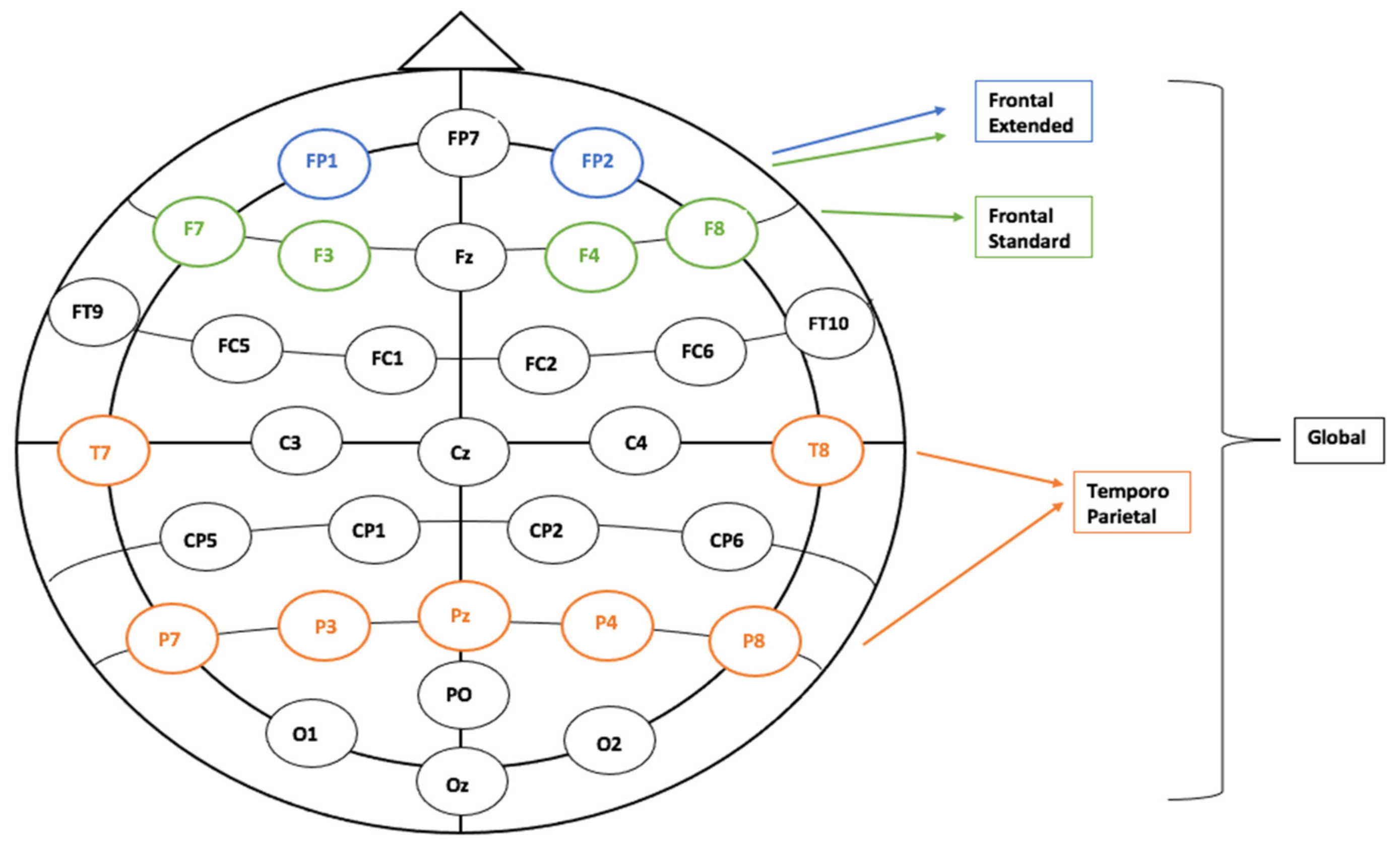
2.2. EEG Signal Acquisition, Preprocessing, and Analysis
3. Results
4. Discussion
5. Conclusions
Author Contributions
Funding
Institutional Review Board Statement
Informed Consent Statement
Data Availability Statement
Conflicts of Interest
References
- Johnson, W.; Onuma, O.; Owolabi, M.; Sachdev, S. Stroke: A Global Response Is Needed. Bull. World Health Organ. 2016, 94, 634–634A. [Google Scholar] [CrossRef] [PubMed]
- Stein, L.A.; Goldmann, E.; Zamzam, A.; Luciano, J.M.; Messé, S.R.; Cucchiara, B.L.; Kasner, S.E.; Mullen, M.T. Association Between Anxiety, Depression, and Post-Traumatic Stress Disorder and Outcomes After Ischemic Stroke. Front. Neurol. 2018, 9, 890. [Google Scholar] [CrossRef] [PubMed]
- Khedr, E.M.; Abdelrahman, A.A.; Desoky, T.; Zaki, A.F.; Gamea, A. Post-Stroke Depression: Frequency, Risk Factors, and Impact on Quality of Life among 103 Stroke Patients—Hospital-Based Study. Egypt. J. Neurol. Psychiatry Neurosurg. 2020, 56, 66. [Google Scholar] [CrossRef]
- Stern, A.F. The Hospital Anxiety and Depression Scale. Occup. Med. 2014, 64, 393–394. [Google Scholar] [CrossRef] [PubMed]
- Ayis, S.A.; Ayerbe, L.; Ashworth, M.; DA Wolfe, C. Evaluation of the Hospital Anxiety and Depression Scale (HADS) in Screening Stroke Patients for Symptoms: Item Response Theory (IRT) Analysis. J. Affect. Disord. 2018, 228, 33–40. [Google Scholar] [CrossRef]
- Towfighi, A.; Ovbiagele, B.; El Husseini, N.; Hackett, M.L.; Jorge, R.E.; Kissela, B.M.; Mitchell, P.H.; Skolarus, L.E.; Whooley, M.A.; Williams, L.S. Poststroke Depression: A Scientific Statement for Healthcare Professionals From the American Heart Association/American Stroke Association. Stroke 2017, 48, e30–e43. [Google Scholar] [CrossRef]
- Roth, M. The Natural History of Mental Disorder in Old Age. J. Ment. Sci. 1955, 101, 281–301. [Google Scholar] [CrossRef]
- Barkercollo, S. Depression and Anxiety 3 Months Post Stroke: Prevalence and Correlates. Arch. Clin. Neuropsychol. 2007, 22, 519–531. [Google Scholar] [CrossRef]
- Townend, B.S.; Whyte, S.; Desborough, T.; Crimmins, D.; Markus, R.; Levi, C.; Sturm, J.W. Longitudinal Prevalence and Determinants of Early Mood Disorder Post-Stroke. J. Clin. Neurosci. 2007, 14, 429–434. [Google Scholar] [CrossRef]
- Folstein, M.F.; Maiberger, R.; McHugh, P.R. Mood Disorder as a Specific Complication of Stroke. J. Neurol. Neurosurg. Psychiatry 1977, 40, 1018–1020. [Google Scholar] [CrossRef]
- Tang, W.K.; Wong, E.; Chiu, H.F.K.; Ungvari, G.S. Rasch Analysis of the Scoring Scheme of the HADS Depression Subscale in Chinese Stroke Patients. Psychiatry Res. 2007, 150, 97–103. [Google Scholar] [CrossRef]
- Wutzl, B.; Golaszewski, S.M.; Leibnitz, K.; Langthaler, P.B.; Kunz, A.B.; Leis, S.; Schwenker, K.; Thomschewski, A.; Bergmann, J.; Trinka, E. Narrative Review: Quantitative EEG in Disorders of Consciousness. Brain. Sci. 2021, 11, 697. [Google Scholar] [CrossRef]
- Wade, E.C.; Iosifescu, D.V. Using Electroencephalography for Treatment Guidance in Major Depressive Disorder. Biol. Psychiatry Cogn. Neurosci. Neuroimaging 2016, 1, 411–422. [Google Scholar] [CrossRef]
- Bares, M.; Brunovsky, M.; Novak, T.; Kopecek, M.; Stopkova, P.; Sos, P.; Krajca, V.; Höschl, C. The Change of Prefrontal QEEG Theta Cordance as a Predictor of Response to Bupropion Treatment in Patients Who Had Failed to Respond to Previous Antidepressant Treatments. Eur. Neuropsychopharmacol. 2010, 20, 459–466. [Google Scholar] [CrossRef]
- de la Salle, S.; Jaworska, N.; Blier, P.; Smith, D.; Knott, V. Using Prefrontal and Midline Right Frontal EEG-Derived Theta Cordance and Depressive Symptoms to Predict the Differential Response or Remission to Antidepressant Treatment in Major Depressive Disorder. Psychiatry Res. Neuroimaging 2020, 302, 111109. [Google Scholar] [CrossRef]
- Finnigan, S.P.; Walsh, M.; Rose, S.E.; Chalk, J.B. Quantitative EEG Indices of Sub-Acute Ischaemic Stroke Correlate with Clinical Outcomes. Clin. Neurophysiol. 2007, 118, 2525–2532. [Google Scholar] [CrossRef]
- Finnigan, S.P.; Rose, S.E.; Walsh, M.; Griffin, M.; Janke, A.L.; McMahon, K.L.; Gillies, R.; Strudwick, M.W.; Pettigrew, C.M.; Semple, J.; et al. Correlation of Quantitative EEG in Acute Ischemic Stroke With 30-Day NIHSS Score. Stroke 2004, 35, 899–903. [Google Scholar] [CrossRef]
- Chen, S.-T.; Ku, L.-C.; Chen, S.-J.; Shen, T.-W. The Changes of QEEG Approximate Entropy during Test of Variables of Attention as a Predictor of Major Depressive Disorder. Brain Sci. 2020, 10, 828. [Google Scholar] [CrossRef]
- Livint Popa, L.; Dragos, H.; Pantelemon, C.; Rosu, O.V.; Strilciuc, S. The Role of Quantitative EEG in the Diagnosis of Neuropsychiatric Disorders. J. Med. Life 2020, 13, 8–15. [Google Scholar] [CrossRef]
- Finnigan, S.; van Putten, M.J.A.M. EEG in Ischaemic Stroke: Quantitative EEG Can Uniquely Inform (Sub-)Acute Prognoses and Clinical Management. Clin. Neurophysiol. 2013, 124, 10–19. [Google Scholar] [CrossRef]
- Finnigan, S.; Wong, A.; Read, S. Defining Abnormal Slow EEG Activity in Acute Ischaemic Stroke: Delta/Alpha Ratio as an Optimal QEEG Index. Clin. Neurophysiol. 2016, 127, 1452–1459. [Google Scholar] [CrossRef] [PubMed]
- Zakaria, H.; Valentine, O.; Mayza, A. Analysis of Quantitative EEG (QEEG) Parameters on the Effect of Transcranial Direct Current Stimulation (TDCS) on Post-Stroke Patients; AIP Publishing LLC: Depok, Indonesia, 2021; p. 050001. [Google Scholar]
- Schleiger, E.; Sheikh, N.; Rowland, T.; Wong, A.; Read, S.; Finnigan, S. Frontal EEG Delta/Alpha Ratio and Screening for Post-Stroke Cognitive Deficits: The Power of Four Electrodes. Int. J. Psychophysiol. 2014, 94, 19–24. [Google Scholar] [CrossRef] [PubMed]
- Balea, M.; Birle, C.; Costin, C.; Marton, J.; Muresanu, I.A.; Jemna, N.; Popa, L.L.; Slavoaca, D.; Rosu, O.V.; Stan, A.; et al. Effects of N-Pep-12 Dietary Supplementation on Neurorecovery after Ischemic Stroke. Neurol. Sci. 2021, 42, 2031–2037. [Google Scholar] [CrossRef] [PubMed]
- Popa, L.L.; Iancu, M.; Livint, G.; Balea, M.; Dina, C.; Vacaras, V.; Vladescu, C.; Balanescu, L.; Buzoianu, A.D.; Strilciuc, S.; et al. N-Pep-12 Supplementation after Ischemic Stroke Positively Impacts Frequency Domain QEEG. Neurol. Sci. 2021, 43, 1115–1125. [Google Scholar] [CrossRef] [PubMed]
- Jordan, K.G. Emergency EEG and Continuous EEG Monitoring in Acute Ischemic Stroke. J. Clin. Neurophysiol. 2004, 21, 341–352. [Google Scholar]
- Tolonen, U.; Sulg, I.A. Comparison of Quantitative EEG Parameters from Four Different Analysis Techniques in Evaluation of Relationships between EEG and CBF in Brain Infarction. Electroencephalogr. Clin. Neurophysiol. 1981, 51, 177–185. [Google Scholar] [CrossRef]
- Schleiger, E.; Wong, A.; Read, S.; Rowland, T.; Finnigan, S. Poststroke QEEG Informs Early Prognostication of Cognitive Impairment. Psychophysiology 2017, 54, 301–309. [Google Scholar] [CrossRef]
- Burghaus, L.; Hilker, R.; Dohmen, C.; Bosche, B.; Winhuisen, L.; Galldiks, N.; Szelies, B.; Heiss, W.-D. Early Electroencephalography in Acute Ischemic Stroke: Prediction of a Malignant Course? Clin. Neurol. Neurosurg. 2007, 109, 45–49. [Google Scholar] [CrossRef]
- Begić, D.; Popović-Knapić, V.; Grubišin, J.; Kosanović-Rajačić, B.; Filipčić, I.; Telarović, I.; Jakovljević, M. Quantitative Electroencephalography in Schizophrenia and Depression. Psychiatr. Danub. 2011, 23, 355–362. [Google Scholar]
- Zheng, Y.-P.; Wang, F.-X.; Zhao, D.-Q.; Wang, Y.-Q.; Zhao, Z.-W.; Wang, Z.-W.; Liu, J.; Wang, J.; Luan, P. Predictive Power of Abnormal Electroencephalogram for Post-Cerebral Infarction Depression. Neural. Regen. Res. 2018, 13, 304–308. [Google Scholar] [CrossRef]
- Davidson, R. Anterior Cerebral Asymmetry and the Nature of Emotion. Brain Cogn. 1992, 20, 125–151. [Google Scholar] [CrossRef]
- Carter, A.R.; Shulman, G.L.; Corbetta, M. Why Use a Connectivity-Based Approach to Study Stroke and Recovery of Function? NeuroImage 2012, 62, 2271–2280. [Google Scholar] [CrossRef]
- Veer, I.M.; Beckmann, C.F.; van Tol, M.-J.; Ferrarini, L.; Milles, J.; Veltman, D.J.; Aleman, A.; van Buchem, M.A.; van der Wee, N.J.; Rombouts, S.A.R.B. Whole Brain Resting-State Analysis Reveals Decreased Functional Connectivity in Major Depression. Front. Syst. Neurosci. 2010, 4, 41. [Google Scholar] [CrossRef]
- Corbetta, M.; Shulman, G.L. Control of Goal-Directed and Stimulus-Driven Attention in the Brain. Nat. Rev. Neurosci. 2002, 3, 201–215. [Google Scholar] [CrossRef]


| Demographic Categories | Frequency | Valid Percentage |
|---|---|---|
| Gender | ||
| Female | 10 | 17.5% |
| Male | 47 | 82.5% |
| Age group | ||
| 37–60 | 24 | 42.1% |
| 61–79 | 33 | 57.9% |
| Type of stroke | ||
| Thrombotic | 46 | 80.7% |
| Lacunar | 2 | 3.5% |
| Cardioembolic | 9 | 15.8% |
| Stroke severity | ||
| Minor (NIHSS 1–4 points) | 22 | 38.6% |
| Moderate (NIHSS 5–12 points) | 31 | 54.4% |
| Mild to Severe (NIHSS 17–20 points) | 4 | 7.0% |
| Vascular Territory | ||
| Left Middle Cerebral Artery | 28 | 49.1% |
| Right Middle Cerebral Artery | 25 | 43.9% |
| Left Posterior Cerebral Artery | 3 | 5.3% |
| Right Posterior Cerebral Artery | 1 | 1.8% |
| VISIT 1 | VISIT 2 | |||
|---|---|---|---|---|
| Frequency | Percentage | Frequency | Percentage | |
| No depressive symptoms (<7) | 38 | 66.7% | 43 | 75.4% |
| Mild (8–10) | 13 | 22.8% | 11 | 19.3% |
| Moderate (11–14) | 5 | 8.8% | 3 | 5.3% |
| Severe (15–21) | 1 | 1.8% | 0 | 0.0% |
| Analysis | Time Point | Region | Sub-Type | Correlation Coefficient | Significance |
|---|---|---|---|---|---|
| DTABR COG HADS-D | V1 | Global | COG2 | 0.292 | 0.028 |
| COG3 | 0.202 | 0.027 |
| Analysis | Time Point | Region | Sub-Type | Correlation Coefficient | Significance |
|---|---|---|---|---|---|
| DTABR COG HADS-D | V2 | Frontal Extended | COG2 | −264 | 0.049 |
| COG3 | −392 | 0.003 |
| Analysis | Time Point | Region | Sub-Type | Correlation Coefficient | Significance |
|---|---|---|---|---|---|
| DTABR COG HADS-D | V1 | Global | COG1 | −002 | 0.988 |
| V2 | Frontal Extended | −119 | 0.383 |
| DTAB Ratio Scores | Visit 1 | Visit 2 | ||||
|---|---|---|---|---|---|---|
| COG1 | COG2 | COG3 | COG1 | COG2 | COG3 | |
| Global | 1.516 | 1.500 | 1.440 | 1.508 | 1.468 | 1.432 |
| Frontal Extended | 1.495 | 1.478 | 1.425 | 1.486 | 1.469 | 1.418 |
Disclaimer/Publisher’s Note: The statements, opinions and data contained in all publications are solely those of the individual author(s) and contributor(s) and not of MDPI and/or the editor(s). MDPI and/or the editor(s) disclaim responsibility for any injury to people or property resulting from any ideas, methods, instructions or products referred to in the content. |
© 2022 by the authors. Licensee MDPI, Basel, Switzerland. This article is an open access article distributed under the terms and conditions of the Creative Commons Attribution (CC BY) license (https://creativecommons.org/licenses/by/4.0/).
Share and Cite
Livinț Popa, L.; Chira, D.; Dăbală, V.; Hapca, E.; Popescu, B.O.; Dina, C.; Cherecheș, R.; Strilciuc, Ș.; Mureșanu, D.F. Quantitative EEG as a Biomarker in Evaluating Post-Stroke Depression. Diagnostics 2023, 13, 49. https://doi.org/10.3390/diagnostics13010049
Livinț Popa L, Chira D, Dăbală V, Hapca E, Popescu BO, Dina C, Cherecheș R, Strilciuc Ș, Mureșanu DF. Quantitative EEG as a Biomarker in Evaluating Post-Stroke Depression. Diagnostics. 2023; 13(1):49. https://doi.org/10.3390/diagnostics13010049
Chicago/Turabian StyleLivinț Popa, Livia, Diana Chira, Victor Dăbală, Elian Hapca, Bogdan Ovidiu Popescu, Constantin Dina, Răzvan Cherecheș, Ștefan Strilciuc, and Dafin F. Mureșanu. 2023. "Quantitative EEG as a Biomarker in Evaluating Post-Stroke Depression" Diagnostics 13, no. 1: 49. https://doi.org/10.3390/diagnostics13010049
APA StyleLivinț Popa, L., Chira, D., Dăbală, V., Hapca, E., Popescu, B. O., Dina, C., Cherecheș, R., Strilciuc, Ș., & Mureșanu, D. F. (2023). Quantitative EEG as a Biomarker in Evaluating Post-Stroke Depression. Diagnostics, 13(1), 49. https://doi.org/10.3390/diagnostics13010049