Article

Trends in Infectious Keratitis in Taiwan: An Update on Predisposing Factors, Microbiological and Antibiotic Susceptibility Patterns

1
Department of Ophthalmology, Chiayi Chang Gung Memorial Hospital, Chiayi 61363, Taiwan
2
Department of Nursing, Chang Gung University of Science and Technology, Chiayi 61363, Taiwan
3
School of Traditional Chinese Medicine, College of Medicine, Chang Gung University, Taoyuan 33302, Taiwan
4
Health Information and Epidemiology Laboratory, Chiayi Chang Gung Memorial Hospital, Chiayi 61363, Taiwan
5
Department of Traditional Chinese Medicine, Chang Gung Memorial Hospital, Chiayi 61363, Taiwan
*
Author to whom correspondence should be addressed.
Diagnostics 2022, 12(9), 2095; https://doi.org/10.3390/diagnostics12092095
Submission received: 1 July 2022 / Revised: 12 August 2022 / Accepted: 25 August 2022 / Published: 29 August 2022
(This article belongs to the Special Issue Diagnosis and Management of Eye Infections)

Abstract

:
Infectious keratitis (IK) represents a major cause of corneal blindness. This study aims to investigate the demographics, risk factors, microbiological characteristics and antibiotic susceptibility patterns of IK in Taiwan over the past 15 years. A retrospective population-based study was conducted using the Chang Gung Research Database. Patients with IK were identified by diagnostic codes for corneal ulcer from 2004 to 2019. Of 7807 included subjects, 45.2% of patients had positive corneal cultures. The proportion of contact lens-related IK declined, while that of IK related to systemic diseases grew. The percentage of isolated gram-positive bacteria surpassed that of gram-negative bacteria in the 15-year period. The prevalence of Pseudomonas aeruginosa showed a decreasing trend (p = 0.004), whereas coagulase-negative Staphylococcus (CNS) and Propionibacterium species were increasingly detected (p < 0.001). Overall, the trend of antibiotic susceptibility of both gram-positive and gram-negative bacteria did not change throughout the study period. The susceptibility to the test antibiotics maintained over 90% in gram-negative isolates over 15 years. Vancomycin preserved 100% susceptibility to all gram-positive isolates. Since most tested antibiotics exhibited stable susceptibility over decades, this study reinforced that fluoroquinolones and fortified vancomycin continue to be good empiric therapies for treating bacterial keratitis in Taiwan.

1. Introduction

Infectious keratitis (IK) is one of the leading causes of ocular morbidity and blindness worldwide [1,2]. Complications related to IK not only cause visual disability but also place a heavy socioeconomic burden on the affected individuals and national health care systems [3,4]. The estimated incidence of IK ranges from 2.5 to 799 cases per 100,000 population per year, varying from geographic locations and study designs [5,6]. In Taiwan, a 14-year population-based study reported an increase in the incidence of IK from 8.3 in 2000 to 20.2 per 100,000 person-years in 2013 [3].
IK can be caused by a wide array of organisms, including bacteria, fungi, protozoa and viruses [7,8]. Polymicrobial infection also accounts for 2–15% of all IK cases [9,10]. Common risk factors for IK encompass contact lens wear, ocular trauma, ocular surface diseases (OSD), preceding ocular surgeries and systemic diseases [11,12]. Corneal scraping culture and stain as the current gold standard for diagnosis and determination of causative pathogens in IK, are time-consuming and may yield no useful results [13]. Timely and appropriate antimicrobial therapy is crucial for the eradication of infection and visual recovery. Accordingly, clinicians usually initiate empiric broad-spectrum treatment, either with fluoroquinolone monotherapy or combining with several fortified antimicrobial regimen, on the basis of clinical findings and regional epidemiological data before the results of diagnostic corneal smears and cultures are available [14,15]. Furthermore, a number of reports have demonstrated the development of bacterial strains resistant to commonly used antimicrobial agents [16,17]. The issue of emerging antimicrobial resistance is rising due to widespread and inappropriate use of broad-spectrum antibiotics [18].
Many published studies have been dedicated to comprehensive global data analyses of microbial keratitis [13,19,20]. However, the microbial spectrum and resistance patterns change greatly over time and vary enormously from region to region [19]. The establishment of updated local data of IK is essential to guide clinical practice. Therefore, the purpose of the present study was to investigate the microbiological and epidemiologic characteristics of IK in the recent decade, and to detect the shifting trends in corneal isolates and their antibiotic susceptibility profiles over time in Taiwan using a population-based database.

2. Materials and Methods

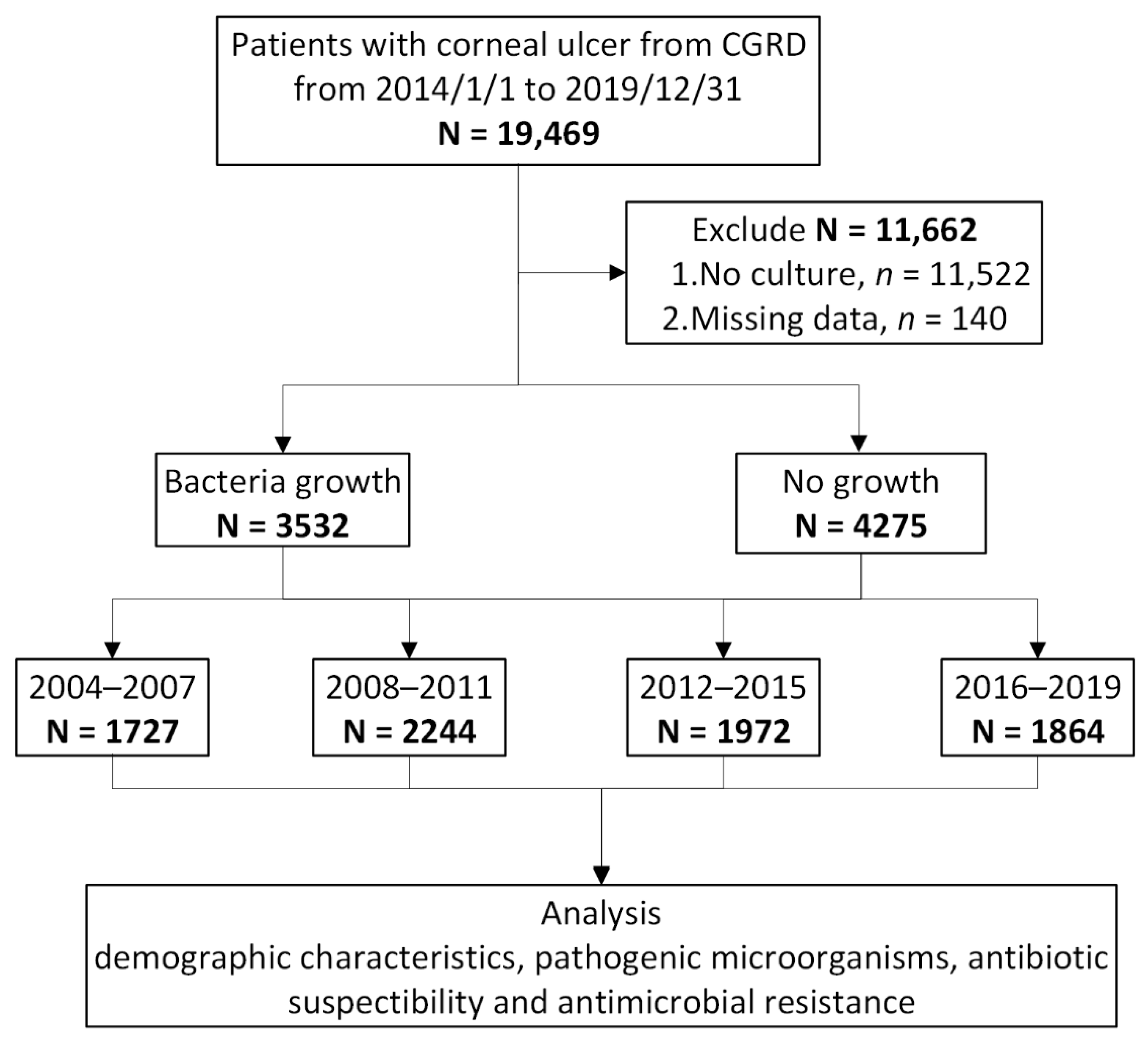
2.1. Study Design and Data Source

This was a hospital-based retrospective study utilizing the Chang Gung Research Database (CGRD), which is the largest multi-institutional de-identified electronic medical records (EMR) database in Taiwan. Overall, the CGRD includes 21.2% of outpatients and 12.4% inpatients in the Taiwanese population. Owing to high overall and disease-specific coverage, the CGRD provides good access for clinical and scientific studies. The patient-level demographics and data on the health conditions are coded according to the International Classification of Diseases, Ninth Revision, Clinical Modification (ICD-9-CM) codes before 2016 and ICD-10-CM codes after 2016.

2.2. Patient Identification

We enrolled patients with IK from 1 January 2004 to 31 December 2019, using ICD-9-CM and ICD-10 codes: 370.00, 370.01, 370.02, 370.03, 370.04, 370.05, 370.06, 370.55 and H16.00, H16.01, H16.02, H16.03, H16.04, H16.06, H16.07, H16.31. Patients with missing demographic information and without corneal culture data were excluded. The corneal scraping samples were obtained within one week of the index date. All cases were included only once; however, recurrent episodes were not involved in this study (Figure 1).

2.3. Determination of Risk Factors

Predisposing factors for IK were determined using the ICD-9-CM and ICD-10 diagnostic codes, procedure codes, and drug prescriptions recorded in the claims data before the diagnosis was recognized (Appendix A). The identifiable risk factors were categorized into five subgroups: contact lens (CL) wear, ocular trauma, recent ocular surgery, OSD and systemic disorders. Without a specific code for contact lens wear, we used diagnostic codes for corneal disorder due to contact lens and contact lens prescription records within 3 months before IK was identified. Ocular trauma was defined by documented presence with a history of traumatic ocular injury within 3 months prior to the diagnosis of IK. Recent ocular surgery performed within 3 months after the diagnosis of IK were considered surgical-related IK. Ocular surface diseases include dry eye, trichiasis, blepharitis, lagophthalmos and exposure keratopathy, neurotrophic keratopathy, corneal transplant status, and chronic topical antiglaucoma agent use for more than 3 months. Systemic disorders with ocular involvement include diabetes mellitus (DM), autoimmune diseases, chronic kidney disease (CKD), atopic dermatitis and human immunodeficiency virus (HIV) infection. These medical comorbidities if appearing at least once in the diagnoses of inpatients or at least three times in the diagnoses of outpatients were included.

2.4. Isolates and Antibiotic Susceptibility

Corneal scraping smears and cultures were routinely processed to identify the causative organisms in patients with IK. Noncorneal samples, such as conjunctival swabs and aqueous taps, were excluded in this study. A bacterium isolated from the same patient on more than one occasion was regarded as 1 isolate if having the same spectrum of antibiotic resistance.
According to the Clinical and Laboratory Standard Institute standards for testing antimicrobial susceptibility, in vitro susceptibility was interpreted based on serum standards due to lack of standardized values for topical antibiotics in ocular tissues. Isolates of intermediate susceptibility were categorized as susceptible organisms, since frequent instillation of fortified antibiotics may yield a higher antibiotic concentration in the corneal stroma than that in the serum after systemic administration.

2.5. Statistical Analysis

For the purpose of comparison and trend analysis, the study was arbitrarily divided into four time periods stratified according to diagnosis during the 15-year periods: 2004 to 2007, 2008 to 2011, 2012 to 2015, 2016 to 2019. Descriptive statistics such as count and percentage were presented for categorical variables. The Cochran–Armitage trend test and the Mann–Kendall trend test were used to detect the trends. All the analyses were performed using SAS software version 9.4 (SAS Institute, Cary, NC, USA), and the threshold for statistical significance was set at p < 0.05.

3. Results

3.1. Demographics of the Study Population

According to the ICD diagnostic codes, 19,469 patients with IK were identified from the CGRD from 1 January 2004 to 31 December 2019, but patients without complete demographic information or culture data were excluded. A total of 7807 patients, including 3809 men (48.8%) and 3998 women (51.2%), were included in this study. The demographic data are summarized in Table 1. The mean age was 46.9 ± 22.1 years. The patients older than 65 years accounted for 22.6%. From 2004 to 2019, no significant differences in sex and age were demonstrated from between-decade comparisons (p = 0.957, p = 0.275, respectively). A total of 7807 records of corneal scraping culture from 7807 IK patients with corneal ulcer were reviewed. Among the selected records, 3532 (45.2%) samples were culture positive. The positivity rate was rising from 40% to 52% over the 15-year period (p < 0.001).

3.2. Predisposing Factors for Infectious Keratitis

Among the patients with recognizable risk factors in the claims data (N = 3268, 41.9%), CL wear accounted for 10%; ocular trauma for 8.8%; recent ocular surgery for 3.2%; OSD for 17.9%; systemic disorders with ocular involvement for 14.2%. From between-decade comparison, an increasing trend in systemic disorder-related IK, particularly in patients with chronic kidney disease, and a decreasing trend in IK associated with prior ocular surgery (p < 0.001) were presented (Table 2). Similarly, a significant changing trend in CL-related IK was observed (p = 0.001) although the coding rate of “corneal disorder due to contact lens” was low. The rate of CL-related IK rose and reached a peak (12.1%) between 2012 and 2015, but subsequently fell to 9.7%. Moreover, IK associated with exposure keratopathy, lagophthalmos, prior corneal transplantation, topical steroid use, Sjögren syndrome and atopy showed a decline over the 15-year period.

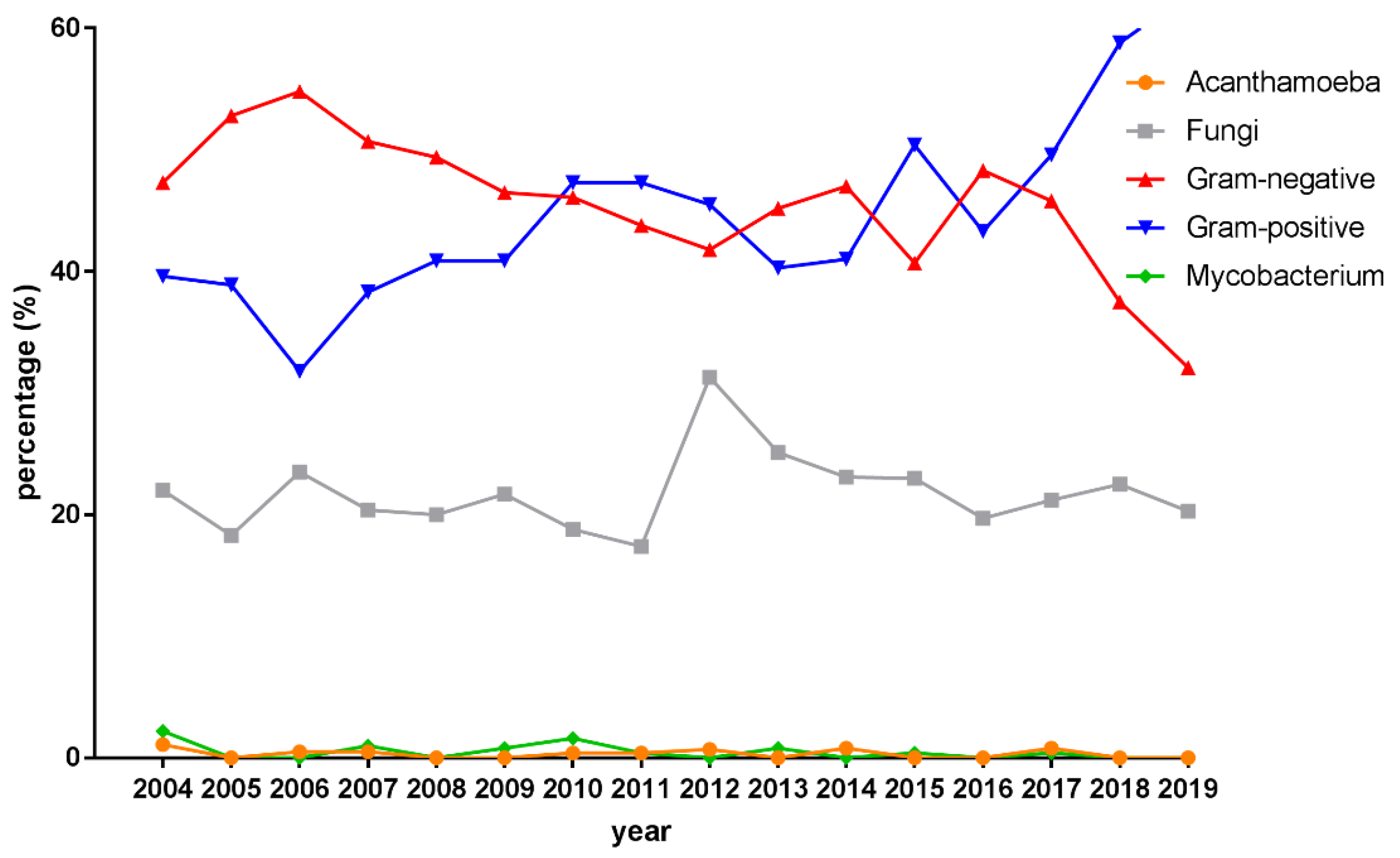
3.3. Microbiological Profiles

Data from 7807 corneal scraping cultures were available for study. A total of 3990 pathogens were recovered with 26.8% polymicrobial infection in the patients with IK. From 2004 to 2019, gram-positive bacteria accounted for 45.6% isolates, followed by gram-negative bacteria (45.1%), fungal isolates (21.5%), Mycobacteria (0.4%) and Acanthamoeba (0.3%). Figure 2 illustrates an increasing trend of gram-positive bacteria and a decreasing trend of gram-negative bacteria over the 15-year period. The upward trend of gram-positive bacteria and the downward trend of gram-negative bacteria were both significant (z = 14.77, p < 0.001; z = −13.13, p < 0.001). In 2012, the proportion of gram-positive bacteria exceeded that of gram-negative bacteria. The percentage of fungal isolates per year ranged from 17.4% to 31.3% for all positive cultures, showing stable over 15 years.
Of all bacterial growths, Pseudomonas aeruginosa was the most commonly identified bacterial isolate (N = 1002, 28.4%), followed by Staphylococcus species (N = 901, 25.5%), Propionibacterium species (N = 264, 7.5%), Streptococcus species (N = 192, 5.4%) and Serratia species (N = 150, 4.2%) (Table 3). Among Staphylococcus species, coagulase-negative Staphylococcus (CNS) (N = 631, 17.9% of total bacterial growths and 70.0% of Staphylococci isolates) ranked first, followed by Staphylococcus aureus (N = 257, 7.3% of total bacterial growths and 28.5% of Staphylococci isolates). A notable increase occurred in the percentage of Staphylococcus species and Propionibacterium species (p < 0.001) over the 15-year period, whereas a significant decrease appeared in the percentage of P. aeruginosa and Streptococcus species (p = 0.004, p = 0.033, respectively). Interestingly, in the subgroup analysis of Staphylococci group, S. epidermis was responsible for the upward trend in the overall proportion, whereas S. aureus remained stable over the 15-year period.

3.4. Antibiotic Susceptibility Patterns

3.4.1. Gram-Negative Isolates

During 2004 to 2019, most isolated gram-negative bacteria were susceptible to β-lactams and fluoroquinolone (piperacillin: 95.4%; ceftazidime: 96%; cefepime: 95.6%; imipenem: 95.7%; meropenem: 95.2%; ciprofloxacin: 95.4%; levofloxacin: 97.1%), followed by aminoglycoside (amikacin: 94.4%; gentamicin: 90.6%), as shown in Table 4. Isolated gram-negative bacteria also continuously exhibited a susceptibility over 90% in all recommended antibiotics in the 15-year period. P. aeruginosa, the most common isolate in this study, exhibited more than 95% in vitro susceptibility to all tested antibiotics. Nearly all P. aeruginosa isolates were susceptible to piperacillin-tazobactam, ceftazidime, cefepime and amikacin, while 96.1% and 97.8% of P. aeruginosa isolates were susceptible to gentamicin and levofloxacin, respectively (Table 5).

3.4.2. Gram-Positive Isolates

As for gram-positive bacteria, more than 95% of isolates were susceptible to glycopeptide, linezolid and fusidic acid (teicoplanin: 99.7%; vancomycin: 99.6%; linezolid: 100%; fusidic acid: 97.3%), while clindamycin covered 75% of the isolates in this study (Table 6). The resistances of oxacillin and erythromycin were observed (63.6%, 877 sensitive in 1379; 52.8%, 970 sensitive in 1837, respectively). Regarding the tested gram-positive isolates, the susceptibility of trimethoprim-sulfamethoxazole (TMP-SMX) increased from 82.8% to 90.2% in the period of 2004–2007 to 2016–2019 (p = 0.025). Fluoroquinolones were not on the recommended list of antibiotics for gram-positive bacteria in our microbiologic laboratory. Furthermore, from 2004 to 2019, all isolated Staphylococcus species showed 100% susceptibility to vancomycin, whereas the percentage of all Staphylococci isolates susceptible to oxacillin was 62.8% (Table 7). The susceptibility of the tested antibiotics against Staphylococcus species had no statistically significant change throughout the four study periods.

3.4.3. Multidrug-Resistant Isolates

During the 15-year period, multidrug-resistant (MDR) bacteria was isolated every year despite representing only a small percentage (N = 144, 4.1%), which was stable without significant change in trend (p = 0.05). Methicillin-resistant S. aureus (MRSA) accounted for 2.3% of all isolated organisms (5.1% of all Gram-positive organisms; 31.9% of S. aureus isolates), followed by vancomycin-resistant Enterococci (VRE) (1.4% of all isolates), Carbapenem-resistant P. aeruginosa (CRPA) (0.2% of all isolates), extended-spectrum β-lactamase-producing Escherichia coli (E. coli-ESBL) (0.1% of all isolates) and multidrug-resistant Acinetobacter baumannii (MDR-AB) (0.1% of all isolates). For the in vitro susceptibility test, vancomycin retained activity (100%) against MRSA (Table S1).

4. Discussion

The spectrum of pathogenic microorganisms and their antimicrobial susceptibility vary with different regions and change over time. Therefore, periodical renewal of the local epidemiology of IK for evidence-based guidance is vital in clinical practice. This study provides updated information about microbiological data and patient demographics of IK in Taiwan.
From our results, the average age (47 years) of the patients with IK was similar to that in previous literature reported worldwide (average age ranges from 42 to 56 years) [20]. Even though no gender predilection was shown, a slight female preponderance was presented through the 15-year period. This finding is consistent with prior domestic studies indicating that IK occurs more commonly in females in Taiwan [3,21]. The data of the Asia Cornea Society Infectious Keratitis Study also suggested that IK was associated with a female predominance in Taiwan, Japan and Singapore [8].
Regarding the risk factors for IK, OSD represented the relatively main identified predisposing factor in this study, which was different from other studies [3,8,10,11,20,22]. Ocular surface diseases have become a popular issue in recent decades. Dysregulation of the ocular surface may lead to ocular surface inflammation and damage of the corneal epithelium, consequently increasing the risk of IK [23]. A 5-year Australian study revealed that IK in patients with a history of ocular surface diseases were more likely to have longer recovery time and less favorable outcomes [10].
Moreover, IK associated with systemic disorders showed a significantly increasing trend, while a decreasing trend of IK presented in patients with preceding ocular surgery. In subgroup analysis, DM and CKD were shown as vital elements contributing to the upward trend. In accordance with our findings, prior studies have reported DM as a risk factor for microbial keratitis [3,24,25]. Existing literature explained that hyperglycemia facilitates microbial growth and inhibits host immune response to infection [26,27]. DM can alter corneal nerve plexuses and affect ocular surface homeostasis, thereby increasing the risk of IK [28,29]. In terms of CKD, a Taiwanese study demonstrated that end-stage renal disease (ESRD) increased 1.17 times of risk to develop corneal ulcer, particularly in patients with DM [30]. The author assumed that compromised ocular surface and quality of tear film may predispose patients to IK [30]. Weng et al. showed that ESRD increases the risk of band keratopathy, which disrupts the regularity of the ocular surface and then prompt colonization of organisms and tissue invasion [31]. Furthermore, several reports suggested that the activation of toll-like receptors expressed on the corneal epithelium may be involved in the pathogenesis of corneal infection by exacerbating various ocular surface inflammation [32].
CL wear had been shown to be the most common risk factor for microbial keratitis in the United State, Europe and Australia [33,34,35]. Previous domestic surveys also indicated that CL wear was the most common documented risk factor of IK with incidence around 31–44% [12,21,36]. Since our results were analyzed based on the ICD diagnostic coding at clinic, we could not accurately identify the exact number of “corneal disorders due to CL use” in the CGRD. In general, the history of CL use was routinely recorded as text rather than code in the medical charts. Intriguingly, in line with previous literature, the number of CL-related IK declined in the present study [21,37]. Liu et al., postulated that the reduction of CL-related IK reflected the rising popularity of daily-disposable lenses or routine application of topical fluoroquinolone eye drops as initial treatment for CL-related ocular disorders [21]. By contrast, in the 2019Think Tank, the American Academy of Optometry (AAO) stated that the rate of CL-related infections had not decreased over three decades despite technology innovation, but they acknowledged that extended wear, such as overnight orthokeratology lenses, increases the risk of IK, whereas daily disposable modalities may minimize the risk of severe corneal infections [38].
The average positive culture rate of the present study was comparable with that in other reports, ranging from 40–68%, and the positive rate increased through the 15-year period [16,19,21,36]. The relatively low rate with a slightly upward trend may be due to the increase of early referral before application of antibiotics [16,21].
Among the isolated microorganisms, the common bacterial isolates were Pseudomonas species, Staphylococcus species, Propionibacterium species, Streptococcus species and Serratia species, which was consistent with other published reports from Taiwan [21,39]. Interestingly, our findings revealed that the percentage of gram-positive bacteria significantly increased, surpassing that of gram-negative isolates for the first time by 2010. Although P. aeruginosa remained the most commonly isolated pathogen during the study period, the percentage declined significantly. The similar phenomenon of shifting trends of isolates was also presented in the studies from the UK and Iran, but not shown in the previous domestic reports [21,36,39,40,41]. Pseudomonas species are responsible for CL- and trauma-related IK in most settings [8]. The decline in the percentage of Pseudomonas species may be attributable to the widespread use of fluoroquinolones and advanced hygiene concepts in contact lens use [19]. The reduction in the rate of corneal scrapes in CL-related infections and early recovery of IK might be another reason for the decreasing percentage of Pseudomonas isolates [37]. On the other hand, coagulase-negative Staphylococcus (CNS), particularly S. epidermis, was the main culprit pathogen of staphylococcal keratitis, the rise of which is one of the leading causes of an increase in the percentage of gram-positive isolates. Likewise, previous studies of bacterial keratitis from India, the UK, New Zealand and Canada, have demonstrated that CNS was the most common causative organism in IK, ranging from 24.8% to 40.8% of the isolates [16,42,43,44]. In a ten-year analysis of microbial keratitis conducted in the UK, Ting et al. consistently observed an increasing trend in Gram-positive organisms, particularly CNS, and a decreasing trend in Gram-negative organisms, particularly Pseudomonas [40]. In contrast, Tam et al. found a decreasing trend in the number of isolates in gram-positive microorganisms over the past 16 years [45].
With respect to the virulence, CNS has always been considered a group of common ocular commensal that opportunistically causes endophthalmitis, keratitis, and blepharoconjunctivitis [46]. However, the identification of CNS as a pathogen varies among laboratories. The increasing trend in the percentage of CNS may reflect both the inherent nature of geographical prevalence in Taiwan and the likelihood of contamination of cornea [39]. We hypothesized that some systemic disorders, such as DM and CKD, may be associated with the increase of CNS isolates in Taiwan. These systemic diseases involving eyes often develop OSD, among which the OSD-related IK was caused by CNS and S. aureus [33,47]. Once normal flora of the ocular surface and a contaminant were considered during scraping, Propionibacterium species implicated in IK showing an increasing trend of isolation has been drawn attention in recent evidence [39,48].
Our study indicated that gram-negative bacteria preserve better susceptibility to the tested antibiotics compared with gram-positive bacteria, supporting evidence from previous reports [16,36,39,44,45,49]. The susceptibility of gram-negative bacteria to tested antibiotics seemed stable and maintained 90% over 15 years [7,16,19,39,45]. Most gram-negative isolates were susceptible to fluoroquinolone (96.3%) and cephalosporin (95.8%), followed by aminoglycoside (92.5%) in the present study. Interestingly, we found a significant increase of susceptibility to amikacin against gram-negative bacteria except P. aeruginosa. Nevertheless, topical fluoroquinolones have substantially replaced combined fortified aminoglycosides and cephalosporins as an empiric treatment for bacterial keratitis, regarding its low ocular toxicity and commercial availability [16,17]. In our institution, our empiric therapy for IK has been changed to levofloxacin in recent years.
Fluoroquinolones are currently widespread used as empiric therapy in bacterial keratitis due to the broad coverage of spectrum, low toxicity and good absorption to the ocular surface [14,15]. However, emerging resistance to the antibiotics has been increasingly reported worldwide over the last two decades [17,49,50]. Nevertheless, susceptibility to levofloxacin in gram-negative bacteria, especially P. aeruginosa, remained high and unchanged, ranging from 96% to 98%, during all study periods in Taiwan. This discrepancy could be attributed to variations between different geographical locations. However, the lack of data hinders further analyses regarding the susceptibility test against fluoroquinolones through the study period.
In gram-positive bacteria, most isolates were resistant to macrolide (52.8% sensitivity of all gram-positive isolates), similar to the global data, 57% [19]. Beyond our expectation, the susceptibility rate to TMP-SMX for gram-positive bacteria increased significantly from 82.1% to 90.2%. The average susceptibility to TMP-SMX was 85.8%, similar to that in reports from Toronto, Canada [45]. Meanwhile, a slight decline trend of antimicrobial resistance against oxacillin and clindamycin, regarded as antibiotic selective pressure, was observed, showing consistency with a previous domestic report [39].
The present study examined that nearly 40% of all Staphylococci were resistant to oxacillin with 31.9% being MRSA, whereas the study by Hsiao et al., showed 40% of CNS and S. aureus isolates were oxacillin-resistant in a 10-year single-center study [39]. Our results presented no significant change in trend regarding the susceptibility to oxacillin for S. aureus in Taiwan. In the Toronto study, Lichtinger et al. found that 29.1% of all gram-positive bacteria were methicillin-resistant isolates, while 43.1% of CNS and 1.3% of S. aureus isolates were oxacillin-resistant [42].
Multiple drug resistance has had a global impact on public health in the field of ophthalmology [51,52]. The development of antimicrobial resistance is multifactorial, including injudicious use of antimicrobial agents, genetic mutational resistance and horizontal gene transfer of microorganisms per se [53]. Several recent studies have demonstrated the emergence of antibiotic resistance in ocular infections [16,19,39,42,45,49,50]. A certain proportion of antimicrobial-resistant bacteria among the isolated strains appeared annually in our study. Reassuringly, the percentage of MDR bacteria, such as MRSA, did not increase over time. All gram-positive bacteria, including methicillin-resistant isolates, were susceptible to vancomycin (100% sensitivity), the last resort for MRSA. In another domestic study in Taiwan, fluoroquinolones were effective against S. aureus [39]. Therefore, we may consider fluoroquinolones as initial empiric treatment for IK and combination regimen with fortified vancomycin for the severe IK cases in Taiwan. Nevertheless, we should appropriately use these second-line antibiotics to prevent the emergence of antimicrobial resistance.
Our study presents several strengths and limitations. To our knowledge, this is the first study to investigate the demographic and microbiological characteristics of IK in Taiwan with the broadest range of study period. Furthermore, the database research contains a large number of cases from multiple hospitals, at least one third of medical coverage of Taiwan; therefore, the findings of this study could represent real-world evidence. However, the retrospective nature restricted detailed review of clinical information such as initial appearance, prior antibiotic use and contact lens wear, which are unavailable in the claims database. CL-related IK was difficult to directly identify in our database since the coding rate of “corneal disorder due to contact lens” was low. The similar problem happened to other predisposing factors, causing underestimate of the incidence. Regarding limitations on the microbiological study, in vitro antibiotic susceptibility interpreted based on serum standards could not provide accurate assessment of antibiotic resistance in ocular strains. To identify whether CNS and Propionibacterium isolates are either pathogens or commensal species from the ocular surface remains a challenge. Therefore, further studies are required to comprehensively clarify species correlations with ocular infections. As with other epidemiological studies, our findings should not be generalized to other geographic regions or populations.

5. Conclusions

The ratio of the isolated gram-positive bacteria and gram-negative bacteria presented a significant cross. Simultaneously, we found an increasing trend in the percentage of IK associated with systemic diseases, particularly CKD and DM, whereas the proportion of CL-related IK declines. As positive culture rate rises, CNS keratitis and Propionibacterium keratitis are regarded as potential ocular surface infections that warrant more attention. However, P. aeruginosa remains the most frequently isolated bacteria responsible for IK in Taiwan. Since no significant change in antibiotic susceptibility and the percentage of MDR strains were noticed, this study highlights that fluoroquinolones and fortified vancomycin continue to be good empiric therapies for treating bacterial keratitis in Taiwan.

Supplementary Materials

The following supporting information can be downloaded at: https://www.mdpi.com/article/10.3390/diagnostics12092095/s1, Table S1: Susceptibility of methicillin-resistant Staphylococcus aureus (MRSA) isolates to the tested antibiotics.

Author Contributions

Conceptualization, J.-J.W. and P.-L.W.; methodology, J.-J.W., M.-H.L. and P.-L.W.; software, C.-Y.L.; validation, M.-H.L. and Y.-H.Y.; formal analysis, C.-Y.L.; resources, J.-J.W. and C.-H.L.; writing—original draft preparation, J.-J.W.; writing—review and editing, M.-H.L., C.-Y.C., C.-H.L. and P.-L.W.; visualization, all authors.; supervision, C.-H.L. and P.-L.W.; project administration, J.-J.W. and P.-L.W.; funding acquisition, J.-J.W. All authors have read and agreed to the published version of the manuscript.

Funding

This research received no external funding.

Institutional Review Board Statement

The study was conducted in accordance with the Declaration of Helsinki, and approved by the Institutional Review Board of Chang Gung Memorial Hospital (IRB number: 202200153B0) on 9 February 2022.

Informed Consent Statement

Patient consent was waived by the Institutional Review Board of Chang Gung Memorial Hospital because personal medication information was encrypted.

Data Availability Statement

The data presented in this study are available upon request from the corresponding author. The data are not publicly available due to ethical restrictions.

Acknowledgments

This study is based in part on data from the Chang Gung Research Database (CGRD) provided by the Chang Gung Memorial Hospital. The authors thank the Health Information and Epidemiology Laboratory, Chiayi Chang Gung Memorial Hospital, for the comments and assistance in data analysis.

Conflicts of Interest

The authors declare no conflict of interest.

Appendix A

Table A1. International Classification of Diseases, 9th Revision, Clinical Modification codes (ICD-9-CM code) and ICD-10 for various conditions.
ConditionsICD-9 CodesICD-10 Codes
Contact lens use (= Corneal disorder due to contact lens)71.82, 367.0, 367.1, 367.2, 367.9H18.82, H52.0X, H52.1, H52.2, H52.7
Ocular trauma18, 918.0, 918.1, 918.2, 918.9S00.2, S00.01, S00.02, S05.00, S05.01, S05.9
Dry eye375.15, 370.33, 710.2H04.12, H16.22, M35.00, M35.01, M35.09
Trichiasis374.05H02.05
Blepharitis372.20, 372.21, 372.22, 373.00, 373.01, 373.02, 373.31, 373.32, 373.33, 373.34H10.50, H10.52, H10.53, H01
Exposure keratopathy, or lagophthalmos370.34, 374.2H16.21, H02.2
Neurotrophic keratopathy 370.35H16.23
Corneal transplant status V42.5Z94.7
Diabetes mellitus 250E11, E10, E09, E13, E08
Sjögren syndrome710.2M35.0
Autoimmune diseases
(Non-Sjörgen)
701.0, 710.1, 710.3, 710.4, 710.5, 710.8, 710.9, 714.0, 714.1, 714.2, 714.30, 714.31, 714.32, 714.33, 720, 725L90.0, L94.0, L94.1, L94.3, M32, M34, M35.1-9, M33, M05, M06, M08, M45, M46, M48, M49
Atopic dermatitis 691.8, 372.05L20.8, L20.9, H10.1
Chronic kidney disease 585N18.4-6, N18.9
Human immunodeficiency virus infection V08, 795.71, 042Z21, Z22.6, R75, B20, B21, B22, B23, B24

References

  1. Whitcher, J.P.; Srinivasan, M.; Upadhyay, M.P. Corneal blindness: A global perspective. Bull. World Health Organ. 2001, 79, 214–221. [Google Scholar] [PubMed]
  2. Flaxman, S.R.; Bourne, R.R.A.; Resnikoff, S.; Ackland, P.; Braithwaite, T.; Cicinelli, M.V.; Das, A.; Jonas, J.B.; Keeffe, J.; Kempen, J.H.; et al. Global causes of blindness and distance vision impairment 1990-2020: A systematic review and meta-analysis. Lancet Glob. Health 2017, 5, e1221–e1234. [Google Scholar] [CrossRef]
  3. Koh, Y.Y.; Sun, C.C.; Hsiao, C.H. Epidemiology and the Estimated Burden of Microbial Keratitis on the Health Care System in Taiwan: A 14-Year Population-Based Study. Am. J. Ophthalmol. 2020, 220, 152–159. [Google Scholar] [CrossRef] [PubMed]
  4. Collier, S.A.; Gronostaj, M.P.; MacGurn, A.K.; Cope, J.R.; Awsumb, K.L.; Yoder, J.S.; Beach, M.J. Estimated burden of keratitis—United States, 2010. MMWR Morb. Mortal Wkly. Rep. 2014, 63, 1027–1030. [Google Scholar]
  5. Upadhyay, M.P.; Karmacharya, P.C.; Koirala, S.; Shah, D.N.; Shakya, S.; Shrestha, J.K.; Bajracharya, H.; Gurung, C.K.; Whitcher, J.P. The Bhaktapur eye study: Ocular trauma and antibiotic prophylaxis for the prevention of corneal ulceration in Nepal. Br. J. Ophthalmol. 2001, 85, 388–392. [Google Scholar] [CrossRef]
  6. Erie, J.C.; Nevitt, M.P.; Hodge, D.O.; Ballard, D.J. Incidence of ulcerative keratitis in a defined population from 1950 through 1988. Arch. Ophthalmol. 1993, 111, 1665–1671. [Google Scholar] [CrossRef] [PubMed]
  7. Tan, S.Z.; Walkden, A.; Au, L.; Fullwood, C.; Hamilton, A.; Qamruddin, A.; Armstrong, M.; Brahma, A.K.; Carley, F. Twelve-year analysis of microbial keratitis trends at a UK tertiary hospital. Eye 2017, 31, 1229–1236. [Google Scholar] [CrossRef] [PubMed]
  8. Khor, W.B.; Prajna, V.N.; Garg, P.; Mehta, J.S.; Xie, L.; Liu, Z.; Padilla, M.D.B.; Joo, C.K.; Inoue, Y.; Goseyarakwong, P.; et al. The Asia Cornea Society Infectious Keratitis Study: A Prospective Multicenter Study of Infectious Keratitis in Asia. Am. J. Ophthalmol. 2018, 195, 161–170. [Google Scholar] [CrossRef]
  9. Ting, D.S.J.; Ho, C.S.; Cairns, J.; Elsahn, A.; Al-Aqaba, M.; Boswell, T.; Said, D.G.; Dua, H.S. 12-year analysis of incidence, microbiological profiles and in vitro antimicrobial susceptibility of infectious keratitis: The Nottingham Infectious Keratitis Study. Br. J. Ophthalmol. 2021, 105, 328–333. [Google Scholar] [CrossRef]
  10. Khoo, P.; Cabrera-Aguas, M.P.; Nguyen, V.; Lahra, M.M.; Watson, S.L. Microbial keratitis in Sydney, Australia: Risk factors, patient outcomes, and seasonal variation. Graefes Arch. Clin. Exp. Ophthalmol. 2020, 258, 1745–1755. [Google Scholar] [CrossRef]
  11. Green, M.; Apel, A.; Stapleton, F. Risk factors and causative organisms in microbial keratitis. Cornea 2008, 27, 22–27. [Google Scholar] [CrossRef] [PubMed]
  12. Lin, T.Y.; Yeh, L.K.; Ma, D.H.; Chen, P.Y.; Lin, H.C.; Sun, C.C.; Tan, H.Y.; Chen, H.C.; Chen, S.Y.; Hsiao, C.H. Risk Factors and Microbiological Features of Patients Hospitalized for Microbial Keratitis: A 10-Year Study in a Referral Center in Taiwan. Medicine 2015, 94, e1905. [Google Scholar] [CrossRef] [PubMed]
  13. Ung, L.; Bispo, P.J.M.; Shanbhag, S.S.; Gilmore, M.S.; Chodosh, J. The persistent dilemma of microbial keratitis: Global burden, diagnosis, and antimicrobial resistance. Surv. Ophthalmol. 2019, 64, 255–271. [Google Scholar] [CrossRef] [PubMed]
  14. Hanet, M.S.; Jamart, J.; Chaves, A.P. Fluoroquinolones or fortified antibiotics for treating bacterial keratitis: Systematic review and meta-analysis of comparative studies. Can. J. Ophthalmol. 2012, 47, 493–499. [Google Scholar] [CrossRef]
  15. Sharma, N.; Goel, M.; Bansal, S.; Agarwal, P.; Titiyal, J.S.; Upadhyaya, A.D.; Vajpayee, R.B. Evaluation of moxifloxacin 0.5% in treatment of nonperforated bacterial corneal ulcers: A randomized controlled trial. Ophthalmology 2013, 120, 1173–1178. [Google Scholar] [CrossRef] [PubMed]
  16. Orlans, H.O.; Hornby, S.J.; Bowler, I.C. In vitro antibiotic susceptibility patterns of bacterial keratitis isolates in Oxford, UK: A 10-year review. Eye 2011, 25, 489–493. [Google Scholar] [CrossRef]
  17. Alexandrakis, G.; Alfonso, E.C.; Miller, D. Shifting trends in bacterial keratitis in south Florida and emerging resistance to fluoroquinolones. Ophthalmology 2000, 107, 1497–1502. [Google Scholar] [CrossRef]
  18. Brown, L. Resistance to ocular antibiotics: An overview. Clin. Exp. Optom. 2007, 90, 258–262. [Google Scholar] [CrossRef]
  19. Zhang, Z.; Cao, K.; Liu, J.; Wei, Z.; Xu, X.; Liang, Q. Pathogens and Antibiotic Susceptibilities of Global Bacterial Keratitis: A Meta-Analysis. Antibiotics 2022, 11, 238. [Google Scholar] [CrossRef]
  20. Ting, D.S.J.; Ho, C.S.; Deshmukh, R.; Said, D.G.; Dua, H.S. Infectious keratitis: An update on epidemiology, causative microorganisms, risk factors, and antimicrobial resistance. Eye 2021, 35, 1084–1101. [Google Scholar] [CrossRef]
  21. Liu, H.Y.; Chu, H.S.; Wang, I.J.; Chen, W.L.; Hu, F.R. Microbial Keratitis in Taiwan: A 20-Year Update. Am. J. Ophthalmol. 2019, 205, 74–81. [Google Scholar] [CrossRef] [PubMed]
  22. Ibrahim, Y.W.; Boase, D.L.; Cree, I.A. Epidemiological characteristics, predisposing factors and microbiological profiles of infectious corneal ulcers: The Portsmouth corneal ulcer study. Br. J. Ophthalmol. 2009, 93, 1319–1324. [Google Scholar] [CrossRef] [PubMed]
  23. Bron, A.J.; de Paiva, C.S.; Chauhan, S.K.; Bonini, S.; Gabison, E.E.; Jain, S.; Knop, E.; Markoulli, M.; Ogawa, Y.; Perez, V.; et al. TFOS DEWS II pathophysiology report. Ocul. Surf. 2017, 15, 438–510. [Google Scholar] [CrossRef] [PubMed]
  24. Dan, J.; Zhou, Q.; Zhai, H.; Cheng, J.; Wan, L.; Ge, C.; Xie, L. Clinical analysis of fungal keratitis in patients with and without diabetes. PLoS ONE 2018, 13, e0196741. [Google Scholar] [CrossRef] [PubMed]
  25. Wang, B.; Yang, S.; Zhai, H.L.; Zhang, Y.Y.; Cui, C.X.; Wang, J.Y.; Xie, L.X. A comparative study of risk factors for corneal infection in diabetic and non-diabetic patients. Int. J. Ophthalmol. 2018, 11, 43–47. [Google Scholar] [CrossRef]
  26. Li, S.; Yi, G.; Peng, H.; Li, Z.; Chen, S.; Zhong, H.; Chen, Y.; Wang, Z.; Deng, Q.; Fu, M. How Ocular Surface Microbiota Debuts in Type 2 Diabetes Mellitus. Front. Cell Infect. Microbiol. 2019, 9, 202. [Google Scholar] [CrossRef]
  27. Hine, J.L.; de Lusignan, S.; Burleigh, D.; Pathirannehelage, S.; McGovern, A.; Gatenby, P.; Jones, S.; Jiang, D.; Williams, J.; Elliot, A.J.; et al. Association between glycaemic control and common infections in people with Type 2 diabetes: A cohort study. Diabet. Med. 2017, 34, 551–557. [Google Scholar] [CrossRef]
  28. Zhu, L.; Titone, R.; Robertson, D.M. The impact of hyperglycemia on the corneal epithelium: Molecular mechanisms and insight. Ocul. Surf. 2019, 17, 644–654. [Google Scholar] [CrossRef]
  29. Szalai, E.; Deak, E.; Modis, L., Jr.; Nemeth, G.; Berta, A.; Nagy, A.; Felszeghy, E.; Kaposzta, R.; Malik, R.A.; Csutak, A. Early Corneal Cellular and Nerve Fiber Pathology in Young Patients With Type 1 Diabetes Mellitus Identified Using Corneal Confocal Microscopy. Invest. Ophthalmol. Vis. Sci. 2016, 57, 853–858. [Google Scholar] [CrossRef]
  30. Jan, R.L.; Tai, M.C.; Weng, S.F.; Chang, C.; Wang, J.J.; Chang, Y.S. Risk of corneal ulcer in patients with end-stage renal disease: A retrospective large-scale cohort study. Br. J. Ophthalmol. 2018, 102, 868–872. [Google Scholar] [CrossRef]
  31. Weng, S.F.; Jan, R.L.; Chang, C.; Wang, J.J.; Su, S.B.; Huang, C.C.; Tseng, S.H.; Chang, Y.S. Risk of Band Keratopathy in Patients with End-Stage Renal Disease. Sci. Rep. 2016, 6, 28675. [Google Scholar] [CrossRef]
  32. Redfern, R.L.; McDermott, A.M. Toll-like receptors in ocular surface disease. Exp. Eye Res. 2010, 90, 679–687. [Google Scholar] [CrossRef] [PubMed]
  33. Sagerfors, S.; Ejdervik-Lindblad, B.; Soderquist, B. Infectious keratitis: Isolated microbes and their antibiotic susceptibility pattern during 2004-2014 in Region Orebro County, Sweden. Acta Ophthalmol. 2020, 98, 255–260. [Google Scholar] [CrossRef] [PubMed]
  34. Ferreira, C.S.; Figueira, L.; Moreira-Goncalves, N.; Moreira, R.; Torrao, L.; Falcao-Reis, F. Clinical and Microbiological Profile of Bacterial Microbial Keratitis in a Portuguese Tertiary Referral Center-Where Are We in 2015? Eye Contact Lens 2018, 44, 15–20. [Google Scholar] [CrossRef] [PubMed]
  35. Truong, D.T.; Bui, M.T.; Memon, P.; Cavanagh, H.D. Microbial Keratitis at an Urban Public Hospital: A 10-Year Update. J. Clin. Exp. Ophthalmol. 2015, 6, 498. [Google Scholar] [CrossRef]
  36. Fong, C.F.; Tseng, C.H.; Hu, F.R.; Wang, I.J.; Chen, W.L.; Hou, Y.C. Clinical characteristics of microbial keratitis in a university hospital in Taiwan. Am. J. Ophthalmol. 2004, 137, 329–336. [Google Scholar] [CrossRef]
  37. Green, M.; Sara, S.; Hughes, I.; Apel, A.; Stapleton, F. Trends in contact lens microbial keratitis 1999 to 2015: A retrospective clinical review. Clin. Exp. Ophthalmol. 2019, 47, 726–732. [Google Scholar] [CrossRef]
  38. Szczotka-Flynn, L.B.; Shovlin, J.P.; Schnider, C.M.; Caffery, B.E.; Alfonso, E.C.; Carnt, N.A.; Chalmers, R.L.; Collier, S.; Jacobs, D.S.; Joslin, C.E.; et al. American Academy of Optometry Microbial Keratitis Think Tank. Optom Vis. Sci. 2021, 98, 182–198. [Google Scholar] [CrossRef]
  39. Hsiao, C.H.; Sun, C.C.; Yeh, L.K.; Ma, D.H.; Chen, P.Y.; Lin, H.C.; Tan, H.Y.; Chen, H.C.; Chen, S.Y.; Huang, Y.C. Shifting Trends in Bacterial Keratitis in Taiwan: A 10-Year Review in a Tertiary-Care Hospital. Cornea 2016, 35, 313–317. [Google Scholar] [CrossRef]
  40. Ting, D.S.J.; Settle, C.; Morgan, S.J.; Baylis, O.; Ghosh, S. A 10-year analysis of microbiological profiles of microbial keratitis: The North East England Study. Eye 2018, 32, 1416–1417. [Google Scholar] [CrossRef]
  41. Al-Dhaheri, H.S.; Al-Tamimi, M.D.; Khandekar, R.B.; Khan, M.; Stone, D.U. Ocular Pathogens and Antibiotic Sensitivity in Bacterial Keratitis Isolates at King Khaled Eye Specialist Hospital, 2011 to 2014. Cornea 2016, 35, 789–794. [Google Scholar] [CrossRef] [PubMed]
  42. Lichtinger, A.; Yeung, S.N.; Kim, P.; Amiran, M.D.; Iovieno, A.; Elbaz, U.; Ku, J.Y.; Wolff, R.; Rootman, D.S.; Slomovic, A.R. Shifting trends in bacterial keratitis in Toronto: An 11-year review. Ophthalmology 2012, 119, 1785–1790. [Google Scholar] [CrossRef] [PubMed]
  43. Green, M.; Carnt, N.; Apel, A.; Stapleton, F. Queensland Microbial Keratitis Database: 2005–2015. Br. J. Ophthalmol. 2019, 103, 1481–1486. [Google Scholar] [CrossRef]
  44. Pandita, A.; Murphy, C. Microbial keratitis in Waikato, New Zealand. Clin. Exp. Ophthalmol. 2011, 39, 393–397. [Google Scholar] [CrossRef] [PubMed]
  45. Tam, A.L.C.; Cote, E.; Saldanha, M.; Lichtinger, A.; Slomovic, A.R. Bacterial Keratitis in Toronto: A 16-Year Review of the Microorganisms Isolated and the Resistance Patterns Observed. Cornea 2017, 36, 1528–1534. [Google Scholar] [CrossRef]
  46. Romanowski, J.E.; Nayyar, S.V.; Romanowski, E.G.; Jhanji, V.; Shanks, R.M.Q.; Kowalski, R.P. Speciation and Antibiotic Susceptibilities of Coagulase Negative Staphylococci Isolated from Ocular Infections. Antibiotics 2021, 10, 721. [Google Scholar] [CrossRef]
  47. Khoo, P.; Cabrera-Aguas, M.; Robaei, D.; Lahra, M.M.; Watson, S. Microbial Keratitis and Ocular Surface Disease: A 5-Year Study of the Microbiology, Risk Factors and Clinical Outcomes in Sydney, Australia. Curr. Eye Res. 2019, 44, 1195–1202. [Google Scholar] [CrossRef]
  48. Ovodenko, B.; Seedor, J.A.; Ritterband, D.C.; Shah, M.; Yang, R.; Koplin, R.S. The prevalence and pathogenicity of Propionibacterium acnes keratitis. Cornea 2009, 28, 36–39. [Google Scholar] [CrossRef]
  49. Shalchi, Z.; Gurbaxani, A.; Baker, M.; Nash, J. Antibiotic resistance in microbial keratitis: Ten-year experience of corneal scrapes in the United Kingdom. Ophthalmology 2011, 118, 2161–2165. [Google Scholar] [CrossRef]
  50. Soleimani, M.; Tabatabaei, S.A.; Masoumi, A.; Mirshahi, R.; Ghahvechian, H.; Tayebi, F.; Momenaei, B.; Mahdizad, Z.; Mohammadi, S.S. Infectious keratitis: Trends in microbiological and antibiotic sensitivity patterns. Eye 2021, 35, 3110–3115. [Google Scholar] [CrossRef]
  51. Asbell, P.A.; Colby, K.A.; Deng, S.; McDonnell, P.; Meisler, D.M.; Raizman, M.B.; Sheppard, J.D., Jr.; Sahm, D.F. Ocular TRUST: Nationwide antimicrobial susceptibility patterns in ocular isolates. Am. J. Ophthalmol. 2008, 145, 951–958. [Google Scholar] [CrossRef] [PubMed]
  52. Haas, W.; Pillar, C.M.; Torres, M.; Morris, T.W.; Sahm, D.F. Monitoring antibiotic resistance in ocular microorganisms: Results from the Antibiotic Resistance Monitoring in Ocular micRorganisms (ARMOR) 2009 surveillance study. Am. J. Ophthalmol. 2011, 152, 567–574.e3. [Google Scholar] [CrossRef] [PubMed]
  53. Laxminarayan, R.; Duse, A.; Wattal, C.; Zaidi, A.K.; Wertheim, H.F.; Sumpradit, N.; Vlieghe, E.; Hara, G.L.; Gould, I.M.; Goossens, H.; et al. Antibiotic resistance-the need for global solutions. Lancet Infect. Dis. 2013, 13, 1057–1098. [Google Scholar] [CrossRef] [Green Version]
Figure 1. Flowchart of study population selection. CGRD, Chang Gung Research Database.
Diagnostics 12 02095 g001
Figure 2. Percentage of microbial isolates from infectious keratitis between 2014 and 2019.
Diagnostics 12 02095 g002
Table 1. Demographic characteristics of patients with infectious keratitis between 2004 to 2019.
2004–2007 N = 17272008–2011 N = 22442012–2015 N = 19722016–2019 N = 1864
n (%)n (%)n (%)n (%)p-Value *
Sex 0.957
    Male810 (46.9%)1146 (51.1%)955 (48.4%)898 (48.2%)
    Female917 (53.1%)1098 (48.9%)1017 (51.6%)966 (51.8%)
Age (years) 0.275
    ≤651300 (75.3%)1698 (75.7%)1487 (75.4%)1375 (73.8%)
    >65427 (24.7%)546 (24.3%)485 (24.6%)489 (26.2%)
Mean ± SD45.5 ± 22.146.0 ± 22.147.5 ± 21.748.5 ± 22.5
Culture rate <0.001
    Number of bacterial growth689 (40%)992 (44%)874 (44%)977 (52%)
    No growth1038 (60%)1252 (56%)1098 (56%)887 (48%)
* Cochran-Armitage trend test.
Table 2. Distribution of identifiable predisposing factors for infectious keratitis.
2004–2007 N = 17272008–2011 N = 22442012–2015 N = 19722016–2019 N = 1864
Variablesn (%)n (%)n (%)n (%)p-Value *
Contact lens wear105 (6.1%)254 (11.3%)238 (12.1%)181 (9.7%)0.001
Ocular trauma138 (8.0%)235 (10.5%)174 (8.8%)141 (7.6%)0.236
Recent ocular surgery82 (4.8%)83 (3.7%)42 (2.1%)43 (2.3%)<0.001
Ocular surface disease312 (18.1%)418 (18.6%)358 (18.2%)313 (16.8%)0.264
    Dry eye104 (6.0%)176 (7.8%)155 (7.9%)147 (7.9%)0.052
    Trichiasis26 (1.5%)37 (1.7%)30 (1.5%)16 (0.9%)0.083
    Blepharitis112 (6.5%)176 (7.8%)139 (7.1%)108 (5.8%)0.226
    Exposure keratopathy, or lagophthalmos21 (1.2%)29 (1.3%)24 (1.2%)7 (0.4%)0.012
    Neurotrophic keratopathy3 (0.2%)7 (0.3%)10 (0.5%)6 (0.3%)0.295
    Corneal transplantation status93 (5.4%)72 (3.2%)58 (2.9%)52 (2.8%)<0.001
Topical antiglaucoma agents60 (3.5%)57 (2.5%)57 (2.9%)65 (3.5%)0.744
Topical steroid46 (2.7%)47 (2.1%)26 (1.3%)31 (1.7%)0.009
    Systemic disorder216 (12.5%)301 (13.4%)292 (14.8%)297 (15.9%)0.001
    Diabetes mellitus152 (8.8%)226 (10.1%)213 (10.8%)197 (10.6%)0.061
    Non-Sjörgen autoimmune dz (RA, SLE, AS, other CTDs…)51 (3.0%)63 (2.8%)47 (2.4%)70 (3.8%)0.255
    Sjögren syndrome44 (2.6%)40 (1.8%)25 (1.3%)27 (1.5%)0.006
    Atopy10 (0.6%)12 (0.5%)16 (0.8%)22 (1.2%)0.020
    Chronic kidney disease27 (1.6%)53 (2.4%)54 (2.7%)67 (3.6%)<0.001
    HIV infection1 (0.1%)3 (0.1%)7 (0.4%)4 (0.2%)0.128
RA, rheumatoid arthritis; SLE, systemic lupus erythematosus; AS, ankylosing spondylitis; CTD, connective tissue disease; HIV, human immunodeficiency virus. * Cochran-Armitage trend test.
Table 3. Most common bacterial isolates from corneal scrapes between 2004–2007, 2008–2011, 2012–2015 and 2016–2019.
2004–2007
N = 689
2008–2011
N = 992
2012–2015
N = 874
2016–2019
N = 977
Total
N = 3532
Bacterian (%)n (%)n (%)n (%)n (%)p-Value *
Staphylococcus spp.140 (20.3)248 (25)193 (22.1)320 (32.8)901 (25.5)<0.001
    S. aureus53 (7.7)68 (6.9)55 (6.3)81 (8.3)257 (7.3)0.619
    S. epidermis26 (3.8)50 (5.0)67 (7.7)169 (17.3)312 (8.8)<0.001
    Other CNS63 (9.1)138 (13.9)67 (7.7)51 (5.2)319 (9)<0.001
Streptococcus spp.55 (8)49 (4.9)36 (4.1)52 (5.3)192 (5.4)0.033
    S. pneumoniae34 (4.9)27 (2.7)13 (1.5)16 (1.6)90 (2.5)<0.001
Propionebacterium spp.14 (2.0)90 (9.1)71 (8.1)89 (9.1)264 (7.5)<0.001
Pseudomonas spp.229 (33.2)283 (28.5)280 (32)254 (26)1046 (29.6)0.012
    P. aureuginosa225 (32.7)277 (27.9)252 (28.8)248 (25.4)1002 (28.4)0.004
Serratia spp.43 (6.2)49 (4.9)29 (3.3)29 (3)150 (4.3)<0.001
CNS, coagulase-negative staphylococcus. * Cochran-Armitage trend test.
Table 4. Summary of in vitro antibiotic susceptibility of gram-negative isolates between 2004–2007, 2008–2011, 2012–2015 and 2016–2019.
2004–20072008–20112012–20152016–2019Total
Antibioticsn (%)n (%)n (%)n (%)n (%)p-Value *
Piperacillin298 (94.6)346 (94.8)302 (97.0)69 (94.2)1015 (95.4)0.365
Ceftazidime324 (96.3)417 (94.7)355 (96.6)349 (96.6)1445 (96.0)0.517
Cefepime260 (95.8)329 (93.9)311 (96.1)278 (96.8)1178 (95.6)0.305
Imipenem313 (97.4)338 (95.3)311 (94.9)278 (95.0)1240 (95.7)0.132
Meropenem220 (95.5)332 (94.0)309 (95.8)270 (95.9)1131 (95.2)0.506
Gemamicin323 (90.1)415 (88.9)355 (92.1)338 (91.7)1431 (90.6)0.231
Amikacin325 (92.0)415 (94.0)355 (94.9)338 (96.5)1433 (94.4)0.012
Ciprofloxacin326 (95.1)414 (94.2)357 (96.6)338 (95.6)1435 (95.4)0.449
Levofloxacin105 (96.2)50 (98.0)235 (97.5)338 (97.0)728 (97.1)0.792
* Cochran-Armitage trend test.
Table 5. Susceptibility of Pseudomonas aeruginosa isolates to the tested antibiotics.
2004–20072008–20112012–20152016–2019Total
Antibioticsn (%)n (%)n (%)n (%)n (%)p-Value *
Piperacillin/tazobacatm 126 (98.4)268 (98.1)245 (98.4)234 (99.6)873 (98.7)0.247
Ceftazidime 214 (99.5)268 (99.6)246 (99.6)235 (99.6)963 (99.6)0.969
Cefepime 214 (99.5)262 (99.2)246 (99.2)235 (99.6)957 (99.4)0.956
Amikacin 214 (99.1)268 (98.9)246 (99.6)235 (100.0)963 (99.4)0.120
Gentamycin 214 (95.8)268 (94.8)246 (97.2)235 (96.6)963 (96.1)0.377
Ciprofloxacin 214 (97.7)268 (97.8)246 (98.8)235 (97.9)963 (98.0)0.685
Levofloxacin 80 (96.3)N/A142 (98.6)235 (97.9)457 (97.8)0.438
N/A = not applicable. * Cochran-Armitage trend test.
Table 6. Summary of in vitro antibiotic susceptibility of gram-positive isolates between 2004–2007, 2008–2011, 2012–2015 and 2016–2019.
2004–20072008–20112012–20152016–2019Total
Antibioticsn (%)n (%)n (%)n (%)n (%)p-Value *
PenicillinN/A10 (90.0)11 (90.9)15 (73.3)36 (83.3)0.238
Oxacillin133 (57.9)229 (63.8)190 (60.0)325 (68.0)877 (63.6)0.062
Teicoplanin170 (100.0)257 (99.6)222 (99.6)361 (99.7)1010 (99.7)0.715
Vancomycin168 (99.4)278 (100.0)232 (100.0)374 (99.2)1052 (99.6)0.383
Linezolid10 (100.0)40 (100.0)39 (100.0)63 (100.0)152 (100.0)NA
Erythromycin158 (51.9)248 (52.0)214 (52.8)350 (53.7)970 (52.8)0.643
Clindamycin172 (71.5)335 (75.5)286 (74.8)434 (76.0)1227 (75.0)0.366
TMP-SMX145 (82.8)233 (84.6)190 (82.1)325 (90.2)893 (85.8)0.025
Fusidic acidNA51 (96.1)51 (98.0)81 (97.5)183 (97.3)0.656
TMP-SMX = trimethoprim-sulfamethoxazole; N/A = not applicable. * Cochran-Armitage trend test.
Table 7. Susceptibility of Staphylococci isolates to the tested antibiotics.
2004–20072008–20112012–20152016–2019Total
Antibioticsn (%)n (%)n (%)n (%)n (%)p-Value *
Penicillin 129 (14.0)229 (20.1)187 (13.4)312 (22.4)857 (18.6)0.099
Oxacillin 133 (57.9)229 (63.8)187 (59.4)312 (66.4)861 (62.8)0.157
Vancomycin 126 (100.0)229 (100.0)187 (100.0)311 (100.0)853 (100.0)NA
Erythromycin 133 (51.9)229 (50.7)187 (52.9)312 (55.1)861 (53.0)0.353
Clindamycin 133 (69.9)229 (68.1)187 (67.9)312 (72.1)861 (69.8)0.467
TMP-SMX133 (85.0)229 (84.7)187 (81.8)312 (90.1)861 (86.1)0.097
TMP-SMX = trimethoprim-sulfamethoxazole. * Cochran-Armitage trend test.
Publisher’s Note: MDPI stays neutral with regard to jurisdictional claims in published maps and institutional affiliations.

