Article

Plasmodium malariae Detected by Microscopy in the International Bordering Area of Mizoram, a Northeastern State of India

1
ICMR—National Institute of Malaria Research, Field Station, Guwahati 781005, India
2
ICMR—National Institute of Malaria Research, New Delhi 110077, India
3
International Centre for Genetic Engineering and Biotechnology, New Delhi 110067, India
*
Author to whom correspondence should be addressed.
Diagnostics 2022, 12(8), 2015; https://doi.org/10.3390/diagnostics12082015
Submission received: 22 June 2022 / Revised: 29 June 2022 / Accepted: 4 July 2022 / Published: 20 August 2022
(This article belongs to the Special Issue Diagnosis and Management of Malaria)

Abstract

:
Northeastern states of India share international borders with Myanmar, China, Bangladesh, and Bhutan, contributing 7.45% of the overall malaria cases in the country. Mizoram accounts for the highest malaria burden in the northeastern states, with perennial transmission in the hilly and deep-forested areas. Plasmodium falciparum (93%) is the most prevalent human Plasmodium species, followed by P. vivax; however, information on P. ovale and P. malariae is negligible. Rapid diagnostic tests (RDTs) are the most preferred malaria diagnostic tool followed by microscopy in this high malaria-endemic region. The present epidemiological study was carried out in July and August 2019 to assess the malaria burden in and around the Chawngte primary health center, Lawngtlai District of Mizoram, using RDTs and microscopy as diagnostic tools. World Health Organization-certified level I microscopists examined the blood smears. Diagnosis using RDTs resulted in 151 malaria cases (P. falciparum: 136; P. vivax: 15) out of 948 screened fever cases. However, blood smear examination detected 179 cases (P. falciparum: 154; P. vivax: 17; mixed P. falciparum + P. vivax infection: 3; P. malariae: 5). Analysis revealed that the risk of malaria infection was higher in the ≥5-year-old subjects than in the under-5 age group. The mean parasite density of P. malariae (1455.00/μL blood) was the lowest; cf. with P. falciparum: 12,275.08/μL blood. Surveillance at the point-of-care level using microscopy was able to detect all the four human Plasmodium species and their mixed infections, including P. malariae, which were missed with RDTs. Thus, the quality of microscopy along with trained manpower should be strengthened to diagnose all human malaria parasite species (particularly P. malariae and P. ovale) until the molecular tools are deployed at the field level to achieve malaria elimination by 2030.

1. Introduction

India accounted for 83% of the estimated malaria cases reported from the World Health Organization (WHO) South-East Asia Region in 2020 [1]. India has varied transmission intensities across its landscape and high malaria endemic states in the center, east, and northeast, contributing 80% of the malaria cases [2]. Northeastern states of India contributed 7.45% of the total malaria cases in 2019. Mizoram alone contributes 2.52% of the overall malaria cases in the country and almost one-third of the patients from northeastern India. Plasmodium falciparum (93.76%) is the most prevalent malaria infection, having perennial transmission in the hilly and deep-forested area of Mizoram. Among 11 districts of the state, Lawngtlai, Lunglei, and Mamit have an annual parasite index (API) of above 10. In 2021, Lawngtlai District reported 2238 positive malaria cases and three deaths due to malaria [3].
Besides P. falciparum and P. vivax, limited data on regional and global transmission patterns of P. malariae, P. ovale, and P. knowlesi are available [4,5,6,7]. Diagnosis and treatment of non-P. falciparum/P. vivax species should also be focused on achieving the malaria elimination target [4,8,9].
In the early phase of malaria elimination, the focus on identifying and quantifying Plasmodium species can help better implement diagnosis strategies and treatment guidelines [10]. Accurate malaria diagnosis is essential for effective disease management. Epy World Health Organization (WHO) recommends universal testing of all suspected malaria cases before initiation of treatment. Rapid diagnostic tests (RDTs) have revolutionized malaria control efforts in the endemic regions where adequate microscopy facilities are unavailable [11]. RDTs may have variable consistency, quality control, and performance capabilities at the temporal and spatial levels [12]. Deletion of the histidine-rich protein 2 (HRP2) gene in P. falciparum results in false-negative RDTs, and a reduction in sensitivity owing to low parasitemia may complicate the diagnosis at the field level [13]. Polymerase chain reaction (PCR)-based diagnosis is the most reliable tool having high sensitivity, high specificity, and capability of diagnosing both human and zoonotic Plasmodium species [14,15]. Tahar et al. (1997) developed an alternative PCR method for identifying P. malariae using the circumsporozoite protein gene as the DNA target. It was reported to be a suitable add-on in clinical diagnosis and feasible for mass screening in the community [16]. Recent advances in molecular diagnostic tools are the most promising in detecting and quantifying human and zoonotic Plasmodium species, their mixed infections, and submicroscopic parasitemia in the community.
However, the need for advanced molecular biology laboratories and skilled biologists limits its application in rugged and inaccessible areas [17]. Microscopic diagnosis of the malaria parasite is a gold-standard technique used since the identification of malaria parasites. Malaria microscopy is easy to use and provides additional information on the gametocyte load in the community, which is critical for malaria transmission. Microscopic diagnosis has great importance in clinical research on the prevalence of non-P. falciparum/non-P. vivax malaria and therapeutic efficacy of antimalarials. However, it is a labor-intensive technique that requires skilled human resources, unable to detect submicroscopic objects and differentiate between recrudescence and reinfection of malaria. P. malariae and their mixed infections have been reported from other northeastern states and central India. In this region, Anopheles baimaii and An. minimus are evidenced as the primary vectors transmitting human malaria [18]. However, the distribution of these species varies considerably throughout the region [18,19]. Therefore, the present study investigated the prevalence of all human malaria parasites, including P. malariae infection, using microscopy from the high endemic district of Mizoram, a northeastern state, in terms of malaria elimination.

2. Materials and Methods

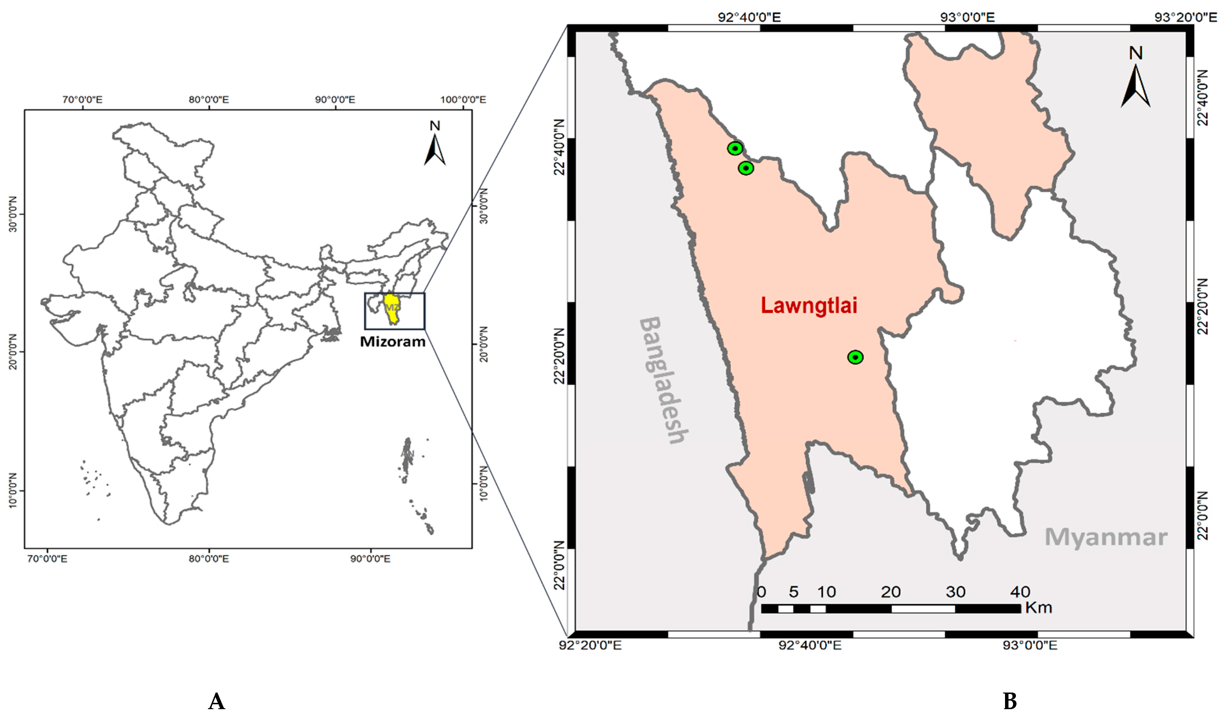
2.1. Description of the Study Area

Mizoram is one of the northeastern states of India, bordering Bangladesh and Myanmar, having a hilly terrain and inaccessible areas [20]. Lawngtlai District is located along the Myanmar and Bangladesh border, with a thick virgin forest and a hilly terrain (Figure 1). Study site “Chawngte” (22°37′10″ N, 92°38′11″ E) has a population of 45,307 and is situated in the western part of Lawngtlai District, which shares an international border with Bangladesh.

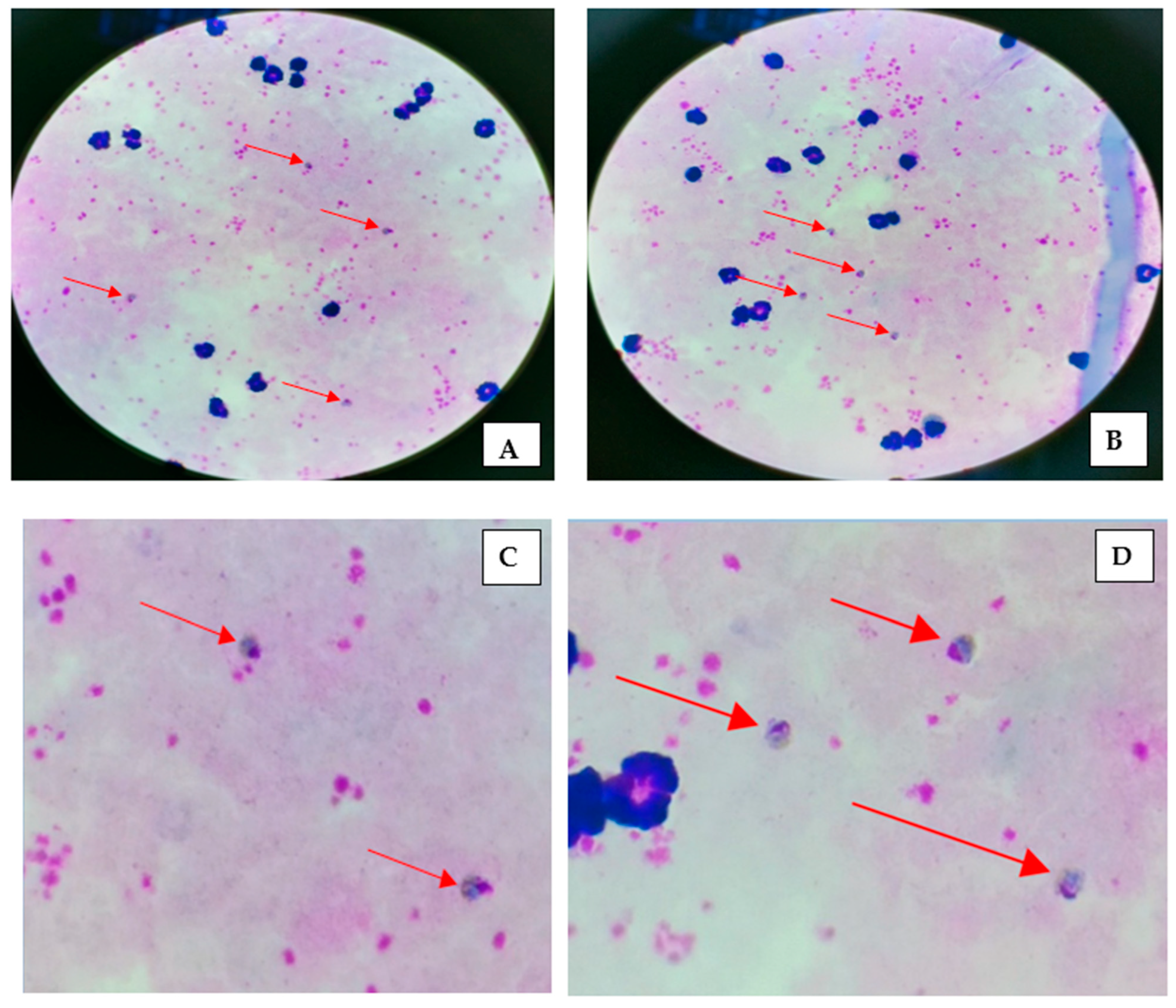
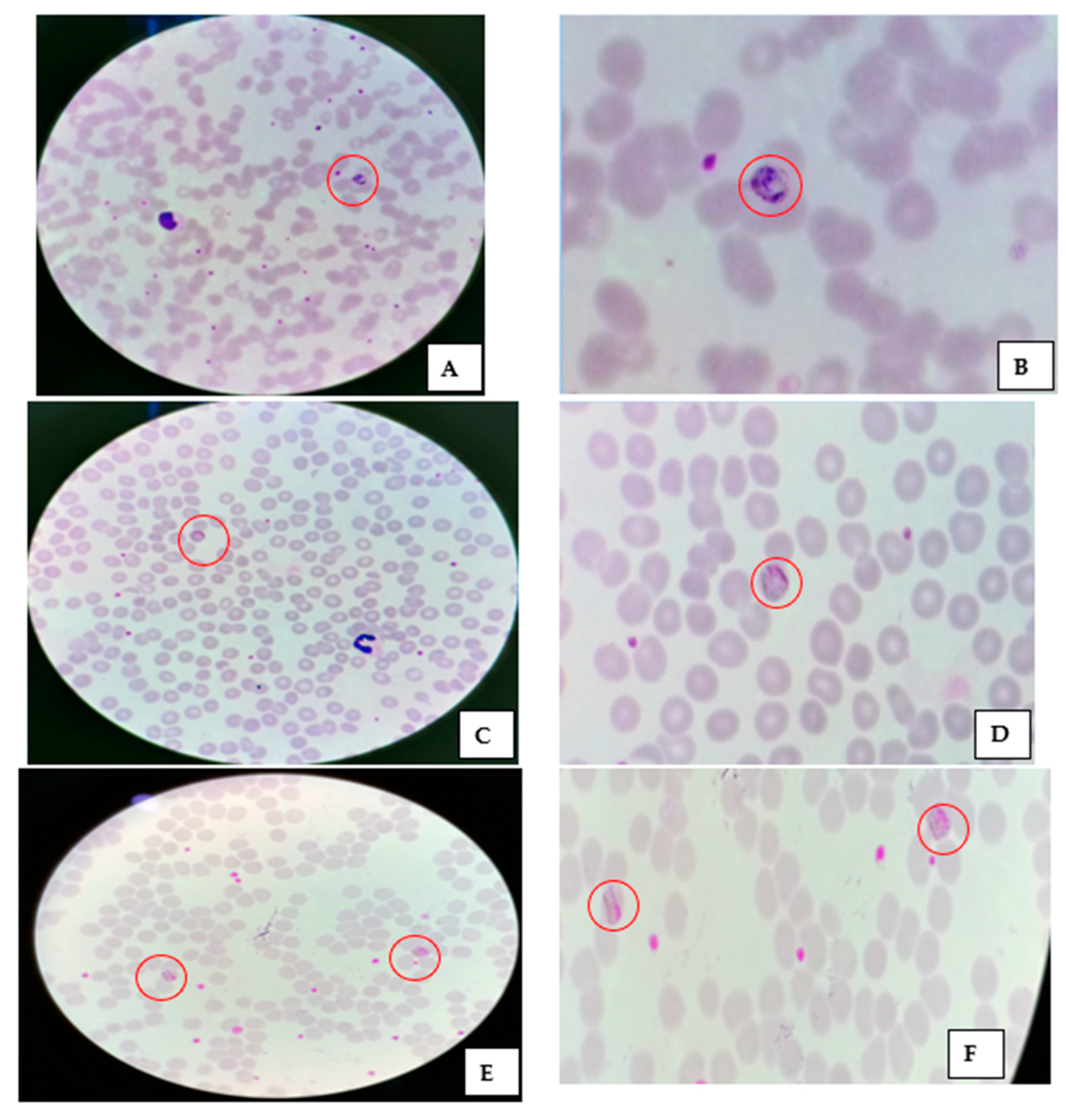
2.2. Study Population and Malaria Diagnosis

Active fever surveys were conducted in July–August 2019 using bivalent RDTs and microscopy to diagnose malaria parasites. After the written consent/assent, thick and thin blood smears were prepared per the method reported elsewhere. The specimens were stained (Giemsa stain) to give the parasites a distinctive appearance. The thick and thin smears were examined with a 100× oil immersion objective. Two WHO-certified microscopists examined the smears at the field level. The third independent microscopist further assessed the thick and thin blood smears for quality control purposes. A blood smear was declared positive only after the concordant findings of two independent microscopists. The positive malaria cases were treated as per the National Drug Policy on Malaria (2013) [21].

2.3. Estimation of Parasitemia

Parasite density per microliter of blood was estimated by counting the number of asexual parasites per 200 (500 in case of low parasitemia) leukocytes in a thick blood smear using Equation (1).
Parasite density per   μ L = Number of parasite count Number of leucocyte count × ( 6000 )

2.4. Quality Control

For quality control, another expert microscopist (WHO level 1), independently and blinded to previous microscopic findings, reassessed 100% of the positive cases and 10% of the negative smears.

2.5. Data Analysis

Statistical data analysis was performed using software package SPSS v.20.0.

3. Results

A total of 948 cases were screened for malaria parasites using RDTs and microscopy, of which 151 were positive as per RDTs and 179 were positive as per microscopy. Using RDTs, 14.34% were detected as P. falciparum, 1.58%—as P. vivax. However, in the microscopic examination, the positivity of P. falciparum and P. vivax was found to be 16.24% and 1.79%, respectively. Further, mixed infection (P. vivax + P. falciparum) was found in three patients (0.32%), and P. malariae was detected in five cases (0.53%) from the study population. All the positive cases diagnosed with RDTs were also found to be positive by malaria microscopy. The results are summarized in Figure 2.
Thick smears showed trophozoites of P. malariae, and their respective magnified images are depicted in Figure 3A–D. A thin smear and its magnified images of P. malariae are shown in Figure 4A–F. The developing schizonts, gametocytes, and band form of trophozoites were confirmed in thin smears. The Plasmodium parasites were differentiated as rings, trophozoites, schizonts, and male/female gametocytes based on their morphological characteristics. P. malariae were distinguished by the features of senescent RBC infection, band-like trophozoites, and rosette forms of merozoites which are lined up around the perimeter of a schizont. The unique part of P. malariae is the band/sash distinctive structure developed as the trophozoite matures. Other Plasmodium species also have specified characteristics, e.g., P. falciparum have thin and delicate ring-form trophozoites that possess one or two chromatin dots, preferably located on the periphery of RBCs and crescent-shaped gametocytes. In P. vivax, the infected erythrocytes are enlarged, and Schüffner’s dots appear in the cytoplasm as the rings mature into trophozoites. P. ovale and P. vivax have similar morphological features, except Schüffner’s dots are distorted into an oval shape and feathering around the edges of the RBCs.
Further analysis revealed that malaria infection is lowest among the under-5 age groups, and no significant difference was observed between the age groups. When the parasite infection was looked into, P. malariae and mixed infections were highest among the 5–15 years group than in other groups. However, P. vivax was most prevalent in the ≥15 years group. Moreover, Plasmodium parasite infections were not significantly different between male and female subjects (Table 1).
The overall risk analysis of malaria infection among the different age groups is shown in Table 2. The 5–15 years and ≥15 years age groups had a 1.67 (95% CI 0.94–2.94) and 1.69 (95% CI 0.99–2.87) times higher risk of malaria infection compared to the under-5 age group. The risk of P. falciparum infection in the 5–15 years and ≥15 years age groups were 1.99 (95% CI 1.06–3.7) and 1.82 (95% CI 1.01–3.29) times higher, respectively, and varied significantly (p < 0.05). Among the three age groups, the risk of non-P. falciparum infection was found to be highest among the ≥15 years group—1.22 (95% CI, 0.40–3.67) times higher compared to the under-5 age group (Table 2).
The mean parasite density of P. falciparum was highest, i.e., 12,275.08 (95% CI, 9056.38–15,493.78) compared to P. vivax and P. malariae, which were 7618.12 (95% CI, 4421.556.10–14,426.30) and 1455.00 (95% CI, 200.22–2709.77), respectively.

4. Discussion

Malaria remains a significant public health problem in Mizoram. The hilly and mountainous terrain, variable ecological habitats, heavy rainfall, and forest-dwelling provide a fertile ground for the prevalence of different human Plasmodium species. P. falciparum is the most prevalent human malaria species in Langwatali District. In the study district, no significant difference (p > 0.05) in the slide positivity rate and the slide Falciparum rate was observed even after the high annual blood examination rate of 29.54. The annual parasite index was between 20.4 to 30.4 from 2017 to 2020 (Figure 5A). The monthly malaria data from 2017 to 2020 showed that malaria transmission was perennial (throughout the year) in Langwatali District (Figure 5B). Early diagnosis, treatment, and introduction of long-lasting insecticidal nets as vector control strategies have significantly reduced malaria incidence in the northeastern states of India. The API of Mizoram decreased from 20.71 to 6.98 (2014–2019). The proper implementation of malaria control strategies by the State Vector-Borne Disease Control Programme has successfully combatted P. falciparum malaria, the deadliest among human Plasmodium species in the hard-to-reach areas of Mizoram.
P. malariae is considered a benign malaria parasite because of low parasite density and coinfections with other Plasmodium species. The density and infectivity of Plasmodium species depend on their potential for invasion into the different age stages of RBCs. The low parasite density of P. vivax is attributed to its invasion capacity only in reticulocytes (<1% of all erythrocytes). P. falciparum is efficient in invading all age stages of RBCs. However, P. malariae preferred to invade mature RBCs [22,23]. Thus, P. malariae infection is considered asymptomatic or sub-patent, and this low parasitemia level probably has substantial chronic transmission potential [24,25]. In the last two decades, the mono-infections of P. malariae and P. ovale and their mixed infections with P. falciparum and P. vivax have been reported in India and the neighboring countries (Table 3). In Myanmar, P. knowlesi and P. malariae were reported in about <3% of the tested cases [26,27], and P. malariae and P. ovale are also prevalent in Bangladesh [28,29]. In India, P. ovale was identified long back in Orissa [30], and P. ovale, as a single case, was reported for the first time from Titabor PHC, Jorhat, Assam, in 2003 [31]. Krishna et al. (2015) also reported the prevalence of P. malariae in many parts of India (Orissa, Chhattisgarh, Maharashtra, Madhya Pradesh, Tripura, Gujarat, and Rajasthan), and of P. ovale in Orissa, Jharkhand, Chhattisgarh, Madhya Pradesh, and Jharkhand. The prevalence of P. ovale was also reported in Jagdalpur, Chhattisgarh [32]. One published report from Mizoram indicates the presence of mixed infections of P. falciparum with other Plasmodium species [6].
Malaria RDTs are the diagnostic tool of choice at points of care in hard-to-access communities and the preferred option for programmatic deployment. However, low-threshold sensitivity, identification, and quantitation of the sexual/asexual stages of Plasmodium have given microscopy an edge over RDTs. RDTs fail to detect low-density parasitemia, deletion of P. falciparum histidine-rich protein 2 (HRP2) leads to false negativity, and the stable HRP2 in the blood results in false positivity. PCR is considered highly sensitive and specific for Plasmodium identification and quantification. However, this technique is expensive and requires regular power backup, trained human resources, high time expenditure, and has only limited application in low-resource settings or emergencies [17]. In summary, microscopy is more reliable in detecting four human Plasmodium species compared to RDTs. Yet, it has the limitations of skilled microscopists, quality of the blood smear, staining, and fixation.
Thus, to target all the Plasmodium species infecting humans, malaria detection using microscopy should be strengthened till the deployment of a molecular tool on the community/primary health center level.

5. Limitations of the Study

In the present study, RDTs and microscopy were used for parasite detection. Molecular analysis was not performed due to the unavailability of blood samples. However, the availability of a WHO-certified level I microscopist strengthened the malaria diagnosis with microscopy.

6. Conclusions

Early and effective diagnosis of human malaria species, including low-density infection and asymptomatic malaria, is crucial in achieving the goal of malaria elimination. Presently, RDTs are used preferably to diagnose malaria, but they have several limitations in identifying P. falciparum, P vivax, and their mixed infection. In the present study, P. malariae cases were detected using microscopy as well as RDTs. Microscopy is the only choice of diagnosis for non-P. falciparum and P. vivax malaria until laboratories are equipped with molecular diagnostic tools at the community and primary health center levels. Thus, it is necessary to strengthen microscopy for accurate and early diagnosis of species-specific malaria until the deployment of molecular tools [5,10,37].

Author Contributions

K.S., P.K.B. and A.S. conceived the study. N.A., K.S. and N.C.D. evaluated the microscopic slides. K.S., N.A. and N.C.D. collected and analyzed the field data. K.S. and P.K.B. drafted the manuscript. A.S. and P.K.B. critically reviewed the manuscript. All authors have read and agreed to the published version of the manuscript.

Funding

The study was part of the study of therapeutic efficacy of artemether and lumefantrine, financially supported by India’s World Health Organization country office (2018/851237).

Institutional Review Board Statement

The study protocol was approved by the Institutional Ethical Review Committee, ICMR National Institute of Malaria Research, New Delhi (ECR/NIMR/EC/2018/28) Dated 26 February 2018, and conducted in accordance with the Declaration of Helsinki.

Informed Consent Statement

The written informed consent was taken from each patient, and assent was obtained from the child’s guardian.

Data Availability Statement

All the data generated or analyzed during this study are included in this published article.

Acknowledgments

The authors would like to thank the State Programme Officer, State Vector-Borne Diseases Control Programme (Mizoram), for logistic support during the study period and data availability. We are thankful to all the study participants and their relatives for providing informed consent/assent, staff of the community health centers/primary health centers, particularly the medical officers and accredited social health activists (ASHA), project staff, and senior technicians for their support in the successful execution of the study at the field and laboratory levels.

Conflicts of Interest

The authors have no conflict of interest.

Abbreviations

Pf: Plasmodium falciparum; RDTs: rapid diagnostic tests, HRP2: histidine-rich protein 2, WHO: World Health Organization, PCR: polymerase chain reaction.

References

  1. World Health Organization. World Malaria Report 2021; World Health Organization: Geneva, Switzerland, 2021; Available online: https://www.who.int/teams/global-malaria-programme/reports/world-malaria-report-2021 (accessed on 3 March 2022).
  2. Dhiman, S.; Veer, V.; Dev, V. Declining Transmission of Malaria in India: Accelerating towards Elimination. In Towards Malaria Elimination—A Leap Forward; Manguin, S., Dev, V., Eds.; IntechOpen: London, UK, 2018; Available online: https://www.intechopen.com/chapters/61663 (accessed on 29 April 2022).
  3. World Malaria Day Observed within Lawngtlai District. Available online: https://lawngtlai.nic.in/world-malaria-day-observed-within-lawngtlai-district/ (accessed on 29 April 2022).
  4. Bharti, P.K.; Chand, S.K.; Singh, M.P.; Mishra, S.; Shukla, M.M.; Singh, R.; Singh, N. Emergence of a new focus of Plasmodium malariae in forest villages of district Balaghat, Central India: Implications for the diagnosis of malaria and its control. Trop. Med. Int. Health 2013, 18, 12–17. [Google Scholar] [CrossRef] [PubMed]
  5. Chaturvedi, R.; Deora, N.; Bhandari, D.; Parvez, S.; Sinha, A.; Sharma, A. Trends of neglected Plasmodium species infection in humans over the past century in India. One Health 2020, 11, 100190. [Google Scholar] [CrossRef]
  6. Nema, S.; Singh, A.; Krishna, S.; Poriya, R.; Dubey, S.; Ali, N.A.; Singh, M.P.; Verma, A.K.; Das, A.; Bharti, P.K. Unreported mixed Plasmodium species infection may increase vivax malaria in India: A challenge for malaria elimination. Trans. R. Soc. Trop. Med. Hyg. 2022; ahead to print. [Google Scholar] [CrossRef] [PubMed]
  7. Gupta, B.; Gupta, P.; Sharma, A.; Singh, V.; Dash, A.P.; Das, A. High proportion of mixed-species Plasmodium infections in India revealed by PCR diagnostic assay. Trop. Med. Int. Health 2010, 15, 819–824. [Google Scholar] [CrossRef] [PubMed]
  8. Lover, A.A.; Baird, J.K.; Gosling, R.; Price, R.N. Malaria elimination: Time to target all species. Am. J. Trop. Med. Hyg. 2018, 99, 17–23. [Google Scholar] [CrossRef]
  9. Collins, W.E.; Jeffery, G.M. Plasmodium malariae: Parasite and disease. Clin. Microbiol. Rev. 2007, 20, 579–592. [Google Scholar] [CrossRef] [Green Version]
  10. Ashraf, S.; Kao, A.; Hugo, C.; Christophel, E.M.; Fatunmbi, B.; Luchavez, J.; Lilley, K.; Bell, D. Developing standards for malaria microscopy: External competency assessment for malaria microscopists in the Asia-Pacific. Malar. J. 2012, 11, 352. [Google Scholar] [CrossRef] [Green Version]
  11. Bharti, P.K.; Silawat, N.; Singh, P.P.; Singh, M.P.; Shukla, M.; Chand, G.; Dash, A.P.; Singh, N. The usefulness of a new rapid diagnostic test, the First Response® Malaria Combo (pLDH/HRP2) card test, for malaria diagnosis in the forested belt of central India. Malar. J. 2008, 7, 126. [Google Scholar] [CrossRef] [Green Version]
  12. Singh, N.; Shukla, M.M.; Shukla, M.K.; Mehra, R.K.; Sharma, S.; Bharti, P.K.; Singh, M.P.; Singh, A.; Gunasekar, A. Field and laboratory comparative evaluation of rapid malaria diagnostic tests versus traditional and molecular techniques in India. Malar. J. 2010, 9, 191. [Google Scholar] [CrossRef] [Green Version]
  13. Bharti, P.K.; Chandel, H.S.; Ahmad, A.; Krishna, S.; Udhayakumar, V.; Singh, N. Prevalence of pfhrp2 and/or pfhrp3 gene deletion in Plasmodium falciparum population in eight highly endemic states in India. PLoS ONE 2016, 11, e0157949. [Google Scholar] [CrossRef]
  14. Ishengoma, D.S.; Lwitiho, S.; Madebe, R.A.; Nyagonde, N.; Persson, O.; Vestergaard, L.S.; Bygbjerg, I.C.; Lemnge, M.M.; Alifrangis, M. Using rapid diagnostic tests as source of malaria parasite DNA for molecular analyses in the era of declining malaria prevalence. Malar. J. 2011, 10, 6. [Google Scholar] [CrossRef] [Green Version]
  15. Komaki-Yasuda, K.; Vincent, J.P.; Nakatsu, M.; Kato, Y.; Ohmagari, N.; Kano, S. A novel PCR-based system for the detection of four species of human malaria parasites and Plasmodium knowlesi. PLoS ONE 2018, 13, e0191886. [Google Scholar] [CrossRef] [PubMed] [Green Version]
  16. Tahar, R.; Ringwald, P.; Basco, L.K. Diagnosis of Plasmodium malariae infection by the polymerase chain reaction. Trans. R. Soc. Trop. Med. Hyg. 1997, 91, 410–411. [Google Scholar] [CrossRef]
  17. Fitri, L.E.; Widaningrum, T.; Endharti, A.T.; Prabowo, M.H.; Winaris, N.; Nugraha, R.Y.B. Malaria diagnostic update: From conventional to advanced method. J. Clin. Lab. Anal. 2022, 36, e24314. [Google Scholar] [CrossRef] [PubMed]
  18. Dev, V.; Manguin, S. Defeating malaria in the North-East region: The forerunner for malaria elimination in India. Acta Trop. 2021, 222, 106040. [Google Scholar] [CrossRef] [PubMed]
  19. Subbarao, S.K.; Nanda, N.; Rahi, M.; Raghavendra, K. Biology and bionomics of malaria vectors in India: Existing information and what more needs to be known for strategizing elimination of malaria. Malar. J. 2019, 18, 1–11. [Google Scholar] [CrossRef] [PubMed]
  20. Lalchhuanawma, R.; Murhekar, M.V. Health-seeking behaviour for febrile illness in malaria-endemic Kolasib district, Mizoram, India. Int. Health 2012, 4, 314–319. [Google Scholar] [CrossRef]
  21. National Drug Policy on Malaria. 2013. Available online: https://nvbdcp.gov.in/Doc/National-Drug-Policy-2013.pdf (accessed on 25 April 2022).
  22. Prajapati, S.K.; Singh, O.P. Insights into the invasion biology of Plasmodium vivax. Front. Cell. Infect. Microbiol. 2013, 3, 8. [Google Scholar] [CrossRef] [Green Version]
  23. Kerlin, D.H.; Gatton, M.L. Preferential invasion by Plasmodium merozoites and the self-regulation of parasite burden. PLoS ONE 2013, 8, e57434. [Google Scholar] [CrossRef] [Green Version]
  24. Roucher, C.; Rogier, C.; Sokhna, C.; Tall, A.; Trape, J.-F. A 20-year longitudinal study of Plasmodium ovale and Plasmodium malariae prevalence and morbidity in a West African population. PLoS ONE 2014, 9, e87169. [Google Scholar] [CrossRef] [Green Version]
  25. Yman, V.; Wandell, G.; Mutemi, D.D.; Miglar, A.; Asghar, M.; Hammar, U.; Karlsson, M.; Lind, I.; Nordfjell, C.; Rooth, I. Persistent transmission of Plasmodium malariae and Plasmodium ovale species in an area of declining Plasmodium falciparum transmission in eastern Tanzania. PLoS Negl. Trop. Dis. 2019, 13, e0007414. [Google Scholar] [CrossRef] [PubMed] [Green Version]
  26. Jiang, N.; Chang, Q.; Sun, X.; Lu, H.; Yin, J.; Zhang, Z.; Wahlgren, M.; Chen, Q. Co-infections with Plasmodium knowlesi and other malaria parasites, Myanmar. Emerg. Infect. Dis. 2010, 16, 1476–1478. [Google Scholar] [CrossRef]
  27. Han, T.Z.; Han, K.T.; Aye, K.H.; Hlaing, T.; Thant, K.Z.; Vythilingam, I. Comparison of microscopy and PCR for the detection of human Plasmodium species and Plasmodium knowlesi in southern Myanmar. Asian Pac. J. Trop. Biomed. 2017, 7, 680–685. [Google Scholar] [CrossRef]
  28. Fuehrer, H.-P.; Starzengruber, P.; Swoboda, P.; Khan, W.A.; Matt, J.; Ley, B.; Thriemer, K.; Haque, R.; Yunus, E.B.; Hossain, S.M. Indigenous Plasmodium ovale malaria in Bangladesh. Am. J. Trop. Med. Hyg. 2010, 83, 75–78. [Google Scholar] [CrossRef] [PubMed] [Green Version]
  29. Fuehrer, H.-P.; Swoboda, P.; Harl, J.; Starzengruber, P.; Habler, V.E.; Bloeschl, I.; Haque, R.; Matt, J.; Khan, W.A.; Noedl, H. High prevalence and genetic diversity of Plasmodium malariae and no evidence of Plasmodium knowlesi in Bangladesh. Parasitol. Res. 2014, 113, 1537–1543. [Google Scholar] [CrossRef] [PubMed]
  30. Jambulingam, P.; Mohapatra, S.; Das, L.; Das, P.; Rajagopalan, P. Detection of Plasmodium ovale in Koraput district, Orissa state. Indian J. Med. Res. 1989, 89, 115–116. [Google Scholar] [PubMed]
  31. Prakash, A.; Mohapatra, P.; Bhattacharyya, D.; Goswami, B.; Mahanta, J. Plasmodium ovale: First case report from Assam, India. Curr. Sci. 2003, 84, 1187–1188. [Google Scholar]
  32. Singh, R.; Jain, V.; Singh, P.; Bharti, P.; Thomas, T.; Basak, S.; Singh, N. First report of detection and molecular confirmation of Plasmodium ovale from severe malaria cases in central India. Trop. Med. Int. Health 2013, 18, 1416–1420. [Google Scholar] [CrossRef]
  33. Krishna, S.; Bharti, P.K.; Chandel, H.S.; Ahmad, A.; Kumar, R.; Singh, P.P.; Singh, M.P.; Singh, N. Detection of mixed infections with Plasmodium spp. by PCR, India, 2014. Emerg. Infect. Dis. 2015, 21, 1853–1857. [Google Scholar] [CrossRef] [Green Version]
  34. Chaturvedi, N.; Bhandari, S.; Bharti, P.K.; Basak, S.K.; Singh, M.P.; Singh, N. Sympatric distribution of Plasmodium ovale curtisi and P. ovale wallikeri in India: Implication for the diagnosis of malaria and its control. Trans. R. Soc. Trop. Med. Hyg. 2015, 109, 352–354. [Google Scholar] [CrossRef]
  35. Mohapatra, P.K.; Prakash, A.; Bhattacharyya, D.R.; Goswami, B.; Ahmed, A.; Sarmah, B.; Mahanta, J. Detection & molecular confirmation of a focus of Plasmodium malariae in Arunachal Pradesh, India. Indian J. Med. Res. 2008, 128, 52–60. [Google Scholar] [PubMed]
  36. Dhangadamajhi, G.; Kar SK, K.; Ranjit, M.R. High prevalence and gender bias in distribution of Plasmodium malariae infection in central east-coast India. Trop. Biomed. 2009, 26, 326–333. [Google Scholar] [PubMed]
  37. Fançony, C.; Sebastião, Y.V.; Pires, J.E.; Gamboa, D.; Nery, S.V. Performance of microscopy and RDTs in the context of a malaria prevalence survey in Angola: A comparison using PCR as the gold standard. Malar. J. 2013, 12, 1–7. [Google Scholar] [CrossRef] [PubMed] [Green Version]
Figure 1. Map showing study sites (A) Mizoram, India; (B) the green circle shows the location of the P. malariae-positive cases detected in Lawngtlai District of Mizoram (India).
Diagnostics 12 02015 g001
Figure 2. Screening of the fever cases and malaria positivity at the study site (Pf = P. falciparum; Pv = P. vivax; Pm = P. malariae).
Diagnostics 12 02015 g002
Figure 3. Thick smear showing Plasmodium malariae trophozoites (A,B) and their respective magnified images (C,D).
Diagnostics 12 02015 g003
Figure 4. Thin smear and its magnified images showing (A,B) a developing schizont; (C,D) a gametocyte; (E,F) the band form of trophozoites.
Diagnostics 12 02015 g004
Figure 5. (A): Malariometric indices (API, SPR, SFR) in Lawngtlai District of the State of Mizoram from 2017 to 2020; (B) monthly total malaria cases from 2017 to 2020 (source: State Vector-Borne Diseases Control Programme, Mizoram). Note: API: annual parasite index, SPR: slide positivity rate, SFR: slide falciparum rate).
Diagnostics 12 02015 g005
Table 1. Age group- and gender-wise malaria cases diagnosis using RDTs and microscopy.
AgeNumber of Fever Cases: 948Rapid Diagnostic Test, n (%)Malaria Microscopy, n (%)
Gender (n)Malaria-Positive (%)Pf (%)Pv (%)Pm (%)Mix (%)Malaria-Positive (%)Pf (%)Pv (%)Pm (%)Mix (%)
Under-5 yearsMale (86)8 (9.30)7(8.14)1 (1.16)009 (10.47)7 (8.14)2 (2.33)00
Female (64)8 (12.50)6(9.38)2 (3.13)009 (14.06)7 (10.94)2 (3.13)00
5–15 yearsMale (164)22 (13.41)21(12.80)1 (0.61)0030 (18.29)27 (16.46)1 (0.61)2 (1.22)0
Female (111)22 (19.81)22 (19.82)00025 (22.52)24 (21.62)001 (0.90)
≥15 yearsMale (262)55 (20.99)49 (18.70)6 (2.29)0065 (24.81)55 (20.99)7 (2.67)1 (0.38)2 (0.76)
Female (261)36 (13.79)31 (11.88)5 (1.92)0041 (15.71)34 (13.03)5 (1.92)2 (0.77)0 (0.00)
TotalMale (512)85 (16.60)77 (15.34)8 (1.56)00104 (20.31)89 (17.38)10 (1.953)3 (0.58)2 (0.39)
Female (436)66 (15.14)59 (13.53)7 (1.60)0075 (17.20)65 (14.91)7 (1.61)2 (0.46)1 (0.23)
Pf: P. falciparum; Pv: P. vivax; Pm: P. malariae; Mix (P. falciparum + P. vivax).
Table 2. Odds ratio of infection of parasite species among different age groups.
Age GroupBSERisk of Malaria InfectionRisk of P. Falciparum * InfectionRisk of Non-P. falciparum **
Infection
+veOdds Ratio (95% CI),
p-Value
PfOdds Ratio (95% CI),
p-Value
Non-PfOdds Ratio (95% CI),
p-Value
Under-5 years15018144
5–15 years275551.67 (0.94–2.94), p = 0.078511.99 (1.06–3.71), p = 0.0340.55 (0.13 to 2.21), p = 0.39
≥15 years5231061.69 (0.99–2.87), p = 0.053891.82 (1.01–3.29), p = 0.046171.22 (0.40 to 3.67), p = 0.73
BSE: blood smear examination, Pf: Plasmodium falciparum *, non-P. falciparum **: other than Plasmodium falciparum, i.e., P. vivax, P. malariae, and P. falciparum + P. vivax.
Table 3. Prevalence of Plasmodium malariae and other neglected Plasmodium species in India and the adjoining countries bordering the northeastern states.
CountryPlasmodium
Species
Study SitePositivityRemarksReference
MyanmarP. knowlesiSouthern Myanmar near Yunnan Province of ChinaPk: 4
Pk + Pf: 13
Pk + Pv: 13
Pk + Pf + Pv: 2
M. nemestrina as reservoir host[26]
P. malariaeRanong province borderPm: 1 Both microscopy and PCR were capable to identify [27]
BangladeshP. malariaeChittagong Hill Tracts in southeastern BangladeshPm: 60P. malariae is highly underestimated in rural Bangladesh[29]
P. malariae and P. ovaleChittagong Hill TractsPm: 3
Po: 3
Both microscopy and PCR were capable to identify[28]
IndiaP. ovaleKoraput District, Orissa state.Po: 3[30]
P. ovaleTitabor PHC, Jorhat (Assam)Po: 1[31]
P. falciparum + P. ovaleOrissa, Jharkhand, Chhattisgarh, Madhya PradeshPf + Po: 6 Mixed infection[33]
P. falciparum + P. malariae + P. ovaleJharkhandPf + Pm + P: 01Mixed infection[33]
P. falciparum + P. malariaeOrissa, Chhattisgarh,
Maharashtra, Madhya Pradesh, Tripura, Gujarat, and Rajasthan
Pf + Pm: 19 Mixed infection [33]
P. ovale curtisi and P. ovale wallikeri Bastar, State of ChhattisgarhPo: 2
Pv + Pf + Po: 1
Mono- and mixed infection of P. ovale curtisi [34]
P. ovaleJagdalpur (Chhattisgarh)Po: 3[32]
P. malariae
(mono- and mixed infections)
Balaghat DistrictPm: 14
Pm + Pv: 2
Pm + Pf: 3
Existence in forest villages of central India[4]
P. malariaeLohit (Arunachal Pradesh)Pm: 9 Confirmed by nested PCR[35]
P. malariaeOrissaPm: 108High prevalence [36]
Pf = Plasmodium falciparum; Pv = P. vivax; Pm = P. malariae; Po = P. ovale; Pk = P. knowlesi.
Publisher’s Note: MDPI stays neutral with regard to jurisdictional claims in published maps and institutional affiliations.

