
Semi-Automated Quantification of Retinal and Choroidal Biomarkers in Retinal Vascular Diseases: Agreement of Spectral-Domain Optical Coherence Tomography with and without Enhanced Depth Imaging Mode

 ,
,  and
and 
Abstract
:1. Introduction
2. Materials and Methods
2.1. Study Cohort
2.2. Optical Coherence Tomography Acquisition and Analysis
2.2.1. Image Acquisition
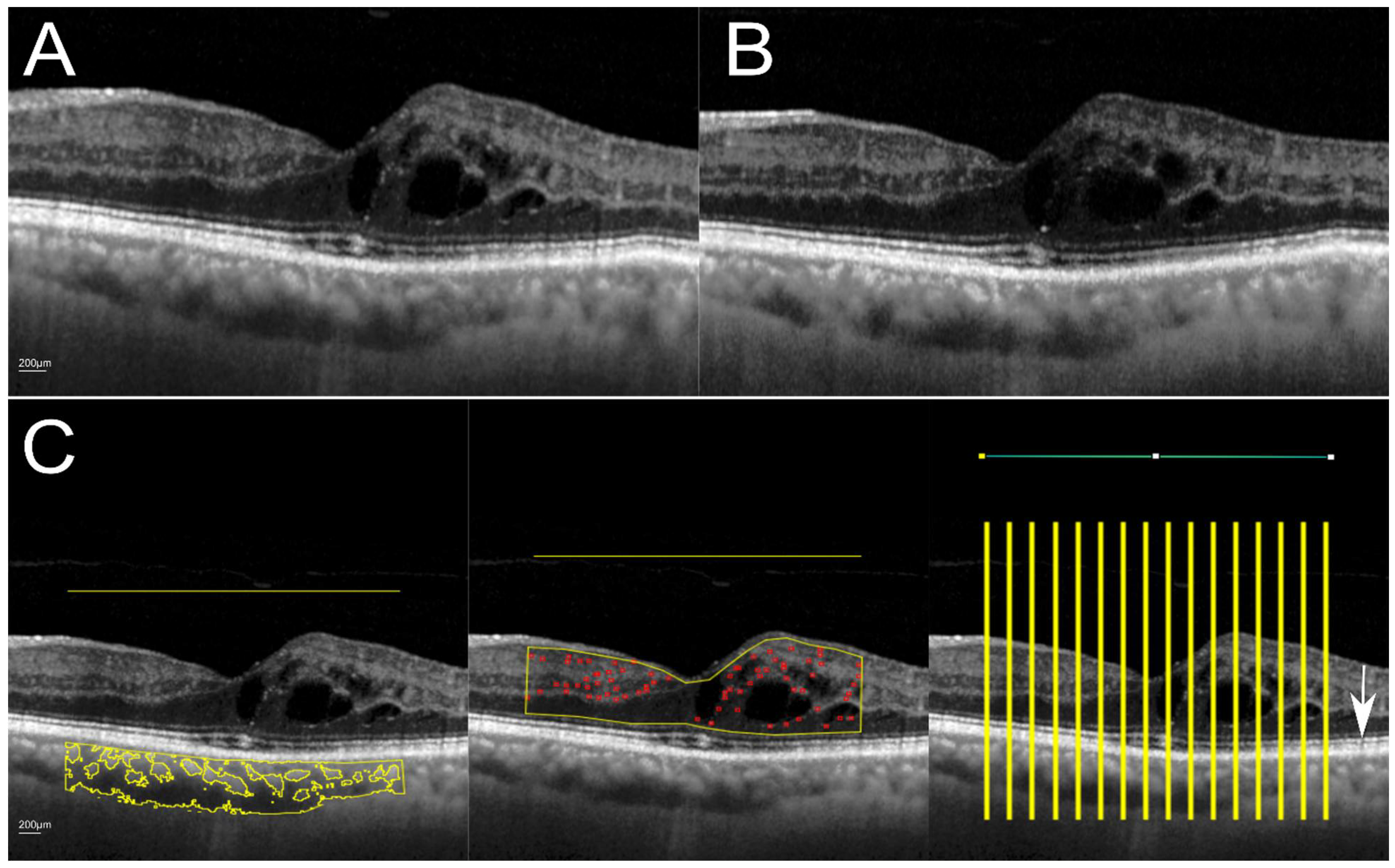
2.2.2. Image Processing and Analysis
2.3. Statistical Analysis
3. Results
4. Discussion
Author Contributions
Funding
Institutional Review Board Statement
Informed Consent Statement
Data Availability Statement
Acknowledgments
Conflicts of Interest
References
- Schuman, J.S. Spectral domain optical coherence tomography for glaucoma (an AOS thesis). Trans. Am. Ophthalmol. Soc. 2008, 106, 426–458. [Google Scholar] [PubMed]
- Schmidt-Erfurth, U.; Garcia-Arumi, J.; Bandello, F.; Berg, K.; Chakravarthy, U.; Gerendas, B.S.; Jonas, J.; Larsen, M.; Tadayoni, R.; Loewenstein, A. Guidelines for the Management of Diabetic Macular Edema by the European Society of Retina Specialists (EURETINA). Ophthalmologica 2017, 237, 185–222. [Google Scholar] [CrossRef] [PubMed]
- Schmidt-Erfurth, U.; Garcia-Arumi, J.; Gerendas, B.S.; Midena, E.; Sivaprasad, S.; Tadayoni, R.; Wolf, S.; Loewenstein, A. Guidelines for the Management of Retinal Vein Occlusion by the European Society of Retina Specialists (EURETINA). Ophthalmologica 2019, 242, 123–162. [Google Scholar] [CrossRef] [PubMed]
- Puzyeyeva, O.; Lam, W.C.; Flanagan, J.G.; Brent, M.H.; Devenyi, R.G.; Mandelcorn, M.S.; Wong, T.; Hudson, C. High-resolution optical coherence tomography retinal imaging: A case series illustrating potential and limitations. J. Ophthalmol. 2011, 2011, 764183. [Google Scholar] [CrossRef] [PubMed] [Green Version]
- Chatziralli, I.P.; Sergentanis, T.N.; Sivaprasad, S. Hyperreflective Foci as an Independent Visual Outcome Predictor in Macular Edema Due to Retinal Vascular Diseases Treated with Intravitreal Dexamethasone or Ranibizumab. Retina 2016, 36, 2319–2328. [Google Scholar] [CrossRef] [PubMed]
- Mo, B.; Zhou, H.Y.; Jiao, X.; Zhang, F. Evaluation of hyperreflective foci as a prognostic factor of visual outcome in retinal vein occlusion. Int. J. Ophthalmol. 2017, 10, 605–612. [Google Scholar] [CrossRef] [PubMed]
- Markan, A.; Agarwal, A.; Arora, A.; Bazgain, K.; Rana, V.; Gupta, V. Novel imaging biomarkers in diabetic retinopathy and diabetic macular edema. Ther. Adv. Ophthalmol. 2020, 12, 2515841420950513. [Google Scholar] [CrossRef]
- Kim, M.; Ha, M.J.; Choi, S.Y.; Park, Y.H. Choroidal vascularity index in type-2 diabetes analyzed by swept-source optical coherence tomography. Sci. Rep. 2018, 8, 70. [Google Scholar] [CrossRef]
- Wang, H.; Tao, Y. Choroidal structural changes correlate with severity of diabetic retinopathy in diabetes mellitus. BMC Ophthalmol. 2019, 19, 186. [Google Scholar] [CrossRef] [Green Version]
- Aribas, Y.K.; Hondur, A.M.; Tezel, T.H. Choroidal vascularity index and choriocapillary changes in retinal vein occlusions. Graefes Arch. Clin. Exp. Ophthalmol. 2020, 258, 2389–2397. [Google Scholar] [CrossRef]
- Kessler, L.J.; Auffarth, G.U.; Bagautdinov, D.; Khoramnia, R. Ellipsoid Zone Integrity and Visual Acuity Changes during Diabetic Macular Edema Therapy: A Longitudinal Study. J. Diabetes Res. 2021, 2021, 8117650. [Google Scholar] [CrossRef] [PubMed]
- Tao, L.W.; Wu, Z.; Guymer, R.H.; Luu, C.D. Ellipsoid zone on optical coherence tomography: A review. Clin. Exp. Ophthalmol. 2016, 44, 422–430. [Google Scholar] [CrossRef] [PubMed]
- Etheridge, T.; Dobson, E.T.A.; Wiedenmann, M.; Oden, N.; VanVeldhuisen, P.; Scott, I.U.; Ip, M.S.; Eliceiri, K.W.; Blodi, B.A.; Domalpally, A. Ellipsoid Zone Defects in Retinal Vein Occlusion Correlates With Visual Acuity Prognosis: SCORE2 Report 14. Transl. Vis. Sci. Technol. 2021, 10, 31. [Google Scholar] [CrossRef] [PubMed]
- De, S.; Saxena, S.; Kaur, A.; Mahdi, A.A.; Misra, A.; Singh, M.; Meyer, C.H.; Akduman, L. Sequential restoration of external limiting membrane and ellipsoid zone after intravitreal anti-VEGF therapy in diabetic macular oedema. Eye 2021, 35, 1490–1495. [Google Scholar] [CrossRef]
- Agrawal, R.; Salman, M.; Tan, K.A.; Karampelas, M.; Sim, D.A.; Keane, P.A.; Pavesio, C. Choroidal Vascularity Index (CVI)--A Novel Optical Coherence Tomography Parameter for Monitoring Patients with Panuveitis? PLoS ONE 2016, 11, e0146344. [Google Scholar] [CrossRef]
- Endo, H.; Kase, S.; Takahashi, M.; Saito, M.; Yokoi, M.; Sugawara, C.; Katsuta, S.; Ishida, S.; Kase, M. Relationship between diabetic macular edema and choroidal layer thickness. PLoS ONE 2020, 15, e0226630. [Google Scholar] [CrossRef] [Green Version]
- Tan, K.A.; Laude, A.; Yip, V.; Loo, E.; Wong, E.P.; Agrawal, R. Choroidal vascularity index—A novel optical coherence tomography parameter for disease monitoring in diabetes mellitus? Acta Ophthalmol. 2016, 94, e612–e616. [Google Scholar] [CrossRef]
- Du, K.F.; Xu, L.; Shao, L.; Chen, C.X.; Zhou, J.Q.; Wang, Y.X.; You, Q.S.; Jonas, J.B.; Wei, W.B. Subfoveal choroidal thickness in retinal vein occlusion. Ophthalmology 2013, 120, 2749–2750. [Google Scholar] [CrossRef]
- Xu, J.; Xu, L.; Du, K.F.; Shao, L.; Chen, C.X.; Zhou, J.Q.; Wang, Y.X.; You, Q.S.; Jonas, J.B.; Wei, W.B. Subfoveal choroidal thickness in diabetes and diabetic retinopathy. Ophthalmology 2013, 120, 2023–2028. [Google Scholar] [CrossRef]
- Spaide, R.F.; Koizumi, H.; Pozzoni, M.C. Enhanced depth imaging spectral-domain optical coherence tomography. Am. J. Ophthalmol. 2008, 146, 496–500. [Google Scholar] [CrossRef]
- Wong, I.Y.; Koizumi, H.; Lai, W.W. Enhanced depth imaging optical coherence tomography. Ophthalmic. Surg. Lasers Imaging 2011, 42, S75–S84. [Google Scholar] [CrossRef] [PubMed] [Green Version]
- Zafar, S.; Siddiqui, M.R.; Shahzad, R. Comparison of choroidal thickness measurements between spectral-domain OCT and swept-source OCT in normal and diseased eyes. Clin. Ophthalmol. 2016, 10, 2271–2276. [Google Scholar] [CrossRef] [PubMed] [Green Version]
- Midena, E.; Torresin, T.; Velotta, E.; Pilotto, E.; Parrozzani, R.; Frizziero, L. OCT Hyperreflective Retinal Foci in Diabetic Retinopathy: A Semi-Automatic Detection Comparative Study. Front. Immunol. 2021, 12, 613051. [Google Scholar] [CrossRef] [PubMed]
- Gin, T.J.; Wu, Z.; Chew, S.K.; Guymer, R.H.; Luu, C.D. Quantitative Analysis of the Ellipsoid Zone Intensity in Phenotypic Variations of Intermediate Age-Related Macular Degeneration. Investig. Ophthalmol. Vis. Sci. 2017, 58, 2079–2086. [Google Scholar] [CrossRef] [Green Version]
- Gong, Y.; Chen, L.J.; Pang, C.P.; Chen, H. Ellipsoid zone optical intensity reduction as an early biomarker for retinitis pigmentosa. Acta Ophthalmol. 2021, 99, e215–e221. [Google Scholar] [CrossRef]
- Gerke, O. Nonparametric Limits of Agreement in Method Comparison Studies: A Simulation Study on Extreme Quantile Estimation. Int. J. Environ. Res. Public Health 2020, 17, 8330. [Google Scholar] [CrossRef]
- Chen, L.A.; Kao, C.L. Parametric and nonparametric improvements in Bland and Altman’s assessment of agreement method. Stat. Med. 2021, 40, 2155–2176. [Google Scholar] [CrossRef]
- Yoshitake, T.; Murakami, T.; Suzuma, K.; Dodo, Y.; Fujimoto, M.; Tsujikawa, A. Hyperreflective Foci in the Outer Retinal Layers as a Predictor of the Functional Efficacy of Ranibizumab for Diabetic Macular Edema. Sci. Rep. 2020, 10, 873. [Google Scholar] [CrossRef] [Green Version]
- Schreur, V.; Altay, L.; van Asten, F.; Groenewoud, J.M.M.; Fauser, S.; Klevering, B.J.; Hoyng, C.B.; de Jong, E.K. Hyperreflective foci on optical coherence tomography associate with treatment outcome for anti-VEGF in patients with diabetic macular edema. PLoS ONE 2018, 13, e0206482. [Google Scholar] [CrossRef]
- Ciulla, T.A.; Kapik, B.; Grewal, D.S.; Ip, M.S. Visual Acuity in Retinal Vein Occlusion, Diabetic, and Uveitic Macular Edema: Central Subfield Thickness and Ellipsoid Zone Analysis. Ophthalmol. Retina 2020, 5, 633–647. [Google Scholar] [CrossRef]
- Ciulla, T.A.; Pollack, J.S.; Williams, D.F. Visual acuity outcomes and anti-VEGF therapy intensity in diabetic macular oedema: A real-world analysis of 28 658 patient eyes. Br. J. Ophthalmol. 2021, 105, 216–221. [Google Scholar] [CrossRef] [PubMed] [Green Version]
- Abraham, J.R.; Boss, J.; Babiuch, A.S.; Singh, R.P.; Srivastava, S.; Reese, J.; Ehlers, J.P. Longitudinal Assessment of Ellipsoid Zone Mapping Parameters in Retinal Venous Occlusive Disease With Associated Macular Edema. J. Vitr. Dis. 2021, 5, 40–45. [Google Scholar] [CrossRef]
- Gonzalez, V.H.; Campbell, J.; Holekamp, N.M.; Kiss, S.; Loewenstein, A.; Augustin, A.J.; Ma, J.; Ho, A.C.; Patel, V.; Whitcup, S.M.; et al. Early and Long-Term Responses to Anti-Vascular Endothelial Growth Factor Therapy in Diabetic Macular Edema: Analysis of Protocol I Data. Am. J. Ophthalmol. 2016, 172, 72–79. [Google Scholar] [CrossRef] [Green Version]
- Ehlers, J.P.; Clark, J.; Uchida, A.; Figueiredo, N.; Babiuch, A.; Talcott, K.E.; Lunasco, L.; Le, T.K.; Meng, X.; Hu, M.; et al. Longitudinal Higher-Order OCT Assessment of Quantitative Fluid Dynamics and the Total Retinal Fluid Index in Neovascular AMD. Transl. Vis. Sci. Technol. 2021, 10, 29. [Google Scholar] [CrossRef] [PubMed]
- Huang, C.H.; Yang, C.H.; Hsieh, Y.T.; Yang, C.M.; Ho, T.C.; Lai, T.T. Hyperreflective foci in predicting the treatment outcomes of diabetic macular oedema after anti-vascular endothelial growth factor therapy. Sci. Rep. 2021, 11, 5103. [Google Scholar] [CrossRef]
- Verner-Cole, E.A.; Campbell, J.P.; Hwang, T.S.; Klein, M.L.; Lauer, A.K.; Choi, D.; Bailey, S.T. Retinal and Choroidal Imaging With 870-nm Spectral-Domain OCT Compared With 1050-nm Spectral-Domain OCT, With and Without Enhanced Depth Imaging. Transl. Vis. Sci. Technol. 2014, 3, 3. [Google Scholar] [CrossRef] [Green Version]
- Hosseini, H.; Razeghinejad, M.R.; Nowroozizadeh, S.; Jafari, P.; Ashraf, H. Effect of macular edema on optical coherence tomography signal strength. Retina 2010, 30, 1084–1089. [Google Scholar] [CrossRef]
- Cao, J.; Wang, P.; Zhang, Y.; Shi, G.; Wu, B.; Zhang, S.; Liu, Y. Methods to improve the performance of the swept source at 1.0 μm based on a polygon scanner. Photon. Res. 2017, 5, 245–250. [Google Scholar] [CrossRef] [Green Version]
- Tan, C.S.; Ngo, W.K.; Cheong, K.X. Comparison of choroidal thicknesses using swept source and spectral domain optical coherence tomography in diseased and normal eyes. Br. J. Ophthalmol. 2015, 99, 354–358. [Google Scholar] [CrossRef]
- Hoseini-Yazdi, H.; Vincent, S.J.; Collins, M.J.; Read, S.A.; Alonso-Caneiro, D. Impact of image averaging on wide-field choroidal thickness measurements using enhanced-depth imaging optical coherence tomography. Clin. Exp. Optom. 2019, 102, 320–326. [Google Scholar] [CrossRef] [Green Version]
- Meleppat, R.K.; Ronning, K.E.; Karlen, S.J.; Burns, M.E.; Pugh, E.N., Jr.; Zawadzki, R.J. In vivo multimodal retinal imaging of disease-related pigmentary changes in retinal pigment epithelium. Sci. Rep. 2021, 11, 16252. [Google Scholar] [CrossRef] [PubMed]
- Meleppat, R.K.; Zhang, P.; Ju, M.J.; Manna, S.K.; Jian, Y.; Pugh, E.N.; Zawadzki, R.J. Directional optical coherence tomography reveals melanin concentration-dependent scattering properties of retinal pigment epithelium. J. Biomed. Opt. 2019, 24, 1–10. [Google Scholar] [CrossRef] [PubMed]
- Chen, H.; Xia, H.; Qiu, Z.; Chen, W.; Chen, X. Correlation of Optical Intensity on Optical Coherence Tomography and Visual Outcome in Central Retinal Artery Occlusion. Retina 2016, 36, 1964–1970. [Google Scholar] [CrossRef] [PubMed]

| Overall (n = 60) | DR (n = 27) | RVO (n = 33) | ||||||
|---|---|---|---|---|---|---|---|---|
| Baseline Variables | Mean (SD) | Median (Q1; Q3) | Mean(SD) | Median (Q1; Q3) | Mean (SD) | Median (Q1; Q3) | p | |
| Age (years) | 64.88 (9.29) | 65.00 (59.25; 72.25) | 62.51 (9.68) | 64.00 (60.00; 67.00) | 66.81 (8.62) | 66.00 (59.00; 74.50) | 0.07 ° | |
| With EDI | frames | 48.97 (7.35) | 50.00 (49.00; 51.00) | 48.70 (7.70) | 50.00 (49.00; 51.00) | 49.18 (7.12) | 50.00 (49.00; 51.00) | 0.80 ° |
| SNR | 31.13 (3.60) | 31.00 (29.00; 33.00) | 30.70 (3.67) | 31.00 (29.00; 33.00) | 31.48 (3.56) | 32 (29.00; 33.50) | 0.40 ° | |
| Without EDI | frames | 48.68 (7.19) | 50.00 (49.00; 50.75) | 48.92 (7.66) | 50.00 (50.00; 51.00) | 48.48 (6.89) | 50 (49.00; 50.00) | 0.82 ° |
| SNR | 31.23 (4.43) | 31.00 (28.25; 34.00) | 31.44 (4,57) | 30.00 (29.00; 34.00) | 31.06 (4.37) | 32 (28.00; 34.50) | 0.74 ° | |
| CMT (µm) | 310.28 (98.99) | 289.50 (252.25; 332.25) | 285.00 (70.11) | 286.00 (239.00; 313.00) | 330.96 (114.43) | 296 (259.00; 358.00) | 0.07 ° | |
| CMV (mm3) | 0.24 (0.078) | 0.23 (0.20; 0.26) | 0.22 (0.05) | 0.22 (0.19; 0.25) | 0.26 (0.09) | 0.23 (0.20; 0.29) | 0.07 ° | |
| n (%) | n (%) | n (%) | ||||||
| Male sex | 33 (55.00) | 15 (55.60) | 18 (54.50) | 0.93 * | ||||
| Study eye OD | 28 (46.70) | 12 (44.40) | 16 (48.50) | 0.75 * | ||||
| Macular edema present | 34 (56.70) | 14 (51.90) | 20 (60.60) | 0.49 * | ||||
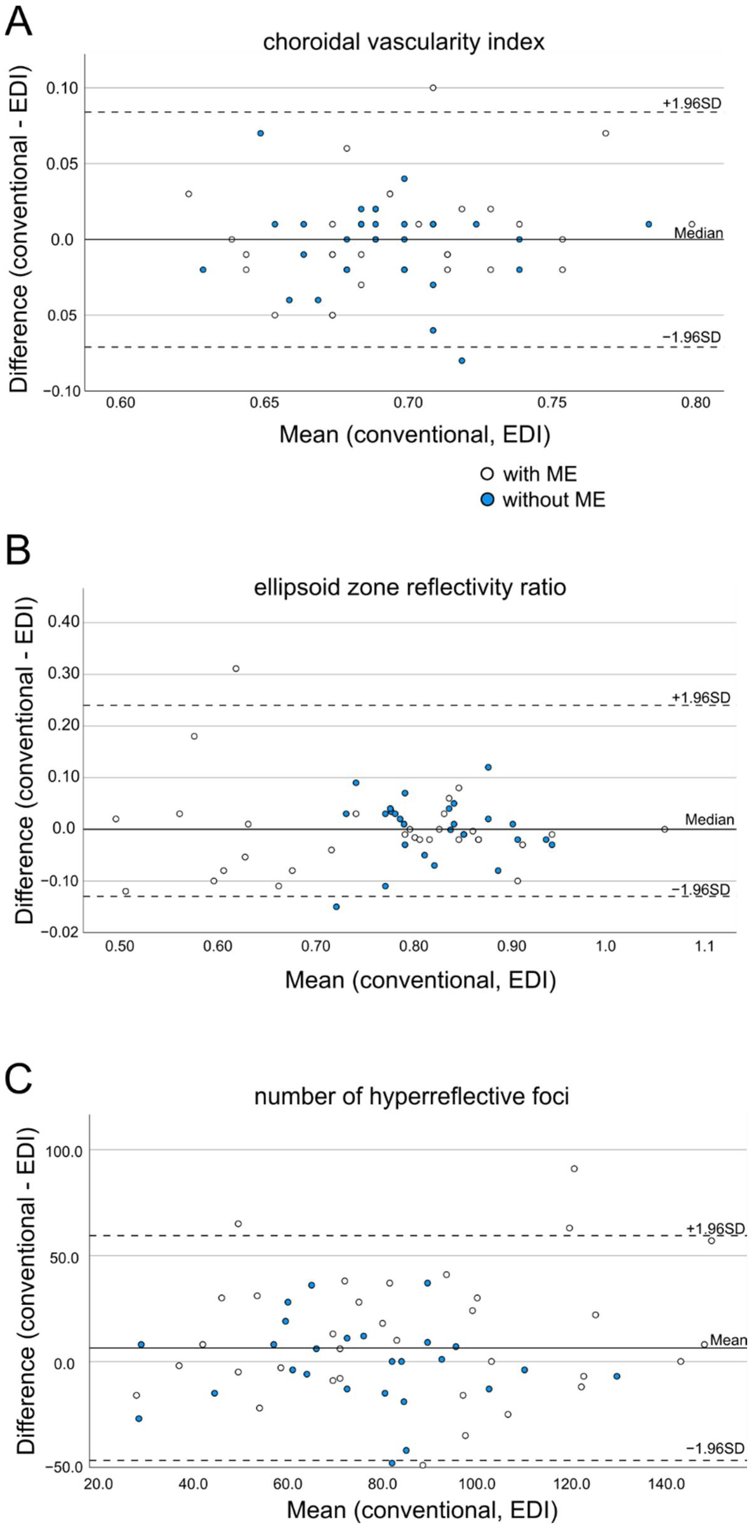
| OCT Biomarker | Group | Without EDI (Mean ± SD) | With EDI (Mean ± SD) | Mean Difference | p-Value * | ICC (95% CI) |
|---|---|---|---|---|---|---|
| Hyperreflective foci (In number) | Overall | 78.30 ± 30.29 | 84.63 ± 33.71 | 6.30 | 0.07 ° | 0.78 (0.62–0.87) |
| With ME | 80.00 ± 33.70 | 92.08 ± 38.11 | 12.09 | 0.03 ° | 0.76 (0.52–0.88) | |
| Without ME | 76.07 ± 25.56 | 74.89 ± 24.30 | −1.19 | 0.76 ° | 0.80 (0.56–0.91) | |
| Ellipsoid zone reflectivity ratio (arbitrary unit) | Overall | 0.78 ± 0.12 | 0.78 ± 0.12 | 0.00 | 0.60 * | 0.90 (0.84–0.94) |
| With ME | 0.76 ± 0.14 | 0.75 ± 0.15 | 0.00 | 0.31 ° | 0.91 (0.83–0.96) | |
| Without ME | 0.82 ± 0.07 | 0.83 ± 0.06 | 0.00 | 0.69 ° | 0.77 (0.47–0.89) | |
| Choroidal vascularity index (arbitrary unit) | Overall | 0.69 ± 0.03 | 0.69 ± 0.04 | 0.00 | 0.91 * | 0.80 (0.67–0.88) |
| With ME | 0.70 ± 0.37 | 0.70 ± 0.45 | 0.00 | 0.39 ° | 0.83 (0.66–0.91) | |
| Without ME | 0.70 ± 0.04 | 0.69 ± 0.03 | 0.00 | 0.50 ° | 0.77 (0.48–0.90) |
Publisher’s Note: MDPI stays neutral with regard to jurisdictional claims in published maps and institutional affiliations. |
© 2022 by the authors. Licensee MDPI, Basel, Switzerland. This article is an open access article distributed under the terms and conditions of the Creative Commons Attribution (CC BY) license (https://creativecommons.org/licenses/by/4.0/).
Share and Cite
Kessler, L.J.; Bagautdinov, D.; Łabuz, G.; Auffarth, G.U.; Khoramnia, R. Semi-Automated Quantification of Retinal and Choroidal Biomarkers in Retinal Vascular Diseases: Agreement of Spectral-Domain Optical Coherence Tomography with and without Enhanced Depth Imaging Mode. Diagnostics 2022, 12, 333. https://doi.org/10.3390/diagnostics12020333
Kessler LJ, Bagautdinov D, Łabuz G, Auffarth GU, Khoramnia R. Semi-Automated Quantification of Retinal and Choroidal Biomarkers in Retinal Vascular Diseases: Agreement of Spectral-Domain Optical Coherence Tomography with and without Enhanced Depth Imaging Mode. Diagnostics. 2022; 12(2):333. https://doi.org/10.3390/diagnostics12020333
Chicago/Turabian StyleKessler, Lucy J., Dmitrii Bagautdinov, Grzegorz Łabuz, Gerd U. Auffarth, and Ramin Khoramnia. 2022. "Semi-Automated Quantification of Retinal and Choroidal Biomarkers in Retinal Vascular Diseases: Agreement of Spectral-Domain Optical Coherence Tomography with and without Enhanced Depth Imaging Mode" Diagnostics 12, no. 2: 333. https://doi.org/10.3390/diagnostics12020333
APA StyleKessler, L. J., Bagautdinov, D., Łabuz, G., Auffarth, G. U., & Khoramnia, R. (2022). Semi-Automated Quantification of Retinal and Choroidal Biomarkers in Retinal Vascular Diseases: Agreement of Spectral-Domain Optical Coherence Tomography with and without Enhanced Depth Imaging Mode. Diagnostics, 12(2), 333. https://doi.org/10.3390/diagnostics12020333