

Right Ventricular Strain by Magnetic Resonance Feature Tracking Is Largely Afterload-Dependent and Does Not Reflect Contractility: Validation by Combined Volumetry and Invasive Pressure Tracings
 , , ,
, , ,
Abstract
:1. Introduction
2. Materials and Methods
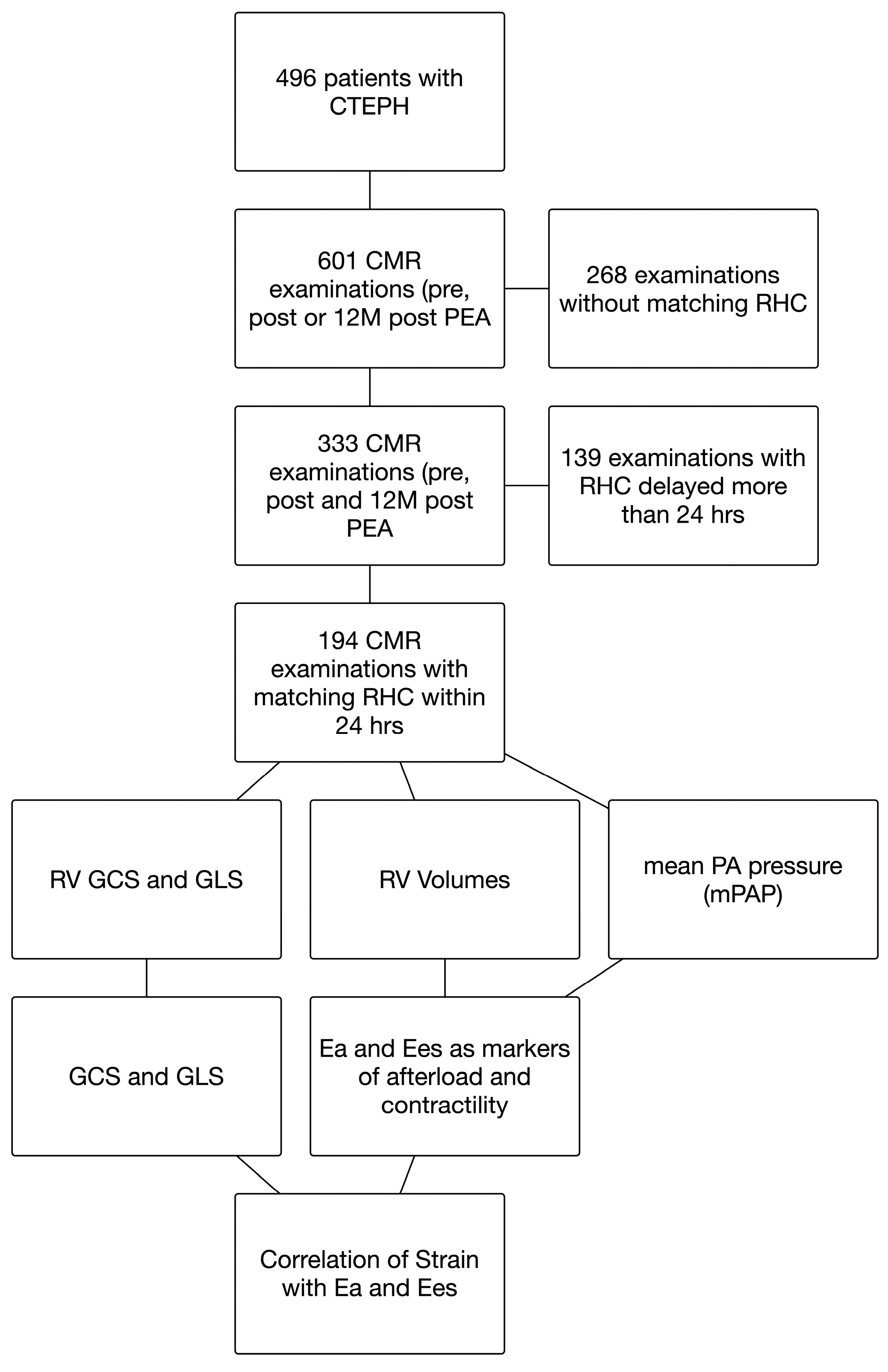
2.1. Patients and Ethics Statement
2.2. Hemodynamic Background and Formulas
2.3. CMR Acquisition
2.4. Feature Tracking
2.5. Right Heart Catheterization
2.6. Statistics
3. Results
3.1. Patient Characteristics
3.2. Association between Strain and Physiological Parameters of Afterload, Contractility, and Coupling
3.3. Association between Strain and Hemodynamic/Volumetric Data or NT-proBNP
4. Discussion
- (1)
- RV longitudinal and circumferential strain correlated well with afterload as represented by Ea in both afterload conditions (before and after PEA);
- (2)
- RV longitudinal and circumferential strain did not correlate with contractility in the whole cohort; however, there was a significant and modest correlation in the setting of elevated pulmonary pressure before PEA. After PEA, there was no correlation between contractility and strain in CTEPH patients;
- (3)
- Longitudinal and circumferential strain correlated well with ventriculoarterial coupling both before and after PEA.
Limitations
5. Conclusions and Clinical Perspective
Supplementary Materials
Author Contributions
Funding
Institutional Review Board Statement
Informed Consent Statement
Data Availability Statement
Acknowledgments
Conflicts of Interest
References
- Galie, N.; Humbert, M.; Vachiéry, J.-L.; Gibbs, S.; Lang, I.M.; Torbicki, A.; Simonneau, G.; Peacock, A.; Noordegraaf, A.V.; Beghetti, M.; et al. 2015 ESC/ERS guidelines for the diagnosis and treatment of pulmonary hypertension. Russ. J. Cardiol. 2016, 37, 5–64. [Google Scholar] [CrossRef] [Green Version]
- Peacock, A.J.; Crawley, S.; McLure, L.; Blyth, K.; Vizza, C.D.; Poscia, R.; Francone, M.; Iacucci, I.; Olschewski, H.; Kovacs, K.; et al. Changes in right ventricular function measured by cardiac magnetic resonance imaging in patients receiving pulmonary arterial hypertension-targeted therapy: The EURO-MR study. Circ. Cardiovasc. Imaging 2014, 7, 107–114. [Google Scholar] [CrossRef] [PubMed] [Green Version]
- Sato, T.; Ambale-Venkatesh, B.; Zimmerman, S.L.; Tedford, R.J.; Hsu, S.; Chamera, E.; Fujii, T.; Mullin, C.J.; Mercurio, V.; Khair, R.; et al. Right ventricular function as assessed by cardiac magnetic resonance imaging-derived strain parameters compared to high-fidelity micromanometer catheter measurements. Pulm. Circ. 2021, 11, 1–10. [Google Scholar] [CrossRef] [PubMed]
- Claus, P.; Omar, A.M.S.; Pedrizzetti, G.; Sengupta, P.P.; Nagel, E. Tissue Tracking Technology for Assessing Cardiac Mechanics: Principles, Normal Values, and Clinical Applications. JACC Cardiovasc. Imaging 2015, 8, 1444–1460. [Google Scholar] [CrossRef] [Green Version]
- Erley, J.; Tanacli, R.; Genovese, D.; Tapaskar, N.; Rashedi, N.; Bucius, P.; Kawaji, K.; Karagodin, I.; Lang, R.M.; Kelle, S.; et al. Myocardial strain analysis of the right ventricle: Comparison of different cardiovascular magnetic resonance and echocardiographic techniques. J. Cardiovasc. Magn. Reson. 2020, 22, 1–12. [Google Scholar] [CrossRef]
- Lahm, T.; Douglas, I.S.; Archer, S.L.; Bogaard, H.J.; Chesler, N.; Haddad, F.; Hemnes, A.R.; Kawut, S.M.; Kline, J.A.; Kolb, T.M.; et al. Assessment of Right Ventricular Function in the Research Setting: Knowledge Gaps and Pathways Forward. An Official American Thoracic Society Research Statement. Am. J. Respir. Crit. Care Med. 2018, 198, e15–e43. [Google Scholar] [CrossRef] [Green Version]
- Qu, Y.-Y.; Li, H.; Rottbauer, W.; Ma, G.-S.; Buckert, D.; Rasche, V. Right ventricular free wall longitudinal strain and strain rate quantification with cardiovascular magnetic resonance based tissue tracking. Int. J. Cardiovasc. Imaging 2020, 36, 1985–1996. [Google Scholar] [CrossRef]
- Rajiah, P.S.; Kalisz, K.; Broncano, J.; Goerne, H.; Collins, J.D.; François, C.J.; Ibrahim, E.-S.; Agarwal, P.P. Myocardial Strain Evaluation with Cardiovascular MRI: Physics, Principles, and Clinical Applications. RadioGraphics 2022, 42, 968–990. [Google Scholar] [CrossRef]
- Pedrizzetti, G.; Claus, P.; Kilner, P.J.; Nagel, E. Principles of cardiovascular magnetic resonance feature tracking and echocardiographic speckle tracking for informed clinical use. J. Cardiovasc. Magn. Reson. 2016, 18, 1–12. [Google Scholar] [CrossRef] [Green Version]
- Freed, B.H.; Collins, J.D.; François, C.J.; Barker, A.J.; Cuttica, M.J.; Chesler, N.C.; Markl, M.; Shah, S.J. MR and CT Imaging for the Evaluation of Pulmonary Hypertension. JACC Cardiovasc. Imaging 2016, 9, 715–732. [Google Scholar] [CrossRef]
- D’Andrea, A.; Stanziola, A.; D’Alto, M.; Di Palma, E.; Martino, M.; Scarafile, R.; Molino, A.; Rea, G.; Maglione, M.; Calabrò, R.; et al. Right ventricular strain: An independent predictor of survival in idiopathic pulmonary fibrosis. Int. J. Cardiol. 2016, 222, 908–910. [Google Scholar] [CrossRef] [PubMed]
- Guerra, F.; Gelardi, C.; Capucci, A.; Gabrielli, A.; Danieli, M.G. Subclinical Cardiac Dysfunction in Polymyositis and Dermatomyositis: A Speckle-tracking Case-control Study. J. Rheumatol. 2017, 44, 815–821. [Google Scholar] [CrossRef] [PubMed]
- Fine, N.M.; Chen, L.; Bastiansen, P.M.; Frantz, R.P.; Pellikka, P.A.; Oh, J.K.; Kane, G.C. Outcome Prediction by Quantitative Right Ventricular Function Assessment in 575 Subjects Evaluated for Pulmonary Hypertension. Circ. Cardiovasc. Imaging 2013, 6, 711–721. [Google Scholar] [CrossRef] [PubMed] [Green Version]
- Moceri, P.; Duchateau, N.; Baudouy, D.; Schouver, E.-D.; Leroy, S.; Squara, F.; Ferrari, E.; Sermesant, M. Three-dimensional right-ventricular regional deformation and survival in pulmonary hypertension. Eur. Hear. J.—Cardiovasc. Imaging 2017, 19, 450–458. [Google Scholar] [CrossRef] [PubMed] [Green Version]
- Bourfiss, M.; Prakken, N.H.J.; James, C.A.; Planken, R.N.; Boekholdt, S.M.; Ahmetagic, D.; Berg, M.P.V.D.; Tichnell, C.; Van der Heijden, J.F.; Loh, P.; et al. Prognostic value of strain by feature-tracking cardiac magnetic resonance in arrhythmogenic right ventricular cardiomyopathy. Eur. Hear. J.—Cardiovasc. Imaging 2022. [Google Scholar] [CrossRef] [PubMed]
- Schmid, J.; Kamml, C.; Zweiker, D.; Hatz, D.; Schmidt, A.; Reiter, U.; Toth, G.G.; Fuchsjäger, M.; Zirlik, A.; Binder, J.S.; et al. Cardiac Magnetic Resonance Imaging Right Ventricular Longitudinal Strain Predicts Mortality in Patients Undergoing TAVI. Front. Cardiovasc. Med. 2021, 8, 644500. [Google Scholar] [CrossRef] [PubMed]
- Shanmuganathan, M.; Rajani, P.; Androulakis, E.; Moledina, S.; Sarri, G.; Robertus, J.; Kiff, K.; Baston, V.; Dar, O.; Riesgo-Gil, F.; et al. Predicting Significant Right Ventricular Failure Post-LVAD Implantation Using CMR Compared to Echocardiography and Right Heart Catheterisation. J. Hear. Lung Transplant. 2021, 40, S438. [Google Scholar] [CrossRef]
- Srinivasan, R.; Faerber, J.A.; DeCost, G.; Zhang, X.; DiLorenzo, M.; Goldmuntz, E.; Fogel, M.; Mercer-Rosa, L. Right Ventricular Strain Is Associated With Increased Length of Stay After Tetralogy of Fallot Repair. J. Cardiovasc. Imaging 2022, 30, 50–58. [Google Scholar] [CrossRef]
- Lejeune, S.; Roy, C.; Ciocea, V.; Slimani, A.; de Meester, C.; Amzulescu, M.; Pasquet, A.; Vancraeynest, D.; Beauloye, C.; Vanoverschelde, J.-L.; et al. Right Ventricular Global Longitudinal Strain and Outcomes in Heart Failure with Preserved Ejection Fraction. J. Am. Soc. Echocardiogr. 2020, 33, 973–984.e2. [Google Scholar] [CrossRef]
- Romano, S.; Dell’atti, D.; Judd, R.M.; Kim, R.J.; Weinsaft, J.W.; Kim, J.; Kim, J.; Heitner, J.F.; Hahn, R.T.; Farzaneh-Far, A. Prognostic Value of Feature-Tracking Right Ventricular Longitudinal Strain in Severe Functional Tricuspid Regurgitation: A Multicenter Study. JACC Cardiovasc. Imaging 2021, 14, 1561–1568. [Google Scholar] [CrossRef]
- Liu, T.; Gao, Y.; Wang, H.; Zhou, Z.; Wang, R.; Chang, S.-S.; Liu, Y.; Sun, Y.; Rui, H.; Yang, G.; et al. Association between right ventricular strain and outcomes in patients with dilated cardiomyopathy. Heart 2020, 107, 1233–1239. [Google Scholar] [CrossRef] [PubMed]
- Reichek, N. Right ventricular strain in pulmonary hypertension: Flavor du jour or enduring prognostic index? Circ. Cardiovasc. Imaging 2013, 6, 609–611. [Google Scholar] [CrossRef] [PubMed] [Green Version]
- Ferferieva, V.; Bergh, A.V.D.; Claus, P.; Jasaityte, R.; Veulemans, P.; Pellens, M.; La Gerche, A.; Rademakers, F.; Herijgers, P.; D’Hooge, J. The relative value of strain and strain rate for defining intrinsic myocardial function. Am. J. Physiol. Circ. Physiol. 2012, 302, H188–H195. [Google Scholar] [CrossRef] [PubMed]
- Weidemann, F.; Jamal, F.; Sutherland, G.R.; Claus, P.; Kowalski, M.; Hatle, L.; De Scheerder, I.; Bijnens, B.; Rademakers, F.E. Myocardial function defined by strain rate and strain during alterations in inotropic states and heart rate. Am. J. Physiol. Circ. Physiol. 2002, 283, H792–H799. [Google Scholar] [CrossRef] [Green Version]
- Tello, K.; Dalmer, A.; Vanderpool, R.; Ghofrani, H.A.; Naeije, R.; Roller, F.; Seeger, W.; Wilhelm, J.; Gall, H.; Richter, M.J. Cardiac Magnetic Resonance Imaging-Based Right Ventricular Strain Analysis for Assessment of Coupling and Diastolic Function in Pulmonary Hypertension. JACC Cardiovasc. Imaging 2019, 12, 2155–2164. [Google Scholar] [CrossRef]
- Rolf, A.; Rixe, J.; Kim, W.K.; Börgel, J.; Möllmann, H.; Nef, H.M.; Liebetrau, C.; Kramm, T.; Guth, S.; Krombach, G.A.; et al. Right ventricular adaptation to pulmonary pressure load in patients with chronic thromboembolic pulmonary hypertension before and after successful pulmonary endarterectomy--a cardiovascular magnetic resonance study. J. Cardiovasc. Magn. Reson. 2014, 16, 96. [Google Scholar] [CrossRef] [Green Version]
- Sanz, J.; García-Alvarez, A.; Friera, L.F.; Nair, A.; Mirelis, J.G.; Sawit, S.T.; Pinney, S.; Fuster, V. Right ventriculo-arterial coupling in pulmonary hypertension: A magnetic resonance study. Heart 2011, 98, 238–243. [Google Scholar] [CrossRef]
- Kuehne, T.; Yilmaz, S.; Steendijk, P.; Moore, P.; Groenink, M.; Saaed, M.; Weber, O.; Higgins, C.B.; Ewert, P.; Fleck, E.; et al. Magnetic resonance imaging analysis of right ventricular pressure-volume loops: In vivo validation and clinical application in patients with pulmonary hypertension. Circulation 2004, 110, 2010–2016. [Google Scholar] [CrossRef] [Green Version]
- Naeije, R.; Huez, S. Right ventricular function in pulmonary hypertension: Physiological concepts. Eur. Hear. J. Suppl. 2007, 9, H5–H9. [Google Scholar] [CrossRef] [Green Version]
- Bishop, A.; White, P.; Oldershaw, P.; Chaturvedi, R.; Brookes, C.; Redington, A. Clinical application of the conductance catheter technique in the adult human right ventricle. Int. J. Cardiol. 1997, 58, 211–221. [Google Scholar] [CrossRef]
- Sagawa, K. The end-systolic pressure-volume relation of the ventricle: Definition, modifications and clinical use. Circulation 1981, 63, 1223–1227. [Google Scholar] [CrossRef] [PubMed] [Green Version]
- Morimont, P.; Lambermont, B.; Ghuysen, A.; Gerard, P.; Kolh, P.; Lancellotti, P.; Tchana-Sato, V.; Desaive, T.; D’Orio, V. Effective arterial elastance as an index of pulmonary vascular load. Am. J. Physiol. Circ. Physiol. 2008, 294, H2736–H2742. [Google Scholar] [CrossRef] [PubMed] [Green Version]
- Marrone, G.; Mamone, G.; Luca, A.; Vitulo, P.; Bertani, A.; Pilato, M.; Gridelli, B. The role of 1.5T cardiac MRI in the diagnosis, prognosis and management of pulmonary arterial hypertension. Int. J. Cardiovasc. Imaging 2010, 26, 665–681. [Google Scholar] [CrossRef] [PubMed]
- Truong, V.T.; Safdar, K.S.; Kalra, D.K.; Gao, X.; Ambach, S.; Taylor, M.D.; Moore, R.; Taylor, R.J.; Germann, J.; Toro-Salazar, O.; et al. Cardiac magnetic resonance tissue tracking in right ventricle: Feasibility and normal values. Magn. Reson. Imaging 2017, 38, 189–195. [Google Scholar] [CrossRef] [Green Version]
- Guihaire, J.; Haddad, F.; Boulate, D.; Decante, B.; Denault, A.Y.; Wu, J.; Hervé, P.; Humbert, M.; Dartevelle, P.; Verhoye, J.-P.; et al. Non-invasive indices of right ventricular function are markers of ventricular-arterial coupling rather than ventricular contractility: Insights from a porcine model of chronic pressure overload. Eur. Hear. J.–Cardiovasc. Imaging 2013, 14, 1140–1149. [Google Scholar] [CrossRef]


| Patient Characteristics | Value 1 |
|---|---|
| General | |
| Age, years | 57.8 (45–71.2) |
| Female sex | 93 (48%) |
| NYHA class ≥ III 2 | 65 (38%) |
| 6 min walking distance, m | 438 (306–530) |
| NT-proBNP, pg/mL | 323 (117–864) |
| Hemodynamics | |
| mPAP, mmHg | 27 (19–42) |
| PVR, dyn/s/cm−5 | 252.5 (150–542) |
| Physiological data | |
| Ea, mmHg/mL | 0.52 (0.33–0.91) |
| Ees, mmHg/mL | 0.29 (0.22–0.43) |
| Ees/Ea ratio | 0.57 (0.37–0.84) |
| CMR strain | |
| GLS, % | −16.7 ± 5.5 |
| GCS, % | −12.1 ± 3.9 |
| CMR volumetry | |
| RV EDVi, mL/m2 | 77.1 (62–96.9) |
| RV ESVi, mL/m2 | 44.6 (35.8–60.1) |
| RV SVi, mL/m2 | 27.4 (21.6–34.8) |
| RV EF, % | 37 (27–46) |
| RVMASS, g | 53 (21–81.3) |
| Patient Characteristics | Before PEA 1 | After PEA | p-Value |
|---|---|---|---|
| General | |||
| Age, years | 57.9 (45.5–71.5) | 57.6 (44.6–70.9) | 0.83 |
| Female sex | 78 (44%) | 56 (46%) | 0.54 |
| NYHA class ≥ III 2 | 63 (88%) | 2 (2%) | 0.0001 |
| 6 min walking distance, m | 390 (285–470) | 480 (385–546) | 0.0003 |
| NT-proBNP, pg/mL | 801 (280–1583) | 226 (101–489) | 0.00001 |
| Hemodynamics | |||
| mPAP, mmHg | 43 (38–50) | 20 (17–27) | 0.00001 |
| PVR, WU | 7.7(5.6–9.8) | 2.2 (1.6–3.1) | 0.00001 |
| Physiological data | |||
| Ea, mmHg/mL | 0.97 (0.68–1.43) | 0.37 (0.28–0.54) | 0.00001 |
| Ees, mmHg/mL | 0.37 (0.26–0.5) | 0.27 (0.19–0.36) | 0.0001 |
| Ees/Ea ratio | 0.37 (0.26–0.6) | 0.68 (0.5–0.88) | 0.00001 |
| CMR strain | |||
| GLS, % | −13.9 ± 4.6 | −18.3 ± 5.4 | 0.00001 |
| GCS, % | −9.2 ± 3.4 | −13.7 ± 3.1 | 0.00001 |
| CMR volumetry | |||
| RV EDVi, mL/m2 | 82.2 (62–105) | 75.6 (61–86.2) | 0.049 |
| RV ESVi, mL/m2 | 57.3 (38.7–79.1) | 41.8 (35–50.3) | 0.0001 |
| RV SVi, mL/m2 | 25.6 (20.3–30.3) | 28.6 (22.1–37.3) | 0.02 |
| RV EF, % | 27 (15.5–37) | 41 (35–48) | 0.00001 |
| RVMASS, g | 81.5 (66–99.5) | 23 (18–41.8) | 0.00001 |
| Association | All Patients 1 | Before PEA | After PEA | |||
|---|---|---|---|---|---|---|
| rho | p | rho | p | rho | p | |
| GLS vs. Ea 2 | 0.5 | 0.00001 | 0.48 | 0.0004 | 0.5 | 0.0002 |
| GCS vs. Ea | 0.49 | 0.00001 | 0.3 | 0.03 | 0.34 | 0.014 |
| GLS vs. Ees | 0.05 | 0.51 | −0.48 | 0.0005 | 0.19 | 0.197 |
| GCS vs. Ees | −0.03 | 0.66 | −0.52 | 0.0001 | −0.1339 | 0.3537 |
| GLS vs. Ees/Ea | −0.56 | 0.00001 | −0.69 | 0.00001 | −0.47 | 0.0006 |
| GLS vs. RV EF | −0.53 | 0.00001 | −0.67 | 0.00001 | −0.37 | 0.008 |
| GCS vs. RV EF | −0.6 | 0.00001 | −0.64 | 0.00001 | −0.51 | 0.0002 |
| GLS vs. RV EDVi | 0.23 | 0.003 | 0.39 | 0.005 | −0.038 | 0.79 |
| GCS vs. RV EDVi | 0.27 | 0.0005 | 0.27 | 0.067 | 0.13 | 0.351 |
| GLS vs. RV ESVi | 0.39 | 0.00001 | 0.61 | 0.00001 | 0.17 | 0.24 |
| GCS vs. RV ESVi | 0.51 | 0.00001 | 0.49 | 0.0003 | 0.39 | 0.005 |
| GLS vs. RV SVi | −0.31 | 0.00001 | −0.48 | 0.0005 | −0.41 | 0.003 |
| GCS vs. RV SVi | −.23 | 0.0012 | −0.3 | 0.03 | −0.29 | 0.04 |
| GLS vs. mPAP | 0.47 | 0.00001 | 0.4 | 0.0045 | 0.34 | 0.016 |
| GCS vs. mPAP | 0.48 | 0.00001 | 0.18 | 0.21 | 0.27 | 0.05 |
| GLS vs. PVR | 0.52 | 0.00001 | 0.4 | 0.0065 | 0.47 | 0.001 |
| GCS vs. PVR | 0.48 | 0.00001 | 0.27 | 0.07 | 0.07 | 0.6 |
| GLS vs. NT-proBNP | 0.45 | 0.00001 | 0.6 | 0.00001 | 0.31 | 0.04 |
| GCS vs. NT-proBNP | 0.37 | 0.00001 | 0.34 | 0.03 | 0.23 | 0.12 |
Publisher’s Note: MDPI stays neutral with regard to jurisdictional claims in published maps and institutional affiliations. |
© 2022 by the authors. Licensee MDPI, Basel, Switzerland. This article is an open access article distributed under the terms and conditions of the Creative Commons Attribution (CC BY) license (https://creativecommons.org/licenses/by/4.0/).
Share and Cite
Rolf, A.; Keller, T.; Wolter, J.S.; Kriechbaum, S.; Weferling, M.; Guth, S.; Wiedenroth, C.; Mayer, E.; Hamm, C.W.; Fischer-Rasokat, U.; et al. Right Ventricular Strain by Magnetic Resonance Feature Tracking Is Largely Afterload-Dependent and Does Not Reflect Contractility: Validation by Combined Volumetry and Invasive Pressure Tracings. Diagnostics 2022, 12, 3183. https://doi.org/10.3390/diagnostics12123183
Rolf A, Keller T, Wolter JS, Kriechbaum S, Weferling M, Guth S, Wiedenroth C, Mayer E, Hamm CW, Fischer-Rasokat U, et al. Right Ventricular Strain by Magnetic Resonance Feature Tracking Is Largely Afterload-Dependent and Does Not Reflect Contractility: Validation by Combined Volumetry and Invasive Pressure Tracings. Diagnostics. 2022; 12(12):3183. https://doi.org/10.3390/diagnostics12123183
Chicago/Turabian StyleRolf, Andreas, Till Keller, Jan Sebastian Wolter, Steffen Kriechbaum, Maren Weferling, Stefan Guth, Christoph Wiedenroth, Eckhard Mayer, Christian W. Hamm, Ulrich Fischer-Rasokat, and et al. 2022. "Right Ventricular Strain by Magnetic Resonance Feature Tracking Is Largely Afterload-Dependent and Does Not Reflect Contractility: Validation by Combined Volumetry and Invasive Pressure Tracings" Diagnostics 12, no. 12: 3183. https://doi.org/10.3390/diagnostics12123183
APA StyleRolf, A., Keller, T., Wolter, J. S., Kriechbaum, S., Weferling, M., Guth, S., Wiedenroth, C., Mayer, E., Hamm, C. W., Fischer-Rasokat, U., & Treiber, J. (2022). Right Ventricular Strain by Magnetic Resonance Feature Tracking Is Largely Afterload-Dependent and Does Not Reflect Contractility: Validation by Combined Volumetry and Invasive Pressure Tracings. Diagnostics, 12(12), 3183. https://doi.org/10.3390/diagnostics12123183