
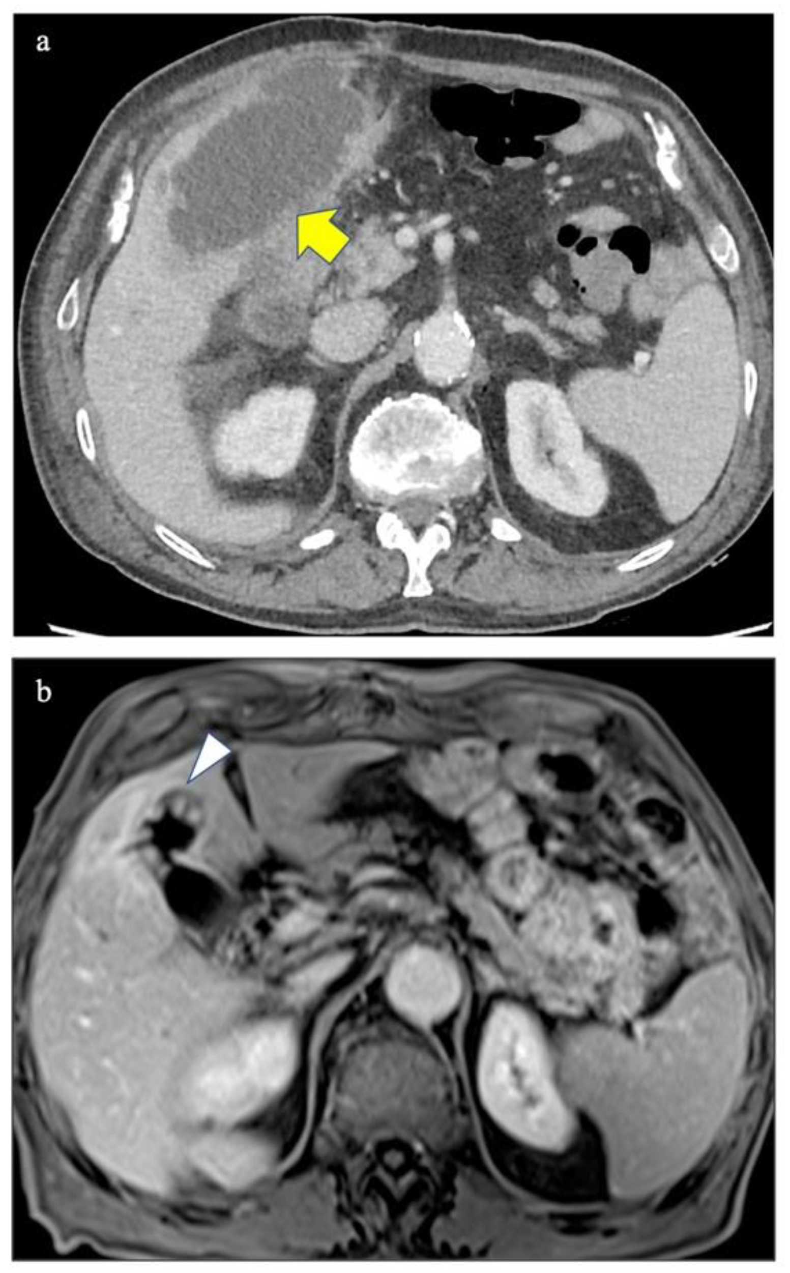
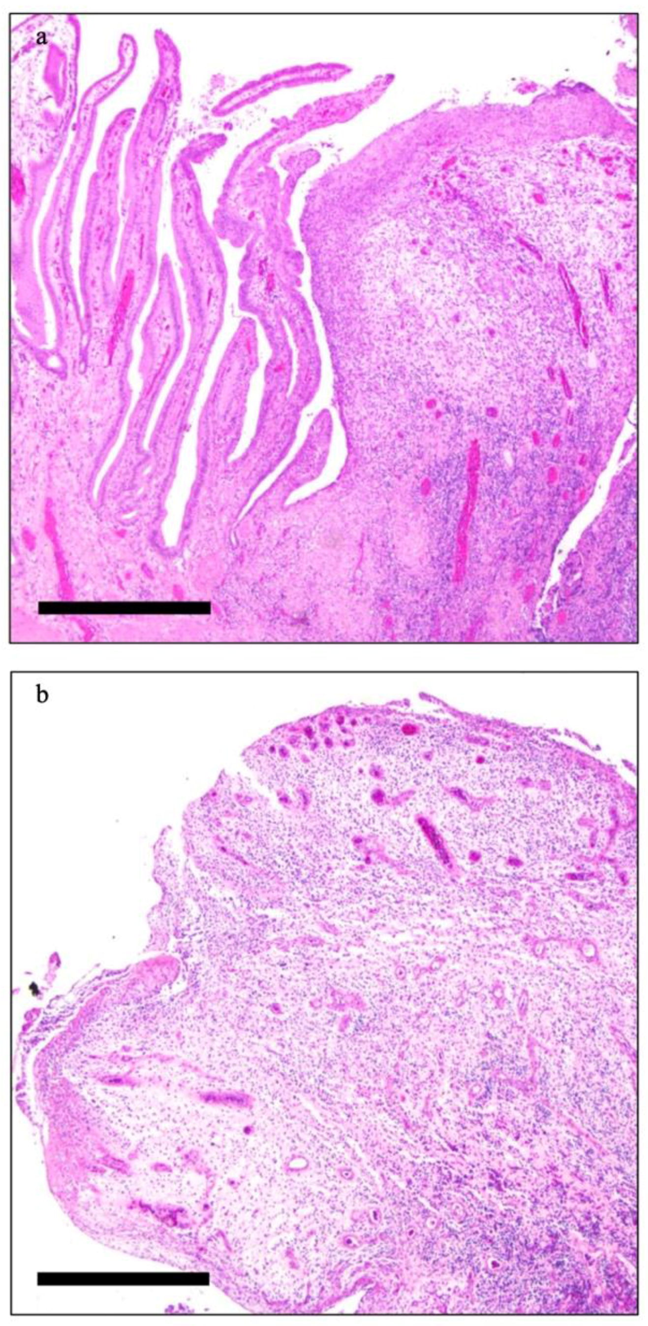
Coronary Artery Disease and Gallbladder Inflammatory Pseudopolyps


 and
and
Abstract
:Author Contributions
Funding
Institutional Review Board Statement
Informed Consent Statement
Conflicts of Interest
References
- Tana, M.; Tana, C.; Cocco, G.; Iannetti, G.; Romano, M.; Schiavone, C. Acute acalculous cholecystitis and cardiovascular disease: A land of confusion. J. Ultrasound 2015, 18, 317–320. [Google Scholar] [CrossRef] [PubMed] [Green Version]
- Cher, D.J. Myocardial Infarction and Acute Cholecystitis: An Application of Sequence Symmetry Analysis. Epidemiology 2000, 11, 446–449. [Google Scholar] [CrossRef] [PubMed]
- Rezkallah, K.N.; Barakat, K.; Farrah, A.; Rao, S.; Sharma, M.; Chalise, S.; Zdunek, T. Acute Acalculous Cholecystitis due to primary acute Epstein-Barr virus infection treated with laparoscopic cholecystectomy: A case report. Ann. Med. Surg. 2018, 35, 189–191. [Google Scholar] [CrossRef] [PubMed]
- Huffmann, J.L.; Schenker, S. Acute acalculous cholecystitis: A review. Clin. Gastroenterol. Hepatol. 2010, 8, 15–22. [Google Scholar] [CrossRef]
- Chen, P.F.; Nimeri, A.; Pham, Q.H.; Yuh, J.N.; Gusz, J.R.; Chung, R.S. The clinical diagnosis of chronic acalculous cholecystitis. Surgery 2001, 130, 578–581. [Google Scholar] [CrossRef]
- Wiles, R.; Thoeni, R.F.; Barbu, S.T.; Vashist, Y.K.; Rafaelsen, S.R.; Dewhurst, C.; Arvanitakis, M.; Lahaye, M.; Soltes, M.; Perinel, J.; et al. Management and follow-up of gallbladder polyps. Eur. Radiol. 2017, 27, 3856–3866. [Google Scholar] [CrossRef]
- Maheshwarappa, R.P.; Menda, Y.; Graham, M.M.; Boukhar, S.A.; Zamba, G.K.D.; Samuel, I. Association of gallbladder hyperkinesia with acalculous chronic cholecystitis: A case-control study. Surgery 2020, 168, 800–808. [Google Scholar] [CrossRef]
- Poddighe, D.; Sazonov, V. Acute acalculous cholecystitis in children. World J. Gastroenterol. 2018, 24, 4870–4879. [Google Scholar] [CrossRef]
- Mellnick, V.M.; Menias, C.O.; Sandrasegaran, K.; Hara, A.K.; Kielar, A.Z.; Brunt, E.M.; Doyle, M.B.M.; Dahiya, N.; Elsayes, K.M. Polypoid lesions of the gallbladder: Disease spectrum with pathologic correlation. Radiographics 2015, 35, 387–399. [Google Scholar] [CrossRef]
- Kwon, W.; Jang, J.Y.; Lee, S.E.; Hwang, D.W.; Kim, S. Clinicopathologic features of polypoid lesions of the gallbladder and risk factors of gallbladder cancer. J. Korean Med. Sci. 2009, 24, 481–487. [Google Scholar] [CrossRef] [Green Version]
- Lee, K.F.; Wong, J.; Li, J.C.; Lai, P.B.S. Polypoid lesions of the gallbladder. Am. J. Surg. 2004, 188, 186–190. [Google Scholar] [CrossRef]
- Welling, R.E.; Rath, R.; Albers, J.E.; Glaser, R.S. Gastrointestinal complications after cardiac surgery. Arch. Surg. 1986, 121, 1178–1180. [Google Scholar] [CrossRef]
- Varma, D.G.; Faust, J.M. Computed tomography of gangrenous acute postoperative acalculous cholecystitis. J. Comput. Tomogr. 1988, 12, 29–31. [Google Scholar] [CrossRef]
- Berger, H.; Pratschke, E.; Arbogast, H.; Stäbler, A. Percutaneous cholecystostomy in acute acalculous cholecystitis. Hepatogastroenterology 1989, 36, 346–348. [Google Scholar] [PubMed]
- Teranishi, K.; Murase, M.; Maeda, M.; Murakami, F. A case of acute hemorrhagic gangrenous acalculous cholecystitis with bile peritonitis during anti-coagulant therapy after coronary-artery bypass grafting. Nihon Kyobu Geka Gakkai 1993, 41, 83–87. [Google Scholar]
- Saito, A.; Shirai, Y.; Ohzeki, H.; Hayashi, J.; Eguchi, S. Acute acalculous cholecystitis after cardiovascular surgery. Surg. Today 1997, 27, 907–909. [Google Scholar] [CrossRef] [PubMed]
- Fujiii, H.; Kubo, S.; Tokuhara, T.; Suehiro, S.; Yamamoto, T.; Kinoshita, H. Acute acalculous cholecystitis complicated by penetration into the liver after coronary artery bypass grafting. Jpn. J. Thorac. Cardiovasc. Surg. 1999, 47, 518–521. [Google Scholar] [CrossRef] [PubMed]
- Funabiki, K.; Masuoka, H.; Shimizu, H.; Emi, Y.; Mori, T.; Ito, M.; Nakano, T. Cholesterol crystal embolization (CCE) after cardiac catheterization: A case report and a review of 36 cases in the Japanese literature. Jpn. Heart J. 2003, 44, 767–774. [Google Scholar] [CrossRef] [Green Version]
- Chen, C.J.; Huang, F.C.; Tiao, M.M.; Huang, Y.H.; Lin, L.Y.; Yu, H.R.; Yang, K.D.; Huang, Y.C.; Chen, C.C.; Chang, W.C.; et al. Sonographic gallbladder ab-normality is associated with intravenous immunoglobulin resistance in Kawasaki disease. Sci. World J. 2012, 2012, 485758. [Google Scholar] [CrossRef] [Green Version]
- Van Steijn, J.H.M.; Roeloffzen, W.W.H.; Appeltans, B.M.G.; Jager, P.L.; Gans, R.O.B.; Bijl, M. Acute acalculous cholecystitis: Not only in the intensive care department. Ned. Tijdschr. Voor Geneeskd. 2002, 146, 1305–1308. [Google Scholar]
- Yi, D.; Kim, J.Y.; Choi, E.Y.; Choi, J.Y.; Yang, H.R. Hepatobiliary risk factors for clinical outcome of Kawasaki disease in children. BMC Pediatr. 2014, 14, 51. [Google Scholar] [CrossRef] [Green Version]
- Kang, D.W.; Kim, S.H. Clinical aspects of splenomegaly as a possible predictive factor of coronary artery changes in Kawasaki disease. Cardiol. Young 2019, 29, 297–302. [Google Scholar] [CrossRef] [PubMed]
- Lipe, D.N.; Bridges, L.C. Kawasaki Disease Presenting as Acute Acalculous Cholecystitis. Clin. Pract. Cases Emerg. Med. 2019, 3, 383–386. [Google Scholar] [CrossRef] [PubMed]
- Chen, B.Q.; Chen, G.D.; Xie, F.; Li, X.; Mao, L.; Jia, B. Percutaneous cholecystostomy as a definitive treatment for moderate and severe acute acalculous cholecystitis: A retrospective observational study. BMC Surg. 2021, 21, 439. [Google Scholar] [CrossRef]
- Shapiro, M.J.; Luchtefeld, W.B.; Kurzweil, S.; Kaminski, D.L.; Durham, R.M.; Mazuski, J.E. Acute acalculous cholecystitis in the critically ill. Am. Surg. 1994, 60, 335–339. [Google Scholar]
- Tsuboi, I.; Hayashi, M.; Miyauchi, Y.; Iwasaki, Y.K.; Yodogawa, K.; Hayashi, H.; Uetake, S.; Takahashi, K.; Shimizu, W. Anatomical factors associated with periesophageal vagus nerve injury after catheter ablation of atrial fibrillation. J. Nippon. Med. Sch. 2014, 81, 248–257. [Google Scholar] [CrossRef] [PubMed] [Green Version]
- Udekwu, P.O.; Sullivan, W.G. Contemporary experience with cholecystectomy: Establishing ‘benchmarks’ two decades after the introduction of laparoscopic cholecystectomy. Am. Surg. 2013, 79, 1253–1257. [Google Scholar] [CrossRef]
- Owen, C.; Bilhartz, L.E. Gallbladder polyps, cholesterolosis, adenomyomatosis, and acute acalculous cholecystitis. Semin. Gastrointest. Dis. 2003, 14, 178–188. [Google Scholar]


| Publication Year | First Author | Title | Study Type | Number of Cases | Main Findings Related to AAC and Coronary Artery Disease |
|---|---|---|---|---|---|
| 1986 | Welling, R.E., et al. [12] | Gastrointestinal complications after cardiac surgery | Original Article | 18 patients out of 1596 who underwent coronary artery bypass or valve replacement. had gastrointestinal complications | One patient underwent cholecystectomy for AAC |
| 1988 | Varma, D.G., et al. [13] | Computed tomography of gangrenous acute postoperative acalculous cholecystitis | Case report | 1 | Gangrenous AAC after two-vessel coronary artery bypass surgery |
| 1989 | Berger, H. et al. [14] | Percutaneous cholecystostomy in acute acalculous cholecystitis | Original Article | 8 | PC was successful in all patients with AAC. One patient had in anamnesis a coronary bypass operation |
| 1993 | Teranishi, K., et al. # [15] | A case of acute hemorrhagic gangrenous acalculous cholecystitis with bile peritonitis during anti-coagulant therapy after coronary-artery bypass grafting | Case report | 1 | A case of acute hemorrhagic, gangrenous acalculous cholecystitis after coronary-artery bypass grafting. Post-operative stasis of bile, swelling of the gallbladder, hypotension during cardiopulmonary bypass, and post-operative anti-coagulant therapy administered after open heart surgery have been proposed as etiological factors |
| 1997 | Saito, A., et al. [16] | Acute acalculous cholecystitis after cardiovascular surgery | Original Article | 6 | Examination of six cases of AAC after cardiovascular surgery and AAC. The authors suggest that post-surgical hypoperfusion of the gallbladder due to various factors may be the cause |
| 1999 | Fujiii, H., et al. [17] | Acute acalculous cholecystitis complicated by penetration into the liver after coronary artery bypass grafting | Case report | 1 | AAC with penetration into the liver in a 71-year-old woman 21 days after coronary artery bypass grafting. At histology, partial obstruction of the cystic artery due to atherosclerosis was found |
| 2003 | Funabiki, K., et al. [18] | Cholesterol crystal embolization (CCE) after cardiac catheterization: a case report and a review of 36 cases in the Japanese literature | Case Report | 1 | A 67-year old man developed AAC 12 days after coronary angiography which followed a previous coronary artery bypass grafting |
| 2012 | Chen, C.J., et al. [19] | Sonographic gallbladder abnormality is associated with intravenous immunoglobulin resistance in Kawasaki disease | Original Article | 93 children with Kawasaki Disease | Five children with KD out of 11 with pathologic findings at abdominal ultrasound had AAC. Overall pathologic findings at US in children with KD seem to be associated with high levels of C-reactive protein, Glutamic-Pyruvic Transaminase, neutrophils and intravenous immunoglobulin resistance |
| 2012 | Van Stejin, J.H.M., et al. # [20] | Acute acalculous cholecystitis: not only in the intensive care department | Case Reports | 2 | Two patients with AAC are reported; one of them admitted to the coronary unit because of atherosclerotic vascular disease then died of sepsis |
| 2014 | Yi, D., et al. [21] | Hepatobiliary risk factors for clinical outcome of Kawasaki disease in children | Original Article | 24 out of 67 children with KD had AAC | Coronary artery abnormalities were more frequent in patients with AAC |
| 2019 | Kang, W.D., et al. [22] | Clinical aspects of splenomegaly as a possible predictive factor of coronary artery changes in Kawasaki disease | Original Article | 77 out of 396 examined patients underwent abdominal ultrasound | There were no cases of AAC at ultrasound among all investigated patients |
| 2019 | Lipe, D.N., et al. [23] | Kawasaki Disease Presenting as Acute Acalculous Cholecystitis | Case report | 1 | Eight-year-old boy affected by KD and with AAC |
| 2021 | Chen, B.Q., et al. [24] | Percutaneous cholecystostomy as a definitive treatment for moderate and severe acute acalculous cholecystitis: a retrospective observational study | Original Article | 44 | In patients with moderate to severe AAC who underwent PC, coronary heart disease or congestive heart failure are independent risk factors for relapse |
Publisher’s Note: MDPI stays neutral with regard to jurisdictional claims in published maps and institutional affiliations. |
© 2022 by the authors. Licensee MDPI, Basel, Switzerland. This article is an open access article distributed under the terms and conditions of the Creative Commons Attribution (CC BY) license (https://creativecommons.org/licenses/by/4.0/).
Share and Cite
Fosio, M.; Cherobin, G.; Stramare, R.; Fassan, M.; Giraudo, C. Coronary Artery Disease and Gallbladder Inflammatory Pseudopolyps. Diagnostics 2022, 12, 155. https://doi.org/10.3390/diagnostics12010155
Fosio M, Cherobin G, Stramare R, Fassan M, Giraudo C. Coronary Artery Disease and Gallbladder Inflammatory Pseudopolyps. Diagnostics. 2022; 12(1):155. https://doi.org/10.3390/diagnostics12010155
Chicago/Turabian StyleFosio, Margherita, Giulia Cherobin, Roberto Stramare, Matteo Fassan, and Chiara Giraudo. 2022. "Coronary Artery Disease and Gallbladder Inflammatory Pseudopolyps" Diagnostics 12, no. 1: 155. https://doi.org/10.3390/diagnostics12010155
APA StyleFosio, M., Cherobin, G., Stramare, R., Fassan, M., & Giraudo, C. (2022). Coronary Artery Disease and Gallbladder Inflammatory Pseudopolyps. Diagnostics, 12(1), 155. https://doi.org/10.3390/diagnostics12010155