
A Systematic Review of Methodology Used in Studies Aimed at Creating Charts of Fetal Brain Structures

 , ,
, ,
Abstract
1. Introduction
2. Materials and Methods
Statistical Analysis
3. Results
4. Discussion
Strengths and Limitations of the Review
5. Conclusions
Supplementary Materials
Author Contributions
Funding
Institutional Review Board Statement
Informed Consent Statement
Conflicts of Interest
References
- ISUOG Committee. ISUOG Practice Guidelines (Updated): Sonographic examination of the fetal central nervous system. Part 1: Performance of screening examination and indications for targeted neurosonography. Ultrasound Obs. Gynecol. 2020, 56, 476–484. [Google Scholar] [CrossRef]
- Namburete, A.I.; Stebbing, R.V.; Kemp, B.; Yaqub, M.; Papageorghiou, A.T.; Noble, A.J. Learning-based prediction of gestational age from ultrasound images of the fetal brain. Med. Image Anal. 2015, 21, 72–86. [Google Scholar] [CrossRef]
- Napolitano, R.; Dhami, J.; Ohuma, E.O.; Ioannou, C.; Conde-Agudelo, A.; Kennedy, S.H.; Villar, J.; Papageorghiou, A.T. Pregnancy dating by fetal crown-rump length: A systematic review of charts. BJOG 2014, 121, 556–565. [Google Scholar] [CrossRef]
- Ioannou, C.; Talbot, K.; Ohuma, E.; Sarris, I.; Villar, J.; Conde-Agudelo, A.; Papageorghiou, A.T. Systematic review of methodology used in ultrasound studies aimed at creating charts of fetal size. BJOG 2012, 119, 1425–1439. [Google Scholar] [CrossRef]
- Papageorghiou, A.T.; Ohuma, E.O.; Altman, D.G.; Todros, T.; Cheikh Ismail, L.; Lambert, A.; Jaffer, Y.A.; Bertino, E.; Gravett, M.G.; Purwar, M.; et al. International Fetal and Newborn Growth Consortium for the 21st Century (INTERGROWTH-21st). International standards for fetal growth based on serial ultrasound measurements: The Fetal Growth Longitudinal Study of the INTERGROWTH-21st Project. Lancet 2014, 384, 869–879. [Google Scholar] [CrossRef]
- Stewart, L.A.; Clarke, M.; Rovers, M.; Riley, R.D.; Simmonds, M.; Stewart, G.; Tierney, J.F. PRISMA-IPD Development Group. Preferred Reporting Items for Systematic Review and Meta-Analyses of individual participant data: The PRISMA-IPD Statement. JAMA 2015, 313, 1657–1665. [Google Scholar] [CrossRef] [PubMed]
- Alagappan, R.; Browning, P.D.; Laorr, A.; McGahan, J.P. Distal lateral ventricular atrium: Reevaluation of normal range. Radiology 1994, 193, 405–408. [Google Scholar] [CrossRef] [PubMed]
- Almog, B.; Gamzu, R.; Achiron, R.; Fainaru, O.; Zalel, Y. Fetal lateral ventricular width: What should be its upper limit? A prospective cohort study and reanalysis of the current and previous data. J. Ultrasound Med. 2003, 22, 39–43. [Google Scholar] [CrossRef] [PubMed]
- Alonso, I.; Borenstein, M.; Grant, G.; Narbona, I.; Azumendi, G. Depth of brain fissures in normal fetuses by prenatal ultrasound between 19 and 30 weeks of gestation. Ultrasound Obstet. Gynecol. 2010, 36, 693–699. [Google Scholar] [CrossRef] [PubMed]
- Alves, C.M.; Araujo Junior, E.; Nardozza, L.M.; Goldman, S.M.; Martinez, L.H.; Martins, W.P.; Oliveira, P.S.; Moron, A.F. Reference ranges for fetal brain fissure development on 3-dimensional sonography in the multiplanar mode. J. Ultrasound Med. 2013, 32, 269–277. [Google Scholar] [CrossRef] [PubMed]
- Araujo Junior, E.; Martins, W.P.; Rolo, L.C.; Pires, C.R.; Zanforlin Filho, S.M. Normative data for fetal cisterna magna length measurement between 18 and 24 weeks of pregnancy. Childs Nerv. Syst. 2014, 30, 9–12. [Google Scholar] [CrossRef] [PubMed]
- Araujo Junior, E.; Martins, W.P.; Nardozza, L.M.; Pires, C.R.; Filho, S.M. Reference range of fetal transverse cerebellar diameter between 18 and 24 weeks of pregnancy in a Brazilian population. J. Child Neurol. 2015, 30, 250–253. [Google Scholar] [CrossRef] [PubMed]
- Brown, R.N. Reassessment of the normal fetal cisterna magna during gestation and an alternative approach to the definition of cisterna magna dilatation. Fetal Diagn. Ther. 2013, 34, 44–49. [Google Scholar] [CrossRef] [PubMed]
- Cardoza, J.D.; Goldstein, R.B.; Filly, R.A. Exclusion of fetal ventriculomegaly with a single measurement: The width of the lateral ventricular atrium. Radiology 1988, 169, 711–714. [Google Scholar] [CrossRef] [PubMed]
- Chang, C.H.; Chang, F.M.; Yu, C.H.; Ko, H.C.; Chen, H.Y. Three-dimensional ultrasound in the assessment of fetal cerebellar transverse and antero-posterior diameters. Ultrasound Med. Biol. 2000, 26, 175–182. [Google Scholar] [CrossRef]
- Chavez, M.R.; Ananth, C.V.; Smulian, J.C.; Lashley, S.; Kontopoulos, E.V.; Vintzileos, A.M. Fetal transcerebellar diameter nomogram in singleton gestations with special emphasis in the third trimester: A comparison with previously published nomograms. Am. J. Obs. Gynecol. 2003, 189, 1021–1025. [Google Scholar] [CrossRef]
- Chen, X.; Li, S.L.; Luo, G.Y.; Norwitz, E.R.; Ouyang, S.Y.; Wen, H.X.; Yuan, Y.; Tian, X.X.; He, J.M. Ultrasonographic Characteristics of Cortical Sulcus Development in the Human Fetus between 18 and 41 Weeks of Gestation. Chin. Med. J. 2017, 130, 920–928. [Google Scholar] [CrossRef] [PubMed]
- Eze, C.U.; Onwuzu, Q.E.; Uchechukwu, N.I. Sonographic Reference Values for Fetal Transverse Cerebellar Diameter in the Second and Third Trimesters in a Nigerian Population. J. Diagn. Med. Sonogr. 2017, 33, 174–181. [Google Scholar] [CrossRef]
- Farrell, T.A.; Hertzberg, B.S.; Kliewer, M.A.; Harris, L.; Paine, S.S. Fetal lateral ventricles: Reassessment of normal values for atrial diameter at US. Radiology 1994, 193, 409–411. [Google Scholar] [CrossRef]
- Goel, P.; Singla, M.; Ghai, R.; Jain, S.; Budhiraja, V.; Babu, C.S.R. Transverse Cerebellar Diameter—A Marker for Estimation of Gestational Age. J. Anat. Soc. India 2010, 59, 158–161. [Google Scholar] [CrossRef]
- Goldstein, I.; Reece, E.A.; Pilu, G.; Bovicelli, L.; Hobbins, J.C. Cerebellar measurements with ultrasonography in the evaluation of fetal growth and development. Am. J. Obstet. Gynecol. 1987, 156, 1065–1069. [Google Scholar] [CrossRef]
- Goldstein, I.; Reece, E.A.; Pilu, G.L.; Hobbins, J.C.; Bovicelli, L. Sonographic evaluation of the normal developmental anatomy of fetal cerebral ventricles: I. The frontal horn. Obstet. Gynecol. 1988, 72, 588–592. [Google Scholar] [PubMed]
- Goldstein, I.; Reece, E.A.; Pilu, G.L.; Hobbins, J.C. Sonographic evaluation of the normal developmental anatomy of fetal cerebral ventricles. IV.: The posterior horn. Am. J. Perinatol. 1990, 7, 79–83. [Google Scholar] [CrossRef] [PubMed]
- Hata, K.; Hata, T.; Senoh, D.; Makihara, K.; Aoki, S.; Takamiya, O.; Kitao, M. Ultrasonographic measurement of the fetal transverse cerebellum in utero. Gynecol. Obstet. Invest. 1989, 28, 111–112. [Google Scholar] [CrossRef] [PubMed]
- Hayata, K.; Hiramatsu, Y.; Masuyama, H.; Etou, E.; Nobumoto, E.; Mitsui, T. Creation of a cerebellar diameter reference standard and its clinical application to the detection of cerebellar hypoplasia unique to trisomy 18. J. Obstet. Gynaecol. Res. 2015, 41, 1899–1904. [Google Scholar] [CrossRef]
- Hilpert, P.L.; Hall, B.E.; Kurtz, A.B. The atria of the fetal lateral ventricles: A sonographic study of normal atrial size and choroid plexus volume. Am. J. Roentgenol. 1995, 164, 731–734. [Google Scholar] [CrossRef][Green Version]
- Ishola, A.; Asaleye, C.M.; Ayoola, O.O.; Loto, O.M.; Idowu, B.M. Reference Ranges of Fetal Cerebral Lateral Ventricle Parameters by Ultrasonography. Rev. Bras. Ginecol. Obs. 2016, 38, 428–435. [Google Scholar] [CrossRef]
- Joshi, B.R. Fetal transcerebellar diameter nomogram in Nepalese population. J. Inst. Med. 2010, 32, 19–23. [Google Scholar] [CrossRef]
- Koktener, A.; Dilmen, G.; Kurt, A. The cisterna magna size in normal second-trimester fetuses. J. Perinat. Med. 2007, 35, 217–219. [Google Scholar] [CrossRef]
- Koktener, A.; Dilmen, G.; Yıldırım, M.; Kösehan, D.; Akın, K.; Çakır, B. Growth of the lateral ventricle in normal second-trimester fetuses: Is a nomogram practical? J. Pediatric Neuroradiol. 2012, 1, 37–41. [Google Scholar]
- Koning, I.V.; Dudink, J.; Groenenberg, I.A.L.; Willemsen, S.P.; Reiss, I.K.M.; Steegers-Theunissen, R.P.M. Prenatal cerebellar growth trajectories and the impact of periconceptional maternal and fetal factors. Hum. Reprod. 2017, 32, 1230–1237. [Google Scholar] [CrossRef] [PubMed]
- Lei, H.; Wen, S.W. Ultrasonographic examination of intrauterine growth for multiple fetal dimensions in a Chinese population. Central-South China Fetal Growth Study Group. Am. J. Obs. Gynecol. 1998, 178, 916–921. [Google Scholar] [CrossRef]
- Mahony, B.S.; Callen, P.W.; Filly, R.A.; Hoddick, W.K. The fetal cisterna magna. Radiology 1984, 153, 773–776. [Google Scholar] [CrossRef] [PubMed]
- Medvedev, M.V.; Kozlova, O.I.; Altynnik, N.A.; Dmitrashchenko, A.A.; Lubashev, A.Y. Normal range of fetal brain structures in the second trimester ultrasound screening. J. Pharm. Sci. Res. 2018, 9, 1156–1159. [Google Scholar]
- Mittal, P.; Goncalves, L.F.; Kusanovic, J.P.; Espinoza, J.; Lee, W.; Nien, J.K.; Soto, E.; Romero, R. Objective evaluation of sylvian fissure development by multiplanar 3-dimensional ultrasonography. J. Ultrasound Med. 2007, 26, 347–353. [Google Scholar] [CrossRef] [PubMed]
- Napolitano, R.; Molloholli, M.; Donadono, V.; Ohuma, E.O.; Wanyonyi, S.Z.; Kemp, B.; Yaqub, M.K.; Ash, S.; Barros, F.C.; Carvalho, M.; et al. International Fetal and Newborn Growth Consortium for the 21st Century (INTERGROWTH-21st). International standards for fetal brain structures based on serial ultrasound measurements from Fetal Growth Longitudinal Study of INTERGROWTH-21. Ultrasound Obs. Gynecol. 2020, 56, 359–370. [Google Scholar] [CrossRef]
- Passos, A.P.; Araujo Júnior, E.; Bruns, R.F.; Nardozza, L.M.; Moron, A.F. Reference ranges of fetal cisterna magna length and area measurements by 3-dimensional ultrasonography using the multiplanar mode. J. Child. Neurol. 2015, 30, 209–215. [Google Scholar] [CrossRef]
- Peixoto, A.B.; Caldas, T.M.; Barbosa, M.F.; Romão, L.A.; Martins, W.P.; Araujo Júnior, E. Reference values for the fetal lateral ventricle atrium measurement in the second and third trimesters of pregnancy in a Brazilian population. J. Matern. Fetal Neonatal Med. 2016, 29, 2337–2340. [Google Scholar] [CrossRef]
- Rodriguez-Sibaja, M.J.; Villar, J.; Ohuma, E.O.; Napolitano, R.; Heyl, S.; Carvalho, M.; Jaffer, Y.A.; Noble, J.A.; Oberto, M.; Purwar, M.; et al. Fetal cerebellar growth and Sylvian fissure maturation: International standards from the Fetal Growth Longitudinal Study of the INTERGROWTH-21. Ultrasound Obs. Gynecol. 2021, 57, 614–623. [Google Scholar] [CrossRef]
- Salomon, L.J.; Bernard, J.P.; Ville, Y. Reference ranges for fetal ventricular width: A non-normal approach. Ultrasound Obs. Gynecol. 2007, 30, 61–66. [Google Scholar] [CrossRef]
- Serhatlioglu, S.; Kocakoc, E.; Kiris, A.; Sapmaz, E.; Boztosun, Y.; Bozgeyik, Z. Sonographic measurement of the fetal cerebellum, cisterna magna, and cavum septum pellucidum in normal fetuses in the second and third trimesters of pregnancy. J. Clin. Ultrasound 2003, 31, 194–200. [Google Scholar] [CrossRef]
- Smith, P.A.; Johansson, D.; Tzannatos, C.; Campbell, S. Prenatal measurement of the fetal cerebellum and cisterna cerebellomedullaris by ultrasound. Prenat. Diagn. 1986, 6, 133–141. [Google Scholar] [CrossRef] [PubMed]
- Snijders, R.J.; Nicolaides, K.H. Fetal biometry at 14–40 weeks’ gestation. Ultrasound Obs. Gynecol. 1994, 4, 34–48. [Google Scholar] [CrossRef]
- Spinelli, M.; Sica, C.; Ghezzi, F.; Cromi, A.; Surbek, D.; Raio, L. Nomograms of the Fetal Sylvian Fissure and Insular Lobe throughout Gestation: A Multicentric, Ultrasonographic Cross-Sectional Study. Fetal Diagn. 2019, 45, 325–331. [Google Scholar] [CrossRef] [PubMed]
- Takano, M.; Nakata, M.; Oji, A.; Nagasaki, S.; Umemura, N.; Maemura, T.; Morita, M. Utility of fetal anteroposterior to transverse cerebellar diameter ratio to exclude cerebellar hypoplasia in trisomy 18. J. Obs. Gynaecol. Res. 2018, 44, 1031–1035. [Google Scholar] [CrossRef] [PubMed]
- Uerpairojkit, B.; Charoenvidhya, D.; Manotaya, S.; Tanawattanachareon, S.; Wacharaprechanont, T.; Tannirandorn, Y. Fetal transverse cerebellar diameter in Thai population. J. Med. Assoc. Thai. 2001, 84, S346–S351. [Google Scholar]
- Verburg, B.O.; Steegers, E.A.; De Ridder, M.; Snijders, R.J.; Smith, E.; Hofman, A.; Moll, H.A.; Jaddoe, V.W.; Witteman, J.C. New charts for ultrasound dating of pregnancy and assessment of fetal growth: Longitudinal data from a population-based cohort study. Ultrasound Obs. Gynecol. 2008, 31, 388–396. [Google Scholar] [CrossRef]
- Vinkesteijn, A.S.; Mulder, P.G.; Wladimiroff, J.W. Fetal transverse cerebellar diameter measurements in normal and reduced fetal growth. Ultrasound Obs. Gynecol. 2000, 15, 47–51. [Google Scholar] [CrossRef] [PubMed]
- Napolitano, R.; Donadono, V.; Ohuma, E.O.; Knight, C.L.; Wanyonyi, S.Z.; Kemp, B.; Norris, T.; Papageorghiou, A.T. Scientific basis for standardization of fetal head measurements by ultrasound: A reproducibility study. Ultrasound Obs. Gynecol. 2016, 48, 80–85. [Google Scholar] [CrossRef]
- Cavallaro, A.; Ash, S.T.; Napolitano, R.; Wanyonyi, S.; Ohuma, E.O.; Molloholli, M.; Sande, J.; Sarris, I.; Ioannou, C.; Norris, T.; et al. Quality control of ultrasound for fetal biometry: Results from the INTERGROWTH-21st Project. Ultrasound Obs. Gynecol. 2018, 52, 332–339. [Google Scholar] [CrossRef]
- Kuklisova-Murgasova, M.; Cifor, A.; Napolitano, R.; Papageorghiou, A.; Quaghebeur, G.; Rutherford, M.A.; Hajnal, J.V.; Noble, J.A.; Schnabel, J.A. Registration of 3D fetal neurosonography and MRI. Med. Image Anal. 2013, 17, 1137–1150. [Google Scholar] [CrossRef] [PubMed]
- Maraci, M.A.; Bridge, C.P.; Napolitano, R.; Papageorghiou, A.; Noble, J.A. A framework for analysis of linear ultrasound videos to detect fetal presentation and heartbeat. Med. Image Anal. 2017, 37, 22–36. [Google Scholar] [CrossRef] [PubMed]


| Study | Year | Country | Structure | Plane | Calipers Methods | TA or Tva | 2D or 3D Acquisition | Study Design | Data Collection | Observation Period (Months) | Sample Size | GA Range (Weeks) | Quality Score |
|---|---|---|---|---|---|---|---|---|---|---|---|---|---|
| Alagappan [7] | 1994 | USA | PV | TV | NA | NA | 2D | NA | R | NA | 500 | 11–40 | 2 |
| Almog [8] | 2003 | Israel | PV | TV | inn to inn | TA | 2D | CS | P | NA | 427 | 20–40 | 14 |
| Alonso [9] | 2010 | Spain | POF SF | NA TV | NA NA | TA | 2D | NA | P | NA | 180 | 19–30 | 10 |
| Alves [10] | 2013 | Brazil | POF SF | TV TV | out to inn† inn to inn | TA | 3D | CS | P | 17 | 393 | 22–33 | 16 |
| Araujo Jùnion [11] | 2014 | Brazil | CM | TC | inn to inn | TA | 2D | CS | R | 83 | 3862 | 18–24 | 14 |
| Araujo Jùnion [12] | 2015 | Brazil | TCD | TC | out to out | TA | 2D | CS | R | 84 | 3772 | 18–24 | 14 |
| Brown [13] | 2013 | Canada | CM | TC | NA | TA | 2D | CS | R | 24 | 4750 | 15–32 | 14 |
| Cardoza [14] | 1988 | USA | PV | TV | NA | TA | 2D | NA | R | NA | 100 | 14–38 | 2 |
| Chang [15] | 2000 | Taiwan | TCD | NA | NA | TA | 3D | CS | P | NA | 223 | 20–40 | 10 |
| Chavez [16] | 2003 | USA | TCD | NA | out to out | NA | 2D | CS | R | 96 | 2010 | 14–38 | 12 |
| Chen [17] | 2017 | China | POF SF | NA TV | on to inn on to inn | TA | 2D | CS | P | 12 | 746 | 18–41 | 7 |
| Eze [18] | 2017 | Nigeria | TCD | TC | out to out | TA | 2D | CS | P | 10 | 697 | 14–40 | 8 |
| Farrell [19] | 1994 | USA | PV | TV | on to on | NA | 2D | NA | P | NA | 662 | 12–40 | 3 |
| Goel [20] | 2010 | India | TCD | TC | out to out | NA | 2D | NA | P | NA | 50 | 14–40 | 6 |
| Goldstein [21] | 1987 | USA/Italy | TCD | TC | out to out | NA | 2D | CS | P | NA | 335 | 13–40 | 8 |
| Goldstein [22] | 1988 | USA | AV | TV | inn to inn | NA | 2D | CS | P | NA | 179 | 15–40 | 9 |
| Goldstein [23] | 1990 | USA | PV | TV | on to on | NA | 2D | NA | P | NA | 160 | 15–40 | 7 |
| Hata [24] | 1989 | Japan | TCD | TC | NA | NA | 2D | NA | P | NA | 116 | 17–40 | 8 |
| Hayata [25] | 2015 | Japan | TCD | TC | NA | TA | 2D | NA | P | 8 | 150 | 14–36 | 9 |
| Hilpert [26] | 1995 | USA | PV | NA | on to on | NA | 2D | NA | P | 6 | 537 | 13–42 | 2 |
| Ishola [27] | 2016 | Nigeria | PV | TV | NA | TA | 2D | CS | P | 12 | 400 | 14–40 | 12 |
| Joshi [28] | 2010 | Nepal | TCD | NA | out to out | NA | 2D | CS | P | 12 | 594 | 15–38 | 10 |
| Köktener [29] | 2007 | Turkey | CM | NA | inn to inn | TA | 2D | CS | P | NA | 194 | 16–24 | 9 |
| Köktener [30] | 2012 | Turkey | PV | TV | NA | TA | 2D | NA | P | NA | 338 | 15–25 | 7 |
| Koning [31] | 2017 | Netherlands | TCD | NA | NA | TA and Tva | 3D | L | P | 17 | 166 | 9–32 | 16 |
| Lei [32] | 1998 | China | PV TCD | NA TC | NA out to out | NA | 2D | CS | P | 24 | 5496 | 16–40 | 8 |
| Mahony [33] | 1984 | USA | CM | TC | inn to inn | NA | 2D | CS | P | 1 | 155 | 15–36 | 4 |
| Medvedev [34] | 2017 | Russia | PV TCD CM | TV TC TC | inn to inn out to out inn to inn | TA | 3D | NA | R | NA | 385 | 16–28 | 3 |
| Mittal [35] | 2007 | USA | SF | NA | on to inn | TA | 3D | CS | R | 54 | 200 | 12–41 | 12 |
| Napolitano [36] | 2020 | Brazil, India, Kenya, Italy, UK | POF SF AV PV CM | TT TT TV TV TC | inn to inn inn to inn inn to inn inn to inn inn to inn | TA | 3D | L | P | 84 | 442 | 14–42 | 27 |
| Passos [37] | 2015 | Brazil | CM | TC | inn to inn | NA | 3D | CS | P | 17 | 224 | 17–29 | 15 |
| Peixoto [38] | 2016 | Brazil | PV | TV | inn to inn | TA | 2D | CS | R | 36 | 512 | 16–41 | 15 |
| Rodriguez-Sibaja [39] | 2020 | Brazil, India, Kenya, Italy, UK | TCD | TC | out to out | TA | 3D | L | P | 84 | 1130 | 14–42 | 25 |
| Salomon [40] | 2007 | France | PV | TV | inn to inn | TA and Tva | 2D | CS | P | 49 | 4769 | 17–36 | 13 |
| Serhatlioglu [41] | 2003 | Turkey | TCD CM | TC TC | NA inn to inn | TA | 2D | CS | P | NA | 130 | 16–38 | 8 |
| Smith [42] | 1986 | UK | TCD CM | TC TC | NA inn to inn | NA | 2D | CS | P | NA | 107 | 14–32 | 12 |
| Snijders [43] | 1994 | UK | AV PV TCD CM | TV TV TC TC | NA NA NA NA | NA | 2D | CS | R | 72 | 1040 | 14–40 | 12 |
| Spinelli [44] | 2019 | Italy, Switzerland | SF | TT | inn to inn | TA | 2D | CS | P | NA | 329 | 18–33 | 16 |
| Takano [45] | 2018 | Japan | TCD | TC | out to out | NA | 2D | CS | R | 60 | 340 | 14–40 | 10 |
| Uerpairojkit [46] | 2001 | Thailand | TCD | TC | out to out | NA | 2D | L | P | 12 | 153 | 14–40 | 8 |
| Verburg [47] | 2008 | Netherlands | TCD | TC | out to out | TA | 2D | L | P | 46 | 8313 | 16–36 | 17 |
| Vinkesteijn [48] | 2000 | Netherlands | TCD | TC | out to out | NA | 2D | CS | R | 24 | 360 | 17–34 | 10 |
| 20 Weeks | 28 Weeks | 32 Weeks | ||||||||||||
|---|---|---|---|---|---|---|---|---|---|---|---|---|---|---|
| Centile | SD | Centile | SD | Centile | SD | |||||||||
| Study | GA Included | Score | 5 | 50 | 95 | 5 | 50 | 95 | 5 | 50 | 95 | |||
| POF studies | ||||||||||||||
| Napolitano [36] 2020 | 14–42 | 27 | 2.7 | 4.2 | 5.8 | 0.95 | 4.1 | 6.1 | 8.2 | 1.27 | 4.6 | 6.8 | 9.0 | 1.34 |
| Alves [10] 2013 | 22–36 | 16 | 15 | 16.7 | 18.3 | 1.65 | 17.8 | 19.4 | 21.1 | 1.65 | ||||
| Alonso [9] 2010 | 19–30 | 10 | 1.8 | 4.0 | 1.35 | 6.6 | 8.8 | 11.1 | 1.35 | |||||
| SF studies | ||||||||||||||
| Napolitano [36] 2020 | 14–42 | 27 | 3.5 | 5.9 | 8.2 | 1.42 | 8.0 | 11 | 14.1 | 1.85 | 9.9 | 13.1 | 16.3 | 1.96 |
| Alves [10] 2013 | 22–36 | 16 | 8.0 | 10.4 | 12.8 | 1.65 | 9.5 | 12.3 | 15.0 | 1.65 | ||||
| Spinelli [44] 2019 | 18–33 | 16 | 5.7 | 6.9 | 8.1 | 0.7 | 10.3 | 12.1 | 13.9 | 1.1 | 11.4 | 13.5 | 15.6 | 1.3 |
| Mittal [35] 2007 | 12–41 | 12 | 4.9 | 5.5 | 6.1 | 8.2 | 8.8 | 9.5 | 9.8 | 10.5 | 11.2 | |||
| Alonso [9] 2010 | 19–30 | 10 | 4.7 | 6.6 | 8.5 | 1.15 | 10.5 | 12.4 | 14.3 | 1.15 | ||||
| AV studies | ||||||||||||||
| Napolitano [36] 2020 | 14–42 | 27 | 4.9 | 6.9 | 8.9 | 1.2 | 5.8 | 7.8 | 9.8 | 1.2 | 6.5 | 8.4 | 10.4 | 1.2 |
| Snijders [43] 1994 | 14–40 | 12 | 5.9 | 7.4 | 8.9 | 0.90 | 7.0 | 8.4 | 9.9 | 0.90 | 7.5 | 9.0 | 10.4 | 0.90 |
| Goldstein [22] 1988 | 15–40 | 9 | 6.6 | 8.2 | 9.9 | 7.8 | 9.5 | 11.1 | 8.8 | 10.5 | 12.1 | |||
| PV studies | ||||||||||||||
| Napolitano [36] 2020 | 14–42 | 27 | 4.1 | 6.3 | 8.5 | 1.36 | 3.1 | 5.8 | 8.5 | 1.63 | 2.7 | 5.7 | 8.6 | 1.77 |
| Peixoto [38] 2016 | 16–41 | 15 | 3.7 | 5.5 | 7.3 | 2.9 | 5.1 | 7.3 | 2.5 | 4.9 | 7.3 | |||
| Almog [8] 2003 | 20–40 | 14 | 4.8 | 5.9 | 8.5 | 1.12 | 4.4 | 6.4 | 9.1 | 1.26 | 4.2 | 6.8 | 8.9 | 1.46 |
| Salomon [40] 2007 | 17–36 | 13 | 6.8 | 8.8 | 0.15 | 6.1 | 8.8 | 0.22 | 6.5 | 9.1 | 0.21 | |||
| Ishola [27] 2016 | 14–40 | 12 | 5.2 | 6.2 | 7.4 | 5.5 | 6.7 | 8.0 | 5.7 | 6.9 | 8.3 | |||
| Snijders [43] 1994 | 14–40 | 12 | 5.6 | 7.2 | 8.9 | 1.02 | 6.2 | 7.9 | 9.6 | 1.02 | 6.6 | 8.3 | 9.9 | 1.02 |
| Goldstein [23] 1990 | 15–40 | 7 | 4.9 | 6.5 | 8.2 | 5.6 | 7.2 | 8.9 | 6.2 | 7.8 | 9.4 | |||
| Köktener [30] 2012 | 15–25 | 7 | 5.1 | 6.7 | 8.4 | |||||||||
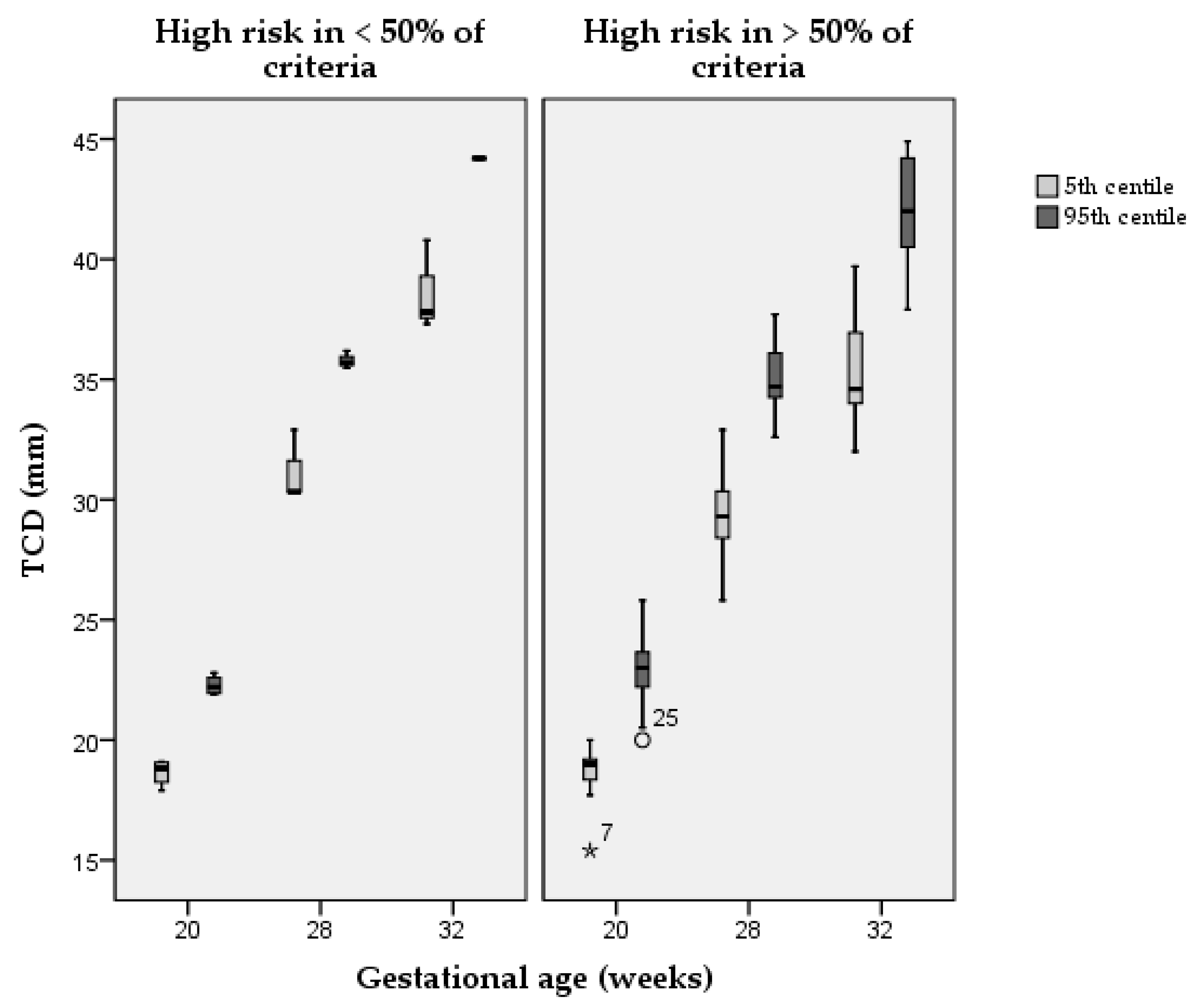
| TCD studies | ||||||||||||||
| Rodriguez-Sibaja [39] 2020 | 14–42 | 25 | 19 | 20.5 | 21.9 | 0.87 | 30.3 | 32.9 | 35.5 | 1.57 | 37.3 | 40.8 | 44.2 | 2.11 |
| Verburg [47] 2008 | 16–36 | 17 | 18.6 | 20.3 | 22.0 | 1.03 | 30.3 | 33.0 | 35.7 | 1.64 | 37.8 | 41.0 | 44.2 | 1.95 |
| Koning [31] 2017 | 9–32 | 16 | 19.1 | 20.8 | 22.4 | 32.9 | 34.6 | 36.2 | 40.8 | 42.4 | 44.1 | |||
| Araujo Jùnior [12] 2015 | 18–24 | 14 | 17.9 | 19.9 | 22.8 | |||||||||
| Chavez [16] 2003 | 14–38 | 12 | 17.7 | 20.4 | 23.0 | 0.20 | 28.2 | 32.4 | 36.6 | 0.35 | 34.4 | 39.5 | 44.7 | 0.46 |
| Smith [42] 1986 | 14–32 | 12 | 18.8 | 20.5 | 22.1 | 29.4 | 31.0 | 32.6 | 34.6 | 36.3 | 37.9 | |||
| Snijders [43] 1994 | 14–40 | 12 | 19.0 | 21.0 | 24.0 | 0.03 | 29.0 | 32.0 | 36.0 | 0.03 | 34.0 | 37.0 | 42.0 | 0.03 |
| Chang [15] 2000 | 20–40 | 10 | 15.4 | 20.3 | 25.3 | 27.8 | 32.7 | 37.7 | 34.0 | 38.9 | 43.8 | |||
| Joshi [28] 2010 | 15–38 | 10 | 19.1 | 20.7 | 22.3 | 31.3 | 32.9 | 34.6 | 37.4 | 39.1 | 40.7 | |||
| Takano [45] 2018 | 14–40 | 10 | 17.9 | 19 | 20 | 0.6 | 28.6 | 31.2 | 33.7 | 1.6 | 34 | 37.3 | 40.6 | 2 |
| Vinkesteijn [48] 2000 | 17–34 | 10 | 18.8 | 20.7 | 22.8 | 29.3 | 32.3 | 35.7 | 36.7 | 40.4 | 44.6 | |||
| Hayata [25] 2015 | 14–36 | 9 | 19.3 | 19.9 | 20.5 | 0.36 | 29.4 | 31.7 | 34.0 | 1.40 | 35.8 | 38.1 | 40.4 | 1.40 |
| Goldstein [21] 1987 | 13–40 | 8 | 20.0 | 31.0 | 38.0 | |||||||||
| Hata [24] 1989 | 17–40 | 8 | 19.9 | 21.5 | 23.2 | 31.4 | 33.0 | 34.7 | 37.2 | 38.8 | 40.4 | |||
| Lei [32] 1998 | 16–40 | 8 | 19.0 | 21.0 | 25.8 | 25.8 | 36.6 | 34.5 | 32.0 | 43.0 | 44.9 | |||
| Serhatlioglu [41] 2003 | 16–38 | 8 | 20.0 | 21.7 | 23.3 | 32.9 | 34.6 | 36.2 | 39.7 | 41.3 | 43.0 | |||
| CM studies | ||||||||||||||
| Napolitano [36] 2020 | 14–42 | 27 | 3.0 | 4.5 | 6.8 | 1.39 | 4.0 | 6.0 | 8.9 | 1.76 | 4.3 | 6.4 | 9.5 | 1.87 |
| Passos [37] 2015 | 17–29 | 15 | 4.3 | 6.0 | 8.0 | 5.6 | 8.0 | 11.1 | ||||||
| Araujo Jùnior [11] 2014 | 18–24 | 14 | 2.9 | 4.7 | 6.5 | |||||||||
| Brown [13] 2013 | 15–32 | 14 | 4.3 | 5.9 | 7.9 | 1.23 | 6.3 | 7.9 | 9.9 | 1.23 | 7.0 | 8.7 | 10.3 | 1.23 |
| Snijders [43] 1994 | 14–40 | 12 | 3.3 | 5.1 | 7.2 | 0.04 | 4.7 | 6.8 | 9.1 | 0.04 | 5.2 | 7.3 | 9.7 | 0.04 |
| Smith [42] 1986 | 14–32 | 12 | 4.2 | 5.8 | 7.5 | 6.7 | 8.3 | 10.0 | 7.9 | 9.6 | 11.2 | |||
| Köktener [29] 2007 | 16–24 | 9 | 2.7 | 4.3 | 6.0 | |||||||||
| Serhatlioglu [41] 2003 | 16–38 | 8 | 3.0 | 4.7 | 6.3 | 4.7 | 6.3 | 7.9 | 4.9 | 6.6 | 8.2 | |||
Publisher’s Note: MDPI stays neutral with regard to jurisdictional claims in published maps and institutional affiliations. |
© 2021 by the authors. Licensee MDPI, Basel, Switzerland. This article is an open access article distributed under the terms and conditions of the Creative Commons Attribution (CC BY) license (https://creativecommons.org/licenses/by/4.0/).
Share and Cite
Donadono, V.; Cavallaro, A.; Roberts, N.W.; Ioannou, C.; Papageorghiou, A.T.; Napolitano, R. A Systematic Review of Methodology Used in Studies Aimed at Creating Charts of Fetal Brain Structures. Diagnostics 2021, 11, 916. https://doi.org/10.3390/diagnostics11060916
Donadono V, Cavallaro A, Roberts NW, Ioannou C, Papageorghiou AT, Napolitano R. A Systematic Review of Methodology Used in Studies Aimed at Creating Charts of Fetal Brain Structures. Diagnostics. 2021; 11(6):916. https://doi.org/10.3390/diagnostics11060916
Chicago/Turabian StyleDonadono, Vera, Angelo Cavallaro, Nia W. Roberts, Christos Ioannou, Aris T. Papageorghiou, and Raffaele Napolitano. 2021. "A Systematic Review of Methodology Used in Studies Aimed at Creating Charts of Fetal Brain Structures" Diagnostics 11, no. 6: 916. https://doi.org/10.3390/diagnostics11060916
APA StyleDonadono, V., Cavallaro, A., Roberts, N. W., Ioannou, C., Papageorghiou, A. T., & Napolitano, R. (2021). A Systematic Review of Methodology Used in Studies Aimed at Creating Charts of Fetal Brain Structures. Diagnostics, 11(6), 916. https://doi.org/10.3390/diagnostics11060916