Application and Performance of Artificial Intelligence Technology in Oral Cancer Diagnosis and Prediction of Prognosis: A Systematic Review
Abstract
1. Introduction
2. Materials and Methods
2.1. Search Strategy
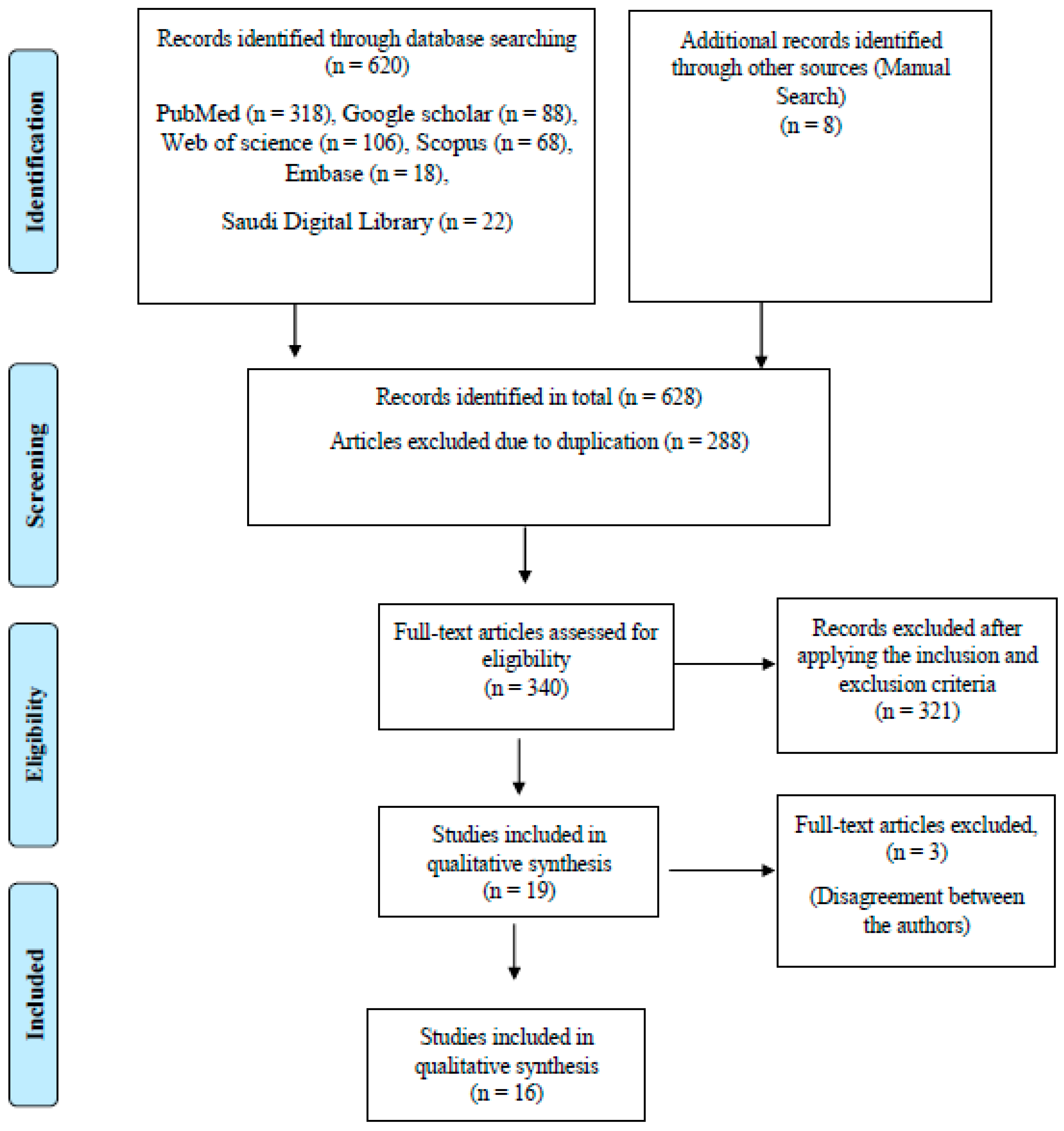
2.2. Study Selection
2.3. Inclusion and Exclusion Criteria
2.4. Data Extraction
3. Results
3.1. Qualitative Synthesis of the Included Studies
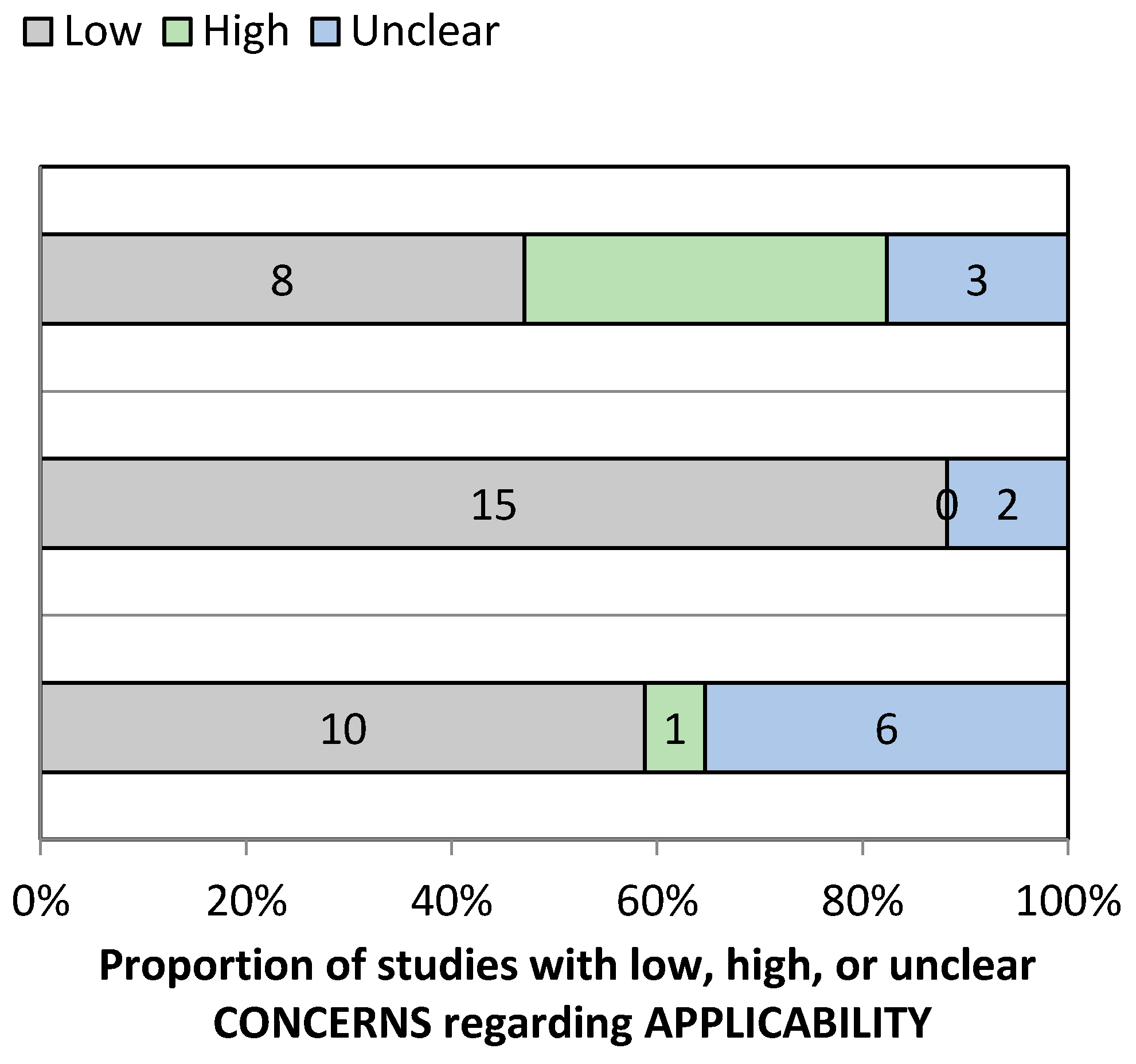
3.2. Risk of Bias Assessment and Applicability Concerns
4. Discussion
4.1. Artificial Intelligence in Detecting and Diagnosing Oral Cancer
4.2. Artificial Intelligence in Predicting the Occurrence of Oral Cancer
5. Conclusions
Supplementary Materials
Author Contributions
Funding
Data Availability Statement
Conflicts of Interest
References
- World Health Organization. WHO Report on Cancer: Setting Priorities, Investing Wisely and Providing Care for All; Technical Report; World Health Organization: Geneva, Switzerland, 2020. [Google Scholar]
- Sinevici, N.; O’sullivan, J. Oral cancer: Deregulated molecular events and their use as biomarkers. Oral Oncol. 2016, 61, 12–18. [Google Scholar] [CrossRef]
- Lewin, F.; Norell, S.; Johansson, H.; Gustavsson, P.; Wennerberg, J.; Biörklund, A.; Rutqvist, L. Smoking Tobacco, Oral Snuff, and Alcohol in the Etiology of Squamous Cell Carcinoma of the Head and Neck: A Population-Based Case-Referent Study in Sweden. Cancer 1998, 82, 1367–1375. [Google Scholar] [CrossRef]
- Ilhan, B.; Lin, K.; Guneri, P.; Wilder-Smith, P. Improving Oral Cancer Outcomes with Imaging and Artificial Intelligence. J. Dent. Res. 2020, 99, 241–248. [Google Scholar] [CrossRef] [PubMed]
- Dhanuthai, K.; Rojanawatsirivej, S.; Thosaporn, W.; Kintarak, S.; Subarnbhesaj, A.; Darling, M.; Kryshtalskyj, E.; Chiang, C.P.; Shin, H.I.; Choi, S.Y.; et al. Oral cancer: A multicenter study. Med. Oral Patol. Oral Cir. Bucal 2018, 23, e23–e29. [Google Scholar] [CrossRef] [PubMed]
- Lavanya, L.; Chandra, J. Oral Cancer Analysis Using Machine Learning Techniques. Int. J. Eng. Res. Technol. 2019, 12, 596–601. [Google Scholar]
- Kearney, V.; Chan, J.W.; Valdes, G.; Solberg, T.D.; Yom, S.S. The application of artificial intelligence in the IMRT planning process for head and neck cancer. Oral Oncol. 2018, 87, 111–116. [Google Scholar] [CrossRef] [PubMed]
- Hamet, P.; Tremblay, J. Artificial intelligence in medicine. Metabolism. 2017, 69, S36–S40. [Google Scholar] [CrossRef]
- Kaladhar, D.; Chandana, B.; Kumar, P. Predicting Cancer Survivability Using Classification Algorithms. Books 1 View project Protein Interaction Networks in Metallo Proteins and Docking Approaches of Metallic Compounds with TIMP and MMP in Control of MAPK Pathway View project Predicting Cancer. Int. J. Res. Rev. Comput. Sci. 2011, 2, 340–343. [Google Scholar]
- Kalappanavar, A.; Sneha, S.; Annigeri, R.G. Artificial intelligence: A dentist’s perspective. Pathol. Surg. 2018, 5, 2–4. [Google Scholar] [CrossRef]
- Krishna, A.B.; Tanveer, A.; Bhagirath, P.V.; Gannepalli, A. Role of artificial intelligence in diagnostic oral pathology-A modern approach. JOMFP 2020, 24, 152–156. [Google Scholar]
- Kareem, S.A.; Pozos-Parra, P.; Wilson, N. An application of belief merging for the diagnosis of oral cancer. Appl. Soft Comput. J. 2017, 61, 1105–1112. [Google Scholar] [CrossRef]
- Arbes, S.J. Factors contributing to the poorer survival of black Americans diagnosed with oral cancer (United States). Cancer Causes Control 1999, 10, 513–523. [Google Scholar] [CrossRef]
- de Melo, G.M.; Ribeiro, K.; Kowalski, L.; Deheinzelin, D. Risk Factors for Postoperative Complications in Oral Cancer and Their Prognostic Implications. Arch. Otolaryngol. Head Neck Surg. 2001, 127, 828–833. [Google Scholar]
- Bànkfalvi, A.; Piffkò, J. Prognostic and predictive factors in oral cancer: The role of the invasive tumour front. J. Oral Pathol. Med. 2000, 29, 291–298. [Google Scholar] [CrossRef] [PubMed]
- Schliephake, H. Prognostic relevance of molecular markers of oral cancer—A review. Int. J. Oral Maxillofac. Surg. 2003, 32, 233–245. [Google Scholar] [CrossRef] [PubMed]
- Kann, B.H.; Aneja, S.; Loganadane, G.V.; Kelly, J.R.; Smith, S.M.; Decker, R.H.; Yu, J.B.; Park, H.S.; Yarbrough, W.G.; Malhotra, A.; et al. Pretreatment Identification of Head and Neck Cancer Nodal Metastasis and Extranodal Extension Using Deep Learning Neural Networks. Sci. Rep. 2018, 8, 1–11. [Google Scholar] [CrossRef] [PubMed]
- Whiting, P.F.; Rutjes, A.W.S.; Westwood, M.E.; Mallett, S.; Deeks, J.J.; Reitsma, J.B.; Leeflang, M.M.G.; Sterne, J.A.C.; Bossuyt, P.M.M. Quadas-2: A revised tool for the quality assessment of diagnostic accuracy studies. Ann. Intern. Med. 2011, 155, 529–536. [Google Scholar] [CrossRef]
- Nayak, G.S.; Kamath, S.; Pai, K.M.; Sarkar, A.; Ray, S.; Kurien, J.; D’Almeida, L.; Krishnanand, B.R.; Santhosh, C.; Kartha, V.B.; et al. Principal component analysis and artificial neural network analysis of oral tissue fluorescence spectra: Classification of normal premalignant and malignant pathological conditions. Biopolymers 2006, 82, 152–166. [Google Scholar] [CrossRef] [PubMed]
- Jubair, F.; Al-karadsheh, O.; Malamos, D.; Al Mahdi, S.; Saad, Y.; Hassona, Y. A novel lightweight deep convolutional neural network for early detection of oral cancer. Oral Dis. 2021, 1–8. [Google Scholar] [CrossRef]
- Musulin, J.; Štifanić, D.; Zulijani, A.; Ćabov, T.; Dekanić, A.; Car, Z. An enhanced histopathology analysis: An ai-based system for multiclass grading of oral squamous cell carcinoma and segmenting of epithelial and stromal tissue. Cancers 2021, 13, 1784. [Google Scholar] [CrossRef]
- Kirubabai, M.P.; Arumugam, G. View of Deep Learning Classification Method to Detect and Diagnose the Cancer Regions in Oral MRI Images. Med. Legal Update 2021, 21, 462–468. [Google Scholar]
- Rathod, J.; Sherkay, S.; Bondre, H.; Sonewane, R.; Deshmukh, D. Oral Cancer Detection and Level Classification Through Machine Learning. Int. J. Adv. Res. Comput. Commun. Eng. 2020, 9, 177–182. [Google Scholar] [CrossRef]
- Rosma, M.D.; Sameem, A.K.; Basir, A.; Siti Mazlipah, I.; Norzaidi, M.D. The use of artificial intelligence to identify people at risk of oral cancer: Empirical evidence in Malaysian university. Int. J. Sci. Res. Educ. 2010, 3, 10–20. [Google Scholar]
- Alhazmi, A.; Alhazmi, Y.; Makrami, A.; Masmali, A.; Salawi, N.; Masmali, K.; Patil, S. Application of artificial intelligence and machine learning for prediction of oral cancer risk. J. Oral Pathol. Med. 2021, 1–7. [Google Scholar] [CrossRef]
- Chu, C.S.; Lee, N.P.; Adeoye, J.; Thomson, P.; Choi, S. Machine learning and treatment outcome prediction for oral cancer. J. Oral Pathol. Med. 2020, 49, 977–985. [Google Scholar] [CrossRef] [PubMed]
- Tseng, W.T.; Chiang, W.F.; Liu, S.Y.; Roan, J.; Lin, C.N. The Application of Data Mining Techniques to Oral Cancer Prognosis. J. Med. Syst. 2015, 39, 59–66. [Google Scholar] [CrossRef] [PubMed]
- Uthoff, R.D.; Song, B.; Sunny, S.; Patrick, S.; Suresh, A.; Kolur, T.; Keerthi, G.; Spires, O.; Anbarani, A.; Wilder-Smith, P.; et al. Point-of-care, smartphone-based, dual-modality, dual-view, oral cancer screening device with neural network classification for low-resource communities. PLoS ONE 2018, 13, 1–21. [Google Scholar] [CrossRef] [PubMed]
- Sunny, S.; Baby, A.; James, B.L.; Balaji, D.; N. V., A.; Rana, M.H.; Gurpur, P.; Skandarajah, A.; D’Ambrosio, M.; Ramanjinappa, R.D.; et al. A smart tele-cytology point-of-care platform for oral cancer screening. PLoS ONE 2019, 14, 1–16. [Google Scholar] [CrossRef]
- Jeyaraj, P.R.; Samuel Nadar, E.R. Computer-assisted medical image classification for early diagnosis of oral cancer employing deep learning algorithm. J. Cancer Res. Clin. Oncol. 2019, 145, 829–837. [Google Scholar] [CrossRef] [PubMed]
- Shams, W.K.; Htike, Z.Z. Oral Cancer Prediction Using Gene Expression Profiling and Machine Learning. Int. J. Appl. Eng. Res. 2017, 12, 4893–4898. [Google Scholar]
- Karadaghy, O.A.; Shew, M.; New, J.; Bur, A.M. Development and Assessment of a Machine Learning Model to Help Predict Survival among Patients with Oral Squamous Cell Carcinoma. JAMA Otolaryngol. Head Neck Surg. 2019, 145, 1115–1120. [Google Scholar] [CrossRef] [PubMed]
- Alabi, R.O.; Elmusrati, M.; Sawazaki-Calone, I.; Kowalski, L.P.; Haglund, C.; Coletta, R.D.; Mäkitie, A.A.; Salo, T.; Almangush, A.; Leivo, I. Comparison of supervised machine learning classification techniques in prediction of locoregional recurrences in early oral tongue cancer. Int. J. Med. Inform. 2020, 136, 104068. [Google Scholar] [CrossRef] [PubMed]
- Kim, D.W.; Lee, S.; Kwon, S.; Nam, W.; Cha, I.H.; Kim, H.J. Deep learning-based survival prediction of oral cancer patients. Sci. Rep. 2019, 9, 1–10. [Google Scholar] [CrossRef] [PubMed]
- Warnakulasuriya, S. Global epidemiology of oral and oropharyngeal cancer. Oral Oncol. 2009, 45, 309–316. [Google Scholar] [CrossRef]
- Gupta, N.; Gupta, R.; Acharya, A.K.; Patthi, B.; Goud, V.; Reddy, S.; Garg, A.; Singla, A. Changing Trends in oral cancer—A global scenario. Nepal J. Epidemiol. 2017, 6, 613–619. [Google Scholar] [CrossRef]
- Dhage, S.N. A Review on Early Detection of Oral Cancer using ML Techniques. Int. J. Sci. Prog. Res. 2019, 158, 1–5. [Google Scholar]
- Chan, C.H.; Huang, T.T.; Chen, C.Y.; Lee, C.C.; Chan, M.Y.; Chung, P.C. Texture-Map-Based Branch-Collaborative Network for Oral Cancer Detection. IEEE Trans. Biomed. Circuits Syst. 2019, 13, 766–780. [Google Scholar] [CrossRef]
- Bur, A.M.; Holcomb, A.; Goodwin, S.; Woodroof, J.; Karadaghy, O.; Shnayder, Y.; Kakarala, K.; Brant, J.; Shew, M. Machine learning to predict occult nodal metastasis in early oral squamous cell carcinoma. Oral Oncol. 2019, 92, 20–25. [Google Scholar] [CrossRef]
- Kan, C.W.; Nieman, L.T.; Sokolov, K.; Markey, M.K. AI in clinical decision support: Applications in optical spectroscopy for cancer detection and diagnosis. Stud. Comput. Intell. 2008, 107, 27–49. [Google Scholar] [CrossRef]
- Chang, S.W.; Abdul-Kareem, S.; Merican, A.F.; Zain, R.B. Oral cancer prognosis based on clinicopathologic and genomic markers using a hybrid of feature selection and machine learning methods. BMC Bioinform. 2013, 14, 170–185. [Google Scholar] [CrossRef] [PubMed]
- Lucheng, Z.; Wenhua, L.; Meng, S.; Hangping, W.; Juan, W.; Xuebang, Z.; Changlin, Z. Comparison between artificial neural network and Cox regression model in predicting the survival rate of gastric cancer patients. Biomed. Rep. 2013, 1, 757–760. [Google Scholar] [CrossRef]
- Zheng, B.; Yoon, S.W.; Lam, S.S. Breast cancer diagnosis based on feature extraction using a hybrid of K-means and support vector machine algorithms. Expert Syst. Appl. 2014, 41, 1476–1482. [Google Scholar] [CrossRef]


| Research question | What are the applications and performance of the artificial intelligence models that have been widely used in oral cancer diagnosis, and predicting the prognosis. |
| Population | Patients, clinical images, radiographs, datasets, and histological images. |
| Intervention | AI-based models for oral cancer diagnosis and predicting prognosis. |
| Comparison | Expert opinions and reference standards. |
| Outcome | Measurable or predictive outcomes such as accuracy, sensitivity, specificity, ROC = Receiver Operating Characteristic curve, AUC = Area Under the Curve, ICC = Intra-class Correlation Coefficient, PPV = Positive Predictive Values, and NPV = Negative Predictive Values. |
| Sr. No. | Authors | Year of Publication | Algorithm Architecture | Study Design | Objective of the Study | No. of Images/Photographs for Testing | Study Factor | Modality | Comparison, If Any | Evaluation Accuracy/Average Accuracy | Results (+) Effective, (−) Non Effective (N) Neutral | Outcomes | Authors Suggestions/Conclusions |
|---|---|---|---|---|---|---|---|---|---|---|---|---|---|
| 1 | Nayak et al. [19] | 2005 | ANNs | Cross sectional study | Discriminating normal, potentially malignant, and malignant conditions using principal component analysis (PCA) and artificial neural network (ANN) | 50 | Differentiating normal, potentially malignant, and malignant | Recorded spectra | Principal component analysis (PCA) | Accuracy 98.3%, specificity of 100% and sensitivity 96.5% | (+) Effective | ANN is found to be slightly better than PCA | This model is efficient for real-time application. |
| 2 | Tseng et al. [27] | 2015 | ANNs | Cohort study | ANN for predicting oral cancer prognosis | - | Determining the differences between the symptoms shown in past cases | Datasets | Decision tree (DT) | Not Mentioned | (+) Effective | Both decision tree and artificial neural network models showed superiority to the traditional statistical model. | Decision tree models are relatively easier to interpret compared to artificial neural network models. |
| 3 | Uthoff et al. [28] | 2017 | CNN’s | Crosssectional study | AI-based deep (CNNs) for early detection of pre-cancerous and cancerous lesions | 170 | Detection of pre-cancerous and cancerous lesions | Autofluorescence imaging (AFI) and white light imaging (WLI) | Specialist’s diagnosis | Sensitivities 85%, specificities 88.75%, positive predictive values 87.67%, and negative predictive values 85.49 | (+) Effective | CNN achieving high values of sensitivity, specificity, PPV, and NPV compared to the on-site specialist gold standard. | Performance should increase as additional images are collected. |
| 4 | Shams et al. [31] | 2017 | CNN’s | Cross sectional comparative study | Deep Neural Network (DNN) for predicting the possibility of oral cancer development in Oral potentially malignant lesion patients | 10 | Oral cancer development in Oral potentially malignant lesion patients | Datasets | Support Vector Machine (SVM), Regularized Least Squares (RLS), Multi-Layer Perception (MLP) | High accuracy 96% | (+) Effective | The results show high accuracy using DNN than SVM and MLP | None |
| 5 | Jeyaraj et al. [30] | 2019 | CNN’s | Cross sectional comparative study | Deep learning algorithm for an automated, computer-aided oral cancer-detecting system | 100 | Detection of pre-cancerous as benign and post cancerous as malignant region | Hyperspectral images | The traditional medical image classification algorithm | Accuracy of 91.4%, sensitivity 94% and a specificity of 91% | (+) Effective | The quality of diagnosis is increased by proposed regression-based partitioned CNN learning algorithm for a complex medical image of oral cancer diagnosis | This deep learning the algorithm can be easily deployed for providing an automatic medical image classifier without expert knowledge. |
| 6 | Fahed Jubair et al. [20] | 2020 | CNN’s | Crosssectional study | Develop a lightweight deep CNN using Efficient net-B0 transfer model CNN for binary classification of oral lesions into benign and malignant or potentially malignant using standard real-time clinical images | 716 | Detecting oral cancer | Clinical images | None | accuracy was 85.0% specificity, 84.5%, sensitivity 86.7% | (+) Effective | AI can improve the quality and reach of oral cancer screening and early detection. | This model of being small in size and need small computation power and memory capacity. |
| 7 | Sunny et al. [29] | 2019 | ANNs | Cross sectional comparative study | Artificial Neural Network (ANN) based risk-stratification model for early detection of oral potentially malignant (OPML)/malignant lesion. | 82 | Oral potentially malignant (OPML)/malignant lesion. | High-resolution cytology images | Conventional cytology and histology | 84–86% Accuracy Sensitivity 93% | (+) Effective | ANN-based risk stratification model improved the detection sensitivity of malignant lesions (93%) and high-grade OPML (73%), increasing the overall accuracy by 30%. | This model can be an invaluable Point-of-Care (POC) tool for early detection/screening in oral cancer. |
| 8 | Jelena Musulin et al. [21] | 2021 | ANNs | Cross sectional comparative study | Diagnosing OC using the histological image of a biopsy | 322 | Detecting oral cancer | Histological image | ResNet50, ResNet101 Xception MobileNetv2 | Xception and SWT resulted in the highest classification value of 0.96 (σ = 0.042) AUCmacro | (+) Effective | The AI-based system has great potential in the diagnosis of OSCC | This cell shape and size, pathological mitoses, tumor-stroma ratio and the distinction between early and advanced-stage OSCCs |
| 9 | M. Praveena Kirubabai et al. [22] | 2021 | CNN | Cross sectional study | To classify the oral images into either normal or abnormal images and diagnosed into ‘Mild’ or ‘Severe’ using a deep learning algorithm | 160 | Detecting oral cancer | Oral images | None | accuracy was 99.7%, 98.6% of sensitivity, 99.1% of specificity, and 99.7% | (+) Effective | CNN has high accuracy in detecting OC | None |
| 10 | Jyoti Rathod et al. [23] | 2019 | CNN’s | Cross sectional comparative study | Classify different stages of oral cancer using machine learning techniques | - | Diagnosing and classifying the premalignant lesion | Data set | SVM, KNN, MLP RSF, and Logistic Regression | DT 90.68%, RSF 91%, SVM 88%, KNN 85%, MLP 81% and Logistic Regression gives 80% of accuracy | (+) Effective | DT and RSF produced the same accuracy results | classification of oral cancer can be classified efficiently with help of Random Forest and Decision Tree |
| 11 | Alabi et al. [33] | 2019 | ANNs | Cross sectional comparative study | Comparing the performance of four machine learning Models (ML) for Predicting Risk of recurrence of oral tongue squamous cell carcinoma (OTSCC) | 311 | Prediction of reoccurrence | Patient datasets | 5 Prognostic significance of the depth of invasion (DOI). | Accuracy of 68% for Support Vector Machine (SVM), 70% Naive Bayes (NB), 81% Boosted Decision Tree (BDT) and 78% Decision Forest (DF) | (+) Effective | Best classification accuracy was achieved with the boosted decision tree algorithm. These models outperformed the DOI-based approach | Machine algorithms should be considered in medical applications. |
| 12 | Kim et al. [35] | 2019 | CNNs | Retrospective study | Deep learning-based survival prediction method in oral squamous cell carcinoma (SCC) patients | 255 | Survival prediction | Datasets | Random Survival Forest (RSF) and the Cox proportional hazard model (CPH) | c-index of testing sets reaching 0.781 | (+) Effective | This AI model displayed the best performance among the three models | This model can be effective in predicting with higher accuracy and can guide clinicians both in choosing treatment options and avoiding unnecessary treatments |
| 13 | Anwar Alhazmi et al. [25] | 2020 | ANNs | Crosssectional study | To develop (ANN) based model in predicting OC | 73 | Predicting risk of developing OC | Datasets | None | Accuracy of 78.95% | (+) Effective | ANN could perform well in estimating the probability of malignancy | More cohort studies are required based on this model |
| 14 | Chui S. Chu et al. [26] | 2021 | CNN’s | Cross sectional comparative study | To evaluate the ability of supervised machine learning models to predict disease outcome | 467 | Predicting risk of developing OC | Clinicopathological data | linear regression (LR), DT, SVM, and k-nearest neighbors (KNN) models | 70.59% accuracy (AUC 0.67), 41.98% sensitivity, and a high specificity of 84.12%. | (+) Effective | CNN’s DT model was most successful in identifying “true positive” progressive disease | AI models in this study have shown promise in predicting progressive OSCC disease outcomes |
| 15 | Rosma et al. [24] | 2010 | ANNs | Cross sectional comparative study | Performances of the two artificial Intelligent prediction models when compared with a group of oral cancer clinicians. | 171 | Predicting the likelihood of an individual developing oral cancer | Datasets | 27 oral cancer clinicians | Mean accuracy, sensitivity, and specificity of the models were 59.9, 45.5, and 85.3 for fuzzy neural network models; 63.1, 54.2, and 78.6 for oral cancer clinicians predictions and 67.5, 69.0 and 64.7 for fuzzy regression prediction models. | (+) Effective | Fuzzy regression and fuzzy neural network performed better than oral cancer clinicians | These neural network models provide a suitable alternative to human expert prediction in predicting oral cancer susceptibility. |
| 16 | Omar A. Karadaghy et al. [32] | 2019 | CNN’s | Crosssectional study | To develop a prediction DT model using machine learning for 5-year overall survival among patients with OSCC | 33, 065 | Predicting OSCC | Dataset | None | accuracy was 71%, precision was 71%, | (+) Effective | AI better in predicting OSCC | AI learning may play in individual patient risk estimation in the the era of big data. |
Publisher’s Note: MDPI stays neutral with regard to jurisdictional claims in published maps and institutional affiliations. |
© 2021 by the authors. Licensee MDPI, Basel, Switzerland. This article is an open access article distributed under the terms and conditions of the Creative Commons Attribution (CC BY) license (https://creativecommons.org/licenses/by/4.0/).
Share and Cite
Khanagar, S.B.; Naik, S.; Al Kheraif, A.A.; Vishwanathaiah, S.; Maganur, P.C.; Alhazmi, Y.; Mushtaq, S.; Sarode, S.C.; Sarode, G.S.; Zanza, A.; et al. Application and Performance of Artificial Intelligence Technology in Oral Cancer Diagnosis and Prediction of Prognosis: A Systematic Review. Diagnostics 2021, 11, 1004. https://doi.org/10.3390/diagnostics11061004
Khanagar SB, Naik S, Al Kheraif AA, Vishwanathaiah S, Maganur PC, Alhazmi Y, Mushtaq S, Sarode SC, Sarode GS, Zanza A, et al. Application and Performance of Artificial Intelligence Technology in Oral Cancer Diagnosis and Prediction of Prognosis: A Systematic Review. Diagnostics. 2021; 11(6):1004. https://doi.org/10.3390/diagnostics11061004
Chicago/Turabian StyleKhanagar, Sanjeev B., Sachin Naik, Abdulaziz Abdullah Al Kheraif, Satish Vishwanathaiah, Prabhadevi C. Maganur, Yaser Alhazmi, Shazia Mushtaq, Sachin C. Sarode, Gargi S. Sarode, Alessio Zanza, and et al. 2021. "Application and Performance of Artificial Intelligence Technology in Oral Cancer Diagnosis and Prediction of Prognosis: A Systematic Review" Diagnostics 11, no. 6: 1004. https://doi.org/10.3390/diagnostics11061004
APA StyleKhanagar, S. B., Naik, S., Al Kheraif, A. A., Vishwanathaiah, S., Maganur, P. C., Alhazmi, Y., Mushtaq, S., Sarode, S. C., Sarode, G. S., Zanza, A., Testarelli, L., & Patil, S. (2021). Application and Performance of Artificial Intelligence Technology in Oral Cancer Diagnosis and Prediction of Prognosis: A Systematic Review. Diagnostics, 11(6), 1004. https://doi.org/10.3390/diagnostics11061004


