Article

Machine Learning Study in Caries Markers in Oral Microbiota from Monozygotic Twin Children

by
Esther Alia-García
1,
Manuel Ponce-Alonso
2,†,
Claudia Saralegui
2,†,
Ana Halperin
2,
Marta Paz Cortés
1,
María Rosario Baquero
1,
David Parra-Pecharromán
1,3,
Javier Galeano
4,* and
Rosa del Campo
1,2,†
1
Facultad de Ciencias de la Salud, Universidad Alfonso X El Sabio, Villanueva de la Cañada, 28691 Madrid, Spain
2
Servicio de Microbiología, Hospital Universitario Ramón y Cajal and Instituto Ramón y Cajal de Investigaciones Sanitarias (IRYCIS), 28034 Madrid, Spain
3
Departamento de Biología, Servicio de Criminalística, Dirección General de la Guardia Civil, 28003 Madrid, Spain
4
Complex Systems Group, Universidad Politécnica de Madrid, 28040 Madrid, Spain
*
Author to whom correspondence should be addressed.
Spanish Network for Research in Infectious Diseases (REIPI).
Diagnostics 2021, 11(5), 835; https://doi.org/10.3390/diagnostics11050835
Submission received: 13 April 2021 / Revised: 30 April 2021 / Accepted: 3 May 2021 / Published: 6 May 2021
(This article belongs to the Special Issue Artificial Intelligence in Oral Health)

Abstract

:
In recent years, the etiology of caries has evolved from a simplistic infectious perspective based on Streptococcus mutans and/or Lactobacillus activity, to a multifactorial disease involving a complex oral microbiota, the human genetic background and the environment. The aim of this work was to identify bacterial markers associated with early caries using massive 16S rDNA. To minimize the other factors, the composition of the oral microbiota of twins in which only one of them had caries was compared with their healthy sibling. Twenty-one monozygotic twin pairs without a previous diagnosis of caries were recruited in the context of their orthodontic treatment and divided into two categories: (1) caries group in which only one of the twins had caries; and (2) control group in which neither of the twins had caries. Each participant contributed a single oral lavage sample in which the bacterial composition was determined by 16S rDNA amplification and further high-throughput sequencing. Data analysis included statistical comparison of alpha and beta diversity, as well as differential taxa abundance between groups. Our results show that twins of the control group have a closer bacterial composition than those from the caries group. However, statistical differences were not detected and we were unable to find any particular bacterial marker by 16S rDNA high-throughput sequencing that could be useful for prevention strategies. Although these results should be validated in a larger population, including children from other places or ethnicities, we conclude that the occurrence of caries is not related to the increase of any particular bacterial population.

1. Introduction

The microbial colonization of the oral cavity starts immediately after birth, differencing among early colonizers (Streptococcus, Veillonella and Lactobacillus), constant (Gemella, Granulicatella, Haemophilus and Rothia) and late colonizers (Actinomyces, Porphyromonas, Abiotrophia and Neisseria) [1,2,3]. The establishment of this ecosystem and its further composition is influenced by numerous factors as the mode of delivery, diet and antibiotic consumption [4]. Oral health is not only a local stomatological problem, but also an important driver of systemic health, as it has been linked to numerous disorders of the digestive, cardiovascular and genitourinary tracts [5,6,7].
Caries is the most prevalent human disease worldwide, although its incidence varies according to geography and ethnicity [8], and it has conventionally been attributed to the direct action of acidogenic bacteria such as Streptococcus mutans, Lactobacillus and Bifidobacterium since these microorganisms have been isolated from the lesions. The application of molecular tools based on high-throughput sequencing of the 16S rDNA gene has revealed that microbiota associated to caries is a much more complex ecosystem than expected (http://www.homd.org/, accessed on 5 May 2021) [9]. While traditional studies classified bacteria as pathogens or commensals according to their potential etiological role on diseases, greater focus has been put on the new concepts of eubiosis/dysbiosis and the disbalance of alkalinogenic/acidogenic bacteria in the caries [3,4]. In addition, metatranscriptomic analyses have permitted to extend the cause of oral diseases as periodontitis from the action of a single microorganism to the metabolic activity of the entire ecosystem [10]. Consequently, the number of microorganisms linked to caries has increased considerably in the last decade [11,12,13], including Streptococcus, Lactobacillus, Veillonella, Actinomyces, Granulicatella, Leptotrichia, Megasphaera, Olsenella, Shuttleworthia and, most recently, Scardovia, Atopobium and Selemonas [14]. One of the major challenges is to identify early markers of caries in order to monitor and prevent this disease during childhood. The exploration of biomarkers in saliva has already demonstrated its usefulness in other pathologies [7,15].
Due to all this complexity in the detection of caries markers, we think that a predictive analysis using machine learning tools can be a good starting point in the study of caries using the oral microbiota. Despite the fact that the implementation of Artificial Intelligence (AI) is still far from being completely common in oral health, some studies highlight the improvements that its use would imply in different areas [16].
The rationale of the present work was to identify bacterial biomarkers in saliva for early caries detection. For this purpose, we explored by massive 16S rDNA sequencing combined with robust bioinformatics tools, statistical analysis and machine learning the oral microbiota of monozygotic twins with and without caries.

2. Materials and Methods

2.1. Patients and Samples

Twenty-one pairs of monozygotic twins were recruited by the first author EAG and divided into two categories: (1) caries group where only one of the twins had caries (22 infants, 73% females, median age of 9 years, range from 6 to 12 years); and (2) control group where neither of them had caries (20 infants, 70% females, median age of 6.7 years, range from 4 to 12 years) (Table 1). Infants were enrolled in 2018 from January to May in four different dental clinics of Madrid (Spain) within the context of their orthodontic treatment. Each child contributed with a single oral lavage sample after 5 min of vigorous rising with 10 mL of sterile water. Samples were immediately frozen after collection and stored at −80 C until processing. The inclusion criteria were twins aged 4–12 years whose parents and they accepted to participate in the study. In the caries group kids with clear lesions as well as pre-cavity lesions, mainly white spots, were included, whereas other types of lesions were excluded. All participants were adequately instructed to avoid teeth brushing, food and sugar drinks intake during the 2 h before sampling.

2.2. Oral Microbiota Characterization

Oral lavages were slowly defrosted at −20 C during 24 h, followed by another 24 h at 4 C, and centrifuged at 14,000 r.p.m. for 15 min discharging the supernatant. Total DNA was obtained from the pellet with the Speedtools tissue DNA extraction kit (Biotools), determining their concentration and quality by Qubit fluorometer (Thermo Fisher Scientific, MA, USA). DNA samples were sent to FISABIO (Valencia, Spain) for massive sequencing (2 × 300 bp, MiSeq, Illumina. Cod. 15044223 Rev. A) of the V3 and V4 regions of the 16S rRNA gene, which were amplified with the following primers: Forward Primer: 5 -TCGTCGGCAGCGTCAGATGTGTATAAGAGACAGC CTACGGGNGGCWGCAG; and Reverse Primer: 5 -GTCTCGTGGGCTCGGAGATGTGTATAAGAGACAGGACTACHVGGGT ATCTAATCC. Sequence quality was measured according to the following parameters: minimum length, 250 bp; trimming quality measure type, mean; trimming quality number from 3’ extreme, 30; and trimming quality window, 10 bp. Shannon–Weaver and Chao1 indexes were used for bacterial alpha diversity estimation excluding taxa with three or fewer reads. Taxonomic affiliations were assigned using the Silva 119 database, and reads with an RDP score below 0.8 were assigned to the upper taxonomic rank, leaving the last rank as unidentified. Relative abundance and contingency tables of the operational taxonomic units (OTUs) included singletons and very low-represented taxa.

2.3. Statistical Analysis and Machine Learning Modeling

Statistical analysis was performed using R statistical software v3.5.3. Quantitative data of the reads were homogenized using their relative percentage from the total reads of each sample to allow the comparison between samples. Finally, the Galaxy Huttenhower Platform (http://huttenhower.sph.harvard.edu/galaxy, accessed on 5 May 2021) was used to calculate the Linear Discriminant Effect Size Analysis (LEfSe) algorithm to identify which microbial taxa explain significant differences among groups of samples [17]. The PCoA analyses were performed by Past 3.0 software. Raw sequences were deposited in the GenBank database as Bioproject PRJNA643173.
Simultaneously, we carried out a statistical exploratory analysis to later search for a machine learning model for a possible caries prediction. To carry out this analysis, we ruled out bacterial species with fewer than 50 data with non-zero values. Exploratory analysis was performed using own software in Python. Machine Learning models were developed with Orange3 v3.27 [18]. We carried out different classification models in two ways. In the first case, we used healthy, control and cavity sample labels. In the second case, we only used healthy and caries labels to classify our samples. Using k-cross validation (k = 10), we tested five different classification model: Random Forest, Neural Network, Support Vector Machine, KNN model and a logistic regression.
To evaluate the results of the used algorithms, we used:
  • Classification accuracy is the proportion of correctly classified examples.
  • F-1 is a weighted harmonic mean of precision and recall.
  • Precision is the proportion of true positives among instances classified as positive, e.g., the proportion of cavity correctly identified as cavity.
  • Recall is the proportion of true positives among all positive instances in the data, e.g., the number of cavity among all diagnosed as cavity.

3. Results

Both groups of participants were comparable in demographic and anthropometric terms, and all were recruited during their orthodontic treatment without previous suspicion of caries. Oral lavages were processed in a single session and the 16S rDNA massive sequencing was developed successfully, passing the quality filters with adequate negative controls. The numbers of read counts were comparable for all samples. The alpha diversity was analyzed by the Shannon–Weaver and Chao 1 alpha diversity indexes showed no significant differences between groups, but more disperse values were detected in the caries group (Figure 1).
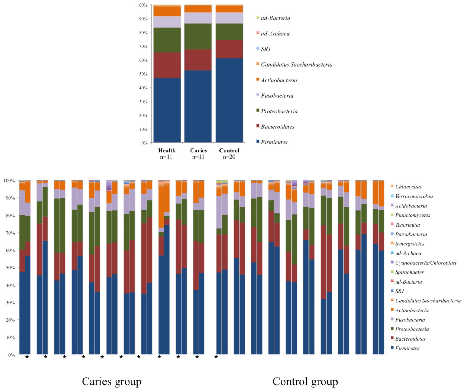
Phyla distributions showed a preserved pattern for each pair of twins, including those from the caries group (Figure 2). Children with caries had a similar phyla distribution to their healthy siblings, whereas controls presented higher proportions of Firmicutes and lower proportions of Proteobacteria.
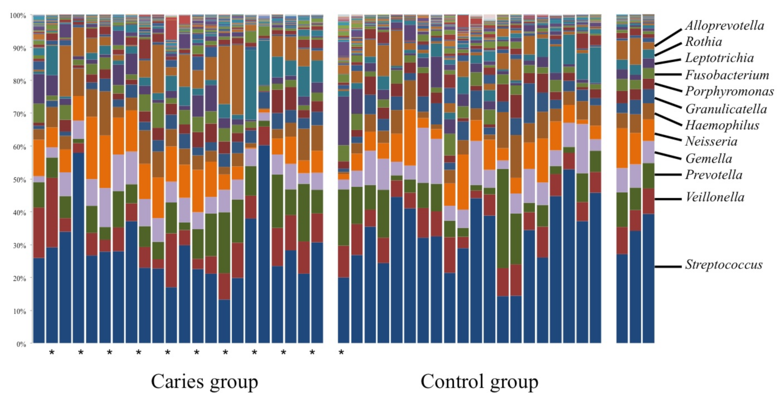
Up to 119 genera were identified, although 13 of them accounted for 90% of the total abundance [Streptococcus (≈30%), Prevotella (≈10 %), Neisseria (≈9%), Veillonella (≈8%), Gemella (≈7%), Haemophilus (≈6%), Alloprevotella (≈5%), Rothia (≈5%), Porphyromonas (≈2%), Fusobacterium (≈2%), Leptotrichia (≈2%), Granucalicatella (≈2%) and Actinomyces (≈2%)]. The remaining genera represented 10% of the abundance, comprising 106 genera with a total population density less than 1 for each one (Figure 3).
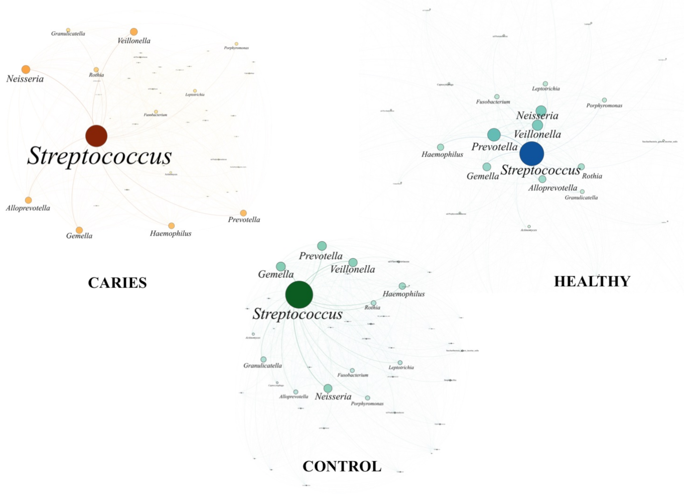
To obtain a global overview of the oral microbiome complexity, we designed an interaction network representing all taxa detected for each subject in circles proportional to their frequency and joined the circles, called nodes, proportional to their frequency joining the circles by lines, called links, to build a network per sample. Subsequently, we superimposed all the individual networks to define the core of the microbiome of each condition (caries, health and controls), and the thickness of the links between nodes is the accumulated number of lectures in all samples, representing the stability in the coexistence of the connecting taxa (Figure 4).
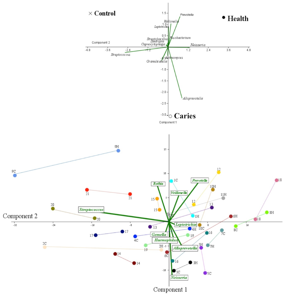
PCoA analysis separated the healthy, caries and control groups (Figure 5), showing a higher level of Veillonella, Prevotella and Fusobacterium genera linked to a healthy status, whereas Alloprevotella and Granullicatella were the most differentiated genera among children with cavities. The control group was allocated in a separate quadrant marked by the abundance of Capnocytophaga, Lautropia and Streptobacillus. Curiously, most of the control group twins were located on the same quadrant (8 out of 10 pairs), three pairs being located in Quadrant 3 (dominated by Gemella and Haemophilus), three in Quadrant 4 (Streptococcus and Rothia) and two in Quadrant 1 (Prevotella and Veillonella). The remaining two pairs of control twins were located on separated coordinates (Quadrants 1–3 and 2–3). Considering the twins of the caries group, only 6 out of the 11 pairs had both children located in the same quadrant: one in Quadrant 1 (Prevotella and Veillonella), three in Quadrant 2 (Neisseria, Alloprevotela and Leptotrichia), one in Quadrant 3 (Gemella and Haemophilus) and one in Quadrant 4 (Streptococcus and Rothia). The remaining five pairs were distributed in separated quadrants: two pairs in Quadrants 1–2, one pair in Quadrants 2–3, one in Quadrants 1–4 and one in Quadrants 1–3.
Furthermore, differential abundance analysis on microbiota composition by LEfSE in relation to the group, age and the sex of children did not obtain any significant result.
Finally, to address the possibility to predict the cavity in patients, we developed five classification models using machine learning tools (Table 2). The model that showed the highest classification accuracy (CA) was the Random Forest model with a value of 0.881 followed by the Neural Network with 0.810. Studying the confusion matrix, from the point of view of caries, the Random Forest model does not produce false positives but does generate quite a few false negatives (54.5%). On the contrary, Neural Network model produce 16.1% false positives but a lower percentage, with respect to the Random Forest, of false negatives (27.3%).

4. Discussion

In the last years, the etiology of caries has evolved from a simplistic infectious perspective (S. mutans and/or Lactobacillus colonization) to a multifactorial disease involving oral microbiota, human genetic background and environment [3]. In accordance, research tools have evolved from culturomics to metagenomics, transcriptomics and proteomics. Diet continues to be one of the most decisive factors in caries incidence and accounts for the individual susceptibility in relation to carbohydrates intake and bacterial fermentation [19,20], whereas human genetic background seems to be not so relevant, as previously expected [4,21]. The main objective of our work was twofold, on the one hand, to detect early metagenomic markers based on the abundance of particular genera in the oral microbiota associated with caries in the context of monozygotic twins with the same genetic, dietary and environmental context, and, on the other hand, address a classification model to predict caries using this microbiota of patients. When the participation in the project was offered to children, none of them had been previously diagnosed of caries. The bioinformatic analyses showed a more uniform microbiota in the control twin group, but without statistical significance. We were unable to identify any bacterial taxon exclusive of participants with caries, discarding the contribution of oral bacteria microbiota at the initial cariogenic process.
To obtain a representative sample of the entire oral microbiota, all children refrained from tooth brush and avoided food intake for at least 2 h. Despite the high and continuous contamination of the oral microbiota with foreign environmental microorganisms, intra-individual particularities of saliva microbiota have been postulated as a forensic marker to identify subjects, even for twins [22]. Some studies perform the sampling directly from the lesion or at the supragingival plaque, but we decided to use rinsing of the total oral microbiota as a representative sample easily collected by children with the absence of macroscopically visible lesions, which seems to be the most suitable option for surveillance purposes. Even though saliva and supragingival plaque are different in terms of bacterial composition [23], saliva has been used in similar studies, providing differentiation between subjects with and without caries [24]. The use of saliva in the identification of biomarkers associated to both local and general health was previously validated [7,15].
Previous studies on monozygotic and dizygotic twins reported discordant results regarding the incidence of caries and the oral microbiota composition [4,21,23,25,26], although those studies have been conducted in different age groups and using different microbiological methodologies, which could explain the lack of reproducibility. In the last years, tools for massive sequencing data analysis have been evolving considerably, allowing us to applied some of those novel tools to our data, including LEfSE and network analysis of the ecosystem.
Our PCoA analysis consistently associates a higher abundance of Alloprevotella in subjects with cavities, whereas in their healthy counterparts Prevotella was the most differential marker. Surprisingly, both genera belong to the same family and might have synonymous metabolic functions, although we cannot rule out synergistic effects of combination of microorganisms [27] and, most notably, the interaction of particular bacterial genera with fungi or virus, which has not been extensively explored. Most of the published studies using the entire oral microbiota with a metagenomic approach failed to find significant differences among healthy and caries status in non-related subjects [4,11,24,28,29]. However, a structural conservation between twins can be observed in our PCoA analysis, where control subjects are also more homogeneous (8/10 in the same quadrant) than the caries group (6/11), suggesting an incipient diversification on the oral microbiome.
As in other human ecosystems, the oral microbiome is usually constant and specific in each individual, but may be influenced by ethnicity [30]. A higher prevalence of caries has been described in a group of subjects from China, with special enrichment of Scardovia [24], whereas this genus is not particularly abundant in our population. In the study by Yasunaga et al., individuals without caries had more diverse communities, with a significantly higher proportion of the genus Porphyromonas, in particular Porphyromonas pasteri [31]. Belstrøm et al. also observed a higher alpha diversity in subjects with caries and an enrichment of Neisseria, Haemophilus and Fusobacterium compared to individuals without caries [29]. In contrast, in our study, both bacterial density and alpha diversity parameters are similar in children of both groups and conditions. An important point is that the composition of the community does not necessarily reflect its metabolic activity [10], particularly in microorganisms represented in low proportions, and it could have essential metabolic activities for the community [5].
Extremely high levels of S. mutans have been associated with caries, and, whereas Streptococcus was the majoritarian genera, we cannot investigate this point since our metagenomic approach is not able to assign to the level of species, due to the short length of the sequences obtained by massive sequencing. The dominance of Streptococcus in the oral cavity can be found in both patients with caries and in controls [29,32], as observed in our case, but being more abundant in the healthy control group than among the cavity group. A protective effect of some streptococcal species has been demonstrated [33], and, beyond the microbiota composition, there is an increasing emphasis on the global ecosystem richness, distribution and functionality [34]. However, in our case, LEfSE analysis failed in the discrimination of children by their cavity status, age, or sex, being the oral microbiota of all participants comparable.
In this study, the classification models showed relatively good precision in predicting caries in our data set (Table 2).The best performing classification models were Random Forest that showed the highest classification accuracy (CA) of a value of 0.881 and closely followed by Neural Networks with an CA of 0.810. One of the most interesting points of the Random Forest model was that it did not produce false positives. However, the worst aspect of these models was the percentage of false negatives (54.5%). On the contrary, the Neural Network model produced 16.1% false positives but a lower percentage, with respect to the Random Forest model, of false negatives (27.3%). This problem could be overcome with a large data-set of caries patients.
The major strength and, at the same time, the major limitation of our study is the inclusive criteria of children, which were enrolled during their orthodontic treatment independently of their caries’ status. In fact, all the detected lesions were small and superficial, corresponding probably to the onset of the disease. Of course, we are unable to ascertain if this was also the case of the control group, where caries were not observed in any children but could be closed to appearing. The oral microbiota could be also implicated in the tooth development [8], and likewise the age of the patient might be considered, since different composition of the oral microbiota has been related to it. Finally, our results show that machine learning models could help us in caries prevention using microbiota data, although they are still far from having good accuracy. In summary, our results demonstrate that composition of oral microbiota in twins is highly conserved independently of their cariogenic process. We were unable to find any bacterial marker by 16S rDNA massive sequencing associated to caries; on the contrary, Isola et al. demonstrated a significant relationship between the salivary IL-6 concentration and existence of periodontitis [7,15].

Author Contributions

E.A.-G., M.R.B., D.P.-P. and R.d.C., study design, data analysis and drafting of the paper; M.P.-A., C.S., A.H. and J.G., data analysis and drafting of the paper; and M.P.C., data collection and drafting of the paper. All authors have read and agreed to the published version of the manuscript.

Funding

Claudia Saralegui was financed by a project of the Fundación Mutua Madrileña (grant to Rosa del Campo achieved in 2017 call with reference number AP165902017). This study was supported by Instituto de Salud Carlos III and the European Regional Development Fund (ERDF, ‘A way to achieve Europe’) (PI17/115 to Rosa del Campo) and Fundación Universidad Alfonso X El Sabio – Santander Universidades (Project number 1.010.811).

Institutional Review Board Statement

The study protocol was approved by the Ethical Committee of the Alfonso X El Sabio University (reference CE2017_0011) approved on 23 February 2017. The research was conducted ethically, in accordance with the World Medical Association Declaration of Helsinki. All participants and their parents/legal guardians signed a written informed consent form. Children received a report of their oral health status and were referred to dental treatment when necessary.

Informed Consent Statement

Informed consent was obtained from all subjects involved in the study.

Data Availability Statement

Data supporting our study can be found in the GenBank database as Bioproject PRJNA643173.

Conflicts of Interest

The authors declare no conflict of interest. The funders had no role in the design of the study; in the collection, analyses, or interpretation of data; in the writing of the manuscript, or in the decision to publish the results

References

  1. Dzidic, M.; Collado, M.C.; Abrahamsson, T.; Artacho, A.; Stensson, M.; Jenmalm, M.C.; Mira, A. Oral microbiome development during childhood: An ecological succession influenced by postnatal factors and associated with tooth decay. ISME J. 2018, 12, 2292–2306. [Google Scholar] [CrossRef] [PubMed]
  2. Fakhruddin, K.S.; Ngo, H.C.; Samaranayake, L.P. Cariogenic microbiome and microbiota of the early primary dentition: A contemporary overview. Oral Dis. 2019, 25, 982–995. [Google Scholar] [CrossRef] [PubMed]
  3. Lamont, R.J.; Koo, H.; Hajishengallis, G. The oral microbiota: Dynamic communities and host interactions. Nat. Rev. Microbiol. 2018, 16, 745–759. [Google Scholar] [CrossRef] [PubMed]
  4. Gomez, A.; Nelson, K.E. The oral microbiome of children: Development, disease, and implications beyond oral health. Microb. Ecol. 2017, 73, 492–503. [Google Scholar] [CrossRef] [Green Version]
  5. Hajishengallis, G.; Darveau, R.P.; Curtis, M.A. The keystone-pathogen hypothesis. Nat. Rev. Microbiol. 2012, 10, 717–725. [Google Scholar] [CrossRef]
  6. Peres, M.A.; Macpherson, L.M.D.; Weyant, R.J.; Daly, B.; Venturelli, R.; Mathur, M.R.; Listl, S.; Celeste, R.K.; Guarnizo-Herreño, C.C.; Kearns, C.; et al. Oral diseases: A global public health challenge. Lancet 2019, 394, 249–260. [Google Scholar] [CrossRef]
  7. Isola, G.; Polizzi, A.; Alibrandi, A.; Williams, R.; Leonardi, R. Independent impact of periodontitis and cardiovascular disease on elevated soluble urokinase-type plasminogen activator receptor (suPAR) levels. J. Periodontol. 2020. [Google Scholar] [CrossRef]
  8. Chen, T.; Shi, Y.; Wang, X.; Wang, X.; Meng, F.; Yang, S.; Yang, J.; Xin, H. High-throughput sequencing analyses of oral microbial diversity in healthy people and patients with dental caries and periodontal disease. Mol. Med. Rep. 2017, 16, 127–132. [Google Scholar] [CrossRef] [Green Version]
  9. Fernández-Escapa, I.; Chen, T.; Huang, Y.; Gajare, P.; Dewhirst, F.E.; Lemon, K.P. New insights into human nostril microbiome from the expanded Human Oral Microbiome Database (eHOMD): A resource for species-level identification of microbiome data from the aerodigestive tract. mSystems 2018, 3, e00187-18. [Google Scholar]
  10. Solbiati, J.; Frías-Lopez, J. Metatranscriptome of the oral microbiome in health and disease. J. Dent. Res. 2018, 97, 492–500. [Google Scholar] [CrossRef]
  11. Jiang, W.; Ling, Z.; Lin, X.; Chen, Y.; Zhang, J.; Yu, J.; Xiang, C.; Chen, H. Pyrosequencing analysis of oral microbiota shifting in various caries states in childhood. Microb. Ecol. 2014, 67, 962–969. [Google Scholar] [CrossRef]
  12. Xu, H.; Tian, J.; Hao, W.; Zhang, Q.; Zhou, Q.; Shi, W.; Qin, M.; He, X.; Chen, F. Oral Microbiome shifts from caries-free to caries-affected status in 3-year-old chinese children: A longitudinal study. Front. Microbiol. 2018, 9, 2009. [Google Scholar] [CrossRef] [Green Version]
  13. Xu, L.; Chen, X.; Wang, Y.; Jiang, W.; Wang, S.; Ling, Z.; Chen, H. Dynamic alterations in salivary microbiota related to dental caries and age in preschool children with deciduous dentition: A 2-year follow-up study. Front. Physiol. 2018, 9, 342. [Google Scholar] [CrossRef]
  14. Kressirer, C.A.; Smith, D.J.; King, W.F.; Dobeck, J.M.; Starr, J.R.; Tanner, A.C.R. Scardovia wiggsiae and its potential role as a caries pathogen. J. Oral Biosci. 2017, 59, 135–141. [Google Scholar] [CrossRef]
  15. Isola, G.; Giudice, A.L.; Polizzi, A.; Alibrandi, A.; Murabiato, P.; Indelicato, F. Identification of the different salivary Interleukin-6 profiles in patients with periodontitis: A cross-sectional study. Arch. Oral Biol. 2021. [Google Scholar] [CrossRef]
  16. Schwendicke, F.; Samek, W.; Krois, J. Artificial Intelligence in Dentistry: Chances and Challenges. J. Dent. Res. 2020, 99, 769–774. [Google Scholar] [CrossRef]
  17. Segata, N.; Izard, J.; Waldron, L.; Gevers, D.; Miropolsky, L.; Garrett, W.S.; Huttenhower, C. Metagenomic biomarker discovery and explanation. Genome Biol. 2011, 12, R60. [Google Scholar] [CrossRef] [Green Version]
  18. Demsar, J.; Curk, T.; Erjavec, A.; Gorup, C.; Hocevar, T.; Milutinovic, M.; Mozina, M.; Polajnar, M.; Toplak, M.; Staric, A.; et al. Orange: Data Mining Toolbox in Python. J. Mach. Learn. Res. 2013, 14, 2349–2353. [Google Scholar]
  19. Ribeiro, A.A.; Azcarate-Peril, M.A.; Cadenas, M.B.; Butz, N.; Paster, B.J.; Chen, T.; Bair, E.; Arnold, R.R. The oral bacterial microbiome of occlusal surfaces in children and its association with diet and caries. PLoS ONE 2017, 12, e0180621. [Google Scholar] [CrossRef]
  20. Ledder, R.G.; Kampoo, K.; Teanpaisan, R.; McBain, A.J. Oral microbiota in severe early childhood caries in Thai children and their families: A pilot study. Front. Microbiol. 2018, 9, 2420. [Google Scholar] [CrossRef]
  21. Silva, M.J.; Kilpatrick, N.M.; Craig, J.M.; Manton, D.J.; Leong, P.; Burgner, D.P.; Scurrah, K.J. Genetic and early-life environmental influences on dental caries risk: A twin study. Pediatrics 2019, 2019. 143, e20183499. [Google Scholar] [CrossRef]
  22. Stahringer, S.S.; Clemente, J.C.; Corley, R.P.; Hewitt, J.; Knights, D.; Walters, W.A.; Knight, R.; Krauter, K.S. Nurture trumps nature in a longitudinal survey of salivary bacterial communities in twins from early adolescence to early adulthood. Genome Res. 2012, 22, 2146–2152. [Google Scholar] [CrossRef] [Green Version]
  23. Wu, H.; Zeng, B.; Li, B.; Ren, B.; Zhao, J.; Li, M.; Peng, X.; Feng, M.; Li, J.; Wei, H.; et al. Research on oral microbiota of monozygotic twins with discordant caries experience—In vitro and in vivo study. Sci. Rep. 2018, 8, 7267. [Google Scholar] [CrossRef]
  24. Zhou, J.; Jiang, N.; Wang, S.; Hu, X.; Jiao, K.; He, X.; Li, Z.; Wang, J. Exploration of human salivary microbiomes–insights into the novel characteristics of microbial community structure in caries and caries-free subjects. PLoS ONE 2016, 11, e0147039. [Google Scholar] [CrossRef]
  25. Lovelina, F.D.; Shastri, S.M.; Kumar, P.D. Assessment of the oral health status of monozygotic and dizygotic twins—A comparative study. Oral Health Prev. Dent. 2012, 10, 135–139. [Google Scholar]
  26. Zheng, Y.; Zhang, M.; Li, J.; Li, Y.; Teng, F.; Jiang, H.; Du, M. Comparative analysis of the microbial profiles in supragingival plaque samples obtained from twins with discordant caries phenotypes and their mothers. Front Cell Infect Microbiol. 2018, 8, 361. [Google Scholar] [CrossRef] [Green Version]
  27. Simón-Soro, A.; Mira, A. Solving the etiology of dental caries. Trends Microbiol. 2015, 23, 76–82. [Google Scholar] [CrossRef] [PubMed]
  28. Jiang, S.; Gao, X.; Jin, L.; Lo, E.C. Salivary microbiome diversity in caries-free and caries-affected children. Int. J. Mol. Sci. 2016, 17, 1978. [Google Scholar] [CrossRef] [PubMed] [Green Version]
  29. Belstrom, D.; Constancias, F.; Liu, Y.; Yang, L.; Drautz-Moses, D.I.; Schuster, S.C.; Kohli, G.S.; Jakobsen, T.H.; Holmstrup, P.; Givskov, M. Metagenomic and metatranscriptomic analysis of saliva reveals disease-associated microbiota in patients with periodontitis and dental caries. NPJ Biofilms Microbiomes 2017, 3, 23. [Google Scholar] [CrossRef] [PubMed] [Green Version]
  30. Richards, V.P.; Alvarez, A.J.; Luce, A.R.; Bedenbaugh, M.; Mitchell, M.L.; Burne, R.A.; Nascimento, M.M. Microbiomes of site-specific dental plaques from children with different caries status. Infect. Immun. 2017, 85. [Google Scholar] [CrossRef] [Green Version]
  31. Yasunaga, H.; Takeshita, T.; Shibata, Y.; Furuta, M.; Shimazaki, Y.; Akifusa, S.; Ninomiya, T.; Kiyohara, Y.; Takahashi, I.; Yamashita, Y. Exploration of bacterial species associated with the salivary microbiome of individuals with a low susceptibility to dental caries. Clin. Oral Investig. 2017, 21, 2399–2406. [Google Scholar] [CrossRef]
  32. Wang, Y.; Zhang, J.; Chen, X.; Jiang, W.; Wang, S.; Xu, L.; Tu, Y.; Zheng, P.; Wang, Y.; Lin, X.; et al. Profiling of oral microbiota in early childhood caries using single-molecule real-time sequencing. Front. Microbiol. 2017, 8, 2244. [Google Scholar] [CrossRef]
  33. López-López, A.; Camelo-Castillo, A.; Ferrer, M.D.; Simon-Soro, A.; Mira, A. Health-associated niche inhabitants as oral probiotics: The case of Streptococcus dentisani. Front. Microbiol. 2017, 8, 379. [Google Scholar] [CrossRef]
  34. Kim, B.S.; Han, D.H.; Lee, H.; Oh, B. Association of salivary microbiota with dental caries incidence with dentine involvement after 4 years. J. Microbiol. Biotechnol. 2018, 28, 454–464. [Google Scholar] [CrossRef] [Green Version]
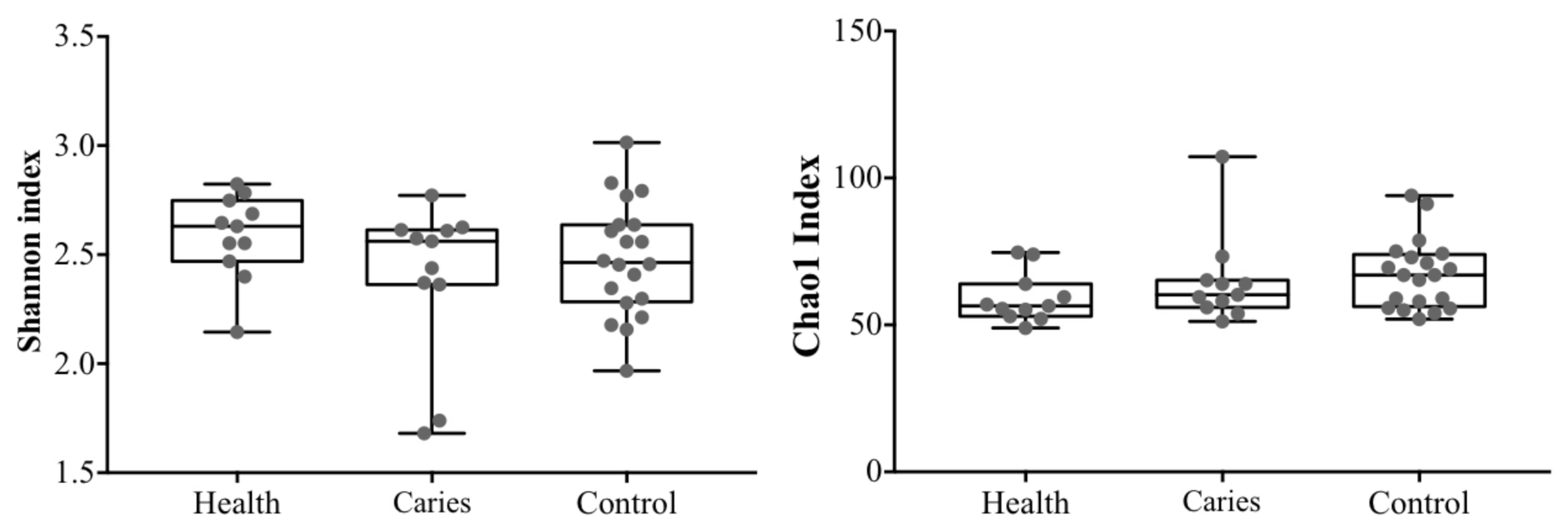
Figure 1. Alpha diversity indexes in all samples. Statistical differences were not detected.
Diagnostics 11 00835 g001
Figure 2. Phyla distribution: (top) the median values for each phyla and group; and (bottom) all individual values. * Represents Children with caries.
Diagnostics 11 00835 g002
Figure 3. Distribution of the major bacterial genera among all participants.* Represents Children with caries.
Diagnostics 11 00835 g003
Figure 4. Complex networks core microbiota for the three differenced subjects using Gephi.
Diagnostics 11 00835 g004
Figure 5. PCoA analysis: (top) the median value for the three categories of subjects respect to the abundance bacterial genera; and (bottom) the same analysis but considering each of the children and in relation to their sibling. The pairs of twins are linked by colored lines. Random colors are used to highlight the two pairs of twins.
Diagnostics 11 00835 g005
Table 1. Main characteristics of the 42 participants. C, caries; H, healthy.
Caries Group 22 InfantsSexAge
1C/1Hfemales12
2C/1Hfemales7
3C/1Hfemales8
4C/1Hfemales6
5C/1Hfemales11
6C/1Hmales9
7C/1Hfemales8
8C/1Hmales10
9C/1Hfemales12
10C/1Hfemales9
11C/1Hmales9
Control Group 20 InfantsSexAge
12females12
13females7
14males4
15females8
16females9
17females5
18females9
19males4
20females4
21males5
Table 2. Results of the accuracy of the 5 model used in this study. We have used different measures: Classification accuracy (CA), F1, precision, and recall.
ModelCAF1PrecisionRecall
kNN0.6660.6550.6460.665
SVM0.7380.6270.5450.738
Random Forest0.8810.8740.8800.881
Neural Network0.8100.8140.8230.810
Logistic Regression0.5950.6010.6070.595
Publisher’s Note: MDPI stays neutral with regard to jurisdictional claims in published maps and institutional affiliations.