Impact of [18F]FDG-PET and [18F]FLT-PET-Parameters in Patients with Suspected Relapse of Irradiated Lung Cancer
Abstract
1. Introduction
2. Materials and Methods
2.1. Patients
2.2. Imaging
2.3. Image Analyses
2.4. Outcome
2.5. Statistics
3. Results
3.1. FDG-SUVmax and FLT-SUVmax
3.2. Relapse Detection
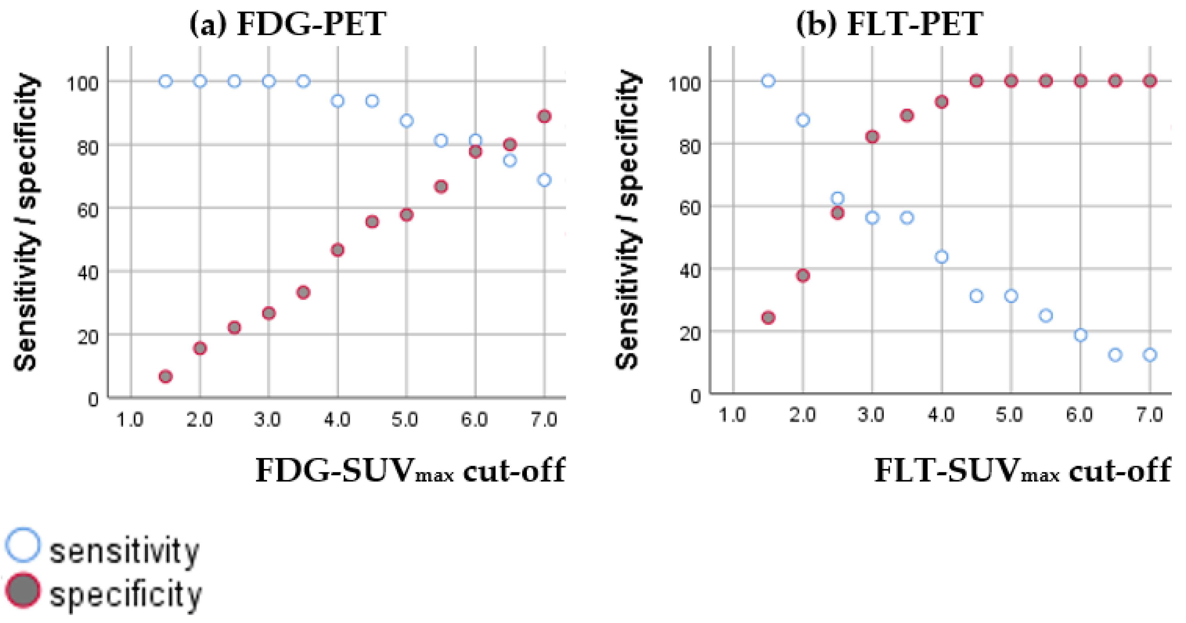
3.2.1. Diagnostic Accuracy of FDG-SUVmax and FLT-SUVmax
3.2.2. Diagnostic Value of Combining FDG-SUVmax and FLT-SUVmax
3.3. Prognosis
4. Discussion
5. Conclusions
Supplementary Materials
Author Contributions
Funding
Institutional Review Board Statement
Informed Consent Statement
Data Availability Statement
Acknowledgments
Conflicts of Interest
References
- Matsuo, Y. A Systematic Literature Review on Salvage Radiotherapy for Local or Regional Recurrence After Previous Stereotactic Body Radiotherapy for Lung Cancer. Technol. Cancer Res. Treat. 2018, 17, 1533033818798633. [Google Scholar] [CrossRef]
- Sheikhbahaei, S.; Mena, E.; Yanamadala, A.; Reddy, S.; Solnes, L.B.; Wachsmann, J.; Subramaniam, R.M. The Value of FDG PET/CT in Treatment Response Assessment, Follow-Up, and Surveillance of Lung Cancer. AJR Am. J. Roentgenol. 2017, 208, 420–433. [Google Scholar] [CrossRef] [PubMed]
- Postmus, P.E.; Kerr, K.M.; Oudkerk, M.; Senan, S.; Waller, D.A.; Vansteenkiste, J.; Escriu, C.; Peters, S.; Committee, E.G. Early and locally advanced non-small-cell lung cancer (NSCLC): ESMO Clinical Practice Guidelines for diagnosis, treatment and follow-up. Ann. Oncol. 2017, 28 (Suppl. 4), iv1–iv21. [Google Scholar] [CrossRef]
- Christensen, T.N.; Langer, S.W.; Persson, G.F.; Larsen, K.R.; Loft, A.; Amtoft, A.G.; Berthelsen, A.K.; Johannesen, H.H.; Keller, S.H.; Kjaer, A.; et al. (18)F-FLT-PET/CT adds value to (18)F-FDG-PET/CT for diagnosing relapse after definitive radiotherapy in patients with lung cancer. Results of a prospective clinical trial. J. Nucl. Med. 2020. [Google Scholar] [CrossRef]
- Inoue, T.; Kim, E.E.; Komaki, R.; Wong, F.C.; Bassa, P.; Wong, W.H.; Yang, D.J.; Endo, K.; Podoloff, D.A. Detecting recurrent or residual lung cancer with FDG-PET. J. Nucl. Med. 1995, 36, 788–793. [Google Scholar] [PubMed]
- Nakajima, N.; Sugawara, Y.; Kataoka, M.; Hamamoto, Y.; Ochi, T.; Sakai, S.; Takahashi, T.; Kajihara, M.; Teramoto, N.; Yamashita, M.; et al. Differentiation of tumor recurrence from radiation-induced pulmonary fibrosis after stereotactic ablative radiotherapy for lung cancer: Characterization of 18F-FDG PET/CT findings. Ann. Nucl. Med. 2013, 27, 261–270. [Google Scholar] [CrossRef]
- Matsuo, Y.; Nakamoto, Y.; Nagata, Y.; Shibuya, K.; Takayama, K.; Norihisa, Y.; Narabayashi, M.; Mizowaki, T.; Saga, T.; Higashi, T.; et al. Characterization of FDG-PET images after stereotactic body radiation therapy for lung cancer. Radiother. Oncol. 2010, 97, 200–204. [Google Scholar] [CrossRef] [PubMed]
- Hoopes, D.J.; Tann, M.; Fletcher, J.W.; Forquer, J.A.; Lin, P.F.; Lo, S.S.; Timmerman, R.D.; McGarry, R.C. FDG-PET and stereotactic body radiotherapy (SBRT) for stage I non-small-cell lung cancer. Lung Cancer 2007, 56, 229–234. [Google Scholar] [CrossRef]
- Hoeben, B.A.; Troost, E.G.; Bussink, J.; van Herpen, C.M.; Oyen, W.J.; Kaanders, J.H. 18F-FLT PET changes during radiotherapy combined with cetuximab in head and neck squamous cell carcinoma patients. Nukl. Nucl. Med. 2014, 53. [Google Scholar] [CrossRef]
- Hoshikawa, H.; Mori, T.; Kishino, T.; Yamamoto, Y.; Inamoto, R.; Akiyama, K.; Mori, N.; Nishiyama, Y. Changes in (18)F-fluorothymidine and (18)F-fluorodeoxyglucose positron emission tomography imaging in patients with head and neck cancer treated with chemoradiotherapy. Ann. Nucl. Med. 2013, 27, 363–370. [Google Scholar] [CrossRef]
- Hoeben, B.A.; Troost, E.G.; Span, P.N.; van Herpen, C.M.; Bussink, J.; Oyen, W.J.; Kaanders, J.H. 18F-FLT PET during radiotherapy or chemoradiotherapy in head and neck squamous cell carcinoma is an early predictor of outcome. J. Nucl. Med. Off. Publ. Soc. Nucl. Med. 2013, 54, 532–540. [Google Scholar] [CrossRef] [PubMed]
- Cho, L.P.; Kim, C.K.; Viswanathan, A.N. Pilot study assessing (18)F-fluorothymidine PET/CT in cervical and vaginal cancers before and after external beam radiation. Gynecol. Oncol. Rep. 2015, 14, 34–37. [Google Scholar] [CrossRef]
- Everitt, S.J.; Ball, D.L.; Hicks, R.J.; Callahan, J.; Plumridge, N.; Collins, M.; Herschtal, A.; Binns, D.; Kron, T.; Schneider, M.; et al. Differential (18)F-FDG and (18)F-FLT Uptake on Serial PET/CT Imaging Before and During Definitive Chemoradiation for Non-Small Cell Lung Cancer. J. Nucl. Med. 2014, 55, 1069–1074. [Google Scholar] [CrossRef]
- Vera, P.; Bohn, P.; Edet-Sanson, A.; Salles, A.; Hapdey, S.; Gardin, I.; Menard, J.F.; Modzelewski, R.; Thiberville, L.; Dubray, B. Simultaneous positron emission tomography (PET) assessment of metabolism with (1)(8)F-fluoro-2-deoxy-d-glucose (FDG), proliferation with (1)(8)F-fluoro-thymidine (FLT), and hypoxia with (1)(8)fluoro-misonidazole (F-miso) before and during radiotherapy in patients with non-small-cell lung cancer (NSCLC): A pilot study. Radiother. Oncol. 2011, 98, 109–116. [Google Scholar] [CrossRef] [PubMed]
- Everitt, S.; Hicks, R.J.; Ball, D.; Kron, T.; Schneider-Kolsky, M.; Walter, T.; Binns, D.; Mac Manus, M. Imaging cellular proliferation during chemo-radiotherapy: A pilot study of serial 18F-FLT positron emission tomography/computed tomography imaging for non-small-cell lung cancer. Int. J. Radiat. Oncol. Biol. Phys. 2009, 75, 1098–1104. [Google Scholar] [CrossRef] [PubMed]
- Trigonis, I.; Koh, P.K.; Taylor, B.; Tamal, M.; Ryder, D.; Earl, M.; Anton-Rodriguez, J.; Haslett, K.; Young, H.; Faivre-Finn, C.; et al. Early reduction in tumour [18F]fluorothymidine (FLT) uptake in patients with non-small cell lung cancer (NSCLC) treated with radiotherapy alone. Eur. J. Nucl. Med. Mol. Imaging 2014, 41, 682–693. [Google Scholar] [CrossRef]
- Wang, Z.; Wang, Y.; Sui, X.; Zhang, W.; Shi, R.; Zhang, Y.; Dang, Y.; Qiao, Z.; Zhang, B.; Song, W.; et al. Performance of FLT-PET for pulmonary lesion diagnosis compared with traditional FDG-PET: A meta-analysis. Eur. J. Radiol. 2015, 84, 1371–1377. [Google Scholar] [CrossRef]
- Hiniker, S.M.; Sodji, Q.; Quon, A.; Gutkin, P.M.; Arksey, N.; Graves, E.E.; Chin, F.T.; Maxim, P.G.; Diehn, M.; Loo, B.W., Jr. FLT-PET-CT for the Detection of Disease Recurrence After Stereotactic Ablative Radiotherapy or Hyperfractionation for Thoracic Malignancy: A Prospective Pilot Study. Front. Oncol. 2019, 9, 467. [Google Scholar] [CrossRef]
- Saga, T.; Koizumi, M.; Inubushi, M.; Yoshikawa, K.; Tanimoto, K.; Fukumura, T.; Miyamoto, T.; Nakajima, M.; Yamamoto, N.; Baba, M. PET/CT with 3’-deoxy-3’-[18F]fluorothymidine for lung cancer patients receiving carbon-ion radiotherapy. Nucl. Med. Commun. 2011, 32, 348–355. [Google Scholar] [CrossRef]
- Lopez Guerra, J.L.; Gladish, G.; Komaki, R.; Gomez, D.; Zhuang, Y.; Liao, Z. Large decreases in standardized uptake values after definitive radiation are associated with better survival of patients with locally advanced non-small cell lung cancer. J. Nucl. Med. 2012, 53, 225–233. [Google Scholar] [CrossRef]
- Bollineni, V.R.; Widder, J.; Pruim, J.; Langendijk, J.A.; Wiegman, E.M. Residual (1)(8)F-FDG-PET uptake 12 weeks after stereotactic ablative radiotherapy for stage I non-small-cell lung cancer predicts local control. Int. J. Radiat. Oncol. Biol. Phys. 2012, 83, e551-5. [Google Scholar] [CrossRef]
- Pierson, C.; Grinchak, T.; Sokolovic, C.; Holland, B.; Parent, T.; Bowling, M.; Arastu, H.; Walker, P.; Ju, A. Response criteria in solid tumors (PERCIST/RECIST) and SUVmax in early-stage non-small cell lung cancer patients treated with stereotactic body radiotherapy. Radiat. Oncol. 2018, 13, 34. [Google Scholar] [CrossRef] [PubMed]
- Lee, J.; Kim, J.O.; Jung, C.K.; Kim, Y.S.; Yoo Ie, R.; Choi, W.H.; Jeon, E.K.; Hong, S.H.; Chun, S.H.; Kim, S.J.; et al. Metabolic activity on [18f]-fluorodeoxyglucose-positron emission tomography/computed tomography and glucose transporter-1 expression might predict clinical outcomes in patients with limited disease small-cell lung cancer who receive concurrent chemoradiation. Clin. Lung Cancer 2014, 15, e13-21. [Google Scholar] [CrossRef]
- Boellaard, R.; Delgado-Bolton, R.; Oyen, W.J.; Giammarile, F.; Tatsch, K.; Eschner, W.; Verzijlbergen, F.J.; Barrington, S.F.; Pike, L.C.; Weber, W.A.; et al. European Association of Nuclear M. FDG PET/CT: EANM procedure guidelines for tumour imaging: Version 2.0. Eur. J. Nucl. Med. Mol. Imaging 2015, 42, 328–354. [Google Scholar] [CrossRef] [PubMed]
- Shusharina, N.; Cho, J.; Sharp, G.C.; Choi, N.C. Correlation of (18)F-FDG avid volumes on pre-radiation therapy and post-radiation therapy FDG PET scans in recurrent lung cancer. Int. J. Radiat. Oncol. Biol. Phys. 2014, 89, 137–144. [Google Scholar] [CrossRef] [PubMed][Green Version]
- Jones, M.P.; Hruby, G.; Metser, U.; Sridharan, S.; Capp, A.; Kumar, M.; Gallagher, S.; Rutherford, N.; Holder, C.; Oldmeadow, C.; et al. FDG-PET parameters predict for recurrence in anal cancer-results from a prospective, multicentre clinical trial. Radiat. Oncol. 2019, 14, 140. [Google Scholar] [CrossRef]
- Boellaard, R.; O’Doherty, M.J.; Weber, W.A.; Mottaghy, F.M.; Lonsdale, M.N.; Stroobants, S.G.; Oyen, W.J.; Kotzerke, J.; Hoekstra, O.S.; Pruim, J.; et al. FDG PET and PET/CT: EANM procedure guidelines for tumour PET imaging: Version 1.0. Eur. J. Nucl. Med. Mol. Imaging 2010, 37, 181–200. [Google Scholar] [CrossRef]
- Van Waarde, A.; Cobben, D.C.; Suurmeijer, A.J.; Maas, B.; Vaalburg, W.; de Vries, E.F.; Jager, P.L.; Hoekstra, H.J.; Elsinga, P.H. Selectivity of 18F-FLT and 18F-FDG for differentiating tumor from inflammation in a rodent model. J. Nucl. Med. 2004, 45, 695–700. [Google Scholar]
- Lee, T.S.; Ahn, S.H.; Moon, B.S.; Chun, K.S.; Kang, J.H.; Cheon, G.J.; Choi, C.W.; Lim, S.M. Comparison of 18F-FDG, 18F-FET and 18F-FLT for differentiation between tumor and inflammation in rats. Nucl. Med. Biol. 2009, 36, 681–686. [Google Scholar] [CrossRef]
- Yue, J.; Chen, L.; Cabrera, A.R.; Sun, X.; Zhao, S.; Zheng, F.; Han, A.; Zheng, J.; Teng, X.; Ma, L.; et al. Measuring tumor cell proliferation with 18F-FLT PET during radiotherapy of esophageal squamous cell carcinoma: A pilot clinical study. J. Nucl. Med. 2010, 51, 528–534. [Google Scholar] [CrossRef]
- Andersen, F.L.; Klausen, T.L.; Loft, A.; Beyer, T.; Holm, S. Clinical evaluation of PET image reconstruction using a spatial resolution model. Eur. J. Radiol. 2013, 82, 862–869. [Google Scholar] [CrossRef]
- Riegler, G.; Karanikas, G.; Rausch, I.; Hirtl, A.; El-Rabadi, K.; Marik, W.; Pivec, C.; Weber, M.; Prosch, H.; Mayerhoefer, M. Influence of PET reconstruction technique and matrix size on qualitative and quantitative assessment of lung lesions on [18F]-FDG-PET: A prospective study in 37 cancer patients. Eur. J. Radiol. 2017, 90, 20–26. [Google Scholar] [CrossRef]
- He, Y.Q.; Gong, H.L.; Deng, Y.F.; Li, W.M. Diagnostic efficacy of PET and PET/CT for recurrent lung cancer: A meta-analysis. Acta Radiol. 2014, 55, 309–317. [Google Scholar] [CrossRef] [PubMed]
- Hellwig, D.; Groschel, A.; Graeter, T.P.; Hellwig, A.P.; Nestle, U.; Schafers, H.J.; Sybrecht, G.W.; Kirsch, C.M. Diagnostic performance and prognostic impact of FDG-PET in suspected recurrence of surgically treated non-small cell lung cancer. Eur. J. Nucl. Med. Mol. Imaging 2006, 33, 13–21. [Google Scholar] [CrossRef] [PubMed]
- Im, H.J.; Pak, K.; Cheon, G.J.; Kang, K.W.; Kim, S.J.; Kim, I.J.; Chung, J.K.; Kim, E.E.; Lee, D.S. Prognostic value of volumetric parameters of (18)F-FDG PET in non-small-cell lung cancer: A meta-analysis. Eur. J. Nucl. Med. Mol. Imaging 2015, 42, 241–251. [Google Scholar] [CrossRef] [PubMed]
- Nygard, L.; Vogelius, I.R.; Fischer, B.M.; Kjaer, A.; Langer, S.W.; Aznar, M.C.; Persson, G.F.; Bentzen, S.M. A Competing Risk Model of First Failure Site after Definitive Chemoradiation Therapy for Locally Advanced Non-Small Cell Lung Cancer. J. Thorac. Oncol. 2018, 13, 559–567. [Google Scholar] [CrossRef] [PubMed]
- Scheffler, M.; Zander, T.; Nogova, L.; Kobe, C.; Kahraman, D.; Dietlein, M.; Papachristou, I.; Heukamp, L.; Buttner, R.; Boellaard, R.; et al. Prognostic impact of [18F]fluorothymidine and [18F]fluoro-D-glucose baseline uptakes in patients with lung cancer treated first-line with erlotinib. PLoS ONE 2013, 8, e53081. [Google Scholar] [CrossRef] [PubMed]


| Patient Characteristics | Number/Median | |
|---|---|---|
| Age, at day of suspicion (median (IQ)) | 70 (65–77) | |
| Sex (male/female) | 34/27 | |
| Histology | Adenocarcinoma | 29 |
| Squamous cell carcinoma | 25 | |
| SCLC | 2 | |
| Not otherwise specified (NOS) | 3 | |
| Mixed NSCLC/SCLC | 2 | |
| Stadium: | IA | 12 |
| Ib | 6 | |
| IIa | 2 | |
| IIb | 5 | |
| IIIa | 14 | |
| IIIb | 16 | |
| IV | 6 | |
| Chemotherapy (yes/no) | 34/27 | |
| Radiotherapy: | SBRT (45-72 Gy) | 28 |
| Normofractionated radiotherapy (66 Gy) | 29 | |
| Hyperfractionated radiotherapy (45-60 Gy) | 3 | |
| Normofractionated radiotherapy (66 Gy) and SBRT (45 Gy) | 1 | |
| Timing between scans | ||
| Time from the end of radiotherapy to relapse suspicion; in months (median (IQ)) | 7 (5–12) | |
| Time from relapse suspicion to FDG-PET/CT; in days (median (IQ)) | 21 (15–27) | |
| Time from relapse suspicion to FLT-PET/CT; in days (median (IQ)) | 23 (21–29) | |
| Time between FLT-PET/CT and FDG-PET/CT; in days (median (IQ)) | 6 (3–9) | |
| Outcome | ||
| Overall relapse (yes/no) | 32/29 | |
| HDV relapse (yes/no) | 16/45 | |
| Intra-pulmonary relapse (yes/no) | 30/31 | |
| Extra-pulmonary relapse (yes/no) | 7/54 | |
| Deceased (yes/no) | 33/28 | |
| Follow-up (from suspicion of relapse); in months (median (IQ)) | 25 (16–44) | |
| FDG-PET-Parameters | All Lesion Median [IQ] (n = 30) | Lesions within HDV Median [IQ] (n = 16) | Lesions Outside of HDV Median [IQ] (n = 14) | p-Value |
|---|---|---|---|---|
| SUVmax | 8.6 [5.1–16.0] | 12.8 [6.4–16.2] | 6.2 [3.3–13.7] | 0.095 |
| SUVpeak | 4.7 [3.1–9.0] | 7.1 [4.1–10.0] | 3.5 [1.9–5.7] | 0.014 * |
| MTV3.0 | 1.7 [0.5–10.5] | 6.6 [1.6–52.2] | 0.9 [0.2–2.0] | 0.016 * |
| MTV80% | 0.1 [0.1–0.5] | 0.2 [0.1–0.6] | 0.1 [0.1–0.2] | 0.113 |
| MTV50% | 1.2 [0.5–3.6] | 2.5 [1.2–7.9] | 0.7 [0.4–1.2] | 0.014 * |
| FLT-PET-parameters | ||||
| SUVmax | 3.7 [2.0–5.1] | 3.9 [1.2–7.9] | 3.3 [1.4–4.4] | 0.188 |
| SUVpeak | 2.3 [1.3–3.3] | 2.5 [1.6–3.5] | 2.0 [1.0–2.9] | 0.145 |
| PTV3.0 | 0.2 [0.0–2.0] | 0.3 [0.0–4.0] | 0.1 [0.0–0.4] | 0.128 |
| PTV80% | 0.2 [0.1–0.3] | 0.3 [0.1–0.5] | 0.1 [0.1–0.2] | 0.311 |
| PTV50% | 1.3 [0.8–4.3] | 2.7 [0.8–10.1] | 1.2 [0.5–1.5] | 0.066 |
| FDG-PET-Parameters in Recurrent Lesions, Continuous Variable | Hazard Ratio [95% CI] | p-Value |
|---|---|---|
| SUVmax (per unit) | 1.02 [0.94–1.10] | 0.675 |
| SUVpeak (per unit) | 1.07 [0.94–1.21] | 0.335 |
| MTV3.0 (per cm3) | 1.01 [0.99–1.02] | 0.485 |
| MTV80% (per cm3) | 3.63 [0.95–13.91] | 0.060 |
| MTV50% (per cm3) | 1.13 [1.00–1.27] | 0.054 |
| FDG-PET-parameters in recurrent lesions, dichotomized variable | ||
| SUVmax (>8.6) | 1.09 [0.43–2.76] | 0.853 |
| SUVpeak (>4.7) | 1.17 [0.46–2.96] | 0.741 |
| MTV3.0 (>1.7 cm3) | 0.81 [0.32–2.07] | 0.665 |
| MTV80% (>0.1 cm3) | 1.43 [0.56–3.64] | 0.454 |
| MTV50% (>1.2 cm3) | 1.56 [0.61–3.97] | 0.355 |
| FLT-PET-parameters in recurrent lesions, continuous variable | ||
| SUVmax (per unit) | 0.93 [0.75–1.15] | 0.496 |
| SUVpeak (per unit) | 0.92 [0.64–1.31] | 0.635 |
| PTV3.0 (per cm3) | 1.05 [0.94–1.17] | 0.411 |
| PTV80% (per cm3) | 4.13 [0.85–20.16] | 0.080 |
| PTV50% (per cm3) | 1.07 [1.01–1.13] | 0.014 * |
| FLT-PET-parameters in recurrent lesions, dichotomized variable | ||
| SUVmax (>3.7) | 0.80 [0.31–2.01] | 0.644 |
| SUVpeak (>2.3) | 0.96 [0.38–2.47] | 0.939 |
| PTV3.0 (>0.2 cm3) | 0.78 [0.30–2.07] | 0.619 |
| PTV80% (>0.2 cm3) | 1.67 [0.65–4.26] | 0.285 |
| PTV50% (>1.3 cm3) | 1.42 [0.56–3.61] | 0.465 |
| Clinical parameters | ||
| Age (at suspicion) (per year) | 1.04 [0.97–1.11] | 0.268 |
| Sex (male) | 2.74 [0.98–7.67] | 0.055 |
| Stadium (III vs. I–II) | 1.18 [0.45–3.14] | 0.736 |
| (IV vs. I–II) | 0.59 [0.07–4.83] | 0.622 |
| Radiotherapy (Conventionally fractionated radiotherapy vs. SBRT) | 0.97 [0.38–2.52] | 0.956 |
| Histology (Squamous cell carcinoma vs. adenocarcinoma) | 1.24 [0.48–3.20] | 0.659 |
| Time since the end of radiotherapy (per month) | 0.99 [0.90–1.09] | 0.837 |
| Site of relapse (within HDV vs. outside HDV) | 1.53 [0.59–3.94] | 0.381 |
| Intention for the relapse treatment (palliation vs. curative) | 1.33 [0.51–3.46] | 0.564 |
| Extra-pulmonary metastases (present) | 2.18 [0.70–6.77] | 0.180 |
| Covariate | Hazard Ratio [95% CI] | p-Value |
|---|---|---|
| MTV50% (per cm3) | 1.19 [0.99–1.44] | 0.063 |
| PTV50% (per cm3) | 1.02 [0.95–1.09] | 0.650 |
| Sex (male) | 4.32 [1.34–13.92] | 0.014 * |
| Age (at suspicion) (per year) | 1.08 [1.00–1.17] | 0.054 |
| Time since the end of radiotherapy (per month) | 0.96 [0.88–1.05] | 0.349 |
Publisher’s Note: MDPI stays neutral with regard to jurisdictional claims in published maps and institutional affiliations. |
© 2021 by the authors. Licensee MDPI, Basel, Switzerland. This article is an open access article distributed under the terms and conditions of the Creative Commons Attribution (CC BY) license (http://creativecommons.org/licenses/by/4.0/).
Share and Cite
Christensen, T.N.; Langer, S.W.; Persson, G.; Larsen, K.R.; Amtoft, A.G.; Keller, S.H.; Kjaer, A.; Fischer, B.M. Impact of [18F]FDG-PET and [18F]FLT-PET-Parameters in Patients with Suspected Relapse of Irradiated Lung Cancer. Diagnostics 2021, 11, 279. https://doi.org/10.3390/diagnostics11020279
Christensen TN, Langer SW, Persson G, Larsen KR, Amtoft AG, Keller SH, Kjaer A, Fischer BM. Impact of [18F]FDG-PET and [18F]FLT-PET-Parameters in Patients with Suspected Relapse of Irradiated Lung Cancer. Diagnostics. 2021; 11(2):279. https://doi.org/10.3390/diagnostics11020279
Chicago/Turabian StyleChristensen, Tine N., Seppo W. Langer, Gitte Persson, Klaus Richter Larsen, Annemarie G. Amtoft, Sune H. Keller, Andreas Kjaer, and Barbara Malene Fischer. 2021. "Impact of [18F]FDG-PET and [18F]FLT-PET-Parameters in Patients with Suspected Relapse of Irradiated Lung Cancer" Diagnostics 11, no. 2: 279. https://doi.org/10.3390/diagnostics11020279
APA StyleChristensen, T. N., Langer, S. W., Persson, G., Larsen, K. R., Amtoft, A. G., Keller, S. H., Kjaer, A., & Fischer, B. M. (2021). Impact of [18F]FDG-PET and [18F]FLT-PET-Parameters in Patients with Suspected Relapse of Irradiated Lung Cancer. Diagnostics, 11(2), 279. https://doi.org/10.3390/diagnostics11020279


