
OCT-Angiography Findings in Patients with Amblyopia: Comparison between Healthy Controls, Treatment-Responsive, and Treatment-Unresponsive Amblyopic Patients

 , ,
, ,  , ,
, , 
Abstract
:1. Introduction
2. Materials and Methods
3. Statistical Analysis
4. Results
5. Discussion
5.1. Vascular Density and Perfusion
5.2. Retinal Thickness
5.3. FAZ
5.4. Limitations
Author Contributions
Funding
Institutional Review Board Statement
Informed Consent Statement
Acknowledgments
Conflicts of Interest
References
- McKean-Cowdin, R.; Cotter, S.A.; Tarczy-Hornoch, K.; Wen, G.; Kim, J.; Borchert, M.; Varma, R.; Multi-Ethnic Pediatric Eye Disease Study Group. Prevalence of amblyopia or strabismus in Asian and non-Hispanic white preschool children: Multi-Ethnic Pediatric Eye Disease Study. Ophthalmology 2013, 120, 2117–2124. [Google Scholar] [CrossRef] [Green Version]
- Friedman, D.S.; Repka, M.X.; Katz, J.; Giordano, L.; Ibironke, J.; Hawse, P.; Tielsch, J. Prevalence of amblyopia and strabismus in white and African American children aged 6 through 71 months the Baltimore Pediatric Eye Disease Study. Ophthalmology 2009, 116, 2128–2134.e2. [Google Scholar] [CrossRef] [Green Version]
- Attebo, K.; Mitchell, P.; Cumming, R.; Smith, W.; Jolly, N.; Sparkes, R. Prevalence and causes of amblyopia in an adult population. Ophthalmology 1998, 105, 154–159. [Google Scholar] [CrossRef]
- Robaei, D.; Kifley, A.; Rose, K.A.; Mitchell, P. Impact of amblyopia on vision at age 12 years: Findings from a population-based study. Eye 2008, 22, 496–502. [Google Scholar] [CrossRef]
- Holmes, J.M.; Clarke, M.P. Amblyopia. Lancet 2006, 367, 1343–1351. [Google Scholar] [CrossRef]
- Simons, K. Amblyopia characterization, treatment, and pro-phylaxis. Surv. Ophthalmol. 2005, 50, 123–166. [Google Scholar] [CrossRef] [PubMed]
- Von Noorden, G.K.; Crawford, M.L. The lateral geniculate nucleus in human strabismic amblyopia. Investig. Ophthalmol. Vis. Sci. 1992, 33, 2729–2732. [Google Scholar]
- Miki, A.; Liu, G.T.; Goldsmith, Z.G.; Liu, C.S.; Haselgrove, J.C. Decreased activation of the lateral geniculate nucleus in a patient with anisometropic amblyopia demonstrated by functional magnetic resonance imaging. Ophthalmologica 2003, 217, 365–369. [Google Scholar] [CrossRef]
- Wiesel, T.N.; Hubel, D.H. Effects of visual deprivation on morphology and physiology of cells in the cats lateral geniculate body. J. Neurophysiol. 1963, 26, 978–993. [Google Scholar] [CrossRef] [PubMed]
- Aygıt, F.D.; Yılmaz, I.; Ozkaya, A.; Alkın, Z.; Gokyigit, B.; Yazıcı, A.T.; Demirok, A. Choroidal thickness of children’s eyes with anisometropic and strabismic amblyopia. J. Am. Assoc. Pediatr. Ophthalmol. Strabismus 2015, 19, 237–241. [Google Scholar] [CrossRef]
- Li, J.; Ji, P.; Yu, M. Meta-analysis of retinal changes in unilateral amblyopia using optical coherence tomography. Eur. J. Ophthalmol. 2015, 25, 400–409. [Google Scholar] [CrossRef]
- Altıntaş, Ö.; Gümüştaş, S.; Cinik, R.; Anık, Y.; Özkan, B.; Karabaş, L. Correlation of the measurements of optical coherence tomography and diffuse tension imaging of optic pathways in amblyopia. Int. Ophthalmol. 2017, 37, 85–93. [Google Scholar] [CrossRef]
- Yassin, S.A.; Al-Tamimi, E.R.; Al-Hassan, S. Macular and retinal nerve fiber thickness in recovered and persistent amblyopia. Int. Ophthalmol. 2015, 35, 833–842. [Google Scholar] [CrossRef]
- Dickmann, A.; Petroni, S.; Perrotta, V.; Salerni, A.; Parrilla, R.; Aliberti, S.; Savastano, M.C.; Centra, D.; Discendenti, S.; Balestrazzi, E. A morpho-functional study of amblyopic eyes with the use of optical coherence tomography and microperimetry. J. Am. Assoc. Pediatr. Ophthalmol. Strabismus 2011, 15, 338–341. [Google Scholar] [CrossRef]
- Dickmann, A.; Petroni, S.; Salerni, A.; Dell’Omo, R.; Balestrazzi, E. Unilateral amblyopia: An optical coherence tomography study. J. Am. Assoc. Pediatr. Ophthalmol. Strabismus 2009, 13, 148–150. [Google Scholar] [CrossRef]
- Spaide, R.F.; Fujimoto, J.G.; Waheed, N.K.; Sadda, S.R.; Staurenghi, G. Optical coherence tomography angiography. Prog. Retin. Eye Res. 2018, 64, 1–55. [Google Scholar] [CrossRef]
- Harris, P.A.; Taylor, R.; Thielke, R.; Payne, J.; Gonzalez, N.; Conde, J.G. Research electronic data capture (REDCap)—A metadata-driven methodology and workflow process for providing translational research informatics support. J. Biomed. Informatics 2009, 42, 377–381. [Google Scholar] [CrossRef] [Green Version]
- Von Noorden, G.K. Histological studies of the visual system in monkeys with experimental amblyopia. Invest. Ophthalmol. 1973, 12, 727–738. [Google Scholar] [PubMed]
- Fifkova, E. Changes in the visual cortex of rats after unilateral deprivation. Nature 1968, 220, 379–381. [Google Scholar] [CrossRef]
- Gaier, E.D.; Gise, R.; Heidary, G. Imaging Amblyopia: Insights from Optical Coherence Tomography (OCT). Semin. Ophthalmol. 2019, 34, 303–311. [Google Scholar] [CrossRef]
- Zhang, T.; Xie, S.; Liu, Y.; Xue, C.; Zhang, W. Effect of amblyopia treatment on macular microvasculature in children with anisometropic amblyopia using optical coherence tomographic angiography. Sci. Rep. 2021, 11, 39. [Google Scholar] [CrossRef]
- Karabulut, M.; Karabulut, S.; Sül, S.; Karalezli, A. Microvascular changes in amblyopic eyes detected by optical coherence tomography angiography. J. Am. Assoc. Pediatr. Ophthalmol. Strabismus 2019, 23, 155.e1–155.e4. [Google Scholar] [CrossRef]
- Yilmaz, I.; Ocak, O.B.; Yilmaz, B.S.; Inal, A.; Gokyigit, B.; Taskapili, M. Comparison of quantitative measurement of foveal avascular zone and macular vessel density in eyes of children with amblyopia and healthy controls: An optical coherence tomography angiography study. J. Am. Assoc. Pediatr. Ophthalmol. Strabismus 2017, 21, 224–228. [Google Scholar] [CrossRef]
- Lonngi, M.; Velez, F.G.; Tsui, I.; Davila, J.P.; Rahimi, M.; Chan, C.; Sarraf, D.; Demer, J.L.; Pineles, S.L. Spectral-Domain Optical Coherence Tomographic Angiography in Children With Amblyopia. JAMA Ophthalmol. 2017, 135, 1086–1091. [Google Scholar] [CrossRef] [PubMed]
- Chen, W.; Lou, J.; Thorn, F.; Wang, Y.; Mao, J.; Wang, Q.; Yu, X. Retinal Microvasculature in Amblyopic Children and the Quantitative Relationship Between Retinal Perfusion and Thickness. Investig. Ophthalmol. Vis. Sci. 2019, 60, 1185–1191. [Google Scholar] [CrossRef] [Green Version]
- Sobral, I.; Rodrigues, T.M.; Soares, M.; Seara, M.; Monteiro, M.; Paiva, C.; Castela, R. OCT angiography findings in children with amblyopia. J. Am. Assoc. Pediatr. Ophthalmol. Strabismus 2018, 22, 286–289.e2. [Google Scholar] [CrossRef] [PubMed]
- Demirayak, B.; Vural, A.; Onur, I.U.; Kaya, F.S.; Yigit, F.U. Analysis of Macular Vessel Density and Foveal Avascular Zone Using Spectral-Domain Optical Coherence Tomography Angiography in Children With Amblyopia. J. Pediatr. Ophthalmol. Strabismus 2019, 56, 55–59. [Google Scholar] [CrossRef] [PubMed]
- Pujari, A.; Chawla, R.; Mukhija, R.; Obedulla, H.; Phuljhele, S.; Saxena, R.; Sharma, P.; Kumar, A. Assessment of macular vascular plexus density using optical coherence tomography angiography in cases of strabismic amblyopia. Indian J. Ophthalmol. 2019, 67, 520–522. [Google Scholar] [CrossRef] [PubMed]
- Araki, S.; Miki, A.; Goto, K.; Yamashita, T.; Yoneda, T.; Haruishi, K.; Ieki, Y.; Kiryu, J.; Maehara, G.; Yaoeda, K. Foveal avascular zone and macular vessel density after correction for magnification error in unilateral amblyopia using optical coherence tomography angiography. BMC Ophthalmol. 2019, 19, 1–6. Available online: https://www.ncbi.nlm.nih.gov/pmc/articles/PMC6683430/ (accessed on 22 August 2021). [CrossRef] [Green Version]
- Huynh, S.C.; Samarawickrama, C.; Wang, X.Y.; Rochtchina, E.; Wong, T.Y.; Gole, G.A.; Rose, K.A.; Mitchell, P. Macular and nerve fiber layer thickness in amblyopia: The Sydney Childhood Eye Study. Ophthalmology 2009, 116, 1604–1609. [Google Scholar] [CrossRef]
- Borrelli, E.; Lonngi, M.; Balasubramanian, S.; Tepelus, T.C.; Baghdasaryan, E.; Pineles, S.L.; Velez, F.G.; Sarraf, D.; Sadda, S.R.; Tsui, I. Increased choriocapillaris vessel density in amblyopic children: A case-control study. J. Am. Assoc. Pediatr. Ophthalmol. Strabismus 2018, 22, 366–370. [Google Scholar] [CrossRef]
- Yilmaz Cinar, F.G.; Ozkan, G. Macular capillary system and ganglion cell-layer complex of the amblyopic eye with optical cohorence tomography angiography and optical cohorence tomography. Int. Ophthalmol. 2021, 41, 675–686. [Google Scholar] [CrossRef]
- Dereli Can, G. Quantitative analysis of macular and peripapillary microvasculature in adults with anisometropic amblyopia. Int. Ophthalmol. 2020, 40, 1765–1772. [Google Scholar] [CrossRef] [PubMed]
- Wong, E.S.; Zhang, X.J.; Yuan, N.; Li, J.; Pang, C.P.; Chen, L.; Tham, C.C.; Cheung, C.Y.; Yam, J.C. Association of Optical Coherence Tomography Angiography Metrics With Detection of Impaired Macular Microvasculature and Decreased Vision in Amblyopic Eyes: The Hong Kong Children Eye Study. JAMA Ophthalmol. 2020, 138, 858–865. [Google Scholar] [CrossRef] [PubMed]
- Pang, Y.; Frantz, K.A.; Block, S.; Goodfellow, G.W.; Allison, C. Effect of amblyopia treatment on macular thickness in eyes with myopic anisometropic amblyopia. Investig. Ophthalmol. Vis. Sci. 2015, 56, 2677–2683. [Google Scholar] [CrossRef] [Green Version]
- Iuvone, P.M.; Tigges, M.; Fernandes, A.; Tigges, J. Dopamine synthesis and metabolism in rhesus monkey retina: Development, aging, and the effects of monocular visual deprivation. Vis. Neurosci. 1989, 2, 465–471. [Google Scholar] [CrossRef]
- Iuvone, P.M. Neurotransmitters and neuromodulators in the retina: Regulation, interactions, and cellular effects. In The Retina: A Model for Cell Biology Studies; Adler, R., Farber, D., Eds.; Academic Press: Cambridge, MA, USA, 1986; pp. 1–72. [Google Scholar]
- Kato, S.; Nakamura, T.; Negishi, K. Postnatal development of dopaminergic cells in the rat retina. J. Comp. Neurol. 1980, 191, 27–36. [Google Scholar] [CrossRef]
- Melamed, E.; Frucht, Y.; Vidauri, J.; Uzzan, A.; Rosenthal, J. Effect of postnatal light deprivation on the ontogenesis of dopamine neurons in rat retina. Brain Res. 1986, 391, 280–284. [Google Scholar] [CrossRef]
- Favard, C.; Simon, A.; Vigny, A.; Nguyen-Legros, J. Ultrastructural evidence for a close relationship between dopamine cell processes and blood capillary walls in Macaca monkey and rat retina. Brain Res. 1990, 523, 127–133. [Google Scholar] [CrossRef]
- Roufail, E.; Stringer, M.; Rees, S. Nitric oxide synthase immunoreactivity and NADPH diaphorase staining are co-localised in neurons closely associated with the vasculature in rat and human retina. Brain Res. 1995, 684, 36–46. [Google Scholar] [CrossRef]
- Provis, J.M. Development of the primate retinal vasculature. Prog. Retin. Eye Res. 2001, 20, 799–821. [Google Scholar] [CrossRef]
- Kornfield, T.E.; Newman, E.A. Regulation of Blood Flow in the Retinal Trilaminar Vascular Network. J. Neurosci. 2014, 34, 11504–11513. [Google Scholar] [CrossRef] [PubMed] [Green Version]
- Son, T.; Alam, M.; Toslak, D.; Wang, B.; Lu, Y.; Yao, X. Functional optical coherence tomography of neurovascular coupling interactions in the retina. J. Biophotonics. 2018, 11, e201800089. [Google Scholar] [CrossRef]


| Total | Ambliopic Eyes | Ex Ambliopic Eyes | Normal Eyes | p | ||
|---|---|---|---|---|---|---|
| N patients | 59 | 16/59 (27.1%) | 15/59 (25.4%) | 28/59 (47.5%) | ||
| Sex | M = 26/59 (44.1%) F = 33/59 (55.9%) | M = 9/16 (56.3%) F = 7/16 (43.7%) | M = 5/15 (33.3%) F = 10/15 (66.7%) | M = 12/28 (42.9%) F = 16/28 (57.1%) | 0.431 | |
| Age | 8.9 ± 2.3 | 7.4 ± 1.6 | 9.3 ± 2.2 | 9.6 ± 2.3 | 0.07 | |
| Eye | R = 28/59 (47.5%) L = 31/59 (52.5%) | R = 10/16 (62.5%) L = 6/16 (37.5%) | R = 4/15 (26.7%) L = 11/15 (73.3%) | R = 14/28 (50%) L = 14/28 (50%) | 0.127 | |
| Spherical equivalent (D) | 1.37 ± 1.63 | 2.33 ± 2.15 | 2.52 ± 1.96 | 0.35 ± 0.24 | 0.048 | |
| Initial BCVA (logMAR) | 0.38 ± 0.34 | 0.40 ± 0.33 | 0.38 ± 0.27 | 0.33 | ||
| Final BCVA (logMAR) | 0.30 ± 0.19 | 0.35 ± 0.23 | 0.04 ± 0.12 | 0.02 ± 0.01 | 0.036 | |
| Duration of treatment (months) | 13.43 ± 1.61 | 13.27 ± 1.21 | 13.64 ± 2.42 | 0.86 | ||
| Central macular thickness | 276.3 ± 27.2 | 269.6 ± 28.4 | 277.8 ± 21.5 | 279.1 ± 24.2 | 0.89 | |
| FAZ area (mm2) | 0.22 ± 0.12 | 0.19 ± 0.13 | 0.25 ± 0.13 | 0.23 ± 0.10 | 0.312 | |
| Density | Central | 10.8 ± 3.25 | 11.84 ± 3.55 | 11.47 ± 2.82 | 9.95 ± 3.01 | 0.112 |
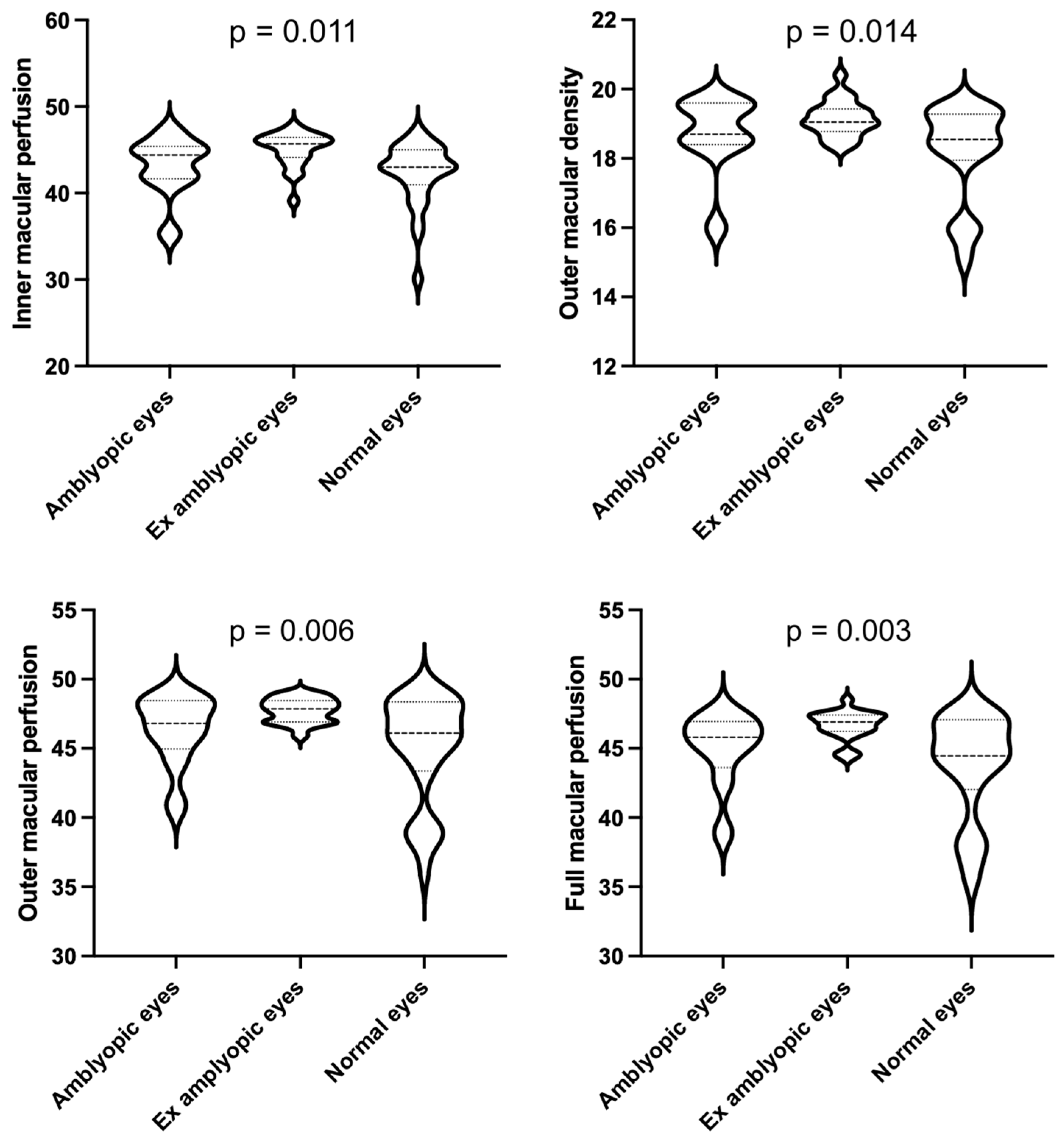
| Inner | 18.1 ± 1.32 | 18.29 ± 1.37 | 18.50 ± 0.78 | 17.75 ± 1.45 | 0.154 | |
| Outer | 18.6 ± 1.26 | 18.70 ± 1.14 | 19.15 ± 0.51 | 18.18 ± 1.40 | 0.014 | |
| Full | 18.3 ± 1.25 | 18.54 ± 0.96 | 18.67 ± 0.83 | 17.86 ± 1.38 | 0.042 | |
| Perfusion (%) | Central | 24.6 ± 7.72 | 27.24 ± 8.42 | 26.21 ± 7.13 | 22.36 ± 7.16 | 0.083 |
| Inner | 43.4 ± 3.46 | 43.11 ± 3.51 | 45.07 ± 2.03 | 42.26 ± 3.69 | 0.011 | |
| Outer | 46.2 ± 3.12 | 46.33 ± 2.53 | 47.74 ± 0.87 | 44.97 ± 3.82 | 0.006 | |
| Full | 45.0 ± 3.18 | 44.96 ± 2.71 | 46.65 ± 1.05 | 43.73 ± 3.73 | 0.003 |
Publisher’s Note: MDPI stays neutral with regard to jurisdictional claims in published maps and institutional affiliations. |
© 2021 by the authors. Licensee MDPI, Basel, Switzerland. This article is an open access article distributed under the terms and conditions of the Creative Commons Attribution (CC BY) license (https://creativecommons.org/licenses/by/4.0/).
Share and Cite
Salerni, A.; Gambini, G.; Fedeli, C.; Paris, L.; Crincoli, E.; Savino, G.; Savastano, M.C.; Bacherini, D.; De Vico, U.; Rizzo, C.; et al. OCT-Angiography Findings in Patients with Amblyopia: Comparison between Healthy Controls, Treatment-Responsive, and Treatment-Unresponsive Amblyopic Patients. Diagnostics 2021, 11, 1751. https://doi.org/10.3390/diagnostics11101751
Salerni A, Gambini G, Fedeli C, Paris L, Crincoli E, Savino G, Savastano MC, Bacherini D, De Vico U, Rizzo C, et al. OCT-Angiography Findings in Patients with Amblyopia: Comparison between Healthy Controls, Treatment-Responsive, and Treatment-Unresponsive Amblyopic Patients. Diagnostics. 2021; 11(10):1751. https://doi.org/10.3390/diagnostics11101751
Chicago/Turabian StyleSalerni, Annabella, Gloria Gambini, Chiara Fedeli, Ludovica Paris, Emanuele Crincoli, Gustavo Savino, Maria Cristina Savastano, Daniela Bacherini, Umberto De Vico, Clara Rizzo, and et al. 2021. "OCT-Angiography Findings in Patients with Amblyopia: Comparison between Healthy Controls, Treatment-Responsive, and Treatment-Unresponsive Amblyopic Patients" Diagnostics 11, no. 10: 1751. https://doi.org/10.3390/diagnostics11101751
APA StyleSalerni, A., Gambini, G., Fedeli, C., Paris, L., Crincoli, E., Savino, G., Savastano, M. C., Bacherini, D., De Vico, U., Rizzo, C., Killian, R., & Rizzo, S. (2021). OCT-Angiography Findings in Patients with Amblyopia: Comparison between Healthy Controls, Treatment-Responsive, and Treatment-Unresponsive Amblyopic Patients. Diagnostics, 11(10), 1751. https://doi.org/10.3390/diagnostics11101751