Article

Coronary Artery Disease is More Severe in Patients with Non-Alcoholic Steatohepatitis than Fatty Liver

1
Department of Gastroenterology and Hepatology, Yokohama City University Graduate School of Medicine, 3-9 Fukuura, Kanazawa-ku, Yokohama 236-0004, Japan
2
Department of Gastroenterology and Hepatology, Omori Red Cross Hospital, 4-30-1, Chuo, Ota-ku 194-0023, Japan
3
Department of Clinical Laboratory, Yokohama City University Hospital, 3-9 Fukuura, Kanazawa-ku, Yokohama 236-0004, Japan
*
Author to whom correspondence should be addressed.
Diagnostics 2020, 10(3), 129; https://doi.org/10.3390/diagnostics10030129
Submission received: 4 February 2020 / Revised: 24 February 2020 / Accepted: 24 February 2020 / Published: 26 February 2020
(This article belongs to the Section Pathology and Molecular Diagnostics)

Abstract

:
Non-alcoholic fatty liver disease (NAFLD) is associated with a higher risk of atherosclerotic disease. However, the relationships between the severity of coronary atherosclerosis and pathologic findings in patients with NAFLD remain unknown. We aimed to characterize the coronary artery lesions in patients with NAFLD using coronary computed tomography angiography (CCTA). Overall, 101 patients with liver biopsy-proven NAFLD who had chest pain or electrocardiographic abnormalities underwent CCTA. Coronary artery lesions, including coronary artery stenosis (CAS), calcium score (CACS, Agatston score), and coronary artery non-calcified plaque were assessed using multi-slice CT. Multivariate analysis showed that age, smoking status, prevalence of dyslipidemia (DLP) and non-alcoholic steatohepatitis (NASH), and stage of fibrosis were independent risk factors for CAS. Age, and the prevalence of DM and DLP, were independent risk factors for CACS, and the prevalence of NASH tended to be an independent risk factor. In addition, the prevalence of DLP and NASH were independent risk factors for non-calcified plaques. Coronary artery lesions are more common in patients with NASH than in those with non-alcoholic fatty liver, suggesting a higher risk in patients with NASH. Therefore, patients with NASH should be closely followed, with particular vigilance for coronary artery diseases.

1. Introduction

Non-alcoholic fatty liver disease (NAFLD) is generally considered to be the hepatic manifestation of metabolic syndrome and is characterized by fat accumulation in the liver in > 5% of hepatocytes, which occurs in the absence of significant alcohol intake, viral infection, or any other specific etiology of liver disease. NAFLD comprises a range of conditions from non-alcoholic fatty liver (NAFL) to non-alcoholic steatohepatitis (NASH) and cirrhosis. NAFLD is associated with a higher prevalence of atherosclerotic diseases, including cardiovascular disease (CVD) and diabetes mellitus (DM) [1,2]. Moreover, patients with NAFLD have higher risks of carotid intima media thickening [3,4], impaired endothelial function [5], coronary artery calcification [6,7], and greater arterial stiffness [8]. However, there are no data regarding the associations between the pathologic features of NAFLD and coronary artery lesions.
Coronary CT angiography (CCTA) has recently been established as a useful technique for evaluating coronary atherosclerosis, including stenosis, plaque characteristics, and calcium score (Agatston calcium score) [9]. Patients with high-risk coronary plaques, including non-calcified plaques, have a high risk of cardiovascular events, and the calcium score is proportional to the risk of such cardiovascular events [10,11]. NAFLD is associated with the progression of coronary calcium scores and high-risk coronary plaques, independent of conventional cardiovascular risk factors [12]. To date, however, no studies have investigated whether the severity of coronary artery lesions differs between patients with NAFL or NASH. We previously reported that the low-density lipoprotein (LDL)-migration index (MI), an indicator of serum small dense LDL (sdLDL) concentration, is higher in patients with NASH than in those with NAFL, which corresponds to the alterations in lipid metabolism involved, and this suggests that the risk of atherosclerotic diseases may be higher in patients with NASH than in those with NAFL [13,14]. Therefore, we hypothesized that there is a higher risk of coronary artery disease (CAD) in patients with NASH than in those with NAFL. This study aimed to assess coronary artery stenosis (CAS), coronary artery calcium score (CACS, Agatston score), and non-calcified plaques using CCTA in patients with NAFL or NASH.

2. Patients and Methods

2.1. Ethical Approval of the Study Protocol

The protocol of this study conforms to the ethical guidelines of the 1975 Declaration of Helsinki as reflected in a priori approval by the institution’s human research committee, and the study was conducted with the approval of the Ethics Committee of Yokohama City University Hospital (Yokohama, Japan) on February 15, 2014. This trial is registered with the University Medical Information Network Clinical Trials Registry as UMIN000009614. All participants provided written informed consent before examination.

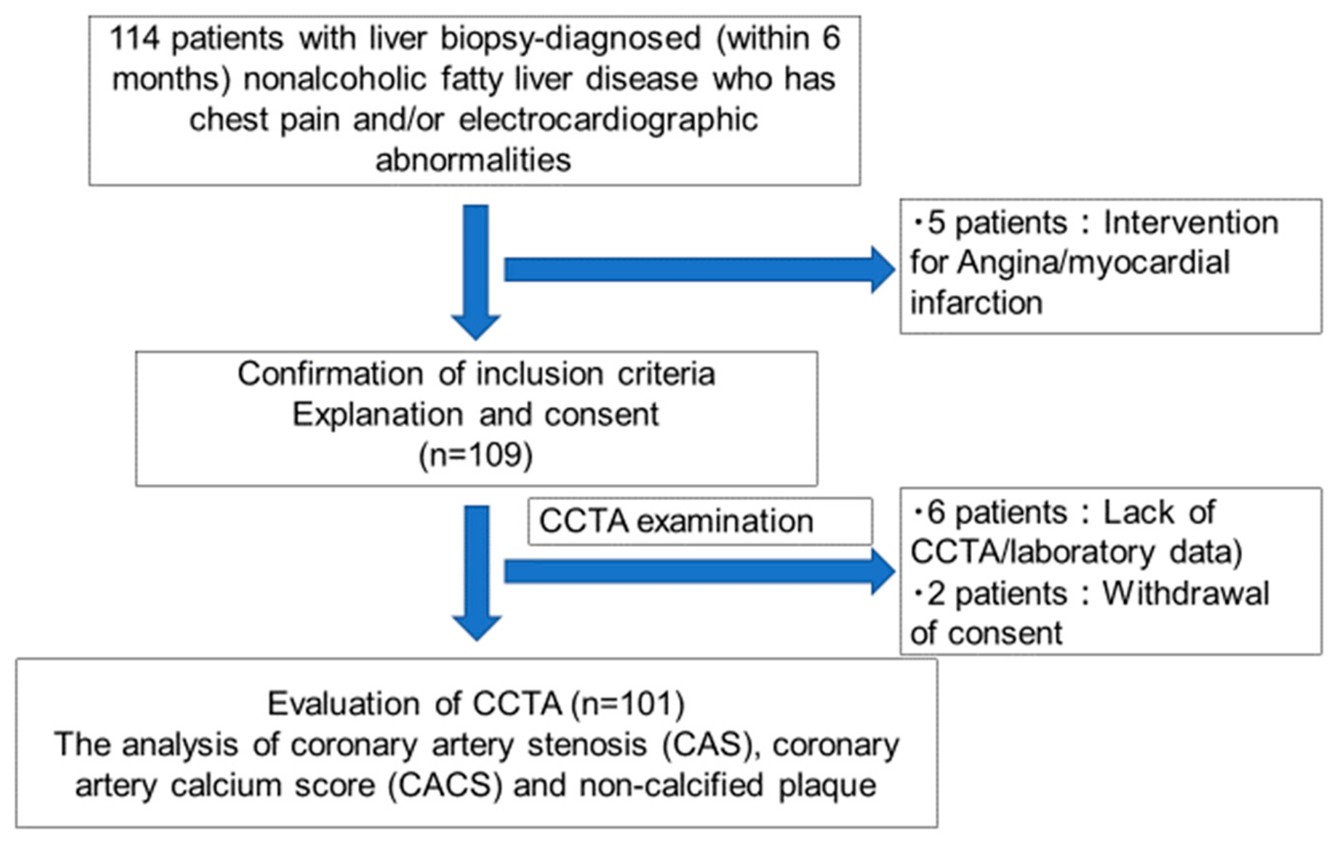
2.2. Study Population

The study population comprised patients who underwent liver biopsy between March 2014 and September 2018 and who were followed up at Yokohama City University Hospital in Japan. This study enrolled 114 patients, of whom five were excluded because they underwent percutaneous coronary intervention before CCTA examinations. We also excluded eight patients because of errors in CCTA examinations (six patients) or withdrawal of consent (two patients). Finally, 101 patients were enrolled in this study. The time interval between CCTA and liver biopsy was < 6 months. The exclusion criteria were as follows: any history of significant alcohol intake (≥210 g/week in men and 140 g/week in women), history of any other specific etiology of liver disease, or history of angina or myocardial infarction. After excluding 13 participants, data of the remaining 101 patients were analyzed (Figure 1).

2.3. Clinical and Laboratory Measurements

Basic demographic data, including the age and sex of study participants, and relevant medical history, including angina, myocardial infarction, renal dysfunction, DM, hypertension (HT), or dyslipidemia (DLP), were acquired from medical records. Smoking status and alcohol intake were obtained from the responses given in a self-reported questionnaire. The smoking score was calculated as the number of cigarettes smoked per day × the number of years as a smoker. Height and body mass were measured with participants wearing light clothing and no shoes. Body mass index (BMI) was calculated as body mass in kilograms divided by the square of height in meters (kg/m2). Blood pressure was measured after the participants rested quietly using an automatic manometer and an appropriate cuff at the time of hospitalization. Blood sampling was performed early in the morning, and the samples were analyzed in the central laboratory. Aspartate aminotransferase, alanine aminotransferase, γ-glutamyl transpeptidase, C-reactive protein, creatinine, fasting blood glucose, fasting insulin, hemoglobin A1c, fasting total cholesterol, triglycerides, high-density lipoprotein-cholesterol, and low-density lipoprotein-cholesterol concentrations were measured. DM was defined as a fasting plasma glucose concentration ≥126 mg/dL, self-reported history of DM, treatment with dietary modification, or use of antidiabetic medication. HT was defined as a blood pressure ≥ 140/90 mmHg or self-reported history of HT and/or the use of antihypertensive medication. DLP was defined as a total cholesterol concentration ≥ 240 mg/dL or the use of antihyperlipidemic medication.

2.4. Histopathologic and Immunohistochemical Evaluations

Liver biopsy specimens were obtained using a 16-gauge needle biopsy kit (Pro-Mag™ Ultra Automatic Biopsy Instrument; Argon Medical Devices, Frisco, TX, USA). Two specimens were obtained from each patient to acquire a sufficient sample size for analysis and to reduce histologic analysis errors. An adequate liver biopsy sample was defined as being > 20 mm in length and/or containing >10 portal tracts. Each specimen was assessed histologically by one pathologist, and the NAFLD activity score (NAS) was calculated [15]. Patients with steatosis, inflammation, and ballooned hepatocytes were classified as having NASH based on the results of the Fatty Liver Inhibition of Progression (FLIP) algorithm [16]. The severity of fibrosis was scored as previously described [17].

2.5. CCTA

CT images were acquired using a single-source 64-detector multi-slice CT with a standard scanning protocol. The protocol comprises a non-enhanced calcium score scan, followed by a contrast-enhanced CCTA scan. The images were then analyzed using a dedicated workstation (Volume analyzer, Synapse Vincent, Fujifilm, city, country). Based on the guidelines of the American Heart Association, a 16-segment coronary artery tree model was used. Using the Agatston score, coronary artery wall calcification was reported as previously described [18]. Agatston score is a semi-automated tool to calculate a score based on the extent of coronary artery calcification detected by an unenhanced CT scan, which is routinely performed in patients undergoing CCTA. This score was calculated using a weighted value assigned to the highest density of calcification in a given coronary artery and was stated in Hounsfield units (HUs). This weighted score was then multiplied by the area (in square millimeters) of the coronary calcification. The recently proposed Society of Cardiovascular Computed Tomography grading scale for CAS severity was used to assess the degree of luminal stenosis [19], in which Grade 0 represented no visible stenosis, Grade 1 represented 1–24% (minimal stenosis), Grade 2 represented 25–49% (mild stenosis), Grade 3 represented 50–69% (moderate stenosis), Grade 4 represented 70–99% (severe stenosis), and Grade 5 represented 100% (occlusion). The features of high-risk (non-calcified) plaques were defined as positive remodeling, CT attenuation < 30 HU, “napkin-ring” sign, and only spots of calcium deposition.

2.6. Statistical Analysis

Continuous variables are summarized as means and standard deviations and categorical variables as frequencies and percentages. Data were expressed as mean ± S.D., unless indicated otherwise. All statistical analyses were performed using SPSS 12.0 software (SPSS Inc., Chicago, IL, USA). The t-test and analysis of variance with Scheffe’s multiple testing correction were used for univariate comparisons between groups. Multivariate analysis was performed to determine factors independently associated with CAS, CACS, or non-calcified plaques. p <0.05 was considered to represent statistical significance.

3. Results

3.1. Baseline Characteristics

The study population comprised 101 patients with NAFLD (41 patients with NAFL and 60 with NASH). Baseline characteristics of each group are shown in Table 1.
The mean age of the NAFL group (52.7 ± 13.9 years) was significantly lower than that of the NASH group (59.6 ± 11.2 years) (p = 0.009). In addition, there were significantly more women in the NASH group (men/women, 26/34) than in the NAFL group (24/17) (p = 0.0471). No differences in other parameters were identified between the NAFL and NASH groups. Histologic characteristics are also summarized in Table 1. The grades for lobular inflammation, hepatocyte ballooning, and fibrosis stage in the NASH group were higher than those in the NAFL group.

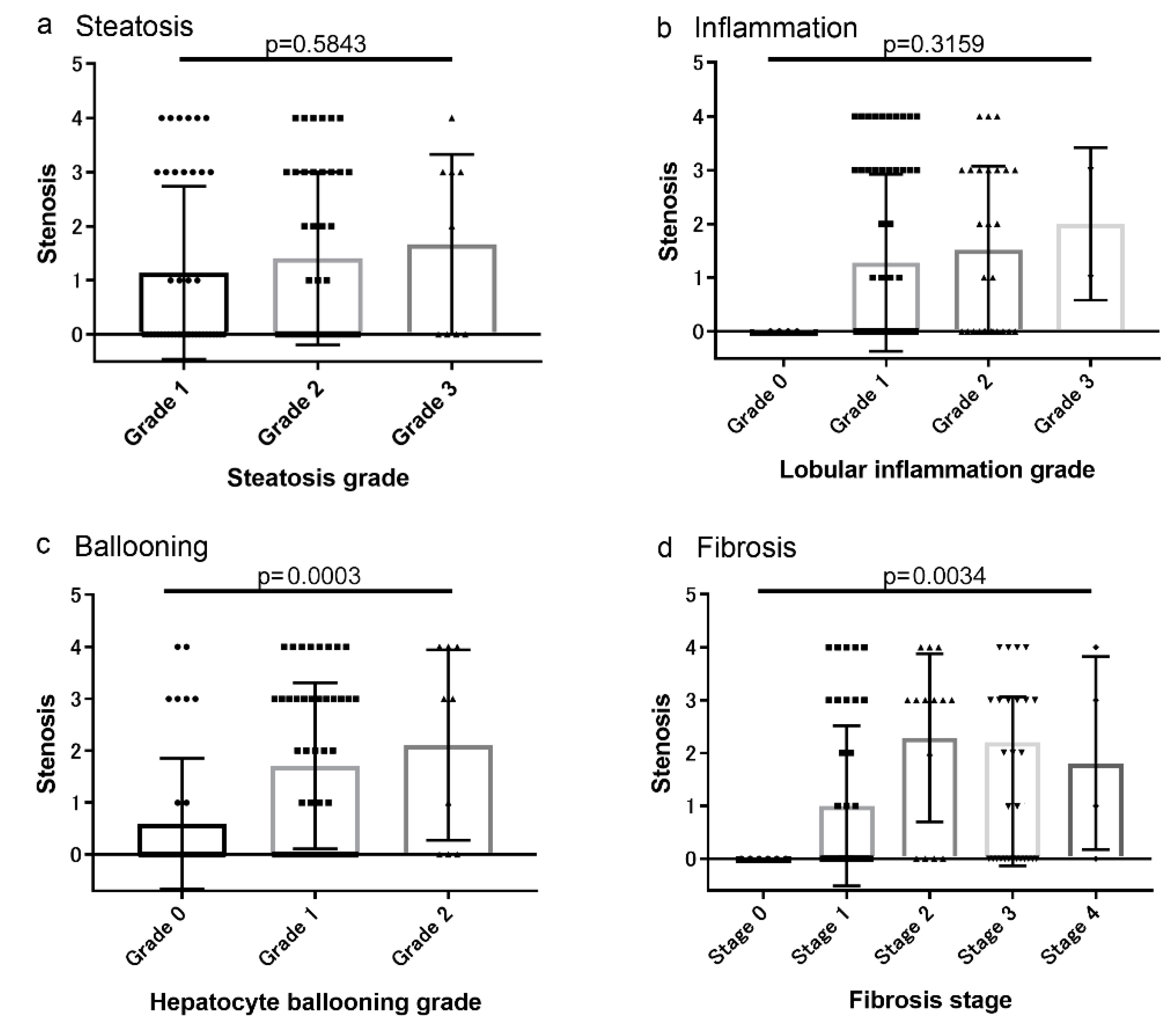
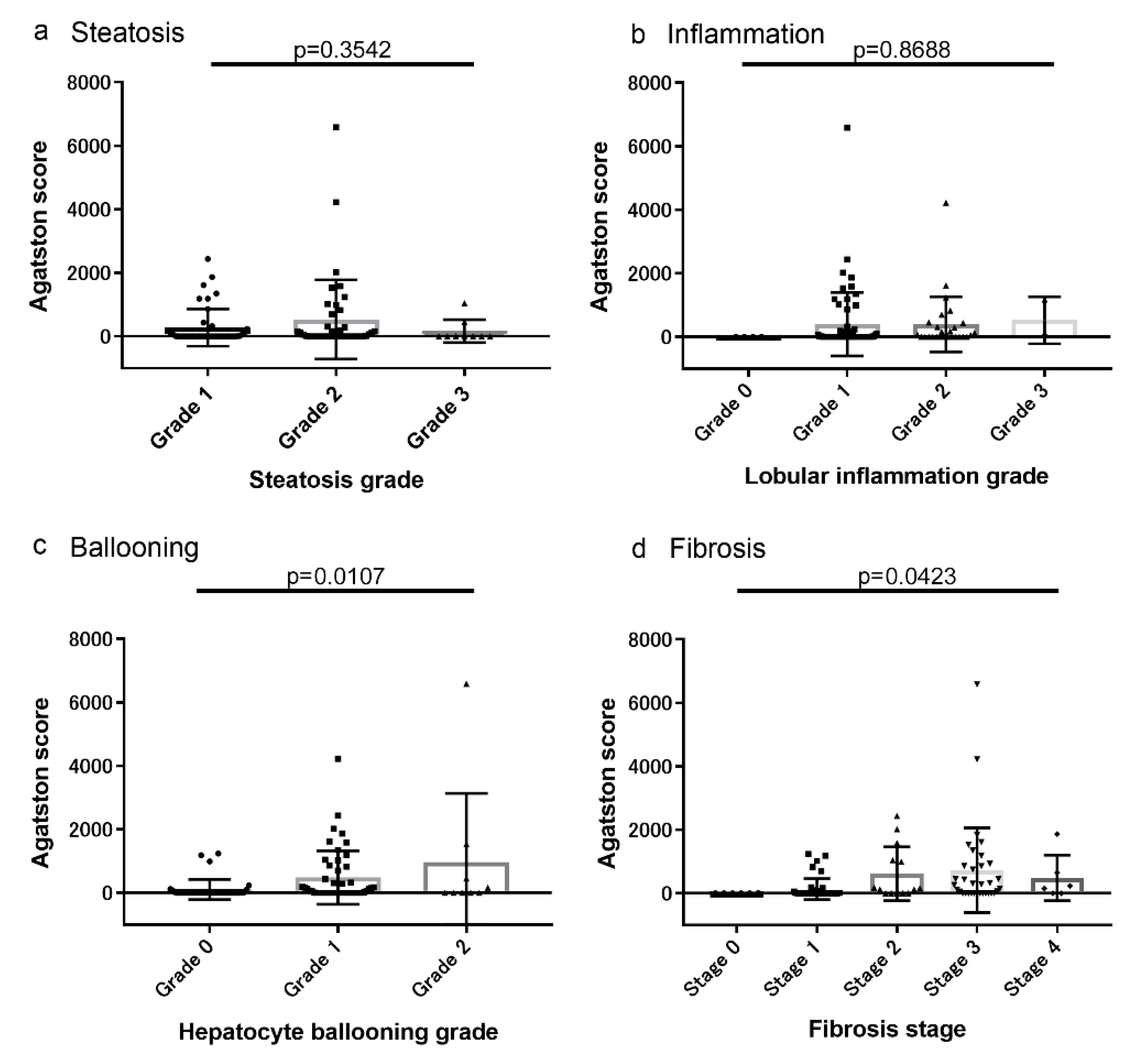
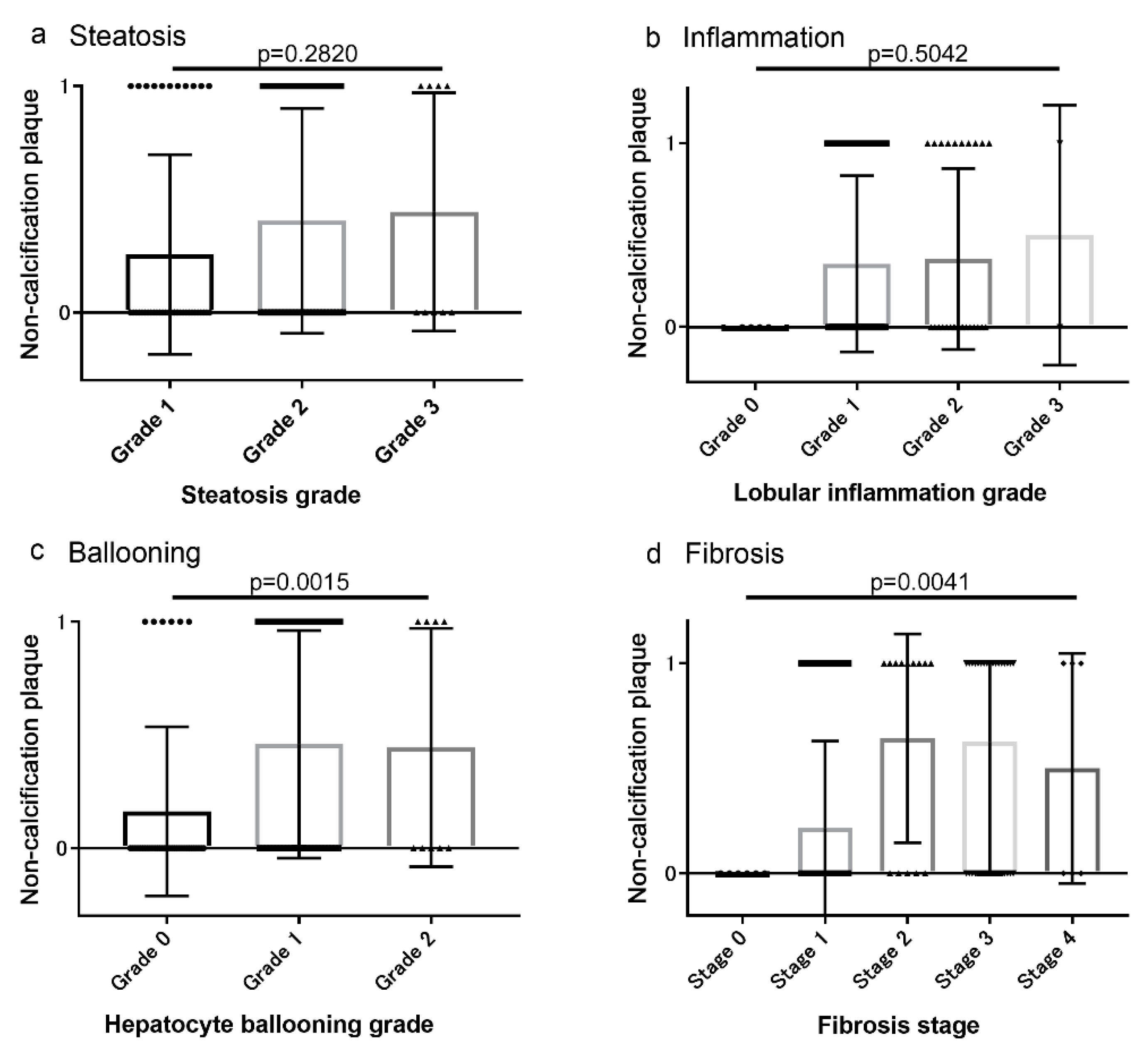
3.2. Associations between Coronary Artery Lesions and Histologic Findings

The associations of CAS, CACS, and non-calcified plaques with histologic characteristics are shown in Figure 2, Figure 3 and Figure 4, respectively. The severity of CAS, CACS, and non-calcified plaques was positively correlated with the hepatocyte ballooning grade and the stage of liver fibrosis (Figure 2, Figure 3 and Figure 4). However, there were no associations between these findings and the grades of steatosis or liver inflammation (Figure 2, Figure 3 and Figure 4).

3.3. Multiple Regression Analysis of Potential Risk Factors for CAS, CACS, and non-Calcified Plaque

The characteristics of NAFLD patients with and without CAS are shown in Table 2. Fifty-one patients had no significant stenosis and 50 had CAS. Univariate analysis showed that age; smoking score; prevalence of DM, HT, DLP, and NASH; and fibrosis stage were higher (p < 0.05 each) in patients with CAS than in those without. Multivariate analysis showed that age, smoking score, prevalence of DLP and NASH, and fibrosis stage were independent risk factors for CAS (all p < 0.05).
Next, the characteristics of NAFLD patients without (score = 0 AU) or with CACS (score >0 AU) were compared (Table 3). In total, 50 patients had no CACS and 51 had CACS. Univariate analysis showed that age; prevalence of DM, HT, DLP, and NASH; and fibrosis stage were higher (all p < 0.05) in patients with CACS than in those without. Multivariate analysis showed that age and the prevalence of DM and DLP were independent risk factors for CACS (p < 0.05 each). NASH tended to be associated with the presence of CACS, although not significantly (p = 0.0987).
Finally, the characteristics of NAFLD patients with and without non-calcified plaque are shown in Table 4. Sixty-three patients had no non-calcified plaque and 38 had non-calcified plaque. Univariate analysis showed that age; smoking score; prevalence of DM, HT, DLP, and NASH; and fibrosis stage were higher (p < 0.05 each) in patients with non-calcified plaque than in those with no non-calcified plaque. Multivariate analysis showed that the prevalence of DLP and NASH were independent risk factors for high-risk plaque (p < 0.05 each).

4. Discussion

This prospective single-center study showed that CAS and non-calcified plaque, a predictor of cardiovascular events, were more frequent in patients with NASH than in those with NAFL, which was diagnosed based on liver biopsy. In addition, NASH tended to be associated with CACS, whereas NAFL was not.
NAFLD may be a cardiovascular risk factor because a higher frequency of CVD was present in patients with NAFLD than in those without [20,21]. The association between NAFLD and atherosclerosis has been investigated in previous studies using the CAC score, which reflects the total coronary atherosclerotic burden. However, inconsistent results have been reported [22,23]. In addition, the absence of coronary calcification does not preclude the presence of significant CAD [12,24]. A recent study investigated the association between NAFLD and coronary atherosclerotic plaque using more comprehensive information regarding coronary atherosclerosis provided by CCTA [25]. However, the participants had not had their NAFLD confirmed by liver biopsy. Therefore, the present study evaluated the effect of the pathology associated with NAFLD on the risk of subclinical coronary atherosclerosis, not only on CACS but also on CAS and non-calcified plaque, as assessed using coronary artery CT.
Lipid disturbances, insulin resistance, and chronic inflammation are common features of the pathogenesis of NAFLD and CAD [26,27,28]. We previously reported that the reduction in the liver secretion of very low-density lipoprotein (VLDL) in patients with NASH is because of the reduction in VLDL synthesis secondary to the lower expression of microsomal triglyceride transfer protein [13,29,30]. The surplus triglycerides in these livers are therefore transported as triglyceride-rich lipoproteins (VLDL1), a precursor of sdLDL particles, which are more potent risk factors than LDL-C for atherosclerotic diseases, including CVD [31,32,33]. In addition, NAFLD and CVD progress because of the combination of insulin resistance, chronic inflammation, and oxidative stress, which are features of NASH rather than of NAFL. Therefore, we hypothesized that CAD is more common in patients with NASH than in those with NAFL, and the present study showed that patients with NASH had significantly worse coronary artery lesions than those with NAFL. This may be explained by higher sdLDL, insulin resistance, and chronic inflammation/oxidative stress in patients with NASH than in those with NAFL.
Coronary arteriosclerosis, evaluated using CACS, correlates with the concentration of a non-invasive fibrosis marker, the FIB-4 index, in patients with NAFLD. Our data show that CAD is worse in NAFLD patients with apparent fibrosis (fibrosis stage > 2). However, the multivariate analysis showed that the presence of NASH was a more significant risk factor for coronary artery lesions than fibrosis stage. Interestingly, a previous report showed that patients with NAFLD cirrhosis predominantly had liver-related events, whereas those with bridging fibrosis (fibrosis stage 3) predominantly had vascular events [34]. These findings suggest that NASH-related liver cirrhosis tends to impair liver function and reduce blood pressure, cholesterol concentration, and body mass, resulting in a lower incidence of vascular events than that observed in NASH patients with fibrosis stages 2–3.
This study had several limitations. First, the use of liver biopsy as the gold standard for assessing liver pathology was associated with sampling errors and intra- and interobserver variability, which are at least partly associated with the biopsy size. Second, the age and the percentage of gender were significantly different between NAFL and NASH groups in this cohort. Additionally, the patients included in the present study were referred from several hepatology centers in Japan that have a particular interest in NAFLD. For these reasons, some selection bias cannot be ruled out. Patient selection bias may also have occurred because of the result of liver biopsies being more likely to be performed for NAFLD patients at a risk of NASH. Finally, the study population could be different from the general population with NAFLD because the enrolled patients had chest pain and/or electrocardiographic abnormalities.

5. Conclusions

In this prospective study of patients with biopsy-diagnosed NAFLD undergoing CCTA, NASH was associated with a higher incidence of CAD than patient with NAFL, which implies a higher risk of cardiovascular events. However, these findings require corroboration in additional studies.

Author Contributions

Study conception and design: T.N., K.I. and A.N. Collection of data: A.O., T.K. (Takashi Kobayashi), M.I., Y.H., Takaomi Kessoku, Y.O., H.K., M.Y. and S.S. Data analysis: T.N., K.I. and A.N. Contribution of reagents/materials/analytical tools: T.N., K.I., M.Y. and A.N. Manuscript preparation: T.N., K.I., T.K. (Takaomi Kessoku), M.Y. and A.N. All authors have read and agreed to the published version of the manuscript.

Funding

Work in the author’s laboratory was supported by the “Step A” program of the Japan Science and Technology Agency (J.S.T.) and Kiban-B, Shingakujuturyouiki. In addition, this work was supported in part by grants-in-aid from the Japanese Ministry of Health, Labour and Welfare.

Acknowledgments

We thank Mark Cleasby, from Edanz Group (www.edanzediting.com/ac) for editing a draft of this manuscript.

Conflicts of Interest

The authors declare no conflict of interest.

Abbreviations

ALTAlanine aminotransferase
ASTAspartate aminotransferase
BMIBody mass index
CACSCoronary artery calcium score
CADCoronary artery disease
CASCoronary artery stenosis
CCTACoronary computed tomography angiography
CVDCardiovascular disease
DLPDyslipidemia
DMDiabetes mellitus
FLIPFatty Liver Inhibition of Progression
GGTGamma-glutamyl transpeptidase
CRPC-reactive protein
HbA1cHemoglobin A1c
HDL-CHigh-density lipoprotein-cholesterol
HTHypertension
HUHounsfield units
LDL-MILow-density lipoprotein-migration index
MSCTMulti-slice computer tomography
NAFLNon-alcoholic fatty liver
NAFLDNon-alcoholic fatty liver disease
NASNon-alcoholic fatty liver disease activity score
NASHNon-alcoholic steatohepatitis
SCCTSociety of Cardiovascular Computed Tomography
SdLDLSmall dense low-density lipoprotein
VLDLVery low-density lipoprotein

References

  1. Brea, A.; Mosquera, D.; Martin, E.; Arizti, A.; Cordero, J.L.; Ros, E. Nonalcoholic fatty liver disease is associated with carotid atherosclerosis: case-control study. Arter. Thromb Vasc Biol 2005, 25, 1045–1050. [Google Scholar] [CrossRef] [PubMed] [Green Version]
  2. Targher, G.; Arcaro, G. Non-alcoholic fatty liver disease and increased risk of cardiovascular disease. Atherosclerosis 2007, 191, 235–240. [Google Scholar] [CrossRef] [PubMed]
  3. Li, X.; Mingfeng, X.; Ma, H.; Hofman, A.; Hu, Y.; Yan, H.; He, W.; Lin, H.; Jeekel, J.; Zhao, N.; et al. Liver fat content is associated with increased carotid atherosclerosis in Chinese middle-agedand elderly population: Shanghai Changfeng study. Atherosclerosis 2012, 224, 480–485. [Google Scholar] [CrossRef] [PubMed]
  4. Targher, G.; Bertolini, L.; Padovani, R.; Rodella, S.; Zoppini, G.; Zenari, L.; Cigolini, M.; Falezza, G.; Arcaro, G. Relations between carotid artery wall thickness and liver histology in subjects with nonalcoholic fatty liver disease. Diabetes Care 2006, 29, 1325–1330. [Google Scholar] [CrossRef] [PubMed] [Green Version]
  5. Schindhelm, R.K.; Diamant, M.; Bakker, S.J.; van Dijk, R.A.; Scheffer, P.G.; Teerlink, T.; Kostense, P.J.; Heine, R.J. Liver alanine aminotransferase, insulin resistance and endothelial dysfunction in normotriglyceridaemic subjects with type 2 diabetes mellitus. Eur. J. Clin Invest. 2005, 35, 369–374. [Google Scholar] [CrossRef]
  6. Chen, C.H.; Nien, C.K.; Yang, C.C.; Yeh, Y.H. Association between nonalcoholic fatty liver disease and coronary artery calcification. Dig. Dis Sci 2010, 55, 1752–1760. [Google Scholar] [CrossRef]
  7. Akabame, S.; Hamaguchi, M.; Tomiyasu, K.; Tanaka, M.; Kobayashi-Takenaka, Y.; Nakano, K.; Oda, Y.; Yoshikawa, T. Evaluation of vulnerable coronary plaques and non-alcoholic fatty liver disease (NAFLD) by 64-detector multislice computed tomography (MSCT). Circ. J. 2008, 72, 618–625. [Google Scholar] [CrossRef] [Green Version]
  8. Kim, B.J.; Kim, N.H.; Kim, B.S.; Kang, J.H. The association between nonalcoholic fatty liver disease, metabolic syndrome and arterial stiffness in nondiabetic, nonhypertensive individuals. Cardiology 2012, 123, 54–61. [Google Scholar] [CrossRef]
  9. Park, G.M.; Yun, S.C.; Cho, Y.R.; Gil, E.H.; Her, S.H.; Kim, S.H.; Jo, M.W.; Lee, M.S.; Lee, S.W.; Kim, Y.H. Prevalence of coronary atherosclerosis in an Asian population: findings from coronary computed tomographic angiography. Int J. Cardiovasc Imaging 2015, 31, 659–668. [Google Scholar] [CrossRef]
  10. Tesche, C.; Plank, F.; De Cecco, C.N.; Duguay, T.M.; Albrecht, M.H.; Varga-Szemes, A.; Bayer, R.R., 2nd; Yang, J.; Jacks, I.L.; Gramer, B.M.; et al. Prognostic implications of coronary CT angiography-derived quantitative markers for the prediction of major adverse cardiac events. J. Cardiovasc Comput Tomogr. 2016, 6, 8–465. [Google Scholar] [CrossRef]
  11. Oudkerk, M.; Stillman, A.E.; Halliburton, S.S.; Kalender, W.A.; Möhlenkamp, S.; McCollough, C.H.; Vliegenthart, R.; Shaw, L.J.; Stanford, W.; Taylor, A.J.; et al. Coronary artery calcium screening: current status and recommendations from the European Society of Cardiac Radiology and North American Society for Cardiovascular Imaging. Int J. Cardiovasc Imaging 2008, 24, 645–671. [Google Scholar] [CrossRef] [PubMed] [Green Version]
  12. Sinn, D.H.; Kang, D.; Chang, Y.; Ryu, S.; Gu, S.; Kim, H.; Seong, D.; Cho, S.J.; Yi, B.K.; Park, H.D. Non-alcoholic fatty liver disease and progression of coronary artery calcium score: a retrospective cohort study. Gut 2017, 66, 323–329. [Google Scholar] [CrossRef] [PubMed]
  13. Fujita, K.; Nozaki, Y.; Wada, K.; Yoneda, M.; Fujimoto, Y.; Fujitake, M.; Endo, H.; Takahashi, H.; Inamori, M.; Kobayashi, N.; et al. Dysfunctional very-low-density lipoprotein synthesis and release is a key factor in nonalcoholic steatohepatitis pathogenesis. Hepatology 2009, 50, 772–780. [Google Scholar] [CrossRef] [PubMed]
  14. Imajo, K.; Hyogo, H.; Yoneda, M.; Honda, Y.; Kessoku, T.; Tomeno, W.; Ogawa, Y.; Taguri, M.; Mawatari, H.; Nozaki, Y.; et al. LDL-migration index (LDL-MI), an indicator of small dense low-density lipoprotein (sdLDL), is higher in non-alcoholic steatohepatitis than in non-alcoholic fatty liver: a multicenter cross-sectional study. Plos One 2014, 9, e115403. [Google Scholar] [CrossRef] [Green Version]
  15. Kleiner, D.E.; Brunt, E.M.; Van Natta, M.; Behling, C.; Contos, M.J.; Cummings, O.W.; Ferrell, L.D.; Liu, Y.C.; Torbenson, M.S.; Unalp-Arida, A.; et al. Design and validation of a histological scoring system for nonalcoholic fatty liver disease. Hepatology 2005, 41, 1313–1321. [Google Scholar] [CrossRef]
  16. Hagström, H.; Nasr, P.; Ekstedt, M.; Kechagias, S.; Stål, P.; Bedossa, P.; Hultcrantz, R. SAF score and mortality in NAFLD after up to 41 years of follow-up. Scand J. Gastroenterol 2017, 52, 87–91. [Google Scholar] [CrossRef]
  17. Brunt, E.M. Nonalcoholic steatohepatitis: definition and pathology. Semin Liver Dis 2001, 21, 3–16. [Google Scholar] [CrossRef]
  18. Agatston, A.S.; Janowitz, W.R.; Hildner, F.J.; Zus- mer, N.R.; Viamonte, M., Jr.; Detrano, R. Quan- tification of coronary artery calcium using ultrafast computed tomography. J. Am. Coll Cardiol 1990, 15, 827–832. [Google Scholar] [CrossRef] [Green Version]
  19. Kumamaru, K.K.; Kondo, T.; Kumamaru, H.; Amanuma, M.; George, E.; Rybicki, F.J. Repeat coronary computed tomographic angiography in patients with a prior scan excluding significant stenosis. Circ Cardiovasc Imaging 2014, 7, 788–795. [Google Scholar] [CrossRef] [Green Version]
  20. Lee, J.I.; Kim, M.C.; Moon, B.S.; Song, Y.S.; Han, E.N.; Lee, H.S.; Son, Y.; Kim, J.; Han, E.J.; Park, H.J.; et al. The Relationship between 10-Year Cardiovascular Risk Calculated Using the Pooled Cohort Equation and the Severity of Non-Alcoholic Fatty Liver Disease. Endocrinol Metab 2016, 3, 86–92. [Google Scholar] [CrossRef]
  21. Lee, M.K.; Park, H.J.; Jeon, W.S.; Park, S.E.; Park, C.Y.; Lee, W.Y.; Oh., K.W.; Park, S.W.; Rhee, E.J. Higher association of coronary artery calcification with non-alcoholic fatty liver disease than with abdominal obesity in middle-aged Korean men: the Kangbuk Samsung Health Study. Cardiovasc. Diabetol. 2015, 14, 88. [Google Scholar] [CrossRef] [PubMed] [Green Version]
  22. Jaruvongvanich, V.; Wirunsawanya, K.; Sanguankeo, A.; Upala, S. Nonalcoholic fatty liver disease is associated with coronary artery calcification: A systematic review and meta-analysis. Dig. Liver Dis 2016, 48, 1410–1417. [Google Scholar] [CrossRef] [PubMed]
  23. Kim, D.; Choi, S.Y.; Park, E.H.; Lee, W.; Kang, J.H.; Kim, W.; Kim, Y.J.; Yoon, J.H.; Jeong, S.H.; Lee, D.H.; et al. Nonalcoholic fatty liver disease is associated with coronary artery calcification. Hepatology 2012, 56, 605–613. [Google Scholar] [CrossRef] [PubMed] [Green Version]
  24. Yoon, Y.E.; Chang, S.A.; Choi, S.I.; Chun, E.J.; Cho, Y.S.; Youn, T.J.; Chung, W.Y.; Chae, I.H.; Choi, D.J.; Chang, H.J. The absence of coronary artery calcification does not rule out the presence of significant coronary artery disease in Asian patients with acute chest pain. Int J. Cardiovasc Imaging 2012, 28, 389–398. [Google Scholar] [CrossRef]
  25. Puchner, S.B.; Lu, M.T.; Mayrhofer, T.; Liu, T.; Pursnani, A.; Ghoshhajra, B.B.; Truong, Q.A.; Wiviott, S.D.; Fleg, J.L.; Hoffmann, U.; et al. High-risk coronary plaque at coronary CT angiography is associated with nonalcoholic fatty liver disease, independent of coronary plaque and stenosis burden: results from the ROMICAT II trial. Radiology 2015, 274, 693–701. [Google Scholar] [CrossRef] [Green Version]
  26. Adams, L.A.; Anstee, Q.M.; Tilg, H.; Targher, G. Non-alcoholic fatty liver disease and its relationship with cardiovascular disease and other extrahepatic diseases. Gut 2017, 66, 1138–1153. [Google Scholar] [CrossRef] [Green Version]
  27. Després, J.P.; Lamarche, B.; Mauriège, P.; Cantin, B.; Dagenais, G.R.; Moorjani, S.; Lupien, P.J. Hyperinsulinemia as an independent risk factor for ischemic heart disease. N Engl J. Med. 1996, 334, 952–957. [Google Scholar] [CrossRef]
  28. Hansson, G.K. Inflammation, atherosclerosis, and coronary artery disease. N Engl J. Med. 2005, 352, 1685–1695. [Google Scholar] [CrossRef] [Green Version]
  29. Imajo, K.; Fujita, K.; Yoneda, M.; Shinohara, Y.; Suzuki, K.; Mawatari, H.; Takahashi, J.; Nozaki, Y.; Sumida, Y.; Kirikoshi, H.; et al. Plasma free choline is a novel non- invasive biomarker for early-stage non-alcoholic steatohepatitis: A multi-center validation study. Hepatol Res. 2012, 42, 757–766. [Google Scholar] [CrossRef]
  30. Imajo, K.; Yoneda, M.; Fujita, K.; Kessoku, T.; Tomeno, W.; Ogawa, Y.; Shinohara, Y.; Sekino, Y.; Mawatari, H.; Nozaki, Y.; et al. Oral choline tolerance test as a novel noninvasive method for predicting nonalcoholic steatohepatitis. J. Gastroenterol 2013, 49, 295–304. [Google Scholar] [CrossRef]
  31. Packard, C.J. Triacylglycerol-rich lipoproteins and the generation of small, dense low-density lipoprotein. Biochem Soc Trans. 2003, 31, 1066–1069. [Google Scholar] [CrossRef] [PubMed]
  32. Krauss, R.M. Lipids and lipoproteins in patients with type 2 diabetes. Diabetes Care 2004, 27, 1496–1504. [Google Scholar] [CrossRef] [PubMed] [Green Version]
  33. Williams, P.T.; Superko, H.R.; Haskell, W.L.; Alderman, E.L.; Blanche, P.J.; Holl, L.G.; Krauss, R.M. Smallest LDL particles are most strongly related to coronary disease progression in men. Arter. Thromb Vasc Biol 2003, 23, 314–321. [Google Scholar] [CrossRef] [PubMed] [Green Version]
  34. Vilar-Gomez, E.; Calzadilla-Bertot, L.; Wong, V.W.S.; Castellanos, M.; Aller-de la Fuente, R.; Metwally, M.; Eslam, M.; Gonzalez-Fabian, L.; Alvarez-Quiñones Sanz, M.; Conde-Martin, A.F.; et al. Fibrosis Severity as a Determinant of Cause-Specific Mortality in Patients With Advanced Nonalcoholic Fatty Liver Disease: A Multi-National Cohort Study. Gastroenterology 2018, 155, 443–457. [Google Scholar] [CrossRef]
Figure 1. Study protocol.
Diagnostics 10 00129 g001
Figure 2. Association between the grade of coronary artery stenosis (CAS) and the histologic characteristics (a) Steatosis, (b) inflammation, (c) Ballooning, and (d) Fibrosis. The CAS grade was positively correlated with the hepatocyte ballooning grade and the stage of liver fibrosis.
Diagnostics 10 00129 g002
Figure 3. Association between the coronary artery calcium score (CACS) and the histologic characteristics (a) Steatosis, (b) inflammation, (c) Ballooning, and (d) Fibrosis. CACS was positively correlated with the hepatocyte ballooning grade and the stage of liver fibrosis.
Diagnostics 10 00129 g003
Figure 4. Association between non-calcified plaques and histologic characteristics (a) Steatosis, (b) inflammation, (c) Ballooning, and (d) Fibrosis. The presence of non-calcified plaques was positively correlated with the hepatocyte ballooning grade and the stage of liver fibrosis.
Diagnostics 10 00129 g004
Table 1. Serological and histological characteristics in patients with NAFLD. *, statistical significance, p value < 0.05 in Univariate analysis.
NAFLNASHp value *
Number (n) 4160
Age (years) 52.7 ± 13.959.6 ± 11.20.0090 *
Gender (male; female) 24; 1726; 340.0471 *
Body mass index (kg/m2) 28.5 ± 4.227.8 ± 3.90.4803
AST (IU/l) 50.9 ± 33.151.1 ± 21.30.9749
ALT (IU/l) 73.6 ± 45.562.7 ± 30.50.1694
C-reactive protein (mg/l)0.22 ± 0.350.19 ± 0.200.6555
Creatine (mg/dl)0.73 ± 0.170.67 ± 0.170.1095
Fasting blood glucose (mg/dl)117.6 ± 25.0118.8 ± 30.00.8435
Fasting insulin (mU/l)13.4 ± 7.0918.6 ± 17.40.0915
HbA1c 6.23 ± 0.976.45 ± 1.080.3405
Diabetes mellitus21 (51.2)42 (68.3)0.4714
Hypertension (%) 16 (39.0)28 (46.7)0.5188
Dyslipidemia (%) 24 (58.5)46 (76.7)0.3008
Steatosis grade 0.5734
5–33%2125
33–66%1729
>66%36
Lobular inflammation <0.0001 *
None60
<2 foci per 200× field3232
2–4 foci per 200× field326
>4 foci per 200× field02
Hepatocyte ballooning <0.0001 *
None380
Few balloon cells351
Many balloon cells09
NAFLD activity score (NAS)2.52 ± 0.864.22 ± 1.17<0.0001 *
Fibrosis stage 0.0009 *
None70
Perisinusoidal or periportal2124
Perisinusoidal and portal/periportal313
Bridging fibrosis919
Cirrhosis14
Table 2. Univariate analysis and multiple regression analysis of potential risk factors for coronary artery stenosis in patients with NAFLD. *, statistical significance, p value < 0.05 in Univariate and Multiple regression analysis, respectively.
FactorsAbsence (n = 51)Presence (n = 50)Univariate
p value * p < 0.05
Multiple
p value
* p < 0.05
Age (years) 52.4 ± 12.662.5 ± 10.80.0001 *0.0049 *
Gender (male; female)22; 2929; 210.2201
Body mass index (kg/m2) 28.0 ± 4.2728.3 ± 3.730.8314
Smoking score120.8 ± 354.3358.2 ± 454.60.0042 *0.0147 *
Serum ALT levels 72.9 ± 44.962.8 ± 26.00.1730
Serum GGT levels90.3 ± 74.566.4 ± 41.50.0647
Renal dysfunction (%)3.99.30.2863
Diabetes mellitus (%)50.974.00.0354 *0.6901
Hypertension (%)33.351.10.0801 *0.8215
Dyslipidemia (%)56.979.00.0208 *0.0373 *
Nonalcoholic steatohepatitis 43.182.00.0085 *0.0102 *
Fibrosis stage1.52 ± 1.062.34 ± 1.000.0002 *0.0450 *
Table 3. Univariate analysis and multiple regression analysis of potential risk factors associated with Agatston score in patients with NAFLD. *, statistical significance, p value < 0.05 in Univariate and Multiple regression analysis, respectively.
FactorsAbsence (score = 0)
(n = 50)
Presence (score > 0)
(n = 51)
Univariate
p value
* p < 0.05
Multiple
p value
* p < 0.05
Age (years)51.6 ± 12.763.1 ± 9.6<0.0001 *0.0023 *
Gender (male; female)23; 2728; 230.3707
Body mass index (kg/m2)28.2 ± 4.1928.1 ± 3.700.9470
Smoking score165.4 ± 377.2309.8 ± 454.40.0857
Serum ALT levels73.1 ± 42.762.8 ± 29.80.1648
Serum GGT levels90.8 ± 74.966.4 ± 41.70.0588
Renal dysfunction (%)3.99.30.2863
Diabetes mellitus (%)44.080.40.0001 *0.0232 *
Hypertension (%)28.058.90.0016 *0.2039
Dyslipidemia (%)56.082.30.0037 *0.0118 *
Nonalcoholic steatohepatitis48.076.40.0029 *0.0987
Fibrosis stage1.62 ± 1.062.23 ± 1.070.0047 *0.1892
Table 4. Univariate analysis and multiple regression analysis of potential risk factors associated with non-calcified coronary artery plaque in patients with NAFLD. *, statistical significance, p value < 0.05 in Univariate and Multiple regression analysis, respectively.
FactorsAbsence
(n = 63)
Presence
(n = 38)
Univariate
p value *
p < 0.05
Multiple
p value *
p < 0.05
Age (years) 54.3 ± 12.862.2 ± 10.80.0012 *0.0688
Gender (male; female)28; 3523; 150.1163
Body mass index (kg/m2) 28.1 ± 3.9128.3 ± 4.010.8136
Smoking score156.5 ± 384.2373.9 ± 451.70.0114 *0.0684
Serum ALT levels 68.8 ± 42.066.4 ± 26.90.7494
Serum GGT levels86.0 ± 69.366.5 ± 44.90.1532
Renal dysfunction (%)4.89.40.4058
Diabetes mellitus (%)55.673.70.0255 *0.9736
Hypertension (%)34.957.90.0240 *0.6271
Dyslipidemia (%)58.786.80.0020 *0.0240 *
Nonalcoholic steatohepatitis49.284.20.0003 *0.0307 *
Fibrosis stage1.67 ± 1.122.37 ± 0.940.0017 *0.0947

