Article

Predictive Value of Carcinoembryonic Antigen in Symptomatic Patients without Colorectal Cancer: A Post-Hoc Analysis within the COLONPREDICT Cohort

by
Noel Pin-Vieito
1,2,3,*,
María José Iglesias
1,2,
David Remedios
1,2,
Victoria Álvarez-Sánchez
4,
Fernando Fernández-Bañares
5,
Jaume Boadas
6,
Eva Martínez-Bauer
7,
Rafael Campo
7,
Luis Bujanda
8,
Ángel Ferrández
9,
Virginia Piñol
10,
Daniel Rodríguez-Alcalde
11,
Martín Menéndez-Rodríguez
12,
Natalia García-Morales
13,
Cristina Pérez-Mosquera
1 and
Joaquín Cubiella
1,2
1
Gastroenterology Department, Complexo Hospitalario Universitario de Ourense, Centro de Investigación Biomédica en Red de Enfermedades Hepáticas y Digestivas (CIBERehd), 32005 Ourense, Spain
2
Instituto de Investigación Biomedica Galicia Sur, 32005 Ourense, Spain
3
Department of Biochemistry, Genetics and Immunology, Faculty of Biology, University of Vigo, 36200 Vigo, Spain
4
Gastroenterology Department, Complejo Hospitalario de Pontevedra, 36001 Pontevedra, Spain
5
Gastroenterology Department, Hospital Universitari Mútua de Terrassa, CIBERehd, 08221 Terrassa, Spain
6
Gastroenterology Department, ConsorciSanitari de Terrassa, 08221 Terrassa, Spain
7
Gastroenterology Department, Hospital de Sabadell, Corporació Sanitàriai Universitària Parc Taulí, 08208 Sabadell, Spain
8
Donostia Hospital, Biodonostia Institute, University of the Basque Country UPV/EHU, CIBERehd, 20010 San Sebastian, Spain
9
Servicio de Aparato Digestivo, Hospital Clínico Universitario, IIS Aragón, University of Zaragoza, CIBERehd, 50009 Zaragoza, Spain
10
Gastroenterology Department, Hospital Dr. Josep Trueta, 17007 Girona, Spain
11
Digestive Disease Section, Hospital Universitario de Móstoles, 28935 Madrid, Spain
12
Lavadores Primary Care Health Centre, Área de Saúde de Vigo, Pontevedra, 36214 Vigo, Spain
13
Gastroenterology Department, Complexo Hospitalario Universitario Vigo, Pontevedra, 36001 Vigo, Spain
*
Author to whom correspondence should be addressed.
Diagnostics 2020, 10(12), 1036; https://doi.org/10.3390/diagnostics10121036
Submission received: 6 October 2020 / Revised: 20 November 2020 / Accepted: 1 December 2020 / Published: 2 December 2020

Abstract

:
We aimed to assess the risk of cancer in patients with abdominal symptoms after a complete colonoscopy without colorectal cancer (CRC), according to the carcinoembryonic antigen (CEA) concentration, as well as its diagnostic accuracy. For this purpose, we performed a post-hoc analysis within a cohort of 1431 patients from the COLONPREDICT study, prospectively designed to assess the fecal immunochemical test accuracy in detecting CRC. Over 36.5 ± 8.4 months, cancer was detected in 115 (8%) patients. Patients with CEA values higher than 3 ng/mL revealed an increased risk of cancer (HR 2.0, 95% CI 1.3–3.1), CRC (HR 4.4, 95% CI 1.1–17.7) and non-gastrointestinal cancer (HR 1.7, 95% CI 1.0–2.8). A new malignancy was detected in 51 (3.6%) patients during the first year and three variables were independently associated: anemia (OR 2.8, 95% CI 1.3–5.8), rectal bleeding (OR 0.3, 95% CI 0.1–0.7) and CEA level >3 ng/mL (OR 3.4, 95% CI 1.7–7.1). However, CEA was increased only in 31.8% (95% CI, 16.4–52.7%) and 50% (95% CI, 25.4–74.6%) of patients with and without anemia, respectively, who would be diagnosed with cancer during the first year of follow-up. On the basis of this information, CEA should not be used to assist in the triage of patients presenting with lower bowel symptoms who have recently been ruled out a CRC.

1. Introduction

Early colorectal cancer (CRC) diagnosis in primary healthcare is challenging. Most CRCs presenting symptoms, if any, are vague, and often shared among different types of cancer [1]. Furthermore, when most types of cancer develop specific symptoms, they have usually progressed to an advanced stage [2].
The quantitative fecal immunochemical test for hemoglobin (FIT) has shown its usefulness in the diagnosis of CRC both in a screening setting and in the assessment of patients with abdominal symptoms [3,4]. Hence, the National Institute for Health and Care Excellence (NICE) recommends FIT in primary healthcare to assist in the triage of patients presenting with lower bowel symptoms who do not meet the criteria for suspected cancer pathway referral [5].
In addition, carcinoembryonic antigen (CEA), a complex intracellular glycoprotein, is one of the most widely used tumor markers. Serum CEA is produced by approximately 90% of colorectal cancers (CRC), and its most common clinical use is monitoring CRC recurrence following curative resection [6]. However, serum CEA levels can also be elevated in other malignancies and have been proven to be useful in decision-making processes in selected clinical situations unrelated to CRC [7,8,9].
Based on the above, general practitioners sometimes incorporate serum CEA unsupported by evidence as part of health testing for asymptomatic individuals. Despite this, serum CEA is not recommended as a screening test [10]. CEA levels can also be elevated under benign conditions (i.e., cirrhosis, ulcerative colitis), and even smoking appears to almost double the CEA serum concentration in healthy subjects [11]. Moreover, the incidence of other gastrointestinal neoplasia which could also elevate CEA levels is low, and serum CEA lacks sensitivity in early stages [12].
Although CRC is the most common gastrointestinal cancer (and the third most common cancer worldwide) [13], other less prevalent cancer diagnoses have been reported in patients with gastrointestinal symptoms, regardless of their FIT result, as a consequence of the previously commented on lack of specificity of symptoms related to cancer diagnosis [14]. Many of these low prevalence cancers could account for elevated serum CEA levels [7,8,9]. Therefore, the incidence of this group of malignancies in the patient with abdominal symptoms could be sufficiently high to consider assessing the value of CEA in this particular clinical situation.
Therefore, the aim of our study is to evaluate the risk of cancer detection and cancer related death in symptomatic patients that underwent a complete colonoscopy with no CRC according to the serum CEA concentration. We will also evaluate which variables are related to cancer diagnosis in the year after the initial evaluation, as well as the diagnostic accuracy of CEA for cancer detection.

2. Materials and Methods

2.1. Study Design

This is a post hoc cohort analysis performed within the COLONPREDICT study, which was designed to prospectively evaluate the accuracy of FIT for CRC diagnosis [15]. The study protocol conforms to the ethical guidelines of the 1975 Declaration of Helsinki and was approved by the Clinical Research Ethics Committee of Galicia (Code 2011/038) under a resolution dated 11 April 2012. Patients provided written informed consent prior to inclusion.
We followed the Strengthening the Reporting of Observational studies in Epidemiology statement to conduct and report our study [16].

2.2. Study Population

The main characteristics of our cohort have been detailed elsewhere [15]. In short, we included in this analysis ambulatory patients referred consecutively from primary and secondary healthcare for the evaluation of gastrointestinal symptoms. Patients included in the study underwent a colonoscopy, serum CEA and a quantitative FIT. Patients were excluded from this post hoc analysis if a CRC was diagnosed in the baseline colonoscopy or if the colonoscopy was incomplete. We also excluded patients either with insufficient follow-up (less than 2 years) or any untreated cancer diagnosis before basal colonoscopy.

2.3. Measurements and Definitions

Serum CEA levels (ng/mL) were measured using a chemiluminescent microparticle immunoassay (UniCel DXI 800; Beckman Coulter, CA, USA). Abnormal levels were defined as levels above 3 ng/mL [15]. Fecal hemoglobin (f-Hb) was measured using OC-SensorTM (Eiken Chemical Co., Tokyo, Japan), as previously reported [17]. Results with f-Hb ≥ 20 µg/g were defined as positive [18].
All the colonoscopies were conducted by endoscopists who perform at least 200 colonoscopies per year [19]. Significant colonic lesion (SCL) was defined as histologically confirmed colitis (any etiology), colonic ulcer, advanced adenoma (any adenoma ≥ 10 mm, with high-grade dysplasia or villous histology), polyposis (>10 polyps of any histology), polyps ≥ 10 mm, bleeding angiodysplasia and complicated diverticular disease (diverticulitis, bleeding). Any diagnosed polyp during baseline colonoscopy was removed either upon that exploration or afterwards.

2.4. Follow-Up and Main Outcome

The main outcomes of the study are cancer detection and its related death. Electronic medical records were reviewed for all patients and cancer diagnoses of any etiology were recorded. We classified esophageal, gastric, intestinal, ampullary and colorectal cancer as gastrointestinal cancer (GIC), and we defined an upper GIC as any GIC located outside the colon. The cause and date of death were recorded. We classified the cause of death as (a) global death (death from any cause) and (b) global cancer (death from any cancer). We also divided the cause of death related with cancer into (a) related to GIC, which is further subdivided into (1) upper GIC and (2) CRC, and (b) other types of cancer (non GIC). Secondary outcomes analyzed were cancer risk the first year after performing colonoscopy and the diagnostic accuracy of CEA for cancer detection in this period.

2.5. Statistical Analysis

Qualitative variables were expressed as absolute numbers and percentages, while quantitative ones were expressed as medians with their interquartile range. We calculated cumulative risk and number of cases per 1000 patient-years (risk density rate) with their 95% confidence interval (CI) according to the CEA concentration. Patients with normal and abnormal CEA levels were compared using the Fisher’s Exact Test and the Mann Whitney U Test for qualitative and quantitative variables, respectively. We analyzed the differences between both groups in cumulative risk and risk density rate using the Chi-square test and Cochran-Mantel-Haenszel statistics expressed as the risk ratio (RR) and incidence ratio (IR), respectively, with their 95% CI. Cox proportional hazard models were used to estimate the adjusted (age, sex and advanced adenoma) hazard ratios (HR) of presenting a main outcome [20].
A multivariable logistic regression model was used to estimate the independent effect of abnormal serum CEA levels on the detection of any cancer during the first year after baseline colonoscopy, as measured by adjusted odds ratio (OR) with a 95% CI. Variables that had a statistically significant association (p < 0.05) with the detection of a new malignancy using Chi-square and Cochran-Mantel-Haenszel statistics were included in the multivariable analysis.
Finally, we assessed the discriminatory ability of serum CEA to detect GIC and non-GIC cancer the first year of follow-up by means of the receiver operating characteristics (ROC) curve and its area under the curve (AUC). Furthermore, we assessed the sensitivity, specificity and positive (PPV) and negative (NPV) predictive value, (positive and negative likelihood ratio and diagnostic odds ratio (DOR) with their 95% CI, using a threshold of 3 ng/mL. Sensitivity analysis was performed using the threshold of 5 ng/mL [21]. Subgroup analysis was conducted to evaluate differences between patients with and without anemia (<11 g hemoglobin per 100 mL in men and <10 g hemoglobin per 100 mL in non-menstruating women), due to its potential association with cancer risk [22,23,24,25]. A p-value of <0.05 was deemed statistically significant. Statistical analysis was performed using SPSS statistical software, version 15.0 (SPSS Inc., Chicago, IL, USA).

3. Results

3.1. Participants

A final sample of 1431 symptomatic patients were included in our analysis (Figure 1).
Of these, 238 (16.6%) had CEA values higher than 3 ng/mL. Patient cohort characteristics are provided in Table 1.

3.2. Cancer Diagnosis

During a mean follow-up of 36.5 ± 8.4 months, cancer was detected in 115 (8.0%) patients. Thirty subjects were diagnosed with GIC, and of these 22 (1.5%) lesions were located outside the colon, while 8 (0.6%) were CRC. Furthermore, 85 patients (5.9%) were diagnosed with cancer located outside the gastrointestinal (GI) tract. Of these, twelve patients were diagnosed with lymphoproliferative syndromes (0.8%) and twelve with skin cancer (0.8%), whilst a solid organ neoplasm was diagnosed in fifty-seven patients (4.0%). Four patients showed cancer from an unknown origin.
The distribution of the different types of diagnosed cancer according to the time elapsed since baseline colonoscopy and CEA result is detailed in Supplementary Table S1.

3.3. Main Outcome

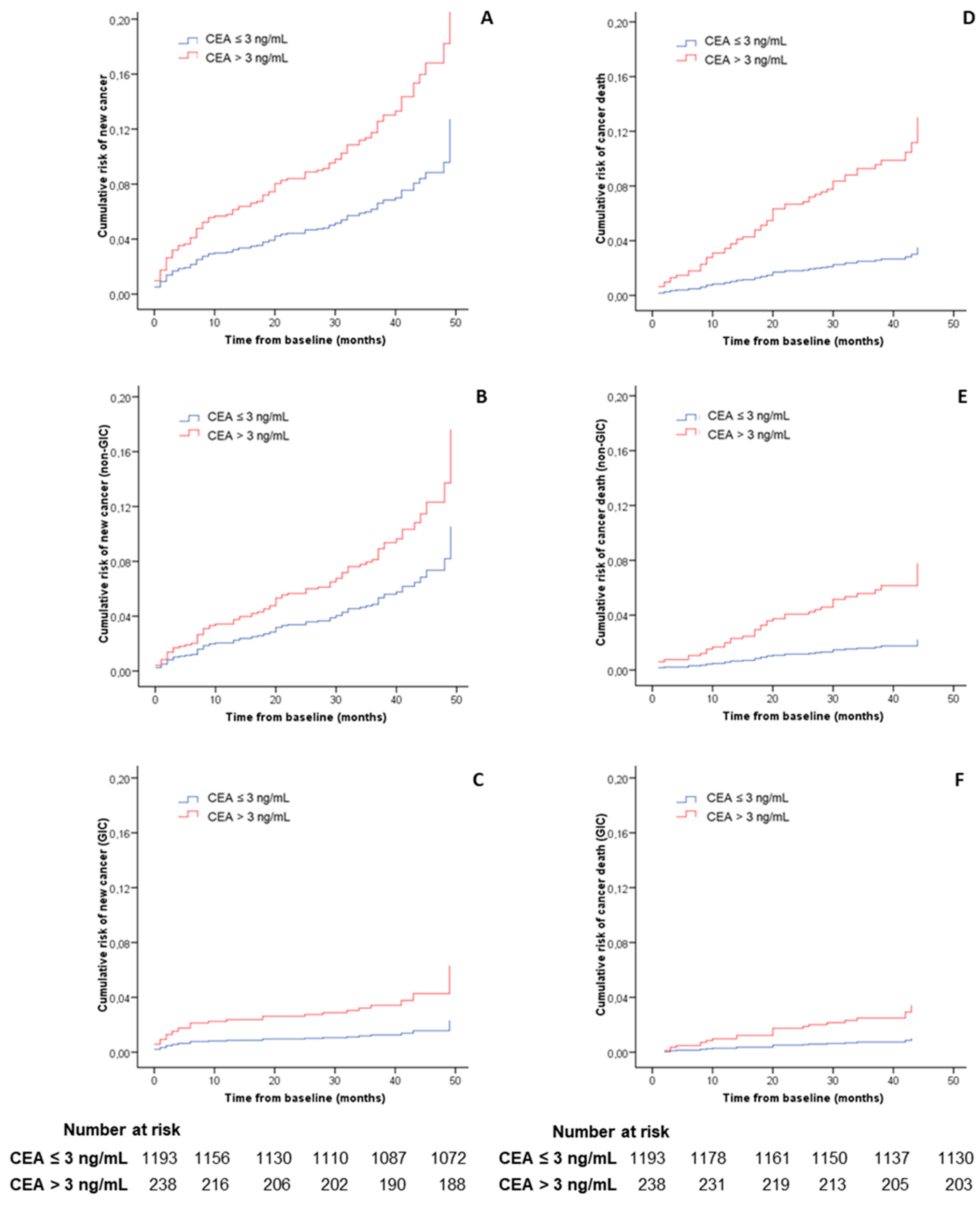
Patients with high CEA values showed an increased risk of death after adjusting for demographic variables and the presence of advanced adenoma (HR 2.6, 95% CI 1.7–3.9). These patients presented both a greater incidence of cancer diagnosis (HR 2.0, 95% CI 1.3–3.1) and cancer-related death (HR 3.7, 95% CI 2.2–6.2) during follow-up. The subgroup analysis also revealed a higher risk for both GIC (HR 2.7, 95% CI 1.3–5.7) and cancer located outside the GI tract (RR 1.7, 95% CI 1.0–2.8). The increased risk of GIC was related to an increased probability of CRC detection (HR 4.4, 95% CI 1.1–17.7) and CRC-related death (HR 8.8, 95% CI 1.6–48.5) in patients with an abnormal CEA concentration. In contrast, the risk of upper GIC and upper GIC-related death did not increase significantly. Table 2 and Figure 2 show the cumulative risk of cancer and related death calculated in Cox’s multivariable regression analysis.

3.4. Risk of Cancer Diagnosis during the First Year of Follow-Up

A cancer was detected in 51 (3.6%) patients during the first year after baseline colonoscopy, which represented 44.3% of all diagnosed cancer during follow-up. Nineteen subjects were diagnosed with GIC over this period (63.3% of all diagnosed GIC in this study). Of these, 16 (84.2%) lesions were located outside the colon, while three (15.8%) were CRC. The percentage of CRC and GIC located outside the colon which were diagnosed over this period was 37.5% and 72.7%, respectively. Two thirds of gastroesophageal cancer were diagnosed during the first year. Only three variables were independently associated with cancer detection: rectal bleeding (OR 0.3, 95% CI 0.1–0.7), anemia (OR 2.8, 95% CI 1.3–5.8) and CEA > 3 ng/mL (OR 3.4, 95% CI 1.7–7.1), as shown in Table 3.

3.5. Diagnostic Accuracy of CEA for Cancer Diagnosis the First Year of Follow-Up

In our cohort, 652 (45.6%) patients did not have anemia, of whom 96 (14.7%) showed CEA values higher than 3 ng/mL. The AUC of CEA for cancer detection during the first year of follow-up was 0.53 (95% CI, 0.38–0.67) and 0.63 (95% CI, 0.42–0.84) for patients with and without anemia, respectively. At a 3 ng/mL threshold, the sensitivity and specificity for detecting a new cancer in patients without anemia was 50.0% (95% CI, 25.4–74.6%) and 85.9% (95% CI, 83.0–88.4%). For patients with anemia, the sensitivity and specificity for detecting a new cancer in patients was 31.8% (95% CI, 16.4–52.7%) and 83.5% (95% CI, 79.1–87.2%). The diagnostic accuracy of CEA to detect different types of cancer based on the presence of anemia using the thresholds of 3 and 5 ng/mL is shown in Table 4.

4. Discussion

4.1. Statement of Principal Findings

In this study, we have evaluated the risk of cancer detection in patients with abdominal symptoms and a complete colonoscopy with no baseline CRC detected according to CEA concentration. The risk of cancer, GIC and CRC detection, but not upper-GIC, as well as their related death, is increased in patients with a CEA > 3 ng/mL. Moreover, we have determined three variables independently associated with cancer detection during the first year, as well as the diagnostic accuracy of CEA. In this sense, although specific, CEA values lack sensitivity to identify those patients who would be diagnosed with cancer during the first year of follow-up regardless of the presence of anemia.

4.2. Strengths and Weaknesses of Our Study

To our knowledge, this is the first cohort study offering information about the utility of CEA to assist in the triage of patients presenting with lower bowel symptoms who have recently been ruled out a CRC but may be suffering (or not) from other multiple types of cancer due the low specificity of digestive symptoms.
Important strengths of our study are (i) a sample size consisting of a significant number of consecutively recruited patients who underwent a colonoscopy, which guaranteed the absence of CRC and (ii) the availability of a sufficient monitoring period to detect different types of cancer. Both characteristics of our study together guarantee that these results reflect the effectiveness of CEA to predict a cancer diagnosis in the foreseeable future in a patient with abdominal symptoms after ruling out CRC.
Nonetheless, our study is not free from bias. The most important bias is a lack of pertinent background information, such as history of tobacco use or other benign conditions, which could account for a high CEA value without subsequent cancer diagnosis in some patients. This bias may have led to understating the role of CEA as a predictor of cancer. However, it should not have an impact on the assessment of CEA’s sensitivity to detect cancer, only CEA’s specificity.
Moreover, symptomatic patients usually present to primary healthcare, although sometimes they are allowed direct access to the specialist. Our results were obtained from a cohort comprised mainly of secondary healthcare patients. These are generally at a higher risk of being diagnosed with cancer than primary healthcare patients. Consequently, the assessment of CEA’s diagnostic accuracy could be lower in a cohort made up solely of patients from that setting.

4.3. Strengths and Weaknesses in Relation to Other Studies

Until now, CEA has been revealed to be mainly related to CRC and our study is along those lines [6]. Even after selecting patients with abdominal symptoms who had undergone a colonoscopy without CRC, a high CEA value only significantly increased the risk of future CRC diagnosis and related death. In this regard, the incidence of CRC during follow-up is very low, within the admissible CRC range values [26].
CEA was previously found to have low sensitivity and specificity to detect CRC in asymptomatic patients. However, it was found that average-risk patients with raised CEA should be investigated, because approximately 9% present some type of cancer. Furthermore, those patients should also be followed up, as another 7% are subsequently diagnosed with some cancer during monitoring [9]. This high cancer incidence could be accounted for the selection process of those patients. Although that study was performed in asymptomatic patients, the reason for which CEA was determined could entail an increased risk of cancer diagnosis. In our prospective cohort, comprised of consecutive patients who complained about abdominal symptoms, 13% of patients were diagnosed with cancer at some time during follow-up. A large proportion of those cancers were detected in the first year of follow-up and were thus likely to be present at the time baseline colonoscopy was performed.
In the past, various CEA thresholds were used. The threshold of 5 ng/mL was the most common during CRC follow-up despite a recommendation to raise this to 10 ng/mL when used in this context as a single test [21]. In this work, we decided to explore a lower threshold, as we are using CEA as a triage test of a group of diseases whose prognosis would benefit from a prompt diagnosis.

4.4. Implications for Clinical Practice and Research

The time between the onset of symptoms and consultation with the physician is a significant proportion of the total time to most cancer diagnosis [27]. Unfortunately, the most frequent initial cancer symptoms are common features of benign conditions and are often shared between different types of cancer [28].
Despite previous work recommending a rational evaluation pathway for a patient with a raised CEA [6], our data reveals that CEA also has a poor sensitivity to detect cancer in the patient with abdominal symptoms. The CEA levels were only higher than 3 ng/mL in about one third and one half of patients—with and without anemia, respectively—who will later be diagnosed with cancer during follow-up. Therefore, even as an independent predictor for future cancer diagnosis in the patient with abdominal symptoms who have ruled out a CRC, the determination of serum CEA should not be used as triage tool to identify who could benefit from further diagnostic tests in this population.

4.5. Unanswered Questions and Future Research

This study has revealed a remarkably high incidence of non-gastrointestinal cancer in a cohort of patients with abdominal symptoms. Furthermore, the incidence of non-gastrointestinal cancer in the subgroup of patients with CEA below the threshold 3 ng/mL and FIT < 20 µg Hb/g feces exceed the threshold risk of cancer of 3% recommended by the 2015 version of the NICE guidelines for investigation [29]. Similarly, other authors have reported that abdominal symptoms are common at presentation in different types of cancer [30]. Further prospective studies are needed in order to better understand the relationship between the presence of some particular abdominal symptoms and the subsequent diagnosis of an apparently unrelated cancer.

4.6. Conclusion

This study assesses the value of CEA in a large cohort of patients with symptoms that could be compatible with multiple tumors after the most common digestive tumor (CRC) had been ruled out with a very reliable test (colonoscopy). In this situation, extremely frequent in clinical practice, some doctors request the determination of serum CEA level to decide whether to perform or not additional work up to assess the possibility of that patient having other types of cancer. Our work, with its limitations, suggests that this practice should be abandoned.

Supplementary Materials

The following are available online at https://www.mdpi.com/2075-4418/10/12/1036/s1. Supplementary Table S1. Time to cancer diagnosis based on CEA level and type of cancer.

Author Contributions

Conceptualization, N.P.-V. and J.C.; methodology, N.P.-V. and J.C.; formal analysis, N.P.-V. and J.C.; investigation, N.P.-V., M.J.I., D.R., V.Á.-S., F.F.-B., J.B., E.M.-B., R.C., L.B., Á.F., V.P., D.R.-A., M.M.-R., N.G.-M., C.P.-M. and J.C.; data curation, N.P.-V.; writing—original draft preparation, N.P.-V. and J.C.; writing—review and editing, M.J.I., D.R., V.Á.-S., F.F.-B., J.B., E.M.-B., R.C., L.B., Á.F., V.P., D.R.-A., M.M.-R., N.G.-M., C.P.-M. and J.C.; visualization, N.P.-V. and J.C.; supervision, N.P.-V. and J.C.; project administration, J.C.; funding acquisition, J.C. All authors have read and agreed to the published version of the manuscript.

Funding

This work was supported by Spain’s Carlos III Healthcare Institute by means of project PI17/00837 (Co-funded by European Regional Development Fund/European Social Fund “A way to make Europe”/“Investing in your future”).

Acknowledgments

Some of our data were accepted for poster presentation at a Spanish scientific meeting (Spanish Gastroenterology Association; November 2020) and at the virtual United European Gastroenterology week to be held in October 2020.

Conflicts of Interest

The authors have no conflict of interest to declare.

References

  1. Koo, M.M.; Hamilton, W.; Walter, F.M.; Rubin, G.P.; Lyratzopoulos, G. Symptom Signatures and Diagnostic Timeliness in Cancer Patients: A Review of Current Evidence. Neoplasia 2018, 20, 165–174. [Google Scholar] [CrossRef]
  2. Koo, M.M.; Swann, R.; McPhail, S.; Abel, G.A.; Elliss-Brookes, L.; Rubin, G.P.; Lyratzopoulos, G. Presenting symptoms of cancer and stage at diagnosis: Evidence from a cross-sectional, population-based study. Lancet Oncol. 2020, 21, 73–79. [Google Scholar] [CrossRef] [Green Version]
  3. Lee, J.K.; Liles, E.G.; Bent, S.; Levin, T.R.; Corley, D.A. Accuracy of fecal immunochemical tests for colorectal cancer: Systematic review and meta-analysis. Ann. Intern. Med. 2014, 160, 171. [Google Scholar] [CrossRef]
  4. Pin-Vieito, N.; Zarraquiños, S.; Cubiella, J. High-risk symptoms and quantitative faecal immunochemical test accuracy: Systematic review and meta-analysis. World J. Gastroenterol. 2019, 25, 2383–2401. [Google Scholar] [CrossRef]
  5. NICE Diagnostics Guidance DG30. Quantitative Faecal Immunochemical Tests to Guide Referral for Colorectal Cancer in Primary Care. 2017. Available online: https://www.nice.org.uk/guidance/dg30 (accessed on 9 May 2019).
  6. Hall, C.; Clarke, L.; Pal, A.; Buchwald, P.; Eglinton, T.; Wakeman, C.; Frizelle, F. A Review of the Role of Carcinoembryonic Antigen in Clinical Practice. Ann. Coloproctol. 2019, 35, 294–305. [Google Scholar] [CrossRef]
  7. Holdenrieder, S.; Wehnl, B.; Hettwer, K.; Simon, K.; Uhlig, S.; Dayyani, F. Carcinoembryonic antigen and cytokeratin-19 fragments for assessment of therapy response in non-small cell lung cancer: A systematic review and meta-analysis. Br. J. Cancer 2017, 116, 1037–1045. [Google Scholar] [CrossRef]
  8. Li, X.; Dai, D.; Chen, B.; Tang, H.; Xie, X.; Wei, W. Clinicopathological and Prognostic Significance of Cancer Antigen 15-3 and Carcinoembryonic Antigen in Breast Cancer: A Meta-Analysis including 12,993 Patients. Dis. Markers 2018, 2018, 9863092. [Google Scholar] [CrossRef] [Green Version]
  9. Kim, J.R.; Jang, J.Y.; Kang, M.J.; Park, T.; Lee, S.Y.; Jung, W.; Chang, J.; Shin, Y.; Han, Y.; Kim, S.W. Clinical implication of serum carcinoembryonic antigen and carbohydrate antigen 19-9 for the prediction of malignancy in intraductal papillary mucinous neoplasm of pancreas. J. Hepatobiliary Pancreat. Sci. 2015, 22, 699–707. [Google Scholar] [CrossRef]
  10. Locker, G.Y.; Hamilton, S.; Harris, J.; Jessup, J.M.; Kemeny, N.; Macdonald, J.S.; Somerfield, M.R.; Hayes, D.F.; Bast, R.C. ASCO 2006 update of recommendations for the use of tumor markers in gastrointestinal cancer. J. Clin. Oncol. 2006, 24, 5313–5327. [Google Scholar] [CrossRef]
  11. Wilson, A.P.M.; van Dalen, A.; Sibley, P.E.C.; Kasper, L.A.; Durham, A.P.; El Shami, A.S. Multicenter tumour marker reference range study. Anticancer Res. 1999, 19, 2749–2752. [Google Scholar]
  12. Lim, Y.K.; Kam, M.H.; Eu, K.W. Carcinoembryonic antigen screening: How far should we go? Singapore Med. J. 2009, 50, 862–865. [Google Scholar]
  13. Bray, F.; Ferlay, J.; Soerjomataram, I.; Siegel, R.L.; Torre, L.A.; Jemal, A. Global cancer statistics 2018: +GLOBOCAN estimates of incidence and mortality worldwide for 36 cancers in 185 countries. CA Cancer J. Clin. 2018, 68, 394–424. [Google Scholar] [CrossRef] [Green Version]
  14. Pin-Vieito, N.; Iglesias, M.J.; Remedios, D.; Rodríguez-Alonso, L.; Rodriguez-Moranta, F.; Álvarez-Sánchez, V.; Fernández-Bañares, F.; Boadas, J.; Martínez-Bauer, E.; Campo, R.; et al. On Behalf of the Colonpredict Study Investigators. Risk of gastrointestinal cancer in a symptomatic cohort after a complete colonoscopy: Role of faecal immunochemical test. World J. Gastroenterol. 2020, 26, 70–85. [Google Scholar] [CrossRef]
  15. Cubiella, J.; Vega, P.; Salve, M.; Díaz-Ondina, M.; Alves, M.T.; Quintero, E.; Álvarez-Sánchez, V.; Fernández-Bañares, F.; Boadas, J.; Campo, R.; et al. COLONPREDICT study investigators. Development and external validation of a faecal immunochemical test-based prediction model for colorectal cancer detection in symptomatic patients. BMC Med. 2016, 14, 128. [Google Scholar] [CrossRef] [PubMed] [Green Version]
  16. Vandenbroucke, J.P.; von Elm, E.; Altman, D.G.; Gøtzsche, P.C.; Mulrow, C.D.; Pocock, S.J.; Poole, C.; Schlesselman, J.J.; Egger, M.; STROBE Initiative. Strengthening the Reporting of Observational Studies in Epidemiology (STROBE): Explanation and elaboration. Epidemiology 2007, 18, 805–835. [Google Scholar] [CrossRef] [Green Version]
  17. Cubiella, J.; Castells, A.; Andreu, M.; Bujanda, L.; Carballo, F.; Jover, R.; Lanas, Á.; Morillas, J.D.; Salas, D.; Quintero, E. COLONPREV study investigators. Correlation between adenoma detection rate in colonoscopy- and fecal immunochemical testing-based colorectal cancer screening programs. United Eur. Gastroenterol. J. 2017, 5, 255–260. [Google Scholar] [CrossRef] [Green Version]
  18. Pin-Vieito, N.; García-Nimo, L.; Bujanda, L.; Román-Alonso, B.; Gutiérrez-Stampa, M.Á.; Aguilar-Gama, V.; Portillo, I.; Cubiella, J. Optimal diagnostic accuracy of quantitative faecal immunochemical test positivity thresholds for colorectal cancer detection in primary health care: A community-based cohort study. United Eur. Gastroenterol. J. 2020, 10. [Google Scholar] [CrossRef]
  19. Jover, R.; Herráiz, M.; Alarcón, O.; Brullet, E.; Bujanda, L.; Bustamante, M.; Campo, R.; Carreño, R.; Castells, A.; Cubiella, J.; et al. Spanish Society of Gastroenterology; Spanish Society of Gastrointestinal Endoscopy Working Group. Clinical practice guidelines: Quality of colonoscopy in colorectal cancer screening. Endoscopy 2012, 44, 444–451. [Google Scholar] [CrossRef] [Green Version]
  20. Kim, N.H.; Lee, M.Y.; Park, J.H.; Park, D.I.; Sohn, C.I.; Choi, K.; Jung, Y.S. Serum CEA and CA 19-9 Levels are Associated with the Presence and Severity of Colorectal Neoplasia. Yonsei Med. J. 2017, 58, 918–924. [Google Scholar] [CrossRef]
  21. Shinkins, B.; Nicholson, B.D.; James, T.; Pathiraja, I.; Pugh, S.; Perera, R.; Primrose, J.; Mant, D. What carcinoembryonic antigen level should trigger further investigation during colorectal cancer follow-up? A systematic review and secondary analysis of a randomised controlled trial. Health Technol. Assess. 2017, 21, 1–60. [Google Scholar] [CrossRef] [Green Version]
  22. Lahner, E.; Capasso, M.; Carabotti, M.; Annibale, B. Incidence of cancer (other than gastric cancer) in pernicious anaemia: A systematic review with meta-analysis. Dig. Liver Dis. 2018, 50, 780–786. [Google Scholar] [CrossRef]
  23. Mashlab, S.; Large, P.; Laing, W.; Ng, O.; D’Auria, M.; Thurston, D.; Thomson, S.; Acheson, A.G.; Humes, D.J.; Banerjea, A.; et al. Anaemia as a risk stratification tool for symptomatic patients referred via the two-week wait pathway for colorectal cancer. Ann. R. Coll. Surg. Engl. 2018, 100, 350–356. [Google Scholar] [CrossRef]
  24. Kok, V.C.; Sung, F.C.; Kao, C.H.; Lin, C.C.; Tseng, C.H. Cancer risk in East Asian patients associated with acquired haemolytic anaemia: A nationwide population-based cohort study. BMC Cancer. 2016, 16, 57. [Google Scholar] [CrossRef] [Green Version]
  25. Hung, N.; Shen, C.C.; Hu, Y.W.; Hu, L.Y.; Yeh, C.M.; Teng, C.J.; Kuan, A.S.; Chen, S.C.; Chen, T.J.; Liu, C.J. Risk of cancer in patients with iron deficiency anemia: A nationwide population-based study. PLoS ONE 2015, 10, e0119647. [Google Scholar] [CrossRef] [Green Version]
  26. Morris, E.J.; Rutter, M.D.; Finan, P.J.; Thomas, J.D.; Valori, R. Post-colonoscopy colorectal cancer (PCCRC)rates vary considerably depending on the method used to calculate them: A retrospective observational population-based study of PCCRC in the English National Health Service. Gut 2015, 64, 1248–1256. [Google Scholar] [CrossRef]
  27. Allgar, V.L.; Neal, R.D. Delays in the diagnosis of six cancers: Analysis of data from the National Survey of NHS Patients: Cancer. Br. J. Cancer 2005, 92, 1959–1970. [Google Scholar] [CrossRef] [Green Version]
  28. Hamilton, W.; Walter, F.M.; Rubin, G.; Neal, R.D. Improving early diagnosis of symptomatic cancer. Nat. Rev. Clin. Oncol. 2016, 13, 740–749. [Google Scholar] [CrossRef]
  29. National Institute for Health and Care Excellence Suspected Cancer: Recognition and Referral. NICE Guideline (NG12). 2015. Available online: https://www.nice.org.uk/guidance/ng12 (accessed on 5 September 2020).
  30. Koo, M.M.; von Wagner, C.; Abel, G.A.; McPhail, S.; Hamilton, W.; Rubin, G.P.; Lyratzopoulos, G. The nature and frequency of abdominal symptoms in cancer patients and their associations with time to help-seeking: Evidence from a national audit of cancer diagnosis. J. Public Health 2018, 40, e388–e395. [Google Scholar] [CrossRef] [Green Version]
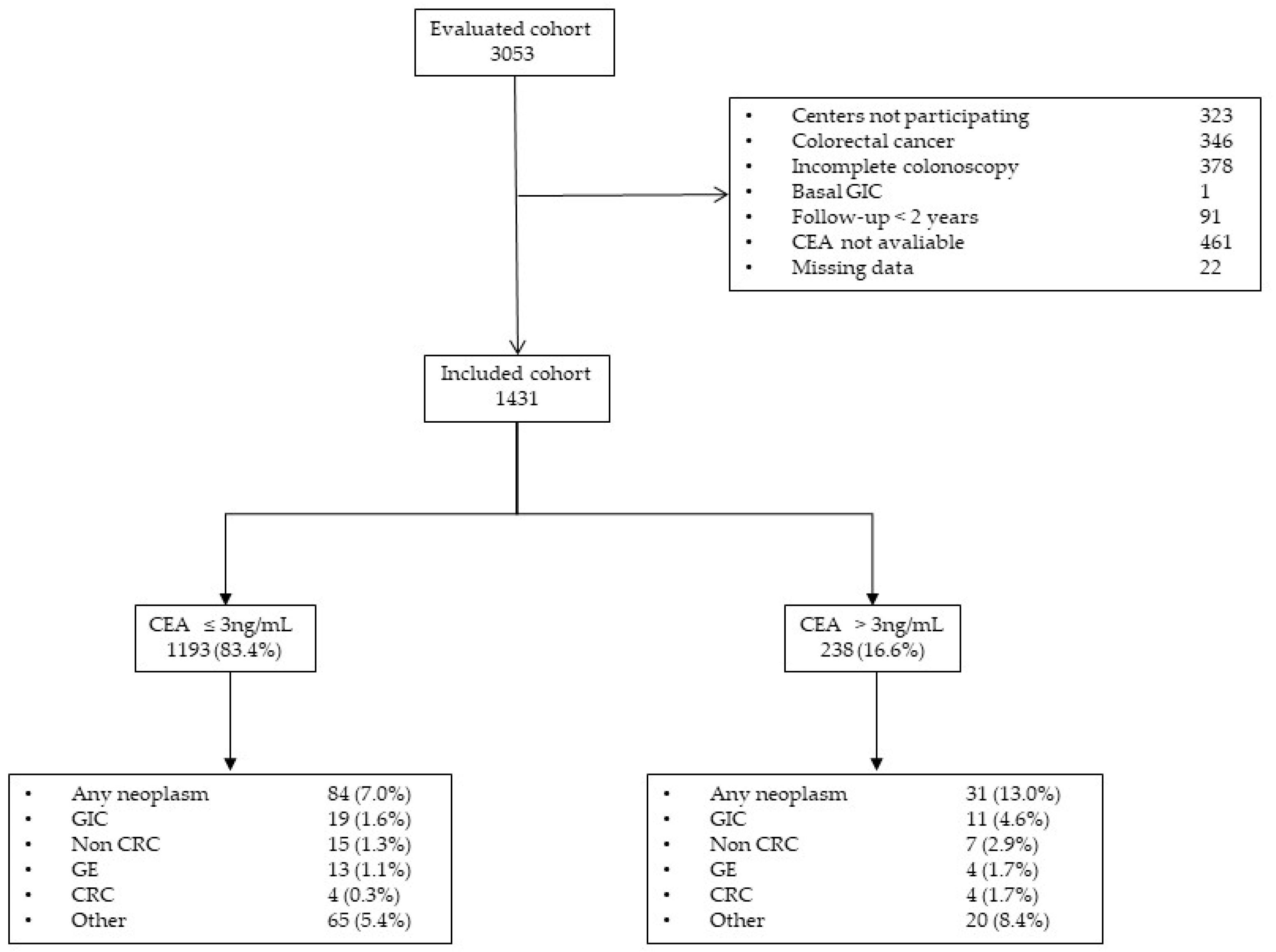
Figure 1. Study population flowchart. CEA = carcinoembryonic antigen; CRC = colorectal cancer; GE = gastroesophageal cancer; GIC = gastrointestinal cancer.
Diagnostics 10 01036 g001
Figure 2. Cumulative risk of cancer and related death. Cumulative risk of cancer and related death during follow-up after baseline evaluation according to carcinoembryonic antigen value and adjusted by sex, age and the presence of advanced adenoma. The figure is calculated with a Cox’s multivariable regression. (A) Risk of cancer diagnosis (global cancer); (B) risk of non-gastrointestinal cancer diagnosis; (C) risk of gastrointestinal cancer diagnosis; (D) risk of new cancer-related death; (E) risk of new non-gastrointestinal cancer death; (F) risk of new gastrointestinal cancer death. Number at risk was calculated for global cancer diagnosis and new cancer related death, respectively.
Diagnostics 10 01036 g002
Table 1. Characteristics of the individuals included in the analysis.
CharacteristicsOverall
(n = 1431)
CEA ≤ 3ng/mL
(n = 1193)
CEA > 3 ng/mL
(n = 238)
p
Demographic
Age, (years)66.7 (20.0)66.2 (19.5)69.1 (22.0)<0.01
Female sex, no. (%)748 (52.3)634 (53.1)114 (47.9)0.16
Primary healthcare referral, no. (%)390 (27.3)335 (28.1)55 (23.1)0.13
Previous colonoscopy, no. (%)398 (27.8)326 (27.3)72 (30.3)0.38
Daily using ASA, no. (%)280 (19.6)223 (18.7)57 (23.9)0.07
f-Hb concentration33.0 (140.0)33.0 (139.5) 31.0 (142.0)0.8
Indications, no. (%)
Rectal bleeding796 (55.6)675 (56.6)121 (50.8)0.12
Change of bowel habit839 (58.6)705 (59.1)134 (56.3)0.43
Anaemia 1338 (34.1)279 (33.4)59 (38.1)0.27
Abdominal pain 1451 (45.6)378 (45.3)73 (47.1)0.73
Weight loss 1240 (24.2)197 (23.6)43 (27.7)0.26
Basal colonoscopy findings, no. (%)
Benign anorectal lesion612 (42.8)521 (43.7)91 (38.2)0.13
Significant colonic lesions277 (19.4)233 (19.5)44 (18.5)0.79
Advanced adenoma206 (14.4)176 (14.8)30 (12.6)0.42
Follow-up, (months)38.0 (10.0)38.0 (10.0)37.0 (11.0)0.03
1 Missing data in 441 subjects; ASA, acetyl salicylic acid; f-Hb, faecal hemoglobin.
Table 2. Risk of cancer and death according to the carcinoembryonic antigen value.
EVENTRiskOverall (n = 1431)CEA ≤ 3 ng/mL (n = 1193)CEA > 3 ng/mL (n = 238)RR/IR (95% CI)pHR (95% CI)p
Gastrointestinal cancerCumulative 12.1 (1.5–3.0)1.6 (1.0–2.5)4.6 (2.6–8.1)2.9 (1.4–6.0)<0.012.7 (1.3–5.7)0.01
Density 26.9 (4.7–9.9)5.1 (2.9–8.0)16.1 (8.0–28.5)3.1 (1.5–6.5)<0.01
Cumulative death 11.5 (1.0–2.2)1.0 (0.6–1.7)3.8 (2.0–7.0)3.8 (1.6–8.8)0.033.4 (1.4–8.1)0.01
Death density 24.7 (2.9–7.3)3.3 (1.8–5.5)12.8 (5.8–24.4)4.0 (1.7–9.4)<0.01
Upper gastrointestinal cancer 3Cumulative 11.5 (1.0–2.3)1.3 (0.8–2.1)2.9 (1.4–5.9)2.3 (1.0–5.7)0.102.2 (0.9–5.4)0.09
Density 25.1 (3.3–7.7)4.0 (2.2–6.6)10.2 (4.0–20.8)2.5 (1.0–6.1)0.04
Cumulative death 11.0 (0.6–1.7)0.8 (0.5–1.5)2.1 (0.9–4.8)2.5 (0.9–7.3)0.162.3 (0.8–6.8)0.13
Death density 23.3 (1.8–5.5)2.6 (1.5–5.1)7.3 (2.2–16.8)2.6 (0.9–7.7)0.06
Colorectal cancerCumulative 10.6 (0.3–1.1)0.3 (0.1–0.9)1.7 (0.7–4.2)5.0 (1.3–19.9)0.044.4 (1.1–17.7)0.04
Density 21.8 (0.7–3.7)1.1 (0.4-2.9)5.8 (1.5–14.6)5.3 (1.3–21.3)<0.01
Cumulative death 10.4 (0.2–0.9)0.2 (0.0–0.6)1.7 (0.7–4.2)10.0 (1.8–54.4)<0.018.8 (1.6–48.5)<0.01
Death density 21.5 (0.4–2.9)0.4 (0.0–1.8)5.8 (1.5–14.6)10.6 (1.9–57.8)<0.01
Non gastrointestinal cancerCumulative 15.9 (4.8–7.3)5.4 (4.3–5.5)8.4 (5.5–12.6)1.5 (1.0–2.5)0.111.7 (1.0–2.8)0.04
Density 219.7 (15.7–24.5)17.9 (13.9–22.6)29.6 (17.9–45.7)1.7 (1.0–2.7)0.05
Cumulative death 12.7 (1.9–3.6)1.8 (1.2–2.8)6.7 (4.2–10.6)3.6 (1.9–6.8)< 0.013.5 (1.8–6.7)<0.01
Death density 28.8 (6.2–11.7)5.8 (3.7–9.1)22.6 (13.1–36.9)3.8 (2.0–7.3)<0.01
CancerCumulative 18.0 (6.7–9.6)7.0 (5.7–8.6)13.0 (9.3–18.0)1.8 (1.3–2.7)<0.012.0 (1.3–3.1)<0.01
Density 227.0 (22.3–32.1)23.4 (18.6–28.9)46.8 (31.8–66.5)2.0 (1.3–3.0)<0.01
Cumulative death 14.1 (3.2–5.3)2.9 (2.0–4.0)10.5 (7.2–15.0)3.7 (2.2–6.1)<0.013.7 (2.2–6.2)<0.01
Death density 213.5 (10.2–17.2)9.1 (6.2–12.8)35.8 (23.0–52.6)3.9 (2.3–6.5)<0.01
DeathCumulative 17.0 (5.8–8.4)5.4 (4.2–6.8)15.1 (11.1–20.2)2.8 (1.9–4.1)<0.012.6 (1.7–3.9)<0.01
Density 222.6 (18.6–27.4)17.2 (13.1–21.9)51.5 (35.8–71.2)3.0 (2.0–4.5)<0.01
1 Cumulative risk is expressed as percentage and its 95% CI; 2 risk density rate is expressed per 1000 patient-years and its 95% CI; 3 defined as a cancer located outside the colon. CI = confidence interval; HR = adjusted hazard ratio; IR = incidence ratio; RR = risk ratio.
Table 3. Factors associated with cancer detection the first year after baseline colonoscopy.
New CancerOdds Ratio (95% CI)Odds Ratio (95% CI) 1
Sex
Male (683)28 (4.0%)1
Female (748)23 (3.1%)0.8 (0.4–1.3)
Age
≤70 years (846)19(2.2%)1
>70 years (585)32(5.5%)2.4 (1.4–4.3)
Primary healthcare referral
No (1041)35 (3.4%)1
Yes (390)16 (4.1%)1.2 (0.7–2.2)
Rectal bleeding
No (635)37 (5.8%)11
Yes (796)14 (1.8%)0.3 (0.2–0.6)0.3 (0.1–0.7)
Change of bowel habit
No (592)25 (4.2%)1
Yes (839)26 (3.1%)0.7 (0.4–1.3)
Anaemia
No (652)12 (1.8%)11
Yes (338)22 (6.5%)3.5 (1.8–7.1)2.8 (1.3–5.8)
Abdominal pain
No (539)21 (3.9%)1
Yes (451)13 (2.9%)0.7 (0.4–1.5)
Weight loss
No (750)25 (3.3%)1
Yes (240)9 (3.4%)1.1 (0.5–2.4)
Faecal immunochemical test
≤10 µg/g (n = 948)30 (3.2%)1
>10 µg/g (n = 483)21 (4.3%)1.4 (0.8–2.4)
Carcinoembryonic antigen value
≤3 ng/mL (n = 1193)31 (2.3%)11
>3 ng/mL (n = 238)20(8.4%)3.2 (1.9–5.6)3.4 (1.7–7.1)
Advanced adenoma
No (1225)43 (3.5%)1
Yes (206)8 (3.9%)1.1 (0.5–2.3)
1 Variables with statistically significant differences were introduced in a multivariable logistic regression analysis stepwise (backward) to identify the prognostic factors associated with cancer detection the first year after baseline colonoscopy; as a result of this method, the dichotomous variable “Age” has finally not entered in the final equation. CI = confidence interval.
Table 4. Diagnostic accuracy of carcinoembryonic antigen for different types of cancer diagnosed in the first year during follow-up.
ThresholdType of CancerAnaemiaPrevalence%ATSensitivity 1Specificity 1NPV ‡,1PPV 1AUCLR+LR−DOR
CEA > 3 ng/mLGI cancerNo0.614.750.0 (15.0–85.0)85.5 (82.6–88.0)99.6 (98.7–99.9)2.1 (0.6–7.3)0.70 (0.41–0.99)3.40.65.82
Yes3.017.530.0 (10.8–60.3)82.9 (78.5–86.6)97.5 (94.9–98.8)5.1 (1.7–13.9)0.57 (0.39–0.74)1.80.82.08
Upper GI cancer 2No0.214.70.0 (0.0–79.3)85.3 (82.3–87.8)99.8 (99.0–99.96)0.0 (0.0–3.8)0.48 (0.44–0.53)0.0NANA
Yes3.017.530.0 (10.8–60.3)82.9 (78.5–86.6)97.5 (94.9–98.8)5.1 (1.7–13.9)0.57 (0.39–0.74)1.80.82.08
Colorectal cancerNo0.514.766.7 (20.8–93.9)85.5 (82.6–88.0)99.8 (99.0–99.96)2.1 (0.6–7.3)0.77 (0.42–1.0)4.60.411.80
Yes0.017.5NANANANANANANANA
Non-GI cancerNo1.214.750.0 (21.5–78.5)85.7 (82.8–88.5)99.3 (98.2–99.7)4.2 (1.6–10.2)0.60 (0.32–0.87)3.50.66.00
Yes3.617.533.3 (13.8–60.9)83.1 (78.7–86.8)97.1 (94.4–98.5)6.8 (2.7–16.2)0.49 (0.28–0.70)2.00.82.46
CancerNo1.814.750.0 (25.4–74.6)85.9 (83.0–88.4)98.9 (97.7–99.5)6.3 (2.9–13.0)0.63 (0.42–0.84)3.60.66.11
Yes6.517.531.8 (16.4–52.7)83.5 (79.1–87.2)94.6 (91.3–96.7)11.9 (5.9–22.5)0.53 (0.38–0.67)1.90.82.37
CEA >5 ng/mLGI cancerNo0.65.150.0 (15.0–85.0)99.2 (93.3–96.6)99.7 (98.8–99.9)6.1 (1.7–19.6)0.70 (0.41–0.99)10.50.519.91
Yes3.08.620.0 (5.7–51.0)91.8 (88.3–94.3)97.4 (95.0–98.7)6.9 (1.9–22.0)0.57 (0.39–0.74)2.40.92.79
Upper GI cancer 2No0.25.10.0 (0.0–79.3)94.9 (93.0–96.4)99.8 (99.1–99.97)0.0 (0.0–10.4)0.48 (0.44–0.53)0.0NANA
Yes3.08.620.0 (5.7–51.0)91.8 (88.3–94.3)97.4 (95.0–98.7)6.9 (1.9–22.0)0.57 (0.39–0.74)2.40.92.79
Colorectal cancerNo0.55.166.7 (20.8–93.9)95.2 (93.3–96.6)99.8 (99.1–99.97)6.1 (1.7–19.6)0.77 (0.42–1.0)14.00.439.88
Yes0.08.6NANANANANANANANA
Non-GI cancerNo1.25.137.5 (13.7–69.4)95.3 (93.4–96.7)99.2 (98.1–99.7)9.1 (3.1–23.6)0.60 (0.32–0.87)8.10.712.27
Yes3.68.616.7 (4.7–44.8)91.7 (88.2–94.2)96.8 (94.1–98.2)6.9 (1.9–22.0)0.49 (0.28–0.70)2.00.92.21
CancerNo1.85.141.7 (19.3–68.0)95.6 (93.8–97.0)98.9 (97.7–99.5)15.2 (6.7–30.9)0.63 (0.42–0.84)9.50.615.61
Yes6.58.618.2 (7.3–38.5)92.1 (88.6–94.6)94.2 (91.0–96.3)13.8 (5.5–30.6)0.53 (0.38–0.67)2.30.92.59
AUC = area under the curve; CEA = carcinoembryonic antigen; DOR = diagnostic odds ratio; GI = gastrointestinal; %AT = percentage of carcinoembryonic antigen above threshold; LR = likelihood ratio; NA = not applicable; NPV = negative predictive value; PPV = positive predictive value; +: positive; −: negative. 1 Values were expressed as percentages and their 95% confidence interval. 2 Defined as a cancer located outside the colon. ‡ Some NPV results were rounded to two decimals, as they could be incorrectly interpreted if they were rounded to one decimal (100.0).
Publisher’s Note: MDPI stays neutral with regard to jurisdictional claims in published maps and institutional affiliations.