


Review

Recent Advances in Biomarkers of Systemic Sclerosis-Associated Interstitial Lung Disease: Clinical Relevance and Therapeutic Perspectives

by
Rasha-Ioana Rămoiu-Shehada
1,
Anca Emanuela Mușetescu
2,
Lucian-Mihai Florescu
3,
Alesandra Florescu
2,* and
Paulina-Lucia Ciurea
2
1
Doctoral School, University of Medicine and Pharmacy of Craiova, 200349 Craiova, Romania
2
Department of Rheumatology, University of Medicine and Pharmacy of Craiova, 200349 Craiova, Romania
3
Department of Radiology and Medical Imaging, University of Medicine and Pharmacy of Craiova, 200349 Craiova, Romania
*
Author to whom correspondence should be addressed.
Life 2026, 16(9), 1420; https://doi.org/10.3390/life16091420
Submission received: 15 July 2026 / Revised: 23 August 2026 / Accepted: 24 August 2026 / Published: 27 August 2026
(This article belongs to the Special Issue Research and Management in Autoimmune Rheumatic Diseases)

Abstract

Systemic sclerosis-associated interstitial lung disease (SSc-ILD) is the leading cause of mortality in patients with systemic sclerosis and a major clinical challenge due to its heterogeneous presentation and variable disease course. Reliable biomarkers may improve disease assessment, risk stratification, and therapeutic decision-making. This narrative review summarizes current evidence on established and emerging serum and molecular biomarkers in SSc-ILD and their potential clinical applications. Recent advances have shifted the focus from single-marker assessment toward integrated biomarker profiling, reflecting the complex pathogenesis of pulmonary fibrosis. KL-6 and surfactant protein D (SP-D) have been associated with pulmonary involvement and radiographic features of SSc-ILD. Inflammatory mediators, including interleukin-6 (IL-6), and autoantibodies such as anti-topoisomerase I (anti-Scl-70) have been associated with disease severity and an increased risk of pulmonary progression. Emerging biomarkers related to epithelial injury, extracellular matrix remodelling, and vascular dysfunction may provide additional insights into the biological mechanisms underlying SSc-ILD. Current evidence highlights the potential of circulating and molecular biomarkers to refine risk stratification and prognostic assessment in SSc-ILD. Prospective validation and methodological standardization remain necessary to determine their clinical utility and potential integration into personalized disease management.

1. Introduction

Systemic sclerosis (SSc) represents a chronic, autoimmune condition defined by fibrosis of the skin and multiple internal organs and blood vessel injury [1]. Immune system dysregulation underlies these pathological processes, sustaining prolonged inflammation and driving the overproduction and accumulation of collagen along with other extracellular matrix components [2]. Based on the extent of skin involvement, SSc is commonly classified into limited cutaneous systemic sclerosis (lcSSc) and diffuse cutaneous systemic sclerosis (dcSSc), which are associated with distinct patterns of organ involvement and disease course. SSc-specific autoantibodies are important serological markers with established roles in disease diagnosis, classification, and clinical phenotyping. Among these, anticentromere antibodies (ACA), anti-topoisomerase I antibodies (ATA; anti-Scl-70), and anti-RNA polymerase III antibodies (anti-RNAP III) are included in the 2013 American College of Rheumatology/European Alliance of Associations for Rheumatology (ACR/EULAR) classification criteria for SSc [3].
Epidemiological studies indicate that male patients with SSc more frequently display a severe disease phenotype, characterised by diffuse cutaneous involvement, greater exposure to smoking and occupational risk factors, and higher prevalence of ATA and anti-U3 RNP antibodies. By contrast, female patients are more commonly associated with limited cutaneous disease, overlap with systemic lupus erythematosus, and ACA positivity [4]. These sex-related differences are clinically relevant, with male patients showing a higher frequency and greater severity of interstitial lung and cardiac involvement, which may contribute to poorer survival, whereas pulmonary arterial hypertension (PAH) represents a more prominent cause of mortality among female patients [5]. Several clinical and serological variables have been associated with the development and progression of ILD in patients with SSc. Among the most consistently reported risk factors are dcSSc, African American ancestry, older age at disease onset, shorter disease duration at the time of evaluation, and seropositivity for ATA antibodies. Collectively, these factors are associated with an increased risk of pulmonary involvement and may identify patients more likely to develop a progressive course of SSc-ILD [6,7].
Pulmonary involvement is widely recognised as a major contributor to morbidity and mortality in SSc, with ILD and PAH representing key factors associated with adverse clinical outcomes [5,8]. While advances in management have mitigated the mortality associated with renal and peripheral vascular complications in SSc, SSc-ILD remains a primary driver of disease-related mortality, frequently associated with progressive functional decline and unfavourable clinical outcomes [9]. Evidence from post mortem investigations suggests that moderate-to-severe pulmonary fibrotic changes are present in approximately 75% of patients with SSc, whereas clinically apparent restrictive ventilatory dysfunction is reported in only about one-third of cases [10,11]. Histopathological examination of lung biopsy specimens from patients with SSC-ILD predominantly reveals a nonspecific interstitial pneumonia (NSIP) pattern, reported in approximately 78% of cases. Usual interstitial pneumonia (UIP), which is more typically associated with idiopathic pulmonary fibrosis (IPF), is less frequently observed in SSc-ILD, with reported frequencies ranging from 10% to 15% [12].
Evidence from large, ethnically diverse multicentre cohorts indicates that African American patients with SSc may exhibit a distinct clinical and immunological phenotype, characterised by earlier disease onset, a higher frequency of diffuse cutaneous involvement (51–57%), and a greater burden of pulmonary fibrosis compared with Caucasian and Hispanic populations [13]. Data from the GENISOS cohort showed that clinically significant pulmonary involvement may develop early in the disease course, affecting approximately 25% of patients within the first three years following diagnosis. Diffuse cutaneous disease was present in more than half of the cohort, while African American patients showed a higher prevalence of pulmonary fibrosis [14]. Findings from the Pittsburgh Scleroderma Databank and the GRASP cohort further identified a serological profile characterised by higher frequencies of ATA antibodies (27–30%), anti-U3 RNP antibodies (16–40%), and anti-U1 RNP antibodies (16–18%), together with a relatively low prevalence of ACA antibodies. African ancestry was also identified as an independent predictor of severe ILD and progressive pulmonary fibrosis, even within comparable autoantibody subsets, suggesting that ancestry-related biological factors may contribute to differences in pulmonary disease severity and outcomes in SSc [15].
High-resolution computed tomography (HRCT) is considered the gold standard for the detection of ILD in patients with SSc, providing detailed characterization of pulmonary parenchymal abnormalities and facilitating the detection of early interstitial changes [16,17]. The diagnostic assessment of SSc-ILD may be further supported by the ILD-RISC score, a validated multiparametric tool integrating clinical, serological, and functional variables to identify patients at increased risk of ILD. By facilitating risk stratification and guiding the selective use of HRCT, the ILD-RISC score may contribute to earlier detection while potentially reducing unnecessary imaging [18]. Recent multicentre evidence indicates that ILD can also develop during later stages of SSc, beyond the early disease period traditionally associated with the highest risk. Although late-onset ILD occurs less frequently, its clinical severity and progression were comparable to those observed in earlier-onset disease, with nearly half of affected patients developing progressive lung involvement. These findings highlight that the risk of clinically significant ILD extends beyond the early disease course and support continued pulmonary surveillance in the patients with SSc [19]. Across published cohorts, the prevalence of ILD detected by HRCT varies considerably, ranging from approximately 22% to 84% with NSIP being more frequently reported than UIP [20].
The prognostic relevance of pulmonary involvement in SSc is further supported by mortality data from the European Alliance of Associations for Rheumatology (EULAR) Scleroderma Trials and Research (EUSTAR) cohort, which prospectively followed 5860 patients with SSc. A total of 284 deaths were recorded, with detailed information on the cause of death available for 234 cases. More than half of the reported deaths (55%) were directly attributed to SSc, whereas 41% were related to non-SSc causes and the remaining 4% could not be classified. Among SSc-related deaths, pulmonary fibrosis accounted for the largest proportion (35%), followed by PAH and cardiac complications, each accounting 26%. Pulmonary restriction and reduced diffusing capacity of the lung were also identified as independent risk factors for mortality. These findings underscore the prognostic relevance of pulmonary involvement in SSc and support the importance of early identification and longitudinal assessment of patients at risk of progressive lung disease [21,22].

2. Research Methods

This narrative review was based on a targeted literature search of PubMed/MEDLINE, Scopus, and Web of Science to identify relevant studies on biomarkers in SSc-ILD published between March 1969 and April 2026. Earlier seminal studies were also considered when relevant to the biological or mechanistic context of specific biomarkers.
The search strategy combined terms related to systemic sclerosis, interstitial lung disease, and biomarkers, including “systemic sclerosis”, “scleroderma”, “SSc-ILD”, “interstitial lung disease”, “biomarker”, “serum biomarker”, “circulating biomarker”, “molecular biomarker”, “autoantibodies”, “cytokines”, “chemokines”, “epithelial injury”, “endothelial dysfunction”, “immune activation”, “extracellular matrix remodelling”, and “fibrosis”. Searches were further refined using the names of individual biomarkers and biomarker families, with Boolean operators (AND and OR) applied as appropriate. Reference lists of key original studies and relevant reviews were manually screened to identify additional publications.
Studies were selected based on their relevance to the objectives of the review, with priority given to original research investigating circulating, serological, and other molecular biomarkers in SSc-ILD. Studies evaluating associations between candidate biomarkers and disease presence, severity, progression, prognosis, or therapeutic response were considered. Prospective and retrospective cohorts, case–control and cross-sectional studies, and clinical trials were included, while systematic reviews and meta-analyses were consulted for contextualization and identification of additional primary studies. Case reports, editorials, commentaries, and publications with insufficient relevant data were excluded from the core evidence synthesis.
Given the narrative design of the review, study selection was guided by relevance, scientific quality, and contribution to the understanding of biomarker biology and clinical utility in SSc-ILD. Biomarkers were synthesized according to their biological and pathophysiological relevance and discussed in relation to their potential diagnostic, prognostic, predictive, and monitoring value, with particular emphasis on emerging biomarkers and multimarker approaches.

3. Pathogenesis

Systemic sclerosis (SSc) is a heterogeneous autoimmune connective tissue disorder characterised by a complex and incompletely elucidated pathogenesis, involving a dynamic interplay between genetic predisposition, environmental exposures, and immune-mediated mechanisms. Current evidence supports a multifactorial model in which genetic, epigenetic, and environmental influences contribute to vascular injury, immune dysregulation, and progressive fibrosis, which are closely interconnected and mutually reinforcing processes. In SSc-ILD, the interplay between these pathogenic mechanisms, particularly immune dysregulation and persistent fibroblast activation, contributes to the development and progression of pulmonary fibrosis [23]. Genetic susceptibility is supported by association with multiple loci, many of which overlap with those implicated in other autoimmune diseases, including HLA class II alleles, as well as PTPN22 [24], STAT4 [25,26], IRF5 [27], and NLRP1. This genetic framework is further modulated by epigenetic mechanisms, particularly non-coding RNAs such as miR-21, miR-29, and miR-155, which have been implicated in the regulation of profibrotic transcriptional programs, extracellular matrix (ECM) turnover, and fibroblast activation. Additional regulatory mechanisms, including chromatin remodelling and bromodomain-mediated transcriptional control, may also contribute to persistent fibrogenesis [28]. Although genetic and epigenetic mechanisms have been implicated in SSc pathogenesis, their specific contribution to the development and progression of SSc-ILD remains incompletely understood.
Environmental and infectious exposures have been investigated as potential disease triggers in genetically susceptible individuals. Occupational exposure to crystalline silica and organic solvents such as trichloroethylene, has been associated with increased risk of SSc. Infectious agents, including cytomegalovirus, Epstein–Barr virus, and parvovirus B19—have also been proposed as potential contributors to disease development; however, the available evidence remains insufficient to establish a causal relationship between these infections and SSc [29,30].
Early abnormalities in SSc occur at the level of microvasculature and are characterised by endothelial cell injury and dysfunction. Endothelial activation results in increased expression of adhesion molecules including ICAM-1 and VCAM-1, enhanced chemokine and cytokine release, and recruitment of inflammatory cells to perivascular tissues. In parallel, dysregulation of vasoactive mediators is characterised by increased endothelin-1 [31] production and reduced synthesis of nitric oxide and prostacyclin, contributing to vasoconstriction and impaired vascular homeostasis. These alterations are accompanied by progressive vascular remodelling, defective angiogenesis, and capillary rarefaction, which may contribute to chronic tissue hypoxia, oxidative stress, and increased production of reactive oxygen species, thereby further promoting inflammation and fibroblast activation [32]. Beyond classical vasoactive mediators, neuropeptide-mediated G protein-coupled receptor signalling may also contribute to cardiovascular homeostasis through the modulation of intracellular calcium dynamics and neuronal excitability [33].
Immune dysregulation represents an important component of the transition from vascular and tissue injury to fibrosis in SSc. Both innate and adaptive immune responses are involved early in the disease course, with perivascular inflammatory infiltrates comprising macrophages, dendritic cells, mast cells, T lymphocytes, and B lymphocytes. Among innate immune populations, alternatively activated M2 macrophages may contribute to fibrogenesis through the release of TGF-β, IL-6, CCL2, and other profibrotic mediators. In parallel, CD4+ T helper 2 (Th2) cells produce cytokines such as IL-4 and IL-13, which promote fibroblast activation, myofibroblast differentiation, and extracellular matrix (ECM) synthesis. B-cell dysregulation [34,35] may further participate in these processes through autoantibody production, cytokine secretion, and profibrotic signalling. The presence of B-cell infiltrates in lung tissue from patients with SSc-ILD provides additional evidence for their involvement in pulmonary immune–fibrotic responses, although these observations do not establish B cells as a primary driver of fibrosis. Innate immune pathways, including Toll-like receptor signalling and type I interferon responses, may further sustain immune activation and the production of inflammatory and profibrotic mediators [2,36,37].
Autoantibodies are detectable in the majority of patients with SSc and have important diagnostic, classification, and prognostic relevance. Among SSc-specific autoantibodies, anticentromere antibodies (ACA), anti-topoisomerase I antibodies (ATA; anti-Scl-70), and anti-RNA polymerase III antibodies (anti-RNAP III) are included in the 2013 American College of Rheumatology/European League Against Rheumatism (ACR/EULAR) classification criteria for SSc [3]. These autoantibodies are associated with distinct clinical phenotypes and patterns of organ involvement. ACAs are predominantly associated with lcSSc, whereas ATAs are more frequently associated with dcSSc and an increased risk of interstitial lung disease. Anti-RNAP III antibodies are predominantly associated with dcSSc and an increased risk of scleroderma renal crisis [38].
Beyond the autoantibodies incorporated into the classification criteria, other SSc-specific or SSc-associated autoantibodies, including anti-U3 RNP, anti-U1 RNP, anti-PM/Scl, anti-Ku, and anti-Th/To antibodies, define additional serological subsets with distinct clinical associations. From an SSc-ILD perspective, ATA are particularly relevant because of their well-established association with pulmonary involvement, whereas the clinical relevance of other autoantibody profiles varies according to the pulmonary outcome considered. Overall, the heterogeneity of SSc autoantibody profiles supports their role in clinical phenotyping and risk stratification, although their utility for detecting established ILD, predicting subsequent progression, or monitoring disease activity should be considered separately according to the available evidence [39].
Persistent fibroblast activation and myofibroblast accumulation are key features of SSc fibrosis and contribute to excessive extracellular matrix (ECM) deposition and progressive tissue remodelling. Multiple signalling pathways are involved in this process, with transforming growth factor-β (TGF-β) acting as an important mediator of fibroblast activation and profibrotic responses. Platelet-derived growth factor (PDGF) signalling may further contribute to fibroblast proliferation and ECM production. Activated fibroblasts and myofibroblasts subsequently produce increased amounts of ECM components, including collagen and fibronectin, thereby promoting progressive fibrotic remodelling [40,41,42].
Within the lung, vascular and epithelial injury, immune dysregulation, and fibroblast activation converge to establish a profibrotic microenvironment characterised by persistent fibroblast activation and excessive ECM deposition. Fibroblasts represent major effector cells in SSc fibrosis and exhibit functional alterations that may contribute to the persistence of the fibrotic response [41]. Sustained fibroblast activation can disrupt the balance between ECM synthesis and degradation, promoting progressive remodelling of pulmonary tissue and contributing to the development and progression of SSc-ILD [43].
Collectively, SSc-ILD develops within a complex pathogenic network in which vascular and epithelial injury, immune dysregulation, and persistent fibroblast activation interact through overlapping pathways. Rather than being attributable to a single pathogenic mechanism, pulmonary fibrosis in SSc appears to result from the convergence of multiple interconnected processes. This framework provides a biological rationale for investigating biomarkers that reflect distinct components of SSc-ILD pathobiology and for evaluating their potential roles in risk stratification, disease monitoring, and assessment of therapeutic response [44] (Figure 1).

4. Biomarkers

4.1. Autoantibodies

Autoantibodies represent central biomarkers in systemic sclerosis, with key roles in diagnosis, disease classification, and prognostic stratification, detectable in over 95% of patients. Most patients are seropositive for antinuclear antibodies and develop disease-specific autoantibodies, often detected even prior to clinical onset, that define distinct clinical subsets and patterns of organ involvement. Among these, the most clinically relevant antibodies include anticentromere antibodies (ACA), anti-topoisomerase I antibodies (anti-Scl-70), and anti-RNA-polymerase III antibodies, which represents the principal serological subsets in systemic sclerosis and form the basis of ACR/EULAR classification criteria [45,46,47].
A large-scale registry-based analysis including 863 patients from the German Network for Systemic Scleroderma Registry demonstrated that a limited panel of five autoantibodies—ACA, ATA, anti-RNA polymerase III, anti-U1 RNP, and anti-PM-Scl—captures over 95% of systemic sclerosis-associated antibody responses in ANA-positive individuals. Antinuclear antibodies were detected in 94.2% of the cohort, while these disease-specific autoantibodies were largely mutually exclusive, with co-occurrence observed in only 1.6% of cases. Importantly, these autoantibody subsets were strongly associated with distinct clinical phenotypes, with ACA linked to limited cutaneous disease and vascular complications, and anti-topoisomerase I antibodies associated with diffuse disease and pulmonary fibrosis [48].
Given that pulmonary complications- specifically ILD and PAH- are the leading cause of mortality in SSc, identifying specific autoantibody profiles is mandatory for early diagnosis and specialised follow-up. According to the comprehensive evidence synthesised by Cavazzana et al., these biomarkers are not merely diagnostic adjuncts but are intrinsically linked to distinct pulmonary trajectories, allowing for the classification of patients into specific fibro-proliferative, vascular, or high-risk mixed phenotypes [49,50], (Table 1).

4.1.1. The Fibro-Proliferative Phenotype: Anti-Topoisomerase I (ATA/Anti-Scl-70)

Anti-topoisomerase I antibodies (ATA; anti-Scl-70) are among the most clinically relevant serological markers associated with SSc-ILD. ATA positivity is more frequently observed in patients with diffuse cutaneous systemic sclerosis (dcSSc) and is associated with an increased risk of ILD and more extensive pulmonary involvement [51]. Longitudinal studies have further associated ATA positivity with a greater risk of pulmonary function deterioration, particularly during the early years of disease, when clinically relevant declines in forced vital capacity (FVC) and diffusing capacity of the lung for carbon monoxide (DLCO) may occur [52,53]. Accordingly, ATA status may contribute to pulmonary risk stratification and, together with clinical characteristics and pulmonary function parameters, help identify patients who warrant careful assessment for SSc-ILD [54]. ATA positivity has been consistently associated with an increased risk of clinically relevant ILD across SSc cutaneous subsets, although pulmonary involvement is more frequently encountered in patients with diffuse cutaneous disease. ATA-positive patients have also been reported to have less favourable survival, particularly in the context of dcSSc [55]. Beyond pulmonary involvement, ATA positivity has been associated with digital vascular and musculoskeletal manifestations, supporting its association with a broader clinical phenotype. Experimental and translational evidence further suggests that immune responses associated with topoisomerase I may participate in fibroblast activation and extracellular matrix remodelling; however, the extent to which these mechanisms independently contribute to progressive pulmonary fibrosis remains incompletely defined [56].
The prognostic implications of ATA positivity appear to depend partly on the clinical context and cutaneous phenotype. In a Japanese SSc cohort, Hamaguchi et al. reported pulmonary fibrosis in 84% of ATA-positive patients, compared with 7% of patients with ACA and 17% of those with anti-RNA polymerase antibodies [57]. ATA-positive patients also had a 10-year cumulative survival rate of approximately 70%, supporting an association between this serological profile, pulmonary involvement, and a less favourable disease course in this population [57]. However, these findings should not be interpreted as establishing ATA as an independent determinant of mortality, as prognosis may also be influenced by disease subtype, extent of organ involvement, and other clinical characteristics.
The relationship between ATA and outcome is further influenced by cutaneous phenotype. In a longitudinal cohort, ATA-positive patients with lcSSc exhibited a substantially higher prevalence of ILD (49%) than ATA-negative patients with lcSSc, approaching that observed in patients with diffuse cutaneous disease. Despite this increased burden of pulmonary involvement, cumulative survival in ATA-positive lcSSc was approximately 75% and was comparable to that of ATA-negative lcSSc, while remaining more favourable than in ATA-positive dcSSc. ATA-positive lcSSc patients were also more likely to develop diffuse cutaneous involvement during follow-up, with most transitions occurring within the first five years. Collectively, these observations suggest that ATA positivity identifies a subgroup at increased risk of ILD even in the presence of a limited cutaneous phenotype, whereas its prognostic implications for survival should be interpreted in conjunction with cutaneous subtype and overall disease phenotype [58].

4.1.2. The Vascular-Predominant Phenotype: Anticentromere Antibodies (ACAs)

In contrast to the predominantly interstitial involvement associated with the fibro-proliferative phenotype, the ACA positivity is characterised by a relative sparing of the lung interstitium and a greater propensity for pulmonary microangiopathy. ACAs are predominantly associated with lcSSc, a lower risk of severe ILD [47,63], and a greater propensity for PAH, an important pulmonary complication of lcSSc [64].
The clinical significance of ACA, however, varies according to the pulmonary outcome considered. Although their presence may facilitate the identification of patients with a predominantly vascular pulmonary phenotype, their prognostic value after PAH has developed appears more limited. Data from the PHAROS registry showed no significant association between ACA positivity and overall survival, and ACA status was not identified as an independent predictor of mortality among patients with SSc-PAH [65]. Thus, ACA may be more informative for phenotypic characterization and risk stratification than for predicting clinical outcomes once PAH is established.
Nevertheless, the relationship between ACA and pulmonary involvement is more complex than a strictly lung-sparing profile. Caetano et al. identified a distinctive phenotype among ACA-positive patients with dcSSc, characterised by a more gradual evolution of cutaneous and internal organ involvement. ILD was more frequent in ACA-positive dcSSc than in ACA-positive lcSSc, but less frequent than in ACA-negative dcSSc. Moreover, ILD development extended over a longer disease course in the ACA-positive diffuse subset, whereas scleroderma renal crisis occurred less frequently and survival was more favourable than in ACA-negative dcSSc. Collectively, these findings suggest that ACA status may contribute to phenotypic stratification even within diffuse cutaneous disease and indicate that ACA positivity does not preclude clinically relevant interstitial lung involvement [63].

4.1.3. The Multisystem Phenotype: Anti-RNA Polymerase III (Anti-RNAP III)

Anti-RNA polymerase III antibodies (anti-RNAP III) characterize a clinical phenotype distinct from that associated with ACA and are particularly linked to rapid disease onset and accelerated progression of skin thickening. Cavazzana et al. reported a shorter interval between the onset of Raynaud’s phenomenon and the first non-Raynaud manifestation in patients with isolated anti-RNAP III compared with those positive for anti-topoisomerase I antibodies, together with more rapid early skin thickening. However, rates of internal organ involvement and survival were similar between the two groups [66].
Beyond the cutaneous phenotype, anti-RNAP III positivity is strongly associated with scleroderma renal crisis [7,67]. Associations with malignancy have also been reported, particularly a close temporal relationship between cancer diagnosis and SSc onset in anti-RNAP III-positive patients [68,69,70]. These distinct systemic associations further support the consideration of anti-RNAP III and ACA as separate serological and clinical phenotypes.
From an SSc-ILD perspective, anti-RNAP III is less strongly associated with interstitial pulmonary involvement than anti-topoisomerase I antibodies (ATA). Severe interstitial fibrosis has been reported less frequently in anti-RNAP III-positive patients [7]; however, this serological profile should not be interpreted as excluding pulmonary parenchymal involvement. Current evidence does not establish anti-RNAP III as an independent predictor of either SSc-ILD development or subsequent progression.
Overall, anti-RNAP III appears to be more informative for clinical and serological phenotyping than as an established prognostic biomarker of SSc-ILD. Its presence identifies a distinct SSc subset characterised particularly by rapidly evolving cutaneous disease and an increased risk of scleroderma renal crisis, together with reported malignancy-related associations. Although pulmonary involvement may occur, available evidence does not currently support anti-RNAP III as an established biomarker of SSc-ILD progression [49].

4.1.4. The Nucleolar and Overlap High-Risk Phenotype: Multi-Organ Vulnerability

The substantial burden of pulmonary involvement observed in patients harbouring antibodies that target nucleolar antigens or reflect overlap syndromes may include both ILD and PAH.
Anti-Th/To antibodies exemplify this profile: despite being associated with limited cutaneous disease, they are linked to a high prevalence of both ILD and PAH, affecting up to 75% of patients. This paradoxical combination of mild skin disease and severe pulmonary involvement has been associated with reduced survival compared to other lcSSc subsets, underscoring the importance of early detection [71].
Overlap-associated autoantibodies further expand this high-risk category. Anti-PM/Scl antibodies are typically associated with SSc–myositis overlap and a high prevalence of ILD; however, the pulmonary course is often more indolent and responsive to therapy, resulting in comparatively favourable outcomes [72,73]. By comparison, in a clinical cohort of 126 European Caucasian patients, the presence of ILD, confirmed by high-resolution computed tomography, was observed in 59% of anti-PM/Scl-positive individuals compared to 51% in antibody-negative patients, while reduced diffusion capacity was noted in 61% of cases. Despite these findings, no statistically significant association was identified between anti-PM/Scl positivity and either ILD occurrence or pulmonary function impairment, suggesting that anti-PM/Scl positivity may not independently identify an increased risk of ILD or pulmonary functional impairment in this cohort [74]. Similarly, anti-Ku antibodies are associated with an overlap phenotype characterised by myositis and ILD, with an intermediate clinical course in which both inflammatory and fibrotic features may be present [75,76]. A prevalence of ILD of approximately 71% has been reported among anti-Ku-positive SSc patients, markedly higher compared to the general SSc population. Radiographic evaluation most frequently demonstrates nonspecific interstitial pneumonia (NSIP) and usual interstitial pneumonia (UIP) patterns. Pulmonary involvement in this subset has been associated with an unfavourable prognosis [77].
Additional markers may further contribute to risk stratification within this group. Anti-U3 RNP (fibrillarin) antibodies are associated with diffuse disease, early onset, and increased risk of PAH and cardiac involvement, particularly in specific ethnic populations. Anti-Ro52 (TRIM21) antibodies, frequently coexisting with other autoantibodies, have emerged as potential markers of disease severity and have been independently associated with ILD progression, PAH, and increased mortality across multiple SSc subsets [78,79,80]. A recent large meta-analysis encompassing 11,751 patients, supported by additional cohort data, indicates that anti-TRIM21 (Ro52) antibodies are present in approximately one-quarter of systemic sclerosis cases. Although these antibodies commonly coexist with other SSc-specific autoantibodies, they have emerged as independent predictors of severe organ involvement. In particular, anti-TRIM21 seropositivity has been significantly associated with the presence of ILD [81]. In an exploratory study, Hamberg et al. (2023) demonstrated that anti-Ro52 antibodies are significantly enriched in the bronchoalveolar lavage (BAL) fluid of patients with new-onset SSc-ILD, often at ratios exceeding 50 times the levels found in serum, raising the possibility of a local pulmonary autoimmune process. Furthermore, longitudinal analysis showed an independent association between anti-Ro52 positivity and accelerated decline in lung function [82,83].
Recent advances have also identified novel autoantibodies that further define high-risk pulmonary phenotypes. Anti-U11/U12 RNP (RNPC3) antibodies have been associated with severe and rapidly progressive ILD, often accompanied by significant reductions in FVC and DLCO and reported associations with malignancy [45,84]. Similarly, anti-BICD2 and anti-eIF2B antibodies have been linked to ILD and overlap features, while anti-RuvBL1/2 antibodies define a multisystem phenotype with myositis, cardiac, and gastrointestinal involvement, occasionally accompanied by pulmonary disease [85].
Beyond classical serological markers, Chepy et al. (2025) highlight emerging antigenic targets, including anti-eIF2B, anti-RNPC3 (U11/U12), anti-BICD2, and telomere-associated antigens such as anti-TERF1, which may refine the classification of previously seronegative subsets and are associated with distinct clinical phenotypes, including severe interstitial lung disease and malignancy. These findings raise the possibility that some autoantibodies may participate in disease-related mechanisms rather than serving solely as diagnostic or prognostic markers. The occurrence of autoantibodies before clinical manifestations, together with reported associations between antibody titres and disease features, provides additional support for a potential contribution of humoral immune mechanisms to SSc pathogenesis. Clinical responses observed with B-cell-targeted therapies, including rituximab and, more recently, anti-CD19 CAR-T cell therapy, further support the involvement of B-cell-mediated immune mechanisms in SSc. Experimental models in which immunization with autoantigens such as topoisomerase I or PRMT5 induces SSc-like cutaneous and pulmonary fibrosis provide additional evidence for a potential contribution of humoral immune dysregulation to fibrotic processes. However, these observations do not establish humoral immunity as a primary driver of fibrosis, and its precise role in the initiation and progression of pulmonary fibrosis remains to be determined [86]. The anti-eIF2B is a rare autoantibody reported in approximately 2.5% of SSc cases. Its presence is strongly associated with the diffuse cutaneous variant of the disease and the development of ILD. Clinically, this specificity may be considered in SSc patients who test negative for traditional antinuclear antibodies (ANA) but display a distinct, high titre cytoplasmic staining pattern (≥1:320) during indirect immunofluorescence [87,88].
Anti-U1 RNA antibodies have been identified in approximately 61% of systemic sclerosis patients with anti-U1 RNP and have been associated with pulmonary involvement. Their presence has also been associated with a higher prevalence of pulmonary fibrosis and impaired gas transfer, suggesting potential value for ILD risk stratification [89].
Taken together, these findings underscore the clinical relevance of autoantibody-defined subsets in systemic sclerosis beyond conventional serological classification. Distinct autoantibody profiles characterise different patterns of ILD involvement and disease course, ranging from more indolent forms to progressive fibrotic phenotypes, and may therefore contribute to risk stratification and longitudinal assessment of SSc-ILD.

4.2. Serum Biomarkers for Interstitial Lung Disease in Systemic Sclerosis

The management of SSc-ILD is increasingly reliant on serum biomarkers that can provide dynamic assessment beyond the static measurements of pulmonary function tests. These molecules, reflecting various facets of alveolar injury and fibro-inflammatory signalling, are essential for identifying “progressors”—patients at high risk for significant decline in forced vital capacity (FVC) and diffusing capacity (DLCO) (Table 2).

4.2.1. Markers of Alveolar Epithelial Injury and Type II Pneumocyte Activity

Krebs von den Lungen-6 (KL-6) and surfactant proteins, particularly surfactant protein D (SP-D), represent circulating biomarkers related to alveolar epithelial injury and type II pneumocyte activity. KL-6 is a high-molecular-weight MUC1 glycoprotein expressed by type II alveolar epithelial cells, with increased circulating concentrations reported in several forms of ILD, including SSc-ILD. Available evidence suggests that KL-6 may provide information on different aspects of pulmonary involvement; however, its association with the presence and extent of ILD should be distinguished from its potential prognostic value for subsequent disease progression [94].
From a cross-sectional perspective, elevated KL-6 concentrations have been associated with greater radiological disease extent and impaired pulmonary function. Sieiro Santos et al. found higher KL-6 concentrations in patients with SSc-ILD than in those without ILD, together with inverse correlations with FVC and DLCO. KL-6 levels were also associated with semiquantitative HRCT extent, with higher concentrations observed in patients with more extensive pulmonary involvement [95]. Consistent with these observations, Lee et al. reported higher serum KL-6 levels in patients with more severe connective tissue disease-associated ILD, further supporting an association between KL-6 and the overall burden of pulmonary involvement [96]. These findings support the potential utility of KL-6 for characterizing the presence and baseline severity of ILD; however, the cross-sectional nature of the available evidence precludes conclusions regarding its prognostic value for subsequent pulmonary deterioration [95,96].
Longitudinal data suggest that KL-6 may provide prognostic information in early SSc-ILD. In the prospective GENISOS cohort, Salazar et al. found that higher baseline KL-6 levels were associated with greater FVC decline over one year, with concentrations above 1273 U/mL corresponding to an approximately 7% greater annualized decline. In contrast, baseline CCL18 was not associated with short-term FVC deterioration, differing from findings from Scleroderma Lung Study II, where higher CCL18 levels were associated with subsequent FVC and DLCO decline. These discrepancies may reflect differences in patient characteristics, disease severity, treatment exposure, follow-up duration, or definitions of progression. Collectively, these findings suggest a potential role for KL-6 in identifying patients at risk of early functional decline, although its prognostic value and proposed thresholds require further evaluation in independent SSc-ILD cohorts [92].
SP-D is another circulating marker of alveolar epithelial and alveolar–capillary injury that has been investigated in SSc-ILD [97]. Elevated serum SP-D concentrations have been associated with pulmonary involvement and greater ILD severity. Hant et al. reported that both SP-D and KL-6 levels correlated with HRCT fibrosis scores, whereas neither marker was significantly associated with the extent of ground-glass opacity [98]. These findings suggest that their relationship with radiological abnormalities may differ according to the imaging features assessed and should not be interpreted as uniformly reflecting all components of ILD severity.
Comparative and longitudinal studies have further explored the potential clinical relevance of SP-D and KL-6. Yanaba et al. reported higher sensitivity for SP-D and greater specificity for KL-6 in detecting pulmonary fibrosis, while serial SP-D measurements appeared to reflect temporal changes in pulmonary involvement [99]. Similarly, Asano et al. found that elevated SP-D levels were associated with pulmonary fibrosis and impaired vital capacity and DLCO [100].
In a retrospective cohort of patients with SSc- or mixed connective tissue disease-associated ILD, Yamakawa et al. observed that longitudinal changes in KL-6 were associated with changes in FVC, whereas higher SP-D levels were associated with subsequent short-term FVC decline. Collectively, these observations suggest that KL-6 and SP-D may provide complementary information regarding longitudinal pulmonary assessment and risk of functional deterioration. However, the limited sample size, retrospective design, and inclusion of a mixed disease population in the latter study warrant caution when extrapolating these findings specifically to SSc-ILD [101].
IL-18 has also been evaluated as a circulating biomarker of pulmonary involvement in SSc. In the cross-sectional study by Sieiro Santos et al., serum IL-18 and KL-6 levels were higher in patients with SSc-ILD than in those without ILD and were inversely associated with FVC and DLCO. Both biomarkers were related to semiquantitative HRCT severity, although the association with extensive lung involvement was observed for KL-6 but not for IL-18. These findings suggest that IL-18 and KL-6 may provide complementary information regarding the presence and baseline severity of SSc-ILD. However, the cross-sectional design precludes conclusions regarding their ability to predict subsequent disease progression, and current evidence does not support their use as alternatives to longitudinal assessment by pulmonary function testing or HRCT [95].
Overall, KL-6 and SP-D are among the most extensively studied circulating markers of alveolar epithelial injury in SSc-ILD. Available evidence indicates associations with the presence and severity of pulmonary involvement, while longitudinal studies suggest that serial or baseline measurements may provide additional information regarding disease course and subsequent functional deterioration [92,94,95,96,97,98,99,101]. However, heterogeneity in study design, patient characteristics, disease stage, treatment exposure, biomarker thresholds, follow-up duration, and definitions of progression limits the comparability and generalizability of these findings. Accordingly, KL-6 and SP-D may currently be considered complementary to established clinical assessment, pulmonary function testing, and HRCT, whereas their independent and incremental value for monitoring and prognostic stratification requires further prospective evaluation.

4.2.2. Chemokines and Macrophage-Driven Fibrogenesis

Chemokines involved in macrophage activation, leukocyte recruitment, and immune–fibrotic signalling have been investigated as circulating biomarkers in SSc-ILD. Among these, CCL18 has been associated with pulmonary fibrotic involvement and impaired lung function. Increased CCL18 concentrations have been reported in serum and bronchoalveolar lavage fluid, with several studies showing inverse associations with FVC and DLCO [102,103,104]. Longitudinal studies have further associated higher baseline CCL18 levels with subsequent deterioration in pulmonary function, progression of lung fibrosis, and reduced survival [103,104]. However, these findings have not been consistent across cohorts. In the prospective GENISOS cohort, Salazar et al. found no significant association between baseline CCL18 and short-term FVC decline in patients with SSc-ILD [92]. Differences in cohort characteristics, disease stage and severity, treatment exposure, follow-up duration, and definitions of progression may contribute to these discrepancies. Thus, CCL18 may provide prognostic information in selected populations, but current evidence does not support its uniform application as an independent predictor of SSc-ILD progression [103,104,105].
CCL2, also known as monocyte chemoattractant protein-1 (MCP-1), has been investigated as a marker of disease activity and subsequent pulmonary deterioration. Hasegawa et al. reported associations between circulating CCL2 levels and clinical disease activity, with longitudinal changes potentially reflecting changes in pulmonary and extrapulmonary manifestations [106]. More specifically, Wu et al. evaluated CCL2 in two independent cohorts of patients with early SSc and found that higher baseline concentrations were associated with greater subsequent FVC decline and increased mortality, with these associations persisting after adjustment for relevant clinical variables. These findings support the potential prognostic relevance of CCL2; however, its incremental value beyond established clinical, functional, and imaging predictors remains to be determined before routine application in SSc-ILD risk stratification [107]. CXCL4, predominantly released by plasmacytoid dendritic cells in SSc, has also been investigated in relation to pulmonary involvement. Van Bon et al. reported higher circulating CXCL4 levels in patients with more severe skin and lung fibrosis, while elevated baseline concentrations were associated with an earlier occurrence of pulmonary fibrosis. These observations suggest that CXCL4 may contribute to the identification of patients at increased risk of pulmonary involvement; however, further longitudinal studies are required to establish its independent and incremental prognostic value specifically in SSc-ILD [108].
CXCL10 has primarily been investigated as a marker of inflammatory activity in SSc. Higher circulating concentrations have been reported in patients with pulmonary and other organ involvement [109], while longitudinal changes in CXCL10 and CCL2 have been proposed to reflect changes in the inflammatory and fibrotic milieu during disease evolution [110]. In contrast, Hasegawa et al. did not identify a significant association between serum CXCL10 levels and the severity of pulmonary involvement. These inconsistent findings may reflect differences in disease stage, clinical phenotype, sample size, treatment exposure, and the pulmonary outcomes assessed. Accordingly, the current evidence is insufficient to define a specific role for CXCL10 in assessing SSc-ILD severity or predicting subsequent progression [106,109,110].
CX3CL1 (fractalkine) has also been investigated as a potential biomarker of pulmonary involvement in SSc. Increased circulating and tissue expression of CX3CL1 has been reported in SSc, with higher concentrations associated with the presence of ILD, greater radiological fibrotic burden, and impaired pulmonary function. Longitudinal observations further suggest an association between elevated CX3CL1 levels and subsequent ILD progression [111,112,113]. Collectively, these findings indicate that CX3CL1 may provide complementary information regarding pulmonary involvement and the risk of disease progression; however, its independent and incremental prognostic value beyond established clinical, functional, and imaging parameters remains to be determined.

4.2.3. Pro-Inflammatory Cytokines and Extracellular Matrix Mediators

Beyond epithelial and macrophage markers, systemic inflammatory mediators such as Interleukin-6 (IL-6) and YKL-40 (Chitinase-3-like protein 1) provide crucial prognostic insights. IL-6 is a pleiotropic cytokine that drives both myofibroblast differentiation and systemic inflammation; elevated serum IL-6 has been robustly associated with worse pulmonary outcomes and increased mortality in SSc cohorts [114]. Interleukin-6 (IL-6) has been identified as a key mediator in the pathogenesis of systemic sclerosis, contributing to a self-sustaining cycle of inflammation, vascular injury, and fibrosis. Elevated serum IL-6 levels have been consistently observed in patients with systemic sclerosis-associated interstitial lung disease and are closely associated with early diffuse disease, increased skin thickening, heightened inflammatory activity, and greater overall disease severity, while also correlating with the extent and progression of both cutaneous and pulmonary involvement [45]. Moreover, increased IL-6 concentrations have been linked to impaired pulmonary function, highlighting its relevance as both a biomarker of disease activity and a promising therapeutic target in systemic sclerosis [115,116,117]. De Lauretis et al. demonstrated that serum interleukin 6 (IL-6) serves as a potent non-invasive biomarker for predicting early functional decline and mortality in patients with SSc-ILD. In an exploratory analysis of eight cytokines, IL-6 emerged as the only independent predictor of lung function deterioration, specifically regarding DLCO decline, in both SSc-ILD and idiopathic pulmonary fibrosis (IPF) cohorts [118]. While therapeutic IL-6 blockade has shown modest effects on skin involvement, its beneficial impact on preserving lung function highlights its potential as a targeted strategy for limiting interstitial lung disease progression in systemic sclerosis [115,119]. Furthermore, YKL-40, a glycoprotein involved in tissue remodelling and inflammation, serves as a significant marker for advanced ILD and poor survival. Clinically, elevated serum YKL-40 levels have been reported in patients with systemic sclerosis compared to healthy controls, with significantly higher concentrations observed in those with SSc-associated interstitial lung disease. A prospective study involving 88 patients identified a serum threshold above 275 mg/L as being associated with more severe pulmonary involvement and increased mortality, suggesting its potential value as a biomarker of disease severity and prognosis [120].

4.2.4. MMPs (Matrix Metalloproteinases)

In systemic sclerosis-associated interstitial lung disease, chronic pulmonary injury promotes excessive collagen deposition and progressive disruption of normal lung architecture through complex interactions between structural and immune cells. Beyond extracellular matrix degradation, matrix metalloproteinases act as important regulators of fibrotic remodelling by modulating cytokine and growth factor activation [90,121].
Circulating MMP-7 (matrilysin) levels have been shown to be elevated in systemic sclerosis, supporting its involvement in fibrotic tissue remodelling. Increased concentrations are associated with pulmonary involvement, including lung fibrosis, dyspnoea, and impaired gas exchange, as well as with the diffuse disease subset and male sex, suggesting a link with more severe disease phenotypes. Although higher levels have also been observed in patients with pulmonary arterial hypertension, this association has not been consistently significant [122,123]. Increased MMP-9 expression appears particularly pronounced in diffuse cutaneous disease and correlates with the extent of skin fibrosis, as reflected by skin thickness scores, as well as with profibrotic signalling pathways, including transforming growth factor-β activity [124]. Manetti et al. (2012) identified matrix metalloproteinase-12 (MMP-12) as a key biomarker reflecting disease severity in SSc. The study found significantly elevated serum MMP-12 levels in SSc patients, which correlated positively with the extent of skin fibrosis (mRSS) and inversely with pulmonary function (%FVC) [125].

4.2.5. Other Molecules

According to the multicentre cohort study conducted by the Canadian Scleroderma Research Group (CSRG), elevated C-reactive protein (CRP) levels were present in approximately 25.7% of patients with SSc. The research demonstrated that CRP elevation is significantly more prevalent in the dcSSc subset, particularly during the early stages of the disease (≤3 years from the first non-Raynaud’s symptom). Furthermore, the study identified that elevated CRP serves as a critical biomarker for higher disease activity, severity, and poor prognosis, showing significant associations with the modified Rodnan skin score (MRSS), reduced pulmonary function (TLC and FVC < 80% predicted), and increased serum creatinine [126,127].

4.3. Immunogenetic and Cellular Biomarkers in SSc-ILD: From Susceptibility to Pulmonary Progression

The immunogenetic background of SSc contributes to disease susceptibility and to the development of distinct clinical and serological phenotypes. Associations involving human leukocyte antigen (HLA) loci have been identified across different SSc subsets, with particularly relevant differences according to autoantibody status. Specific HLA class II variants have been associated with anticentromere antibody (ACA) and anti-topoisomerase I antibody (ATA) positivity, supporting a relationship between genetic background and serological heterogeneity in SSc. Given the markedly different patterns of pulmonary involvement associated with these autoantibody profiles, such immunogenetic associations may contribute indirectly to the heterogeneity observed in SSc-ILD. However, current evidence primarily supports their relevance to SSc susceptibility and serological stratification rather than their use as independent biomarkers for ILD detection or prediction of pulmonary disease progression [128,129].
Additional immunogenetic complexity is suggested by associations involving HLA class I alleles and killer cell immunoglobulin-like receptor (KIR) loci. Hanson et al. reported an association of the HLA-B44:03–HLA-C16:01 haplotype with SSc and described interactions between KIR and HLA class I loci that may contribute to the clinical and immunological heterogeneity of the disease. However, the specific relevance of these immunogenetic associations to pulmonary fibrosis remains unclear, and their potential utility for SSc-ILD risk stratification requires further investigation [130].
More direct evidence for a cellular biomarker relevant to pulmonary involvement was provided by Fava et al. in patients with SSc positive for anti-topoisomerase I antibodies. Circulating topoisomerase I-specific CD4+ T cells exhibited a predominantly Th17-associated phenotype, with higher frequencies observed in patients with ILD. Moreover, increased levels of these autoreactive T cells were related to subsequent deterioration in pulmonary function, including FVC decline, in both retrospective and prospective analyses. These findings suggest that this cellular marker may have potential value for identifying pulmonary involvement and stratifying the risk of subsequent functional deterioration. However, confirmation in larger, independent cohorts is required before topoisomerase I-specific CD4+ T-cell frequency can be considered suitable for clinical risk stratification or longitudinal monitoring in SSc-ILD [131].
Overall, available immunogenetic studies provide insight into the biological heterogeneity of SSc but currently have limited direct applicability to SSc-ILD assessment. HLA and KIR associations appear to be more informative for disease susceptibility and serological or clinical phenotyping, whereas circulating topoisomerase I-specific CD4+ T cells have shown a more direct relationship with both ILD presence and subsequent pulmonary functional decline. Further longitudinal studies are needed to determine whether these emerging cellular and immunogenetic biomarkers provide incremental clinical value beyond established autoantibody profiles, pulmonary function testing, and HRCT-based assessment [128,129,130,131].

5. Biomarkers in Therapeutic Stratification and Response Assessment in SSc-ILD

The expanding therapeutic landscape of SSc-ILD has increased interest in biomarkers that may complement clinical assessment, pulmonary function testing (PFT), and high-resolution computed tomography (HRCT) in evaluating treatment response. Importantly, biomarkers associated with disease severity or spontaneous progression should be distinguished from predictive biomarkers that identify differential responses to specific therapies and from monitoring biomarkers that reflect changes during treatment. Although several circulating, serological, and imaging markers have shown potential in these settings, evidence supporting biomarker-guided therapeutic selection in SSc-ILD remains limited, and most candidates require prospective validation before routine clinical implementation [132].
Evidence from the Scleroderma Lung Studies provides a clinical framework for evaluating potential markers of treatment response. Scleroderma Lung Study I (SLS I) demonstrated a modest beneficial effect of oral cyclophosphamide on pulmonary function in symptomatic SSc-ILD, although the treatment effect was largely no longer apparent one year after therapy was discontinued [133,134]. In Scleroderma Lung Study II (SLS II), mycophenolate mofetil (MMF) and cyclophosphamide were associated with comparable improvements in FVC over 24 months, with MMF showing a more favourable tolerability profile [135]. Quantitative HRCT analyses subsequently demonstrated reductions in radiological ILD burden following immunosuppressive therapy, supporting imaging-derived measures as potential tools for longitudinal assessment of treatment-associated changes rather than as established predictors of therapeutic response [136].
Circulating biomarkers of epithelial injury and inflammation have also been investigated in relation to cyclophosphamide response. Sumida et al. reported that an unfavourable response to intravenous cyclophosphamide was associated with lower pretreatment DLCO and higher baseline concentrations of KL-6, SP-D, CRP. During treatment, persistently elevated KL-6 concentrations were more frequently observed among poor responders, whereas declining SP-D levels were associated with a more favourable therapeutic course. These findings suggest that serial KL-6 and SP-D measurements may provide complementary information for monitoring treatment response. However, given the observational design and limited sample size, the proposed biomarker thresholds should be regarded as exploratory rather than established criteria for therapeutic decision-making [137].
The IL-6 pathway provides another example of the potential relationship between inflammatory phenotype and therapeutic response. In the phase II faSScinate and phase III focuSSced trials, tocilizumab did not meet the primary efficacy endpoints related to cutaneous disease, although preservation of FVC was observed as a secondary pulmonary outcome [138,139]. Subsequent analysis of the focuSSced SSc-ILD population by Ghuman et al. specifically distinguished prognostic factors from potential treatment-predictive marker. Higher baseline IL-6 concentrations were associated with greater subsequent FVC decline in placebo-treated patients, while several clinical and biological variables were investigated in relation to the pulmonary treatment effect of tocilizumab. These observations illustrate the potential value of integrating inflammatory and serological profiles into treatment-response models; however, they do not yet define a biomarker-selected subgroup in which response to tocilizumab can be reliably predicted [140].
B-cell-directed therapy represents another setting in which biomarker-based stratification could potentially refine therapeutic selection. Observational studies have reported stabilization or improvement of pulmonary function following rituximab treatment in some patients with SSc-ILD [141,142,143,144]. These observations support further investigation of B-cell-associated pathways but should not be interpreted as confirming humoral immunity as a primary driver of pulmonary fibrosis. Moreover, no circulating or cellular B-cell biomarker has yet been established for identifying patients with SSc-ILD who are more likely to respond to rituximab. The EVER-ILD trial reported greater improvement in FVC with rituximab combined with MMF than with MMF alone in patients with an NSIP pattern of ILD [145]; however, the heterogeneous underlying diagnoses limit direct extrapolation of these findings specifically to SSc-ILD.
The final RECITAL trial compared rituximab with intravenous cyclophosphamide in patients with severe or progressive connective tissue disease-associated ILD, including SSc. Both treatment groups showed improvement in FVC, but rituximab was not superior to cyclophosphamide for the primary FVC endpoint at 24 weeks. Because RECITAL included patients with SSc, idiopathic inflammatory myositis, and mixed connective tissue disease and did not establish a biomarker capable of predicting differential treatment response, its relevance to biomarker-guided management lies primarily in highlighting the need for markers that can identify patients more likely to benefit from alternative immunomodulatory strategies [146].
Evidence linking biomarkers to antifibrotic treatment response in SSc-ILD remains comparatively limited. The LOTUSS study primarily evaluated the safety and tolerability of pirfenidone in SSc-ILD. More recently, Scleroderma Lung Study III evaluated MMF plus pirfenidone versus MMF plus placebo. No statistically significant difference in the primary FVC endpoint was observed between the treatment groups, although interpretation was limited by substantially lower enrolment than planned and consequent loss of statistical power. Accordingly, current evidence does not establish a circulating biomarker capable of predicting individual response to pirfenidone in SSc-ILD [147,148].
For nintedanib, the SENSCIS trial demonstrated a reduction in the annual rate of FVC decline in patients with SSc-ILD [149], while INBUILD provided evidence for nintedanib across a broader population with progressive fibrosing ILDs [150,151]. However, therapeutic efficacy at the population level should not be interpreted as evidence of an individual treatment-predictive biomarker. Although circulating biomarkers are increasingly being investigated in progressive pulmonary fibrosis, heterogeneity in underlying ILD diagnoses, disease stage, and treatment exposure limits direct extrapolation to SSc-ILD. Identification of biomarkers capable of predicting or dynamically monitoring antifibrotic response therefore remains an important area for prospective investigation.
Emerging molecular approaches may provide additional opportunities to integrate mechanistic biomarkers with targeted treatment. Experimental studies suggest that Janus kinase (JAK) inhibition can modulate inflammatory and profibrotic macrophage pathways relevant to SSc-ILD, while a phase I/II study of tofacitinib demonstrated modulation of interferon-regulated molecular signatures in skin from patients with early diffuse cutaneous SSc. However, these observations neither establish therapeutic efficacy in SSc-ILD nor validate these molecular signatures as pulmonary treatment-response biomarkers. Their relevance to SSc-ILD should therefore be considered mechanistic and hypothesis-generating pending studies incorporating pulmonary-specific molecular and clinical endpoints [152,153].
Overall, current evidence supports a distinction between biomarkers of disease prognosis, biomarkers reflecting longitudinal treatment-associated changes, and biomarkers capable of predicting differential therapeutic response. Serial KL-6 and SP-D measurements during cyclophosphamide therapy and inflammatory or serological profiles evaluated in tocilizumab-treated populations provide preliminary examples of treatment-related biomarker applications, but none currently has sufficient evidence to guide routine therapeutic selection. Future prospective studies should determine whether circulating, cellular, imaging, or composite biomarkers provide incremental information beyond established clinical characteristics, PFT, and HRCT and, critically, whether biomarker-defined subgroups derive differential benefit from specific immunomodulatory or antifibrotic therapies.

6. Conclusions

The landscape of Systemic Sclerosis-associated Interstitial Lung Disease (SSc-ILD) is being fundamentally reshaped by the emergence of high-precision biomarkers. This review highlights that while autoantibody profiling—specifically the presence of anti-topoisomerase I (Scl-70)—remains a cornerstone for initial risk stratification, it is the integration of dynamic biochemical markers that offers the most promising avenue for real-time monitoring.
Epithelial cell-derived proteins, particularly KL-6 and SP-D, have emerged as candidate circulating biomarkers of alveolar epithelial injury and pulmonary fibrosis, with longitudinal studies suggesting potential value for monitoring disease course and stratifying the risk of functional deterioration. In parallel, inflammatory mediators, including IL-6 and CCL18, reflect components of immune activation and profibrotic signalling and may provide complementary information regarding SSc-ILD activity and progression.
Despite these advancements, the transition from “candidate markers” to “validated clinical endpoints” remains incomplete. The next frontier in SSc-ILD research must involve the development of multi-parametric biosignatures that synthesize serological data with genomic and radiomic features. Moving forward, large-scale longitudinal validation within standardized international cohorts is mandatory to establish robust cut-off values. Ultimately, leveraging these molecular insights will be instrumental in achieving the goal of personalized therapeutic stewardship, allowing for the timely initiation of antifibrotic or targeted immunosuppressive regimens before irreversible structural lung damage occurs.

Author Contributions

Conceptualization, R.-I.R.-S. and A.F.; methodology, P.-L.C. and A.E.M.; validation, L.-M.F., A.F. and A.E.M.; investigation, A.F. and R.-I.R.-S.; data curation, A.F. and R.-I.R.-S.; writing—original draft preparation, R.-I.R.-S., A.F. and L.-M.F.; writing—review and editing, A.E.M. and P.-L.C.; visualization, A.F.; supervision, A.E.M. and P.-L.C. All authors have read and agreed to the published version of the manuscript.

Funding

This research was supported by The Ministry of Investments and European Projects, through the Health Program, project DOCMED+, MySMIS code 350696, funded by the European Social Fund Plus (ESF+).

Institutional Review Board Statement

Not applicable.

Informed Consent Statement

Not applicable.

Data Availability Statement

The original contributions presented in this study are included in the article. Further inquiries can be directed to the corresponding author.

Conflicts of Interest

The authors declare no conflicts of interest.

Abbreviations

The following abbreviations are used in this manuscript:
SScSystemic Sclerosis
SSc-ILDSystemic sclerosis- associated interstitial lung disease
ILDInterstitial Lung Disease
dcSScdiffuse cutaneous systemic sclerosis
lcSScLimited cutaneous systemic sclerosis
IL-6Interleukin-6
KL-6Krebs von den Lungen-6
HRCTHigh resolution computed tomography
FVCForced vital capacity
PAHPulmonary arterial hypertension
SP-DSurfactant protein D
CCL18C-C motif chemokine ligand 18
CCL2C-C motif chemokine ligand 2
CXCL4Platelet Factor 4 or PF4
YKL-40Chitinase-3-like protein 1
CXCL8Interleukin-8
CX3CL1Fractalkine
MMFMycophenolate mofetil
NSIPNonspecific interstitial pneumonia
UIPUsual interstitial pneumonia
IPFIdiopathic pulmonary fibrosis
MRSSModified Rodnan skin score
DLCODiffusion Capacity for Carbon Monoxide
PDGFplatelet-derived growth factor
TGF-βtransforming growth factor-β
CTGFconnective tissue growth factor
VEGFvascular endothelial growth factor
CXCL10Interferon gamma-induced protein 10
JAKJanus kinase
CYCcyclophosphamide
MMPMatrix metalloproteinases
STATSignal Transducer and Activator of Transcription

References

  1. Denton, C.P.; Khanna, D. Systemic Sclerosis. Lancet 2017, 390, 1685–1699. [Google Scholar] [CrossRef] [Scilit]
  2. Prajjwal, P.; Marsool, M.D.M.; Yadav, V.; Kanagala, R.S.D.; Reddy, Y.B.; John, J.; Lam, J.R.; Karra, N.; Amiri, B.; Islam, M.U.; et al. Neurological, Cardiac, Musculoskeletal, and Renal Manifestations of Scleroderma along with Insights into Its Genetics, Pathophysiology, Diagnostic, and Therapeutic Updates. Health Sci. Rep. 2024, 7, e2072. [Google Scholar] [CrossRef] [Scilit]
  3. van den Hoogen, F.; Khanna, D.; Fransen, J.; Johnson, S.R.; Baron, M.; Tyndall, A.; Matucci-Cerinic, M.; Naden, R.P.; Medsger, T.A.; Carreira, P.E.; et al. 2013 Classification Criteria for Systemic Sclerosis: An American College of Rheumatology/European League Against Rheumatism Collaborative Initiative. Arthritis Rheum. 2013, 65, 2737–2747. [Google Scholar] [CrossRef]
  4. Peoples, C.; Medsger, T.A.; Lucas, M.; Rosario, B.L.; Feghali-Bostwick, C.A. Gender Differences in Systemic Sclerosis: Relationship to Clinical Features, Serologic Status and Outcomes. J. Scleroderma Relat. Disord. 2016, 1, 204–212. [Google Scholar] [CrossRef] [Scilit]
  5. Steen, V.D.; Medsger, T.A. Changes in Causes of Death in Systemic Sclerosis, 1972–2002. Ann. Rheum. Dis. 2007, 66, 940–944. [Google Scholar] [CrossRef] [Scilit]
  6. Nihtyanova, S.I.; Schreiber, B.E.; Ong, V.H.; Rosenberg, D.; Moinzadeh, P.; Coghlan, J.G.; Wells, A.U.; Denton, C.P. Prediction of Pulmonary Complications and Long-Term Survival in Systemic Sclerosis. Arthritis Rheumatol. 2014, 66, 1625–1635. [Google Scholar] [CrossRef] [Scilit]
  7. Steen, V.D. Autoantibodies in Systemic Sclerosis. Semin. Arthritis Rheum. 2005, 35, 35–42. [Google Scholar] [CrossRef] [Scilit]
  8. Elhai, M.; Meune, C.; Avouac, J.; Kahan, A.; Allanore, Y. Trends in Mortality in Patients with Systemic Sclerosis over 40 Years: A Systematic Review and Meta-Analysis of Cohort Studies. Rheumatology 2012, 51, 1017–1026. [Google Scholar] [CrossRef] [Scilit]
  9. Denton, C.P.; Wells, A.U.; Coghlan, J.G. Major Lung Complications of Systemic Sclerosis. Nat. Rev. Rheumatol. 2018, 14, 511–527. [Google Scholar] [CrossRef] [Scilit]
  10. D’Angelo, W.A.; Fries, J.F.; Masi, A.T.; Shulman, L.E. Pathologic Observations in Systemic Sclerosis (Scleroderma). Am. J. Med. 1969, 46, 428–440. [Google Scholar] [CrossRef] [Scilit]
  11. Konopka, K.E.; Myers, J.L. Interstitial Lung Disease Pathology in Systemic Sclerosis. Ther. Adv. Musculoskelet. Dis. 2021, 13, 1759720X211032437. [Google Scholar] [CrossRef] [Scilit]
  12. Cottin, V.; Brown, K.K. Interstitial Lung Disease Associated with Systemic Sclerosis (SSc-ILD). Respir. Res. 2019, 20, 13. [Google Scholar] [CrossRef] [Scilit]
  13. Steen, V.; Domsic, R.T.; Lucas, M.; Fertig, N.; Medsger, T.A. A Clinical and Serologic Comparison of African American and Caucasian Patients with Systemic Sclerosis. Arthritis Rheum. 2012, 64, 2986–2994. [Google Scholar] [CrossRef] [Scilit]
  14. Mcnearney, T.A.; Reveille, J.D.; Fischbach, M.; Friedman, A.W.; Lisse, J.R.; Goel, N.; Tan, F.K.; Zhou, X.; Ahn, C.; Feghali-Bostwick, C.A.; et al. Pulmonary Involvement in Systemic Sclerosis: Associations with Genetic, Serologic, Sociodemographic, and Behavioral Factors. Arthritis Care Res. 2007, 57, 318–326. [Google Scholar] [CrossRef] [Scilit]
  15. Morgan, N.D.; Shah, A.A.; Mayes, M.D.; Domsic, R.T.; Medsger, T.A.; Steen, V.D.; Varga, J.; Carns, M.; Ramos, P.S.; Silver, R.M.; et al. Clinical and Serological Features of Systemic Sclerosis in a Multicenter African American Cohort. Medicine 2017, 96, e8980. [Google Scholar] [CrossRef] [Scilit]
  16. Hoffmann-Vold, A.-M.; Fretheim, H.; Halse, A.-K.; Seip, M.; Bitter, H.; Wallenius, M.; Garen, T.; Salberg, A.; Brunborg, C.; Midtvedt, Ø.; et al. Tracking Impact of Interstitial Lung Disease in Systemic Sclerosis in a Complete Nationwide Cohort. Am. J. Respir. Crit. Care Med. 2019, 200, 1258–1266. [Google Scholar] [CrossRef] [Scilit]
  17. Johnson, S.R.; Bernstein, E.J.; Bolster, M.B.; Chung, J.H.; Danoff, S.K.; George, M.D.; Khanna, D.; Guyatt, G.; Mirza, R.D.; Aggarwal, R.; et al. 2023 American College of Rheumatology (ACR)/American College of Chest Physicians (CHEST) Guideline for the Screening and Monitoring of Interstitial Lung Disease in People with Systemic Autoimmune Rheumatic Diseases. Arthritis Rheumatol. 2024, 76, 1201–1213. [Google Scholar] [CrossRef] [Scilit]
  18. Bruni, C.; Tofani, L.; Fretheim, H.; Liem, S.I.E.; Velauthapillai, A.; Bjørkekjær, H.; Barua, I.; Galetti, I.; Garaiman, A.; Becker, M.O.; et al. A Screening Tool to Detect Interstitial Lung Disease in Systemic Sclerosis: The ILD-RISC Score. Rheumatology 2025, 64, 6285–6293. [Google Scholar] [CrossRef] [Scilit]
  19. Hoa, S.; Berger, C.; Lahmek, N.; Larché, M.; Osman, M.; Choi, M.; Pope, J.; Thorne, C.; Canadian Scleroderma Research Group; Hudson, M. Characterization of Incident Interstitial Lung Disease in Late Systemic Sclerosis. Arthritis Rheumatol. 2025, 77, 450–457. [Google Scholar] [CrossRef] [Scilit]
  20. Khanna, S.A.; Nance, J.W.; Suliman, S.A. Detection and Monitoring of Interstitial Lung Disease in Patients with Systemic Sclerosis. Curr. Rheumatol. Rep. 2022, 24, 166–173. [Google Scholar] [CrossRef] [Scilit]
  21. Tyndall, A.J.; Bannert, B.; Vonk, M.; Airò, P.; Cozzi, F.; Carreira, P.E.; Bancel, D.F.; Allanore, Y.; Müller-Ladner, U.; Distler, O.; et al. Causes and Risk Factors for Death in Systemic Sclerosis: A Study from the EULAR Scleroderma Trials and Research (EUSTAR) Database. Ann. Rheum. Dis. 2010, 69, 1809–1815. [Google Scholar] [CrossRef] [Scilit]
  22. Walker, U.A.; Tyndall, A.; Czirják, L.; Denton, C.; Farge-Bancel, D.; Kowal-Bielecka, O.; Müller-Ladner, U.; Bocelli-Tyndall, C.; Matucci-Cerinic, M. Clinical Risk Assessment of Organ Manifestations in Systemic Sclerosis: A Report from the EULAR Scleroderma Trials and Research Group Database. Ann. Rheum. Dis. 2007, 66, 754–763. [Google Scholar] [CrossRef] [Scilit]
  23. Fuschiotti, P. Current Perspectives on the Immunopathogenesis of Systemic Sclerosis. ImmunoTargets Ther. 2016, 5, 21–35. [Google Scholar] [CrossRef] [Scilit]
  24. Diaz-Gallo, L.; Gourh, P.; Broen, J.; Simeon, C.; Fonollosa, V.; Ortego-Centeno, N.; Agarwal, S.; Vonk, M.; Coenen, M.; Riemekasten, G.; et al. Analysis of the Influence of PTPN22 Gene Polymorphisms in Systemic Sclerosis. Ann. Rheum. Dis. 2011, 70, 454–462. [Google Scholar] [CrossRef] [Scilit]
  25. Gourh, P.; Agarwal, S.K.; Divecha, D.; Assassi, S.; Paz, G.; Arora-Singh, R.K.; Reveille, J.D.; Shete, S.; Mayes, M.D.; Arnett, F.C.; et al. Polymorphisms in TBX21 and STAT4 Increase the Risk of Systemic Sclerosis: Evidence of Possible Gene–Gene Interaction and Alterations in Th1/Th2 Cytokines. Arthritis Rheum. 2009, 60, 3794–3806. [Google Scholar] [CrossRef] [Scilit]
  26. Barnes, J.; Agarwal, S.K. Targeting STAT4 in Systemic Sclerosis: A Promising New Direction. Expert Rev. Clin. Immunol. 2011, 7, 445–448. [Google Scholar] [CrossRef] [Scilit]
  27. Sharif, R.; Mayes, M.D.; Tan, F.K.; Gorlova, O.Y.; Hummers, L.K.; Shah, A.A.; Furst, D.E.; Khanna, D.; Martin, J.; Bossini-Castillo, L.; et al. IRF5 Polymorphism Predicts Prognosis in Patients with Systemic Sclerosis. Ann. Rheum. Dis. 2012, 71, 1197–1202. [Google Scholar] [CrossRef] [Scilit]
  28. Chen, J.-Q.; Papp, G.; Szodoray, P.; Zeher, M. The Role of MicroRNAs in the Pathogenesis of Autoimmune Diseases. Autoimmun. Rev. 2016, 15, 1171–1180. [Google Scholar] [CrossRef] [Scilit]
  29. Marie, I.; Gehanno, J.-F. Environmental Risk Factors of Systemic Sclerosis. Semin. Immunopathol. 2015, 37, 463–473. [Google Scholar] [CrossRef] [Scilit]
  30. Muntyanu, A.; Milan, R.; Rahme, E.; LaChance, A.; Ouchene, L.; Cormier, M.; Litvinov, I.V.; Hudson, M.; Baron, M.; Netchiporouk, E.; et al. Exposure to Silica and Systemic Sclerosis: A Retrospective Cohort Study Based on the Canadian Scleroderma Research Group. Front. Med. 2022, 9, 984907. [Google Scholar] [CrossRef] [Scilit]
  31. Kawaguchi, Y.; Suzuki, K.; Hara, M.; Hidaka, T.; Ishizuka, T.; Kawagoe, M.; Nakamura, H. Increased Endothelin-1 Production in Fibroblasts Derived from Patients with Systemic Sclerosis. Ann. Rheum. Dis. 1994, 53, 506–510. [Google Scholar] [CrossRef] [Scilit]
  32. Allanore, Y. Physiopathologie de La Sclérodermie Systémique. Médecine/Sciences 2016, 32, 183–191. [Google Scholar] [CrossRef] [Scilit]
  33. Gherghina, F.L.; Tica, A.A.; Deliu, E.; Abood, M.E.; Brailoiu, G.C.; Brailoiu, E. Effects of VPAC1 Activation in Nucleus Ambiguus Neurons. Brain Res. 2017, 1657, 297–303. [Google Scholar] [CrossRef] [Scilit]
  34. Lafyatis, R.; O’Hara, C.; Feghali-Bostwick, C.A.; Matteson, E. B Cell Infiltration in Systemic Sclerosis–Associated Interstitial Lung Disease. Arthritis Rheum. 2007, 56, 3167–3168. [Google Scholar] [CrossRef] [Scilit]
  35. Matsushita, T.; Hasegawa, M.; Yanaba, K.; Kodera, M.; Takehara, K.; Sato, S. Elevated Serum BAFF Levels in Patients with Systemic Sclerosis: Enhanced BAFF Signaling in Systemic Sclerosis B Lymphocytes. Arthritis Rheum. 2006, 54, 192–201. [Google Scholar] [CrossRef] [Scilit]
  36. Cutolo, M.; Soldano, S.; Smith, V. Pathophysiology of Systemic Sclerosis: Current Understanding and New Insights. Expert Rev. Clin. Immunol. 2019, 15, 753–764. [Google Scholar] [CrossRef] [Scilit]
  37. Jin, J.; Chou, C.; Lima, M.; Zhou, D.; Zhou, X. Systemic Sclerosis Is a Complex Disease Associated Mainly with Immune Regulatory and Inflammatory Genes. Open Rheumatol. J. 2014, 8, 29–42. [Google Scholar] [CrossRef] [Scilit]
  38. Clark, K.E.N.; Campochiaro, C.; Host, L.V.; Sari, A.; Harvey, J.; Denton, C.P.; Ong, V.H. Combinations of Scleroderma Hallmark Autoantibodies Associate with Distinct Clinical Phenotypes. Sci. Rep. 2022, 12, 11212. [Google Scholar] [CrossRef] [Scilit]
  39. Sargent, J.L.; Milano, A.; Bhattacharyya, S.; Varga, J.; Connolly, M.K.; Chang, H.Y.; Whitfield, M.L. A TGFβ-Responsive Gene Signature Is Associated with a Subset of Diffuse Scleroderma with Increased Disease Severity. J. Investig. Dermatol. 2010, 130, 694–705. [Google Scholar] [CrossRef] [Scilit]
  40. Khalil, N.; Bereznay, O.; Sporn, M.; Greenberg, A.H. Macrophage Production of Transforming Growth Factor Beta and Fibroblast Collagen Synthesis in Chronic Pulmonary Inflammation. J. Exp. Med. 1989, 170, 727–737. [Google Scholar] [CrossRef] [Scilit]
  41. Denton, C.P.; Abraham, D.J. Transforming Growth Factor-β and Connective Tissue Growth Factor: Key Cytokines in Scleroderma Pathogenesis. Curr. Opin. Rheumatol. 2001, 13, 505–511. [Google Scholar] [CrossRef] [Scilit]
  42. Bonner, J.C. Regulation of PDGF and Its Receptors in Fibrotic Diseases. Cytokine Growth Factor Rev. 2004, 15, 255–273. [Google Scholar] [CrossRef] [Scilit]
  43. Gilbane, A.J.; Denton, C.P.; Holmes, A.M. Scleroderma Pathogenesis: A Pivotal Role for Fibroblasts as Effector Cells. Arthritis Res. Ther. 2013, 15, 215. [Google Scholar] [CrossRef] [Scilit]
  44. Dumoitier, N.; Lofek, S.; Mouthon, L. Pathophysiology of Systemic Sclerosis: State of the Art in 2014. Presse Médicale 2014, 43, e267–e278. [Google Scholar] [CrossRef] [Scilit]
  45. Kuwana, M.; Gil-Vila, A.; Selva-O’Callaghan, A. Role of Autoantibodies in the Diagnosis and Prognosis of Interstitial Lung Disease in Autoimmune Rheumatic Disorders. Ther. Adv. Musculoskelet. Dis. 2021, 13, 1759720X211032457. [Google Scholar] [CrossRef] [Scilit]
  46. Kowalska-Kępczyńska, A. Systemic Scleroderma—Definition, Clinical Picture and Laboratory Diagnostics. J. Clin. Med. 2022, 11, 2299. [Google Scholar] [CrossRef] [Scilit]
  47. Nihtyanova, S.I.; Denton, C.P. Autoantibodies as Predictive Tools in Systemic Sclerosis. Nat. Rev. Rheumatol. 2010, 6, 112–116. [Google Scholar] [CrossRef] [Scilit]
  48. Mierau, R.; Moinzadeh, P.; Riemekasten, G.; Melchers, I.; Meurer, M.; Reichenberger, F.; Buslau, M.; Worm, M.; Blank, N.; Hein, R.; et al. Frequency of Disease-Associated and Other Nuclear Autoantibodies in Patients of the German Network for Systemic Scleroderma: Correlation with Characteristic Clinical Features. Arthritis Res. Ther. 2011, 13, R172. [Google Scholar] [CrossRef] [Scilit]
  49. Cavazzana, I.; Vojinovic, T.; Airo’, P.; Fredi, M.; Ceribelli, A.; Pedretti, E.; Lazzaroni, M.G.; Garrafa, E.; Franceschini, F. Systemic Sclerosis-Specific Antibodies: Novel and Classical Biomarkers. Clin. Rev. Allergy Immunol. 2022, 64, 412–430. [Google Scholar] [CrossRef] [Scilit]
  50. Meier, F.M.P.; Frommer, K.W.; Dinser, R.; Walker, U.A.; Czirjak, L.; Denton, C.P.; Allanore, Y.; Distler, O.; Riemekasten, G.; Valentini, G.; et al. Update on the Profile of the EUSTAR Cohort: An Analysis of the EULAR Scleroderma Trials and Research Group Database. Ann. Rheum. Dis. 2012, 71, 1355–1360. [Google Scholar] [CrossRef] [Scilit]
  51. van Oostveen, W.M.; Huizinga, T.W.J.; Fehres, C.M. Pathogenic Role of Anti-nuclear Autoantibodies in Systemic Sclerosis: Insights from Other Rheumatic Diseases. Immunol. Rev. 2024, 328, 265–282. [Google Scholar] [CrossRef] [Scilit]
  52. Assassi, S.; Sharif, R.; Lasky, R.E.; McNearney, T.A.; Estrada-Y-Martin, R.M.; Draeger, H.; Nair, D.K.; Fritzler, M.J.; Reveille, J.D.; Arnett, F.C.; et al. Predictors of Interstitial Lung Disease in Early Systemic Sclerosis: A Prospective Longitudinal Study of the GENISOS Cohort. Arthritis Res. Ther. 2010, 12, R166. [Google Scholar] [CrossRef] [Scilit]
  53. Hanke, K.; Dähnrich, C.; Brückner, C.S.; Huscher, D.; Becker, M.; Jansen, A.; Meyer, W.; Egerer, K.; Hiepe, F.; Burmester, G.R.; et al. Diagnostic Value of Anti-Topoisomerase I Antibodies in a Large Monocentric Cohort. Arthritis Res. Ther. 2009, 11, R28. [Google Scholar] [CrossRef] [Scilit]
  54. Boonstra, M.; Ninaber, M.K.; Ajmone Marsan, N.; Huizinga, T.W.J.; Scherer, H.U.; de Vries-Bouwstra, J.K. Prognostic Properties of Anti-Topoisomerase Antibodies in Patients Identified by the ACR/EULAR 2013 Systemic Sclerosis Criteria. Rheumatology 2019, 58, 730–732. [Google Scholar] [CrossRef] [Scilit]
  55. Nihtyanova, S.I.; Sari, A.; Harvey, J.C.; Leslie, A.; Derrett-Smith, E.C.; Fonseca, C.; Ong, V.H.; Denton, C.P. Using Autoantibodies and Cutaneous Subset to Develop Outcome-Based Disease Classification in Systemic Sclerosis. Arthritis Rheumatol. 2020, 72, 465–476. [Google Scholar] [CrossRef] [Scilit]
  56. Hu, P.Q.; Fertig, N.; Medsger, T.A.; Wright, T.M. Correlation of Serum Anti-DNA Topoisomerase I Antibody Levels with Disease Severity and Activity in Systemic Sclerosis. Arthritis Rheum. 2003, 48, 1363–1373. [Google Scholar] [CrossRef] [Scilit]
  57. Hamaguchi, Y.; Hasegawa, M.; Fujimoto, M.; Matsushita, T.; Komura, K.; Kaji, K.; Kondo, M.; Nishijima, C.; Hayakawa, I.; Ogawa, F.; et al. The Clinical Relevance of Serum Antinuclear Antibodies in Japanese Patients with Systemic Sclerosis. Br. J. Dermatol. 2008, 158, 487–495. [Google Scholar] [CrossRef] [Scilit]
  58. Kranenburg, P.; van den Hombergh, W.M.T.; Knaapen-Hans, H.K.A.; van den Hoogen, F.H.J.; Fransen, J.; Vonk, M.C. Survival and Organ Involvement in Patients with Limited Cutaneous Systemic Sclerosis and Anti-Topoisomerase-I Antibodies: Determined by Skin Subtype or Auto-Antibody Subtype? A Long-Term Follow-up Study. Rheumatology 2016, 55, 2001–2008. [Google Scholar] [CrossRef] [Scilit]
  59. Hoffmann-Vold, A.-M.; Fretheim, H.; Meier, C.; Maurer, B. Circulating Biomarkers of Systemic Sclerosis—Interstitial Lung Disease. J. Scleroderma Relat. Disord. 2020, 5, 41–47. [Google Scholar] [CrossRef] [Scilit]
  60. Di Maggio, G.; Confalonieri, P.; Salton, F.; Trotta, L.; Ruggero, L.; Kodric, M.; Geri, P.; Hughes, M.; Bellan, M.; Gilio, M.; et al. Biomarkers in Systemic Sclerosis: An Overview. Curr. Issues Mol. Biol. 2023, 45, 7775–7802. [Google Scholar] [CrossRef] [Scilit]
  61. Utsunomiya, A.; Oyama, N.; Hasegawa, M. Potential Biomarkers in Systemic Sclerosis: A Literature Review and Update. J. Clin. Med. 2020, 9, 3388. [Google Scholar] [CrossRef] [Scilit]
  62. Wells, A.U.; Denton, C.P. Interstitial Lung Disease in Connective Tissue Disease—Mechanisms and Management. Nat. Rev. Rheumatol. 2014, 10, 728–739. [Google Scholar] [CrossRef] [Scilit]
  63. Caetano, J.; Nihtyanova, S.I.; Harvey, J.; Denton, C.P.; Ong, V.H. Distinctive Clinical Phenotype of Anti-Centromere Antibody-Positive Diffuse Systemic Sclerosis. Rheumatol. Adv. Pract. 2018, 2, rky002. [Google Scholar] [CrossRef] [Scilit]
  64. Koenig, M.; Dieudé, M.; Senécal, J.-L. Predictive Value of Antinuclear Autoantibodies: The Lessons of the Systemic Sclerosis Autoantibodies. Autoimmun. Rev. 2008, 7, 588–593. [Google Scholar] [CrossRef] [Scilit]
  65. Hinchcliff, M.; Khanna, S.; Hsu, V.M.; Lee, J.; Almagor, O.; Chang, R.W.; Steen, V.; Chung, L. Survival in Systemic Sclerosis–Pulmonary Arterial Hypertension by Serum Autoantibody Status in the Pulmonary Hypertension Assessment and Recognition of Outcomes in Scleroderma (PHAROS) Registry. Semin. Arthritis Rheum. 2015, 45, 309–314. [Google Scholar] [CrossRef] [Scilit]
  66. Cavazzana, I.; Angela, C.; Paolo, A.; Stefania, Z.; Angela, T.; Franco, F. Anti-RNA Polymerase III Antibodies: A Marker of Systemic Sclerosis with Rapid Onset and Skin Thickening Progression. Autoimmun. Rev. 2009, 8, 580–584. [Google Scholar] [CrossRef] [Scilit]
  67. Hamaguchi, Y.; Kodera, M.; Matsushita, T.; Hasegawa, M.; Inaba, Y.; Usuda, T.; Kuwana, M.; Takehara, K.; Fujimoto, M. Clinical and Immunologic Predictors of Scleroderma Renal Crisis in Japanese Systemic Sclerosis Patients with Anti–RNA Polymerase III Autoantibodies. Arthritis Rheumatol. 2015, 67, 1045–1052. [Google Scholar] [CrossRef] [Scilit]
  68. Moinzadeh, P.; Fonseca, C.; Hellmich, M.; Shah, A.A.; Chighizola, C.; Denton, C.P.; Ong, V.H. Association of Anti-RNA Polymerase III Autoantibodies and Cancer in Scleroderma. Arthritis Res. Ther. 2014, 16, R53. [Google Scholar] [CrossRef] [Scilit]
  69. Hoa, S.; Lazizi, S.; Baron, M.; Wang, M.; Fritzler, M.J.; Hudson, M.; Canadian Scleroderma Research Group. Association between Autoantibodies in Systemic Sclerosis and Cancer in a National Registry. Rheumatology 2022, 61, 2905–2914. [Google Scholar] [CrossRef] [Scilit]
  70. Shah, A.A.; Hummers, L.K.; Casciola-Rosen, L.; Visvanathan, K.; Rosen, A.; Wigley, F.M. Examination of Autoantibody Status and Clinical Features Associated with Cancer Risk and Cancer-Associated Scleroderma. Arthritis Rheumatol. 2015, 67, 1053–1061. [Google Scholar] [CrossRef] [Scilit]
  71. Stochmal, A.; Czuwara, J.; Trojanowska, M.; Rudnicka, L. Antinuclear Antibodies in Systemic Sclerosis: An Update. Clin. Rev. Allergy Immunol. 2020, 58, 40–51. [Google Scholar] [CrossRef] [Scilit]
  72. Guillen-Del Castillo, A.; Pilar Simeón-Aznar, C.; Fonollosa-Pla, V.; Alonso-Vila, S.; Reverte-Vinaixa, M.M.; Muñoz, X.; Pallisa, E.; Selva-O׳Callaghan, A.; Fernández-Codina, A.; Vilardell-Tarrés, M. Good Outcome of Interstitial Lung Disease in Patients with Scleroderma Associated to Anti-PM/Scl Antibody. Semin. Arthritis Rheum. 2014, 44, 331–337. [Google Scholar] [CrossRef] [Scilit]
  73. D’Aoust, J.; Hudson, M.; Tatibouet, S.; Wick, J.; Canadian Scleroderma Research Group; Mahler, M.; Baron, M.; Fritzler, M.J. Clinical and Serologic Correlates of Anti-PM/Scl Antibodies in Systemic Sclerosis: A Multicenter Study of 763 Patients. Arthritis Rheumatol. 2014, 66, 1608–1615. [Google Scholar] [CrossRef] [Scilit]
  74. Wielosz, E.; Dryglewska, M.; Majdan, M. The Prevalence and Significance of Anti-PM/Scl Antibodies in Systemic Sclerosis. Ann. Agric. Environ. Med. 2021, 28, 189–192. [Google Scholar] [CrossRef] [Scilit]
  75. Hoa, S.; Hudson, M.; Troyanov, Y.; Proudman, S.; Walker, J.; Stevens, W.; Nikpour, M.; Assassi, S.; Mayes, M.D.; Wang, M.; et al. Single-Specificity Anti-Ku Antibodies in an International Cohort of 2140 Systemic Sclerosis Subjects. Medicine 2016, 95, e4713. [Google Scholar] [CrossRef] [Scilit]
  76. Mimori, T. Clinical Significance of Anti-Ku Autoantibodies—A Serologic Marker of Overlap Syndrome? Intern. Med. 2002, 41, 1096–1098. [Google Scholar] [CrossRef] [Scilit]
  77. Rigolet, A.; Musset, L.; Dubourg, O.; Maisonobe, T.; Grenier, P.; Charuel, J.-L.; Behin, A.; Herson, S.; Amoura, Z.; Benveniste, O. Inflammatory Myopathies with Anti-Ku Antibodies. Medicine 2012, 91, 95–102. [Google Scholar] [CrossRef] [Scilit]
  78. Hudson, M.; Pope, J.; Mahler, M.; Tatibouet, S.; Steele, R.; Baron, M.; Canadian Scleroderma Research Group; Fritzler, M.J. Clinical Significance of Antibodies to Ro52/TRIM21 in Systemic Sclerosis. Arthritis Res. Ther. 2012, 14, R50. [Google Scholar] [CrossRef] [Scilit]
  79. Imai, R.; Bermea, R.S.; Zhao, S.H.; Montesi, S.B.; Singh, A.; Flashner, B.M.; Synn, A.J.; Munchel, J.K.; Rice, M.B.; Soskis, A.; et al. Association of Anti-Ro52 Seropositive Interstitial Lung Disease with a Higher Risk of Disease Progression and Mortality. Chest 2025, 168, 954–966. [Google Scholar] [CrossRef] [Scilit]
  80. Chan, E.K.L. Anti-Ro52 Autoantibody Is Common in Systemic Autoimmune Rheumatic Diseases and Correlating with Worse Outcome When Associated with Interstitial Lung Disease in Systemic Sclerosis and Autoimmune Myositis. Clin. Rev. Allergy Immunol. 2022, 63, 178–193. [Google Scholar] [CrossRef] [Scilit]
  81. Martel, M.-E.; Leurs, A.; Launay, D.; Behal, H.; Chepy, A.; Collet, A.; Sanges, S.; Hachulla, E.; Dubucquoi, S.; Dauchet, L.; et al. Prevalence of Anti-Ro52-KDa/SSA (TRIM21) Antibodies and Associated Clinical Phenotype in Systemic Sclerosis: Data from a French Cohort, a Systematic Review and Meta-Analysis. Autoimmun. Rev. 2024, 23, 103536. [Google Scholar] [CrossRef] [Scilit]
  82. Hamberg, V.; Sohrabian, A.; Volkmann, E.R.; Wildt, M.; Löfdahl, A.; Wuttge, D.M.; Hesselstrand, R.; Dellgren, G.; Westergren-Thorsson, G.; Rönnelid, J.; et al. Anti-Ro52 Positivity Is Associated with Progressive Interstitial Lung Disease in Systemic Sclerosis—An Exploratory Study. Arthritis Res. Ther. 2023, 25, 162. [Google Scholar] [CrossRef] [Scilit]
  83. Nayebirad, S.; Mohamadi, A.; Yousefi-Koma, H.; Javadi, M.; Farahmand, K.; Atef-Yekta, R.; Tamartash, Z.; Jameie, M.; Mohammadzadegan, A.M.; Kavosi, H. Association of Anti-Ro52 Autoantibody with Interstitial Lung Disease in Autoimmune Diseases: A Systematic Review and Meta-Analysis. BMJ Open Respir. Res. 2023, 10, e002076. [Google Scholar] [CrossRef] [Scilit]
  84. Fritzler, M.J.; Bentow, C.; Beretta, L.; Palterer, B.; Perurena-Prieto, J.; Sanz-Martínez, M.T.; Guillen-Del-Castillo, A.; Marín, A.; Fonollosa-Pla, V.; Callejas-Moraga, E.; et al. Anti-U11/U12 Antibodies as a Rare but Important Biomarker in Patients with Systemic Sclerosis: A Narrative Review. Diagnostics 2023, 13, 1257. [Google Scholar] [CrossRef] [Scilit]
  85. Pauling, J.D.; Salazar, G.; Lu, H.; Betteridge, Z.E.; Assassi, S.; Mayes, M.D.; McHugh, N.J. Presence of Anti-Eukaryotic Initiation Factor-2B, Anti-RuvBL1/2 and Anti-Synthetase Antibodies in Patients with Anti-Nuclear Antibody Negative Systemic Sclerosis. Rheumatology 2018, 57, 712–717. [Google Scholar] [CrossRef] [Scilit]
  86. Chepy, A.; Collet, A.; Launay, D.; Dubucquoi, S.; Sobanski, V. Autoantibodies in Systemic Sclerosis: From Disease Bystanders to Pathogenic Players. J. Transl. Autoimmun. 2025, 10, 100272. [Google Scholar] [CrossRef] [Scilit]
  87. Ceribelli, A.; Isailovic, N.; De Santis, M.; Gorlino, C.; Satoh, M.; Selmi, C. Autoantibodies as Biomarkers for Interstitial Lung Disease in Idiopathic Inflammatory Myositis and Systemic Sclerosis: The Case of Anti-EIF2B Antibodies. J. Transl. Autoimmun. 2020, 3, 100049. [Google Scholar] [CrossRef] [Scilit]
  88. Vulsteke, J.-B.; Coutant, F.; Goncalves, D.; Nespola, B.; De Haes, P.; Wuyts, W.A.; Blockmans, D.; De Langhe, E.; Fabien, N.; Bossuyt, X. Detection of Anti-EIF2B Autoantibodies in Systemic Sclerosis by Immunoprecipitation-Mass Spectrometry. Rheumatology 2023, 62, e216–e218. [Google Scholar] [CrossRef] [Scilit]
  89. Asano, Y.; Ihn, H.; Yamane, K.; Kubo, M.; Tamaki, K. The Prevalence and Clinical Significance of Anti-U1 RNA Antibodies in Patients with Systemic Sclerosis. J. Investig. Dermatol. 2003, 120, 204–210. [Google Scholar] [CrossRef] [Scilit]
  90. Bonhomme, O.; André, B.; Gester, F.; de Seny, D.; Moermans, C.; Struman, I.; Louis, R.; Malaise, M.; Guiot, J. Biomarkers in Systemic Sclerosis-Associated Interstitial Lung Disease: Review of the Literature. Rheumatology 2019, 58, 1534–1546. [Google Scholar] [CrossRef] [Scilit]
  91. Khedoe, P.; Marges, E.; Hiemstra, P.; Ninaber, M.; Geelhoed, M. Interstitial Lung Disease in Patients with Systemic Sclerosis: Toward Personalized-Medicine-Based Prediction and Drug Screening Models of Systemic Sclerosis-Related Interstitial Lung Disease (SSc-ILD). Front. Immunol. 2020, 11, 1990. [Google Scholar] [CrossRef] [Scilit]
  92. Salazar, G.A.; Kuwana, M.; Wu, M.; Estrada-Y-Martin, R.M.; Ying, J.; Charles, J.; Mayes, M.D.; Assassi, S. KL-6 but Not CCL-18 Is a Predictor of Early Progression in Systemic Sclerosis-Related Interstitial Lung Disease. J. Rheumatol. 2018, 45, 1153–1158. [Google Scholar] [CrossRef] [Scilit]
  93. Maher, T.M.; Jenkins, R.G.; Bonella, F.; Assassi, S.; Diefenbach, C.; Ittrich, C.; Rohr, K.B.; Kolb, M. Circulating Biomarkers in Subjects with Progressive Pulmonary Fibrosis: Data from the INBUILD Trial. ERJ Open Res. 2026, 12, 00158–2025. [Google Scholar] [CrossRef] [Scilit]
  94. Pugashetti, J.V.; Khanna, D.; Kazerooni, E.A.; Oldham, J. Clinically Relevant Biomarkers in Connective Tissue Disease-Associated Interstitial Lung Disease. Immunol. Allergy Clin. N. Am. 2023, 43, 411–433. [Google Scholar] [CrossRef] [Scilit]
  95. Santos, C.S.; Antolín, S.C.; De la Calle Lorenzo, J.; Garay, C.L.; Morales, C.M.; de Miguel, E.B.; Guerrero, M.R.; Herránz, L.S.; Álvarez, E.D. KL6 and IL-18 Levels Are Negatively Correlated with Respiratory Function Tests and ILD Extent Assessed on HRCT in Patients with Systemic Sclerosis-Related Interstitial Lung Disease (SSc-ILD). Semin. Arthritis Rheum. 2024, 65, 152366. [Google Scholar] [CrossRef] [Scilit]
  96. Lee, J.S.; Lee, E.Y.; Ha, Y.-J.; Kang, E.H.; Lee, Y.J.; Song, Y.W. Serum KL-6 Levels Reflect the Severity of Interstitial Lung Disease Associated with Connective Tissue Disease. Arthritis Res. Ther. 2019, 21, 58. [Google Scholar] [CrossRef] [Scilit]
  97. Kuroki, Y.; Takahashi, H.; Chiba, H.; Akino, T. Surfactant Proteins A and D: Disease Markers. Biochim. Biophys. Acta (BBA) -Mol. Basis Dis. 1998, 1408, 334–345. [Google Scholar] [CrossRef] [Scilit]
  98. Hant, F.N.; Ludwicka-Bradley, A.; Wang, H.-J.; Li, N.; Elashoff, R.; Tashkin, D.P.; Silver, R.M. Surfactant Protein D and KL-6 as Serum Biomarkers of Interstitial Lung Disease in Patients with Scleroderma. J. Rheumatol. 2009, 36, 773–780. [Google Scholar] [CrossRef] [Scilit]
  99. Yanaba, K.; Hasegawa, M.; Takehara, K.; Sato, S. Comparative Study of Serum Surfactant Protein-D and KL-6 Concentrations in Patients with Systemic Sclerosis as Markers for Monitoring the Activity of Pulmonary Fibrosis. J. Rheumatol. 2004, 31, 1112–1120. [Google Scholar]
  100. Asano, Y.; Ihn, H.; Yamane, K.; Yazawa, N.; Kubo, M.; Fujimoto, M.; Tamaki, K. Clinical Significance of Surfactant Protein D as a Serum Marker for Evaluating Pulmonary Fibrosis in Patients with Systemic Sclerosis. Arthritis Rheum. 2001, 44, 1363–1369. [Google Scholar] [CrossRef] [Scilit]
  101. Yamakawa, H.; Hagiwara, E.; Kitamura, H.; Yamanaka, Y.; Ikeda, S.; Sekine, A.; Baba, T.; Okudela, K.; Iwasawa, T.; Takemura, T.; et al. Serum KL-6 and Surfactant Protein-D as Monitoring and Predictive Markers of Interstitial Lung Disease in Patients with Systemic Sclerosis and Mixed Connective Tissue Disease. J. Thorac. Dis. 2017, 9, 362–371. [Google Scholar] [CrossRef] [Scilit]
  102. Prasse, A.; Pechkovsky, D.V.; Toews, G.B.; Schäfer, M.; Eggeling, S.; Ludwig, C.; Germann, M.; Kollert, F.; Zissel, G.; Müller-Quernheim, J. CCL18 as an Indicator of Pulmonary Fibrotic Activity in Idiopathic Interstitial Pneumonias and Systemic Sclerosis. Arthritis Rheum. 2007, 56, 1685–1693. [Google Scholar] [CrossRef] [Scilit]
  103. Hoffmann-Vold, A.-M.; Tennøe, A.H.; Garen, T.; Midtvedt, Ø.; Abraityte, A.; Aaløkken, T.M.; Lund, M.B.; Brunborg, C.; Aukrust, P.; Ueland, T.; et al. High Level of Chemokine CCL18 Is Associated with Pulmonary Function Deterioration, Lung Fibrosis Progression, and Reduced Survival in Systemic Sclerosis. Chest 2016, 150, 299–306. [Google Scholar] [CrossRef] [Scilit]
  104. Tiev, K.P.; Hua-Huy, T.; Kettaneh, A.; Gain, M.; Duong-Quy, S.; Tolédano, C.; Cabane, J.; Dinh-Xuan, A.T. Serum CC Chemokine Ligand-18 Predicts Lung Disease Worsening in Systemic Sclerosis. Eur. Respir. J. 2011, 38, 1355–1360. [Google Scholar] [CrossRef] [Scilit]
  105. Affandi, A.J.; Radstake, T.R.D.J.; Marut, W. Update on Biomarkers in Systemic Sclerosis: Tools for Diagnosis and Treatment. Semin. Immunopathol. 2015, 37, 475–487. [Google Scholar] [CrossRef] [Scilit]
  106. Hasegawa, M.; Fujimoto, M.; Matsushita, T.; Hamaguchi, Y.; Takehara, K.; Sato, S. Serum Chemokine and Cytokine Levels as Indicators of Disease Activity in Patients with Systemic Sclerosis. Clin. Rheumatol. 2011, 30, 231–237. [Google Scholar] [CrossRef] [Scilit]
  107. Wu, M.; Baron, M.; Pedroza, C.; Salazar, G.A.; Ying, J.; Charles, J.; Agarwal, S.K.; Hudson, M.; Pope, J.; Zhou, X.; et al. CCL2 in the Circulation Predicts Long-Term Progression of Interstitial Lung Disease in Patients with Early Systemic Sclerosis: Data from Two Independent Cohorts. Arthritis Rheumatol. 2017, 69, 1871–1878. [Google Scholar] [CrossRef] [Scilit]
  108. van Bon, L.; Affandi, A.J.; Broen, J.; Christmann, R.B.; Marijnissen, R.J.; Stawski, L.; Farina, G.A.; Stifano, G.; Mathes, A.L.; Cossu, M.; et al. Proteome-Wide Analysis and CXCL4 as a Biomarker in Systemic Sclerosis. N. Engl. J. Med. 2014, 370, 433–443. [Google Scholar] [CrossRef] [Scilit]
  109. Tiev, K.P.; Chatenoud, L.; Kettaneh, A.; Tolédano, C.; Bach, J.-F.; Cabane, J. Augmentation de CXCL10 Dans Le Sérum Au Cours de La Pneumopathie Interstitielle de La Sclérodermie Systémique. Rev. Med. Interne 2009, 30, 942–946. [Google Scholar] [CrossRef] [Scilit]
  110. Antonelli, A.; Ferri, C.; Fallahi, P.; Ferrari, S.M.; Giuggioli, D.; Colaci, M.; Manfredi, A.; Frascerra, S.; Franzoni, F.; Galetta, F.; et al. CXCL10 ( ) and CCL2 ( ) Chemokines in Systemic Sclerosis a Longitudinal Study. Rheumatology 2008, 47, 45–49. [Google Scholar] [CrossRef] [Scilit]
  111. Hasegawa, M.; Sato, S.; Echigo, T.; Hamaguchi, Y.; Yasui, M.; Takehara, K. Up Regulated Expression of Fractalkine/CX3CL1 and CX3CR1 in Patients with Systemic Sclerosis. Ann. Rheum. Dis. 2005, 64, 21–28. [Google Scholar] [CrossRef] [Scilit]
  112. Imai, T.; Hieshima, K.; Haskell, C.; Baba, M.; Nagira, M.; Nishimura, M.; Kakizaki, M.; Takagi, S.; Nomiyama, H.; Schall, T.J.; et al. Identification and Molecular Characterization of Fractalkine Receptor CX3CR1, Which Mediates Both Leukocyte Migration and Adhesion. Cell 1997, 91, 521–530. [Google Scholar] [CrossRef] [Scilit]
  113. Hoffmann-Vold, A.-M.; Weigt, S.S.; Palchevskiy, V.; Volkmann, E.; Saggar, R.; Li, N.; Midtvedt, Ø.; Lund, M.B.; Garen, T.; Fishbein, M.C.; et al. Augmented Concentrations of CX3CL1 Are Associated with Interstitial Lung Disease in Systemic Sclerosis. PLoS ONE 2018, 13, e0206545. [Google Scholar] [CrossRef] [Scilit]
  114. Karalilova, R.; Kazakova, M.; Sapundzhieva, T.; Dichev, V.; Batalov, Z.; Sarafian, V.; Batalov, A. Serum YKL-40 and IL-6 Levels Correlate with Ultrasound Findings of Articular and Periarticular Involvement in Patients with Systemic Sclerosis. Rheumatol. Int. 2019, 39, 1841–1848. [Google Scholar] [CrossRef] [Scilit]
  115. Muangchan, C.; Pope, J.E. Interleukin 6 in Systemic Sclerosis and Potential Implications for Targeted Therapy. J. Rheumatol. 2012, 39, 1120–1124. [Google Scholar] [CrossRef] [Scilit]
  116. Kishimoto, T. INTERLEUKIN-6: From Basic Science to Medicine—40 Years in Immunology. Annu. Rev. Immunol. 2005, 23, 1–21. [Google Scholar] [CrossRef] [Scilit]
  117. Sato, S.; Hasegawa, M.; Takehara, K. Serum Levels of Interleukin-6 and Interleukin-10 Correlate with Total Skin Thickness Score in Patients with Systemic Sclerosis. J. Dermatol. Sci. 2001, 27, 140–146. [Google Scholar] [CrossRef] [Scilit]
  118. De Lauretis, A.; Sestini, P.; Pantelidis, P.; Hoyles, R.; Hansell, D.M.; Goh, N.S.L.; Zappala, C.J.; Visca, D.; Maher, T.M.; Denton, C.P.; et al. Serum Interleukin 6 Is Predictive of Early Functional Decline and Mortality in Interstitial Lung Disease Associated with Systemic Sclerosis. J. Rheumatol. 2013, 40, 435–446. [Google Scholar] [CrossRef] [Scilit]
  119. Kang, S.; Tanaka, T.; Narazaki, M.; Kishimoto, T. Targeting Interleukin-6 Signaling in Clinic. Immunity 2019, 50, 1007–1023. [Google Scholar] [CrossRef] [Scilit]
  120. Nordenbæk, C.; Johansen, J.S.; Halberg, P.; Wiik, A.; Garbarsch, C.; Ullman, S.; Price, P.A.; Jacobsen, S. High Serum Levels of YKL-40 in Patients with Systemic Sclerosis Are Associated with Pulmonary Involvement. Scand. J. Rheumatol. 2005, 34, 293–297. [Google Scholar] [CrossRef] [Scilit]
  121. Afratis, N.A.; Selman, M.; Pardo, A.; Sagi, I. Emerging Insights into the Role of Matrix Metalloproteases as Therapeutic Targets in Fibrosis. Matrix Biol. 2018, 68–69, 167–179. [Google Scholar] [CrossRef] [Scilit]
  122. Moinzadeh, P.; Krieg, T.; Hellmich, M.; Brinckmann, J.; Neumann, E.; Müller-Ladner, U.; Kreuter, A.; Dumitrescu, D.; Rosenkranz, S.; Hunzelmann, N. Elevated MMP-7 Levels in Patients with Systemic Sclerosis: Correlation with Pulmonary Involvement. Exp. Dermatol. 2011, 20, 770–773. [Google Scholar] [CrossRef] [Scilit]
  123. Ra, H.-J.; Parks, W.C. Control of Matrix Metalloproteinase Catalytic Activity. Matrix Biol. 2007, 26, 587–596. [Google Scholar] [CrossRef] [Scilit]
  124. Kim, W.U.; Min, S.Y.; Cho, M.L.; Hong, K.H.; Shin, Y.J.; Park, S.H.; Cho, C.S. Elevated Matrix Metalloproteinase-9 in Patients with Systemic Sclerosis. Arthritis Res. Ther. 2005, 7, R71–R79. [Google Scholar] [CrossRef] [Scilit]
  125. Manetti, M.; Guiducci, S.; Romano, E.; Bellando-Randone, S.; Conforti, M.L.; Ibba-Manneschi, L.; Matucci-Cerinic, M. Increased Serum Levels and Tissue Expression of Matrix Metalloproteinase-12 in Patients with Systemic Sclerosis: Correlation with Severity of Skin and Pulmonary Fibrosis and Vascular Damage. Ann. Rheum. Dis. 2012, 71, 1064–1072. [Google Scholar] [CrossRef] [Scilit]
  126. Muangchan, C.; Harding, S.; Khimdas, S.; Bonner, A.; Canadian Scleroderma Research Group; Baron, M.; Pope, J. Association of C-reactive Protein with High Disease Activity in Systemic Sclerosis: Results from the Canadian Scleroderma Research Group. Arthritis Care Res. 2012, 64, 1405–1414. [Google Scholar] [CrossRef] [Scilit]
  127. Ohtsuka, T. Serum Interleukin-6 Level Is Reflected in Elevated High-sensitivity C-reactive Protein Level in Patients with Systemic Sclerosis. J. Dermatol. 2010, 37, 801–806. [Google Scholar] [CrossRef] [Scilit]
  128. Gourh, P.; Safran, S.A.; Alexander, T.; Boyden, S.E.; Morgan, N.D.; Shah, A.A.; Mayes, M.D.; Doumatey, A.; Bentley, A.R.; Shriner, D.; et al. HLA and Autoantibodies Define Scleroderma Subtypes and Risk in African and European Americans and Suggest a Role for Molecular Mimicry. Proc. Natl. Acad. Sci. USA 2020, 117, 552–562. [Google Scholar] [CrossRef] [Scilit]
  129. Acosta-Herrera, M.; Kerick, M.; Lopéz-Isac, E.; Assassi, S.; Beretta, L.; Simeón-Aznar, C.P.; Ortego-Centeno, N.; Proudman, S.M.; Hunzelmann, N.; Moroncini, G.; et al. Comprehensive Analysis of the Major Histocompatibility Complex in Systemic Sclerosis Identifies Differential HLA Associations by Clinical and Serological Subtypes. Ann. Rheum. Dis. 2021, 80, 1040–1047. [Google Scholar] [CrossRef] [Scilit]
  130. Hanson, A.L.; Sahhar, J.; Ngian, G.-S.; Roddy, J.; Walker, J.; Stevens, W.; Nikpour, M.; Assassi, S.; Proudman, S.; Mayes, M.D.; et al. Contribution of HLA and KIR Alleles to Systemic Sclerosis Susceptibility and Immunological and Clinical Disease Subtypes. Front. Genet. 2022, 13, 913196. [Google Scholar] [CrossRef] [Scilit]
  131. Fava, A.; Cimbro, R.; Wigley, F.M.; Liu, Q.R.; Rosen, A.; Boin, F. Frequency of Circulating Topoisomerase-I-Specific CD4 T Cells Predicts Presence and Progression of Interstitial Lung Disease in Scleroderma. Arthritis Res. Ther. 2016, 18, 99. [Google Scholar] [CrossRef] [Scilit]
  132. Cappelli, S.; Bellando Randone, S.; Camiciottoli, G.; De Paulis, A.; Guiducci, S.; Matucci-Cerinic, M. Interstitial Lung Disease in Systemic Sclerosis: Where Do We Stand? Eur. Respir. Rev. 2015, 24, 411–419. [Google Scholar] [CrossRef] [Scilit]
  133. Tashkin, D.P.; Elashoff, R.; Clements, P.J.; Goldin, J.; Roth, M.D.; Furst, D.E.; Arriola, E.; Silver, R.; Strange, C.; Bolster, M.; et al. Cyclophosphamide versus Placebo in Scleroderma Lung Disease. N. Engl. J. Med. 2006, 354, 2655–2666. [Google Scholar] [CrossRef] [Scilit]
  134. Tashkin, D.P.; Elashoff, R.; Clements, P.J.; Roth, M.D.; Furst, D.E.; Silver, R.M.; Goldin, J.; Arriola, E.; Strange, C.; Bolster, M.B.; et al. Effects of 1-Year Treatment with Cyclophosphamide on Outcomes at 2 Years in Scleroderma Lung Disease. Am. J. Respir. Crit. Care Med. 2007, 176, 1026–1034. [Google Scholar] [CrossRef] [Scilit]
  135. Tashkin, D.P.; Roth, M.D.; Clements, P.J.; Furst, D.E.; Khanna, D.; Kleerup, E.C.; Goldin, J.; Arriola, E.; Volkmann, E.R.; Kafaja, S.; et al. Mycophenolate Mofetil versus Oral Cyclophosphamide in Scleroderma-Related Interstitial Lung Disease (SLS II): A Randomised Controlled, Double-Blind, Parallel Group Trial. Lancet Respir. Med. 2016, 4, 708–719. [Google Scholar] [CrossRef] [Scilit]
  136. Goldin, J.G.; Kim, G.H.J.; Tseng, C.-H.; Volkmann, E.; Furst, D.; Clements, P.; Brown, M.; Roth, M.; Khanna, D.; Tashkin, D.P. Longitudinal Changes in Quantitative Interstitial Lung Disease on Computed Tomography after Immunosuppression in the Scleroderma Lung Study II. Ann. Am. Thorac. Soc. 2018, 15, 1286–1295. [Google Scholar] [CrossRef] [Scilit]
  137. Sumida, H.; Asano, Y.; Tamaki, Z.; Aozasa, N.; Taniguchi, T.; Toyama, T.; Takahashi, T.; Ichimura, Y.; Noda, S.; Akamata, K.; et al. Prediction of Therapeutic Response before and during i.v. Cyclophosphamide Pulse Therapy for Interstitial Lung Disease in Systemic Sclerosis: A Longitudinal Observational Study. J. Dermatol. 2018, 45, 1425–1433. [Google Scholar] [CrossRef] [Scilit]
  138. Khanna, D.; Lin, C.J.F.; Furst, D.E.; Goldin, J.; Kim, G.; Kuwana, M.; Allanore, Y.; Matucci-Cerinic, M.; Distler, O.; Shima, Y.; et al. Tocilizumab in Systemic Sclerosis: A Randomised, Double-Blind, Placebo-Controlled, Phase 3 Trial. Lancet Respir. Med. 2020, 8, 963–974. [Google Scholar] [CrossRef] [Scilit]
  139. Khanna, D.; Denton, C.P.; Jahreis, A.; van Laar, J.M.; Frech, T.M.; Anderson, M.E.; Baron, M.; Chung, L.; Fierlbeck, G.; Lakshminarayanan, S.; et al. Safety and Efficacy of Subcutaneous Tocilizumab in Adults with Systemic Sclerosis (FaSScinate): A Phase 2, Randomised, Controlled Trial. Lancet 2016, 387, 2630–2640. [Google Scholar] [CrossRef] [Scilit]
  140. Ghuman, A.; Khanna, D.; Lin, C.J.F.; Furst, D.E.; Raghu, G.; Martinez, F.J.; Zucchetto, M.; Huang, S.; Jennings, A.; Nihtyanova, S.I.; et al. Prognostic and Predictive Markers of Systemic Sclerosis-Associated Interstitial Lung Disease in a Clinical Trial and Long-Term Observational Cohort. Rheumatology 2024, 63, 472–481. [Google Scholar] [CrossRef] [Scilit]
  141. Daoussis, D.; Melissaropoulos, K.; Sakellaropoulos, G.; Antonopoulos, I.; Markatseli, T.E.; Simopoulou, T.; Georgiou, P.; Andonopoulos, A.P.; Drosos, A.A.; Sakkas, L.; et al. A Multicenter, Open-Label, Comparative Study of B-Cell Depletion Therapy with Rituximab for Systemic Sclerosis-Associated Interstitial Lung Disease. Semin. Arthritis Rheum. 2017, 46, 625–631. [Google Scholar] [CrossRef] [Scilit]
  142. Daoussis, D.; Liossis, S.-N.C.; Yiannopoulos, G.; Andonopoulos, A.P. B-Cell Depletion Therapy in Systemic Sclerosis: Experimental Rationale and Update on Clinical Evidence. Int. J. Rheumatol. 2011, 2011, 214013. [Google Scholar] [CrossRef] [Scilit]
  143. Daoussis, D.; Liossis, S.-N.C.; Tsamandas, A.C.; Kalogeropoulou, C.; Kazantzi, A.; Sirinian, C.; Karampetsou, M.; Yiannopoulos, G.; Andonopoulos, A.P. Experience with Rituximab in Scleroderma: Results from a 1-Year, Proof-of-Principle Study. Rheumatology 2010, 49, 271–280. [Google Scholar] [CrossRef] [Scilit]
  144. Elhai, M.; Boubaya, M.; Distler, O.; Smith, V.; Matucci-Cerinic, M.; Alegre Sancho, J.J.; Truchetet, M.-E.; Braun-Moscovici, Y.; Iannone, F.; Novikov, P.I.; et al. Outcomes of Patients with Systemic Sclerosis Treated with Rituximab in Contemporary Practice: A Prospective Cohort Study. Ann. Rheum. Dis. 2019, 78, 979–987. [Google Scholar] [CrossRef] [Scilit]
  145. Mankikian, J.; Caille, A.; Reynaud-Gaubert, M.; Agier, M.-S.; Bermudez, J.; Bonniaud, P.; Borie, R.; Brillet, P.-Y.; Cadranel, J.; Court-Fortune, I.; et al. Rituximab and Mycophenolate Mofetil Combination in Patients with Interstitial Lung Disease (EVER-ILD): A Double-Blind, Randomised, Placebo-Controlled Trial. Eur. Respir. J. 2023, 61, 2202071. [Google Scholar] [CrossRef] [Scilit]
  146. Maher, T.M.; Tudor, V.A.; Saunders, P.; Gibbons, M.A.; Fletcher, S.V.; Denton, C.P.; Hoyles, R.K.; Parfrey, H.; Renzoni, E.A.; Kokosi, M.; et al. Rituximab versus Intravenous Cyclophosphamide in Patients with Connective Tissue Disease-Associated Interstitial Lung Disease in the UK (RECITAL): A Double-Blind, Double-Dummy, Randomised, Controlled, Phase 2b Trial. Lancet Respir. Med. 2023, 11, 45–54. [Google Scholar] [CrossRef] [Scilit]
  147. Khanna, D.; Albera, C.; Fischer, A.; Khalidi, N.; Raghu, G.; Chung, L.; Chen, D.; Schiopu, E.; Tagliaferri, M.; Seibold, J.R.; et al. An Open-Label, Phase II Study of the Safety and Tolerability of Pirfenidone in Patients with Scleroderma-Associated Interstitial Lung Disease: The LOTUSS Trial. J. Rheumatol. 2016, 43, 1672–1679. [Google Scholar] [CrossRef] [Scilit]
  148. Khanna, D.; Spino, C.; Tashkin, D.P.; Bernstein, E.J.; Goldin, J.; Kleerup, E.C.; Harui, A.; Ibrahim, G.; Kim, G.H.J.; Huang, S.; et al. Combining Pirfenidone with Mycophenolate Mofetil for Systemic Sclerosis–Related Interstitial Lung Disease (Scleroderma Lung Study III): An Investigator-Initiated, Multicenter, Randomized, Double-Blind, Placebo-Controlled Phase 2 Trial. ACR Open Rheumatol. 2025, 7, e70126. [Google Scholar] [CrossRef] [Scilit]
  149. Distler, O.; Highland, K.B.; Gahlemann, M.; Azuma, A.; Fischer, A.; Mayes, M.D.; Raghu, G.; Sauter, W.; Girard, M.; Alves, M.; et al. Nintedanib for Systemic Sclerosis–Associated Interstitial Lung Disease. N. Engl. J. Med. 2019, 380, 2518–2528. [Google Scholar] [CrossRef] [Scilit]
  150. Flaherty, K.R.; Wells, A.U.; Cottin, V.; Devaraj, A.; Inoue, Y.; Richeldi, L.; Walsh, S.L.F.; Kolb, M.; Koschel, D.; Moua, T.; et al. Nintedanib in Progressive Interstitial Lung Diseases: Data from the Whole INBUILD Trial. Eur. Respir. J. 2022, 59, 2004538. [Google Scholar] [CrossRef] [Scilit]
  151. Flaherty, K.R.; Wells, A.U.; Cottin, V.; Devaraj, A.; Walsh, S.L.F.; Inoue, Y.; Richeldi, L.; Kolb, M.; Tetzlaff, K.; Stowasser, S.; et al. Nintedanib in Progressive Fibrosing Interstitial Lung Diseases. N. Engl. J. Med. 2019, 381, 1718–1727. [Google Scholar] [CrossRef] [Scilit]
  152. Lescoat, A.; Lelong, M.; Jeljeli, M.; Piquet-Pellorce, C.; Morzadec, C.; Ballerie, A.; Jouneau, S.; Jego, P.; Vernhet, L.; Batteux, F.; et al. Combined Anti-Fibrotic and Anti-Inflammatory Properties of JAK-Inhibitors on Macrophages in Vitro and in Vivo: Perspectives for Scleroderma-Associated Interstitial Lung Disease. Biochem. Pharmacol. 2020, 178, 114103. [Google Scholar] [CrossRef] [Scilit]
  153. Khanna, D.; Padilla, C.; Tsoi, L.C.; Nagaraja, V.; Khanna, P.P.; Tabib, T.; Kahlenberg, J.M.; Young, A.; Huang, S.; Gudjonsson, J.E.; et al. Tofacitinib Blocks IFN-Regulated Biomarker Genes in Skin Fibroblasts and Keratinocytes in a Systemic Sclerosis Trial. JCI Insight 2022, 7, e159566. [Google Scholar] [CrossRef] [Scilit]
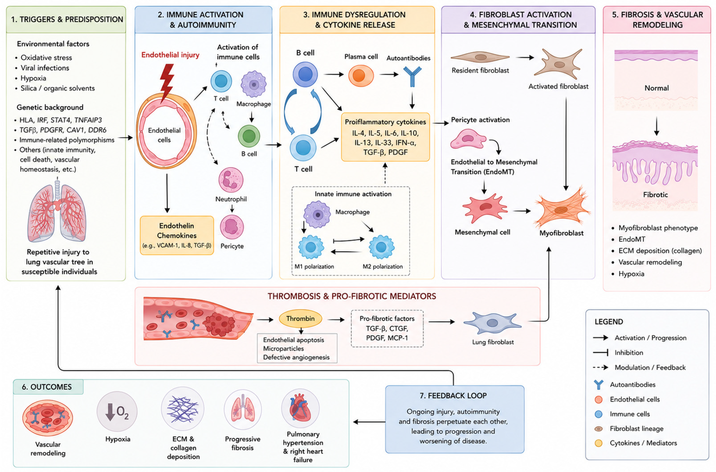
Figure 1. Pathogenesis of systemic sclerosis.
Life 16 01420 g001
Table 1. Autoantibodies in Systemic Sclerosis.
AutoantibodyAntigen TargetCutaneous PhenotypePulmonary InvolvementPrognostic Implications
Autoantibodies included in the 2013 ACR/EULAR classification criteriaAnti-Topoisomerase I (ATA/Scl-70)
[45,46,47,48,49,51,52,53,54,55,56,57,58,59,60,61,62]
DNA Topoisomerase IDiffuseSevere, progressive ILD; early FVC/DLCO declinePoor prognosis; high mortality
Anti-RNA polymerase III (anti-RNAP III)
[46,47,48,49,59,60,61,62]
RNA Polymerase IIIRapid diffuseModerate ILD (~18%); possible PAHAggressive early disease
ACA (anti-centromere)
[45,46,47,48,49,59,61,62]
Centromere proteinsLimitedLow ILD risk; high PAH risk (10–20%), ↓DLCOBetter survival; PAH main mortality driver
Other SSc-specific or SSc-associated autoantibodiesAnti-Th/To [47]RNase MRP complexLimitedHigh ILD + PAH (~70–75%)Reduced survival
Anti-U3 RNP
[47]
FibrillarinDiffuse/mixedIncreased PAH risk; variable ILDSevere phenotype
Anti-RNPC3
[46,48,49]
U11/U12 RNPMixedSevere ILDHigh-risk subset
Anti-U1 RNP
[45,47,48,49,60]
U1 RNP complexOverlapILD and PAHOverlap phenotype
Anti-PM/Scl
[45,47,48,49,60]
PM/Scl complexOverlapILD (often slower progression)More favourable pulmonary outcome
Anti-Ku
[45,47,48,49]
Ku proteinOverlapILDIntermediate prognosis
Anti-Ro52
[45,48,49]
TRIM21Overlap/mixedILD and PAHIncreased mortality risk
Anti-NOR90
[49]
NOR90LimitedPossible ILDGenerally favourable
ANCA
[46]
Neutrophil cytoplasmOverlapILD, vasculitis lung involvementHigher mortality
Anti-RNA Pol I
[46,48]
RPA194MixedModerate involvementPossibly lower cancer risk
Table 2. Clinical roles and potential utility of circulating biomarkers in systemic sclerosis-associated interstitial lung disease (SSc-ILD).
BiomarkerPathophysiological MechanismEarly Detection and Baseline SeverityPrediction of Disease Progression and PrognosisDisease Activity MonitoringTreatment Response/Predictive Utility
KL-6 [9,59,61,62,90,91,92]Type II pneumocyte proliferation and basement membrane disruptionAssociated with greater HRCT extent;
Inversely correlated with baseline FVC and DLCO
Correlates with initial ground-glass opacities
Higher levels have been associated with subsequent functional decline and adverse pulmonary outcomes.
KL-6 has shown comparatively consistent evidence for prediction of progression
Serial changes in KL-6 may provide information on disease evolution; increasing levels have been associated with subsequent ILD progressionChanges in KL-6 levels during treatment may reflect variations in disease activity; however, its utility for predicting treatment response has not been established
CCL18 [59,60,61,90,91,92]M2 Macrophage ActivationAssociated with greater fibrotic burden and pulmonary involvementAssociated with functional deterioration and adverse outcomes, although findings on short-term progression are inconsistentPersistent or changing CCL18 levels may reflect ongoing profibrotic activity associated with macrophage activationChanges in CCL18 levels during treatment have been reported; however, their utility for predicting treatment response remains unestablished
SP-D [59,60,61,62,90,91]Alveolar-capillary barrier disruption; leakage into systemic circulationAssociated with pulmonary involvement and ground-glass abnormalitiesElevated baseline levels have been associated with subsequent functional deterioration in some cohortsLongitudinal changes in SP-D levels may reflect variations in alveolar epithelial injury and pulmonary involvementThe utility of SP-D for predicting treatment response has not been established
IL-6 [9,59,60,61,90]Pro-inflammatory signalling; acute-phase response and systemic immune activationAssociated with early active disease and greater disease severityHigher baseline levels have been associated with early FVC decline and adverse outcomesLongitudinal changes in IL-6 levels may reflect variations in systemic inflammatory activityThe role of IL-6 in predicting therapeutic response remains to be determined
CXCL4 [9,60,61,91]Plasmacytoid dendritic cell activationBiomarker of early systemic disease and pulmonary involvementHigher levels have been associated with disease severity and progression in some cohortsChanges in CXCL4 levels may reflect variations in immune activation, but their value for longitudinal disease monitoring remains uncertainThe role of CXCL4 in predicting treatment response remains unclear
YKL-40 [61,90]Extracellular matrix remodelling, macrophage activation, and tissue repair dysregulationAssociated with established fibrosis and structural lung abnormalitiesHigher levels have been associated with adverse outcomesVariations in YKL-40 levels may reflect changes in extracellular matrix remodelling, although their value for disease monitoring remains uncertainThe predictive value of YKL-40 for treatment response remains uncertain
CRP [45,59,61,90,93]Systemic inflammation- Acute-phase reactantAssociated with systemic inflammation and pulmonary involvement, but lacks specificity for SSc-ILDHigher levels have been associated with functional decline and adverse outcomes in some cohortsChanges in CRP may reflect systemic inflammation, although their value for monitoring SSc-ILD activity remains limitedThe role of CRP in predicting treatment response in SSc-ILD remains unclear
CCL-2 [9,59,60,61,90,91]Fibroblast stimulation, myofibroblasts differentiation, lymphocyte T trafficking and lymphocyte Th2 phenotype polarisationAssociated with pulmonary involvement and impaired lung functionAssociated with progressive functional impairmentChanges in CCL2 levels may reflect variations in inflammatory cell recruitment and profibrotic activityThe role of CCL2 in predicting treatment response remains uncertain
CXCL10 [60,61,90]Promoting leukocyte recruitment-chemoattractant for Th1 lymphocytesAssociated with inflammatory activity in early SScAssociated with subsequent pulmonary functional deteriorationChanges in CXCL10 levels may reflect variations in inflammatory immune activity, although their value for longitudinal disease monitoring remains uncertainWhether CXCL10 can predict treatment response requires further investigation
MMP-7 MMP-9 MMP-12 [60,61,90,91]Extracellular matrix remodellingAssociated with pulmonary fibrosis and disease severityMMP-7 has been associated with impaired lung function and progression; evidence for MMP-9/MMP-12 is less establishedChanges in circulating MMP levels may reflect ongoing extracellular matrix turnover and remodelling, although their value for longitudinal disease monitoring remains uncertainTheir potential role in predicting treatment response remains to be determined
Disclaimer/Publisher’s Note: The statements, opinions and data contained in all publications are solely those of the individual author(s) and contributor(s) and not of MDPI and/or the editor(s). MDPI and/or the editor(s) disclaim responsibility for any injury to people or property resulting from any ideas, methods, instructions or products referred to in the content.


