Article

Preliminary In Silico Evaluation of Extra Virgin Olive Oil-Derived Bioactive Compounds as Multi-Target-Directed Ligands in Metabolic Dysfunction-Associated Steatotic Liver Disease


by
Ludovico Abenavoli
1,2,*,
Maja Milanović
3,
Giuseppe Guido Maria Scarlata
1,2,
Nataša Milošević
3,
Maria Luisa Gambardella
1,2 and
Nataša Milić
3
1
Department of Health Sciences, University “Magna Graecia”, 88100 Catanzaro, Italy
2
Center for Chronic Liver Diseases, “Renato Dulbecco” University Hospital, 88100 Catanzaro, Italy
3
Department of Pharmacy, Faculty of Medicine, University of Novi Sad, Hajduk Veljkova 3, 21000 Novi Sad, Serbia
*
Author to whom correspondence should be addressed.
Life 2026, 16(7), 1146; https://doi.org/10.3390/life16071146
Submission received: 10 June 2026 / Revised: 6 July 2026 / Accepted: 8 July 2026 / Published: 10 July 2026

Abstract

Background: Metabolic dysfunction-associated steatotic liver disease (MASLD) is the most prevalent chronic liver disease worldwide and is driven by complex metabolic and inflammatory disturbances. Extra virgin olive oil (EVOO), a hallmark of the Mediterranean diet, contains numerous bioactive compounds that may exert beneficial effects on liver and cardiometabolic health. This preliminary study investigated the interactions of selected EVOO-derived compounds, with molecular targets implicated in MASLD using an integrated in silico approach. Methods: Phenolic compounds, secoiridoids, fatty acids, sterols, squalene, and vitamin E were evaluated. Physicochemical properties, drug-likeness, and pharmacokinetic profiles were predicted using ADMETlab 3.0. Molecular docking analyses were performed against liver X receptors (LXRα and LXRβ), peroxisome proliferator-activated receptors (PPARα and PPARγ), hydroxymethylglutaryl-CoA reductase, cyclooxygenase-1, and cyclooxygenase-2. Binding modes were further examined by three-dimensional interaction analyses. Results: The investigated compounds displayed heterogeneous physicochemical and pharmacokinetic profiles. Oleuropein, oleacein, and oleocanthal demonstrated the most consistent binding patterns across targets involved in lipid metabolism, inflammation, and cardiometabolic regulation. In contrast, highly lipophilic compounds, including squalene, β-sitosterol, and vitamin E, frequently achieved high docking scores but formed fewer biologically relevant interactions. Conclusions: EVOO phenolics, particularly oleuropein, oleacein, and oleocanthal, emerged as promising multi-target modulators of MASLD-related pathways, supporting the potential role of EVOO in MASLD prevention and management.

1. Introduction

Metabolic dysfunction-associated steatotic liver disease (MASLD) is currently the most prevalent chronic liver disease worldwide, affecting approximately 38% of the adult population and posing a major global health burden [1]. MASLD encompasses a spectrum of progressive liver disorders, ranging from simple steatosis to metabolic dysfunction-associated steatohepatitis, fibrosis, cirrhosis, and hepatocellular carcinoma [2]. Despite its increasing prevalence and substantial clinical impact, effective pharmacological treatment options remain limited, and lifestyle interventions continue to represent the cornerstone of disease prevention and management [3].
Among lifestyle-based intervention, the Mediterranean diet has emerged as one of the most effective nutritional strategies for patients with MASLD. Characterized by a high consumption of plant-derived foods, monounsaturated fatty acids, and bioactive compounds, this dietary pattern has consistently been associated with improvements in metabolic health and reduced cardiovascular risk; conditions that frequently coexist with MASLD [4,5]. Recent clinical evidence suggests that greater adherence to the Mediterranean diet may reduce the risk of advanced liver fibrosis and improved metabolic outcomes in affected individuals [6].
Extra virgin olive oil (EVOO), the principal source of dietary fat in the Mediterranean diet, is considered one of the major contributors to these beneficial effects. Unlike refined olive oils, EVOO retains a complex phenolic fraction rich in hydroxytyrosol, tyrosol, oleuropein, oleocanthal, oleacein, and related secoiridoids [7,8,9]. Experimental and clinical data suggest that these bioactive compounds exert antioxidant, anti-inflammatory, lipid-modulating, and metabolic regulatory effects, potentially influencing multiple molecular pathways involved in MASLD pathogenesis [10].
The pathogenesis of MASLD is highly complex and multifactorial, involving dysregulated lipid metabolism, oxidative stress, chronic inflammation, and progressive fibrogenesis. Accordingly, several EVOO-derived polyphenols have been shown to modulate key molecular pathways implicated in disease progression, including nuclear receptor signaling, inflammatory mediators, and metabolic regulators [11,12,13,14]. Their pleiotropic biological activity suggests that these compounds may act as multi-target-directed ligands, simultaneously modulating multiple interconnected pathogenic mechanisms. Although increasing evidence supports the beneficial role of EVOO and its phenolic constituents in MASLD, comparative investigations exploring their interactions with multiple molecular targets remain scarce [10].
Therefore, the aim of this preliminary study was to evaluate, through an integrated in silico approach combining molecular and physicochemical profiling and molecular docking analysis, the interactions of selected EVOO-derived compounds with key targets involved in MASLD pathogenesis, with the objective of identifying potential multi-target-directed ligands.

2. Materials and Methods

2.1. Ligand Library Preparation

A library of bioactive compounds representative of EVOO was compiled based on the published literature. The selected molecules included phenolic compounds (hydroxytyrosol, tyrosol, and their derivatives), secoiridoids (oleuropein, oleuropein aglycone, oleacein, oleocanthal), fatty acids, sterols, and highly lipophilic molecules such as squalene and vitamin E. SMILES strings of the selected compounds were retrieved from the PubChem database and converted into 2D structures using ChemDraw Professional 16.0.
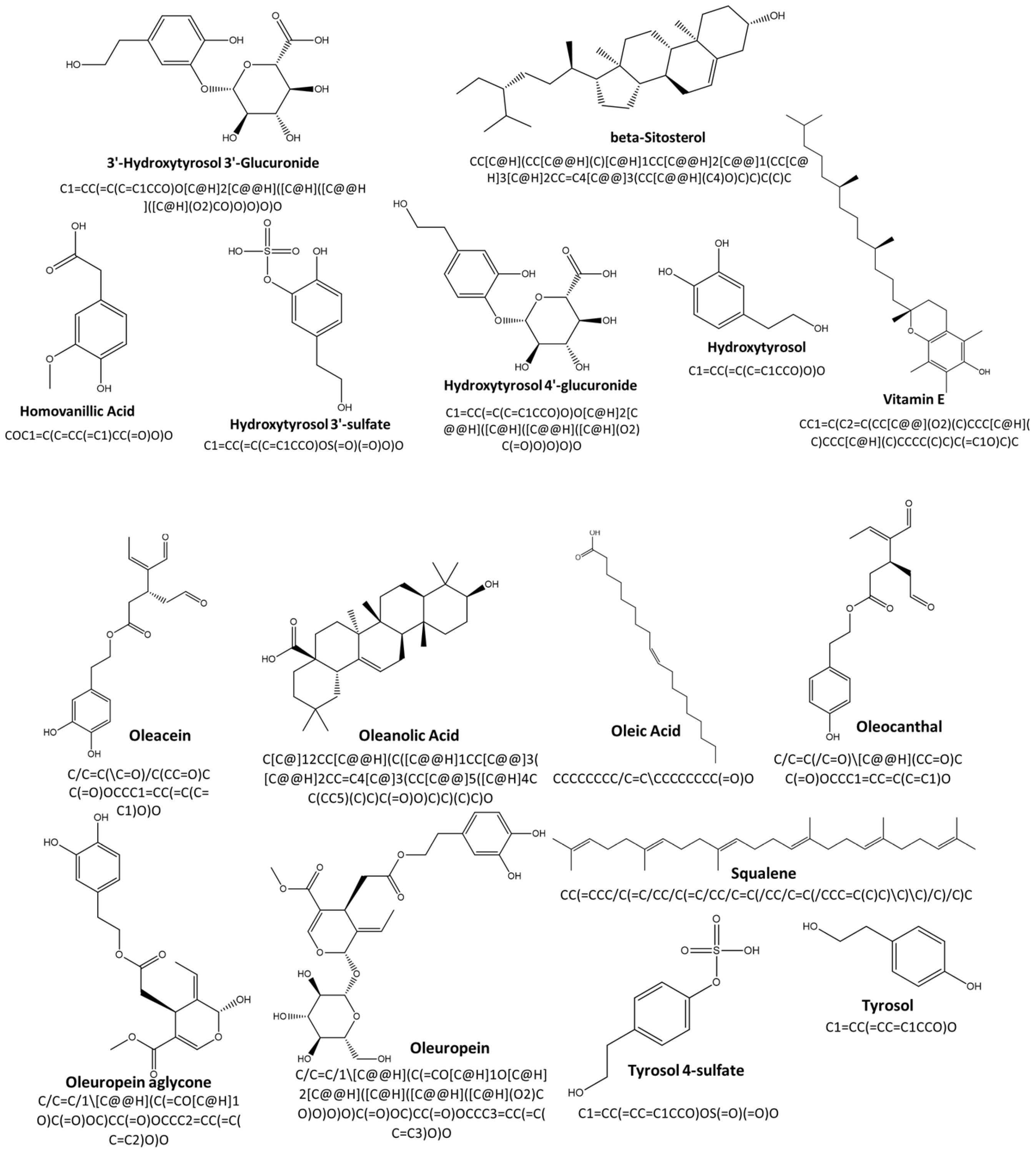
The chemical structures and corresponding SMILES strings of the investigated compounds are presented in Figure 1.

2.2. Molecular Properties and Bioavailability Profiles

Molecular descriptors and physicochemical properties were predicted in silico using ADMETlab 3.0 (https://admetlab3.scbdd.com/ (accessed on 5 April 2026)). The evaluated parameters included molecular weight (MW), volume (V), number of rings (nRing), number of rotatable bonds (nRot), hydrogen-bond donors and acceptors (nHD and nHA), topological polar surface area (TPSA), and fraction of sp3 carbons (Fsp3).
Lipophilicity (logP and logD7.4), aqueous solubility (logS), human intestinal absorption (HIA), and blood–brain barrier (BBB) permeability were also assessed to estimate drug-likeness and oral bioavailability profile. According to Lipinski’s rule of five, compounds with good oral bioavailability generally violate no more than one of the following criteria: MW < 500 Da, nHD ≤ 5, nHA ≤ 10 and logP < 5 [15]. According to Veber’s criteria, favorable oral bioavailability is expected for compounds with nRot ≤ 10 and TPSA ≤ 140 Å2 [16].

2.3. Target Selection and Preparation

Based on their established roles in lipid metabolism, inflammation, and MASLD pathogenesis, in accordance with previously validated multi-target approaches [17], the following targets were selected: liver X receptor alpha (LXRα), liver X receptor beta (LXRβ), peroxisome proliferator-activated receptor alpha (PPARα) and gamma (PPARγ), along with the enzymes hydroxymethylglutaryl-CoA reductase (HMG-CoA reductase), cyclooxygenase-1 (COX-1) and cyclooxygenase-2 (COX-2).
The 3D crystal structures of the selected targets in complex with co-crystallized ligands were retrieved from the RCSB Protein Data Bank (PDB, https://www.rcsb.org/ (accessed on 9 April 2026)) in PDB format (Table 1). The Protein Preparation Wizard incorporated in Genetic Optimisation for Ligand Docking (GOLD, version 2022.3.0) was used for target preparation. All water molecules and co-crystallized ligands were removed, to ensure a standardized preparation protocol, across all tested targets. Missing hydrogen atoms were added to the protein structures prior to docking simulations.

2.4. Docking Protocol and Validation

Molecular docking simulations were performed using a validated protocol in GOLD (version 2022.3.0) based on flexible ligand-rigid receptor docking [17]. The SMILES strings of the EVOO-derived compounds were converted into 3D structures using the Chem3D tool (version 16.0). The MMFF94 force field was applied for the ligand geometry optimization. The ligand-binding domain for each target was defined within a 6 Å radius around the coordinates of the co-crystallized ligand. The docking protocol was validating by redocking the co-crystallized ligands (e.g., T0901317, lanifibranor, GW1929, mevastatin, flurbiprofen, and naproxen) into their respective binding sites. The obtained RMSD values were 1.3404 for LXRα, 1.2824 for LXRβ, 0.8773 for PPARα, 0.7786 for PPARγ, 1.2193 for HMG-CoA reductase and 0.5038 and 0.4001 for COX-1 and COX-2, respectively. All values were within acceptable thresholds (RMSD < 2 Å) for each target, confirming accurate identification of the binding site and reproduction of key interactions [18,19]. For each EVOO-derived compound and each selected target, ten docking poses were generated using the ChemPLP scoring function. The corresponding ChemPLP scores were calculated. The pose with the highest ChemPLP score was selected for subsequent interaction analysis [8,17,18,19].

2.5. Binding-Mode Analysis

Protein–ligand interactions were analyzed, including hydrogen bonding, hydrophobic interactions, π–π interactions (where applicable), and van der Waals interactions. The academic version of Maestro Schrödinger software (v.14.7.) was used for visualization of the selected protein–ligand complexes.

3. Results

3.1. Molecular Properties and Bioavailability Profiles

The analyzed EVOO-derived compounds displayed considerable variability in their molecular properties (Table 2). Phenolic compounds, such as hydroxytyrosol and its conjugates, had low molecular weights and high polarity, whereas sterols and lipidic compounds, such as β-sitosterol, squalene, and vitamin E, were characterized by higher molecular weights, larger molecular volumes, and higher Fsp3 values. Oleuropein was the only polyphenolic compound that had more than 10 rotatable bonds. In addition, the high polarity of oleuropein and hydroxytyrosol glucuronides corresponded to the increased total number of hydrogen-bond donors and acceptors.
Marked differences in lipophilicity and predicted permeability were observed among the analyzed compounds (Table 3). Highly lipophilic compounds (logP/logD7.4 > 5), including squalene, β-sitosterol, and vitamin E, exhibited poor aqueous solubility and limited predicted intestinal absorption. In contrast, conjugated phenolic derivatives exhibited improved solubility but reduced membrane permeability. Among the studied EVOO compounds, only oleanolic acid and vitamin E exhibited permeation through the BBB, while predicted high human intestinal absorption was limited to 3′-hydroxytyrosol 3′-glucuronide, hydroxytyrosol 3′-sulfate, and tyrosol 4-sulfate. To complement the in silico predictions, Lipinski’s and Veber’s empirical rules were applied to further evaluate drug-likeness and oral bioavailability. Although the majority of compounds met criteria set by empirical rules, violations frequently occurred due to high lipophilicity (β-sitosterol, oleic acid, squalene, and vitamin E) and an elevated number of rotatable bonds (oleic acid, oleuropein, squalene, and vitamin E). Interestingly, oleuropein did not meet Lipinski’s and Veber’s criteria due to its high molecular weight (540.18), increased polarity (nHA = 13 and TPSA = 201.67 Å2), and 11 rotatable bonds. Among the other phenolic compounds, only hydroxytyrosol 4′-glucuronide exhibited more than one violation, driven by a high number of hydrogen-bond donors (nHD = 6) and a high TPSA (156.91 Å2).

3.2. Docking Performance and Score Interpretation

Docking simulations demonstrated heterogeneous ChemPLP scores across the analyzed targets. Since the ChemPLP score is a dimensionless scoring function and is based on an empirical fitness function optimized for pose prediction within a specific binding site, the obtained scores were interpreted relative to the scores of co-crystalized ligands within each individual target. Consequently, the docking results were used to rank compounds within each target rather than to compare binding affinities between targets with different pocket properties. (Table 4).
Within individual targets, several highly lipophilic compounds, particularly squalene, vitamin E, and β-sitosterol, achieved high docking scores, in some cases, comparable to those of reference ligands. However, squalene, vitamin E, β-sitosterol and oleic acid exhibited also very high values of lipophilicity (logP > 7, Table 3), which may lead to an overestimation of binding affinity in molecular docking analyses. In contrast, phenolic compounds and secoiridoids with logP values below 3 demonstrated more consistent ChemPLP scores across all examined targets in comparison with co-crystalized ligands.

3.3. Target-Specific Binding-Mode Analysis

Despite the high docking scores, several highly lipophilic compounds did not establish meaningful interactions within the ligand-binding domain of the target proteins, as evidenced by the absence of hydrogen bonds or stable contacts in the 3D representations.
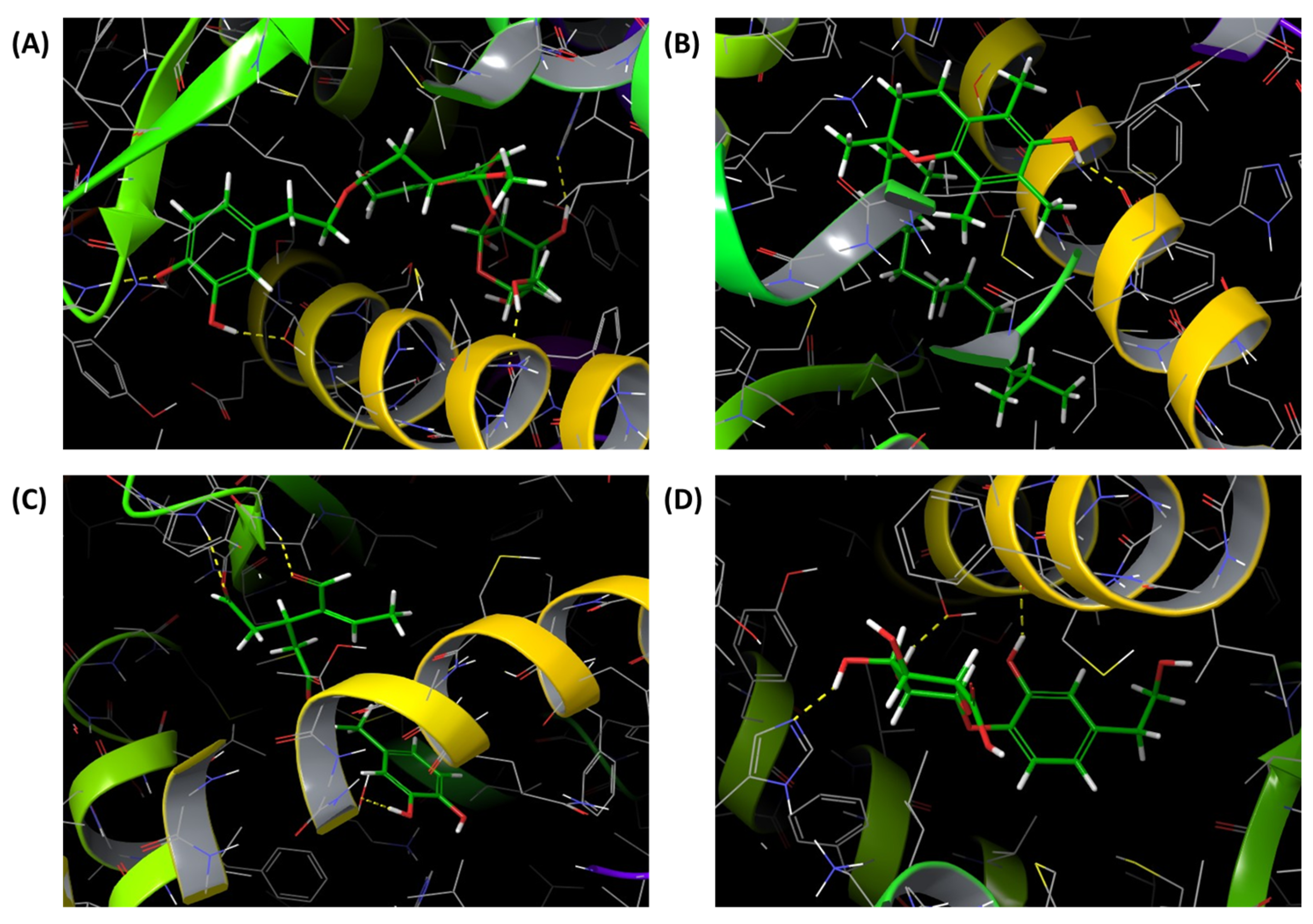
Binding-mode analysis performed on LXRα indicated that oleuropein and vitamin E adopted stable conformations within the ligand-binding domain, forming hydrogen bonds. In contrast, β-sitosterol and squalene, despite high ChemPLP scores, did not establish meaningful interactions. Even the compounds that expressed lower ChemPLP scores—tyrosol, hydroxytyrosol, hydroxytyrosol 4′-glucuronide and hydroxytyrosol 3′-sulfate—like oleuropein, reproduced the hydrogen-bond interaction with His421 observed in co-crystalized ligand, T0901317 (Figure 2).
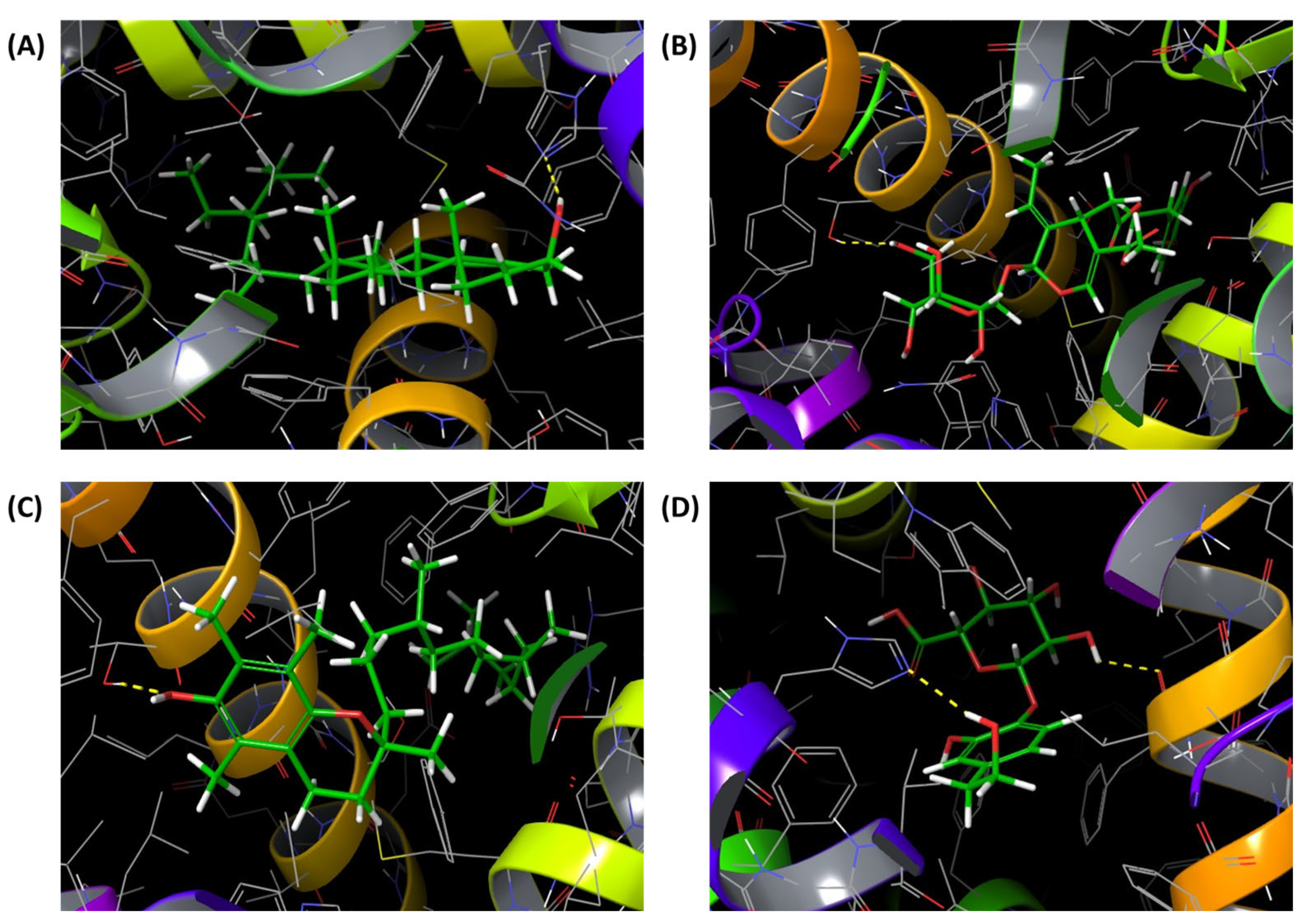
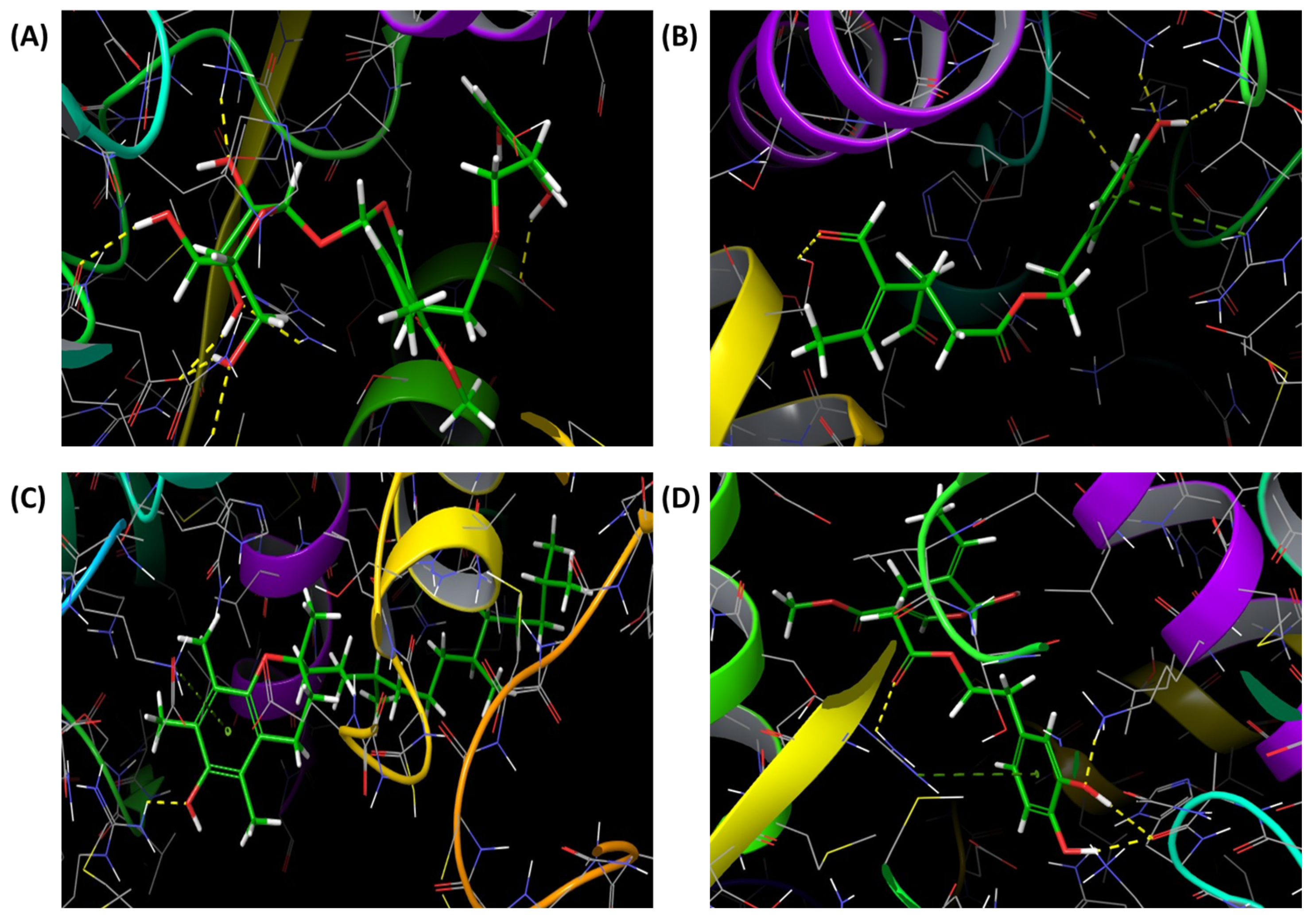
A similar interaction pattern was observed for LXRβ, where oleuropein, β-sitosterol and vitamin E formed stable hydrogen-bond networks within the ligand-binding domain, resulting in more favorable binding modes, while squalene failed to form stabilizing contacts (Figure 3). β-sitosterol reproduced the hydrogen bond with His435 as proven ligand, T0901317, whereas oleuropein and vitamin E formed polar interactions with Thr272. Interestingly, despite its lower ChemPLP values (65.929), oleocanthal established a dense hydrogen-bond network involving Thr272. Hydroxytyrosol 4′-glucuronide and hydroxytyrosol 3′-sulfate were stabilized with the ligand-binding pocket by creating multiple hydrogen bonds, most notably with His435, similar to β-sitosterol.
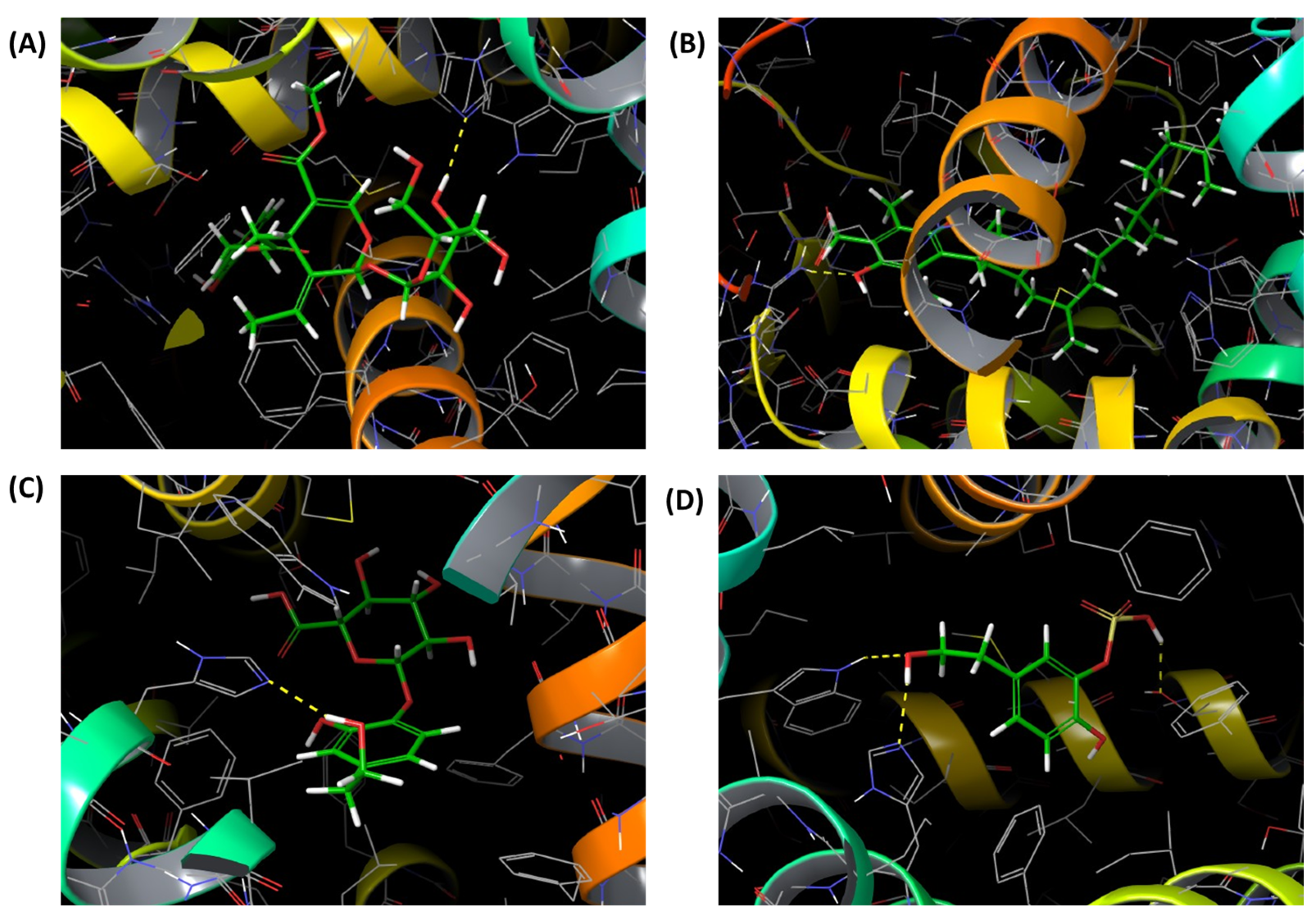
For PPARα, oleuropein and vitamin E indicated more consistent binding modes, whereas oleic acid and squalene did not establish any interactions, suggesting limited biological relevance. The stability of the oleuropein–PPARα complex was attributed to four hydrogen bonds associated with stable binding with PPARα. In addition, phenolic compounds like oleacein, hydroxytyrosol 4′-glucuronide and oleocanthal formed multiple hydrogen bonds within the PPARα pocket (Figure 4).
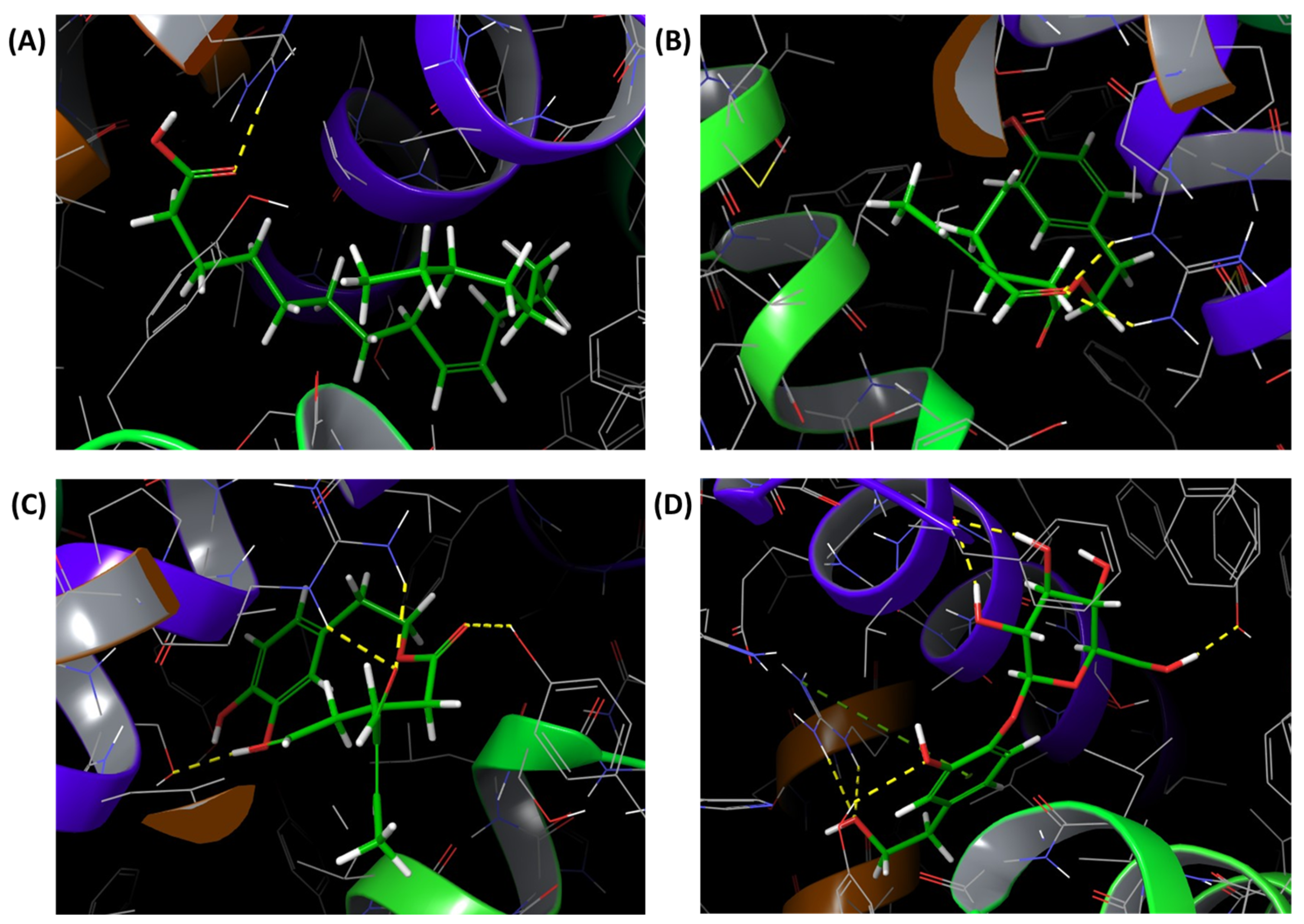
For PPARγ, among the analyzed compounds, oleic acid, oleuropein, oleacein, and vitamin E demonstrated stable binding conformations within the ligand-binding domain (Figure 5). Again, hydrogen bonds were responsible for the stability of the created ligand–target complexes. Even the phenolic compound that exhibited lower ChemPLP scores similar to 3′-hydroxytyrosol 3′-glucuronide, hydroxytyrosol 4′-glucuronide and oleocanthal established multiple hydrogen interactions with the receptor.
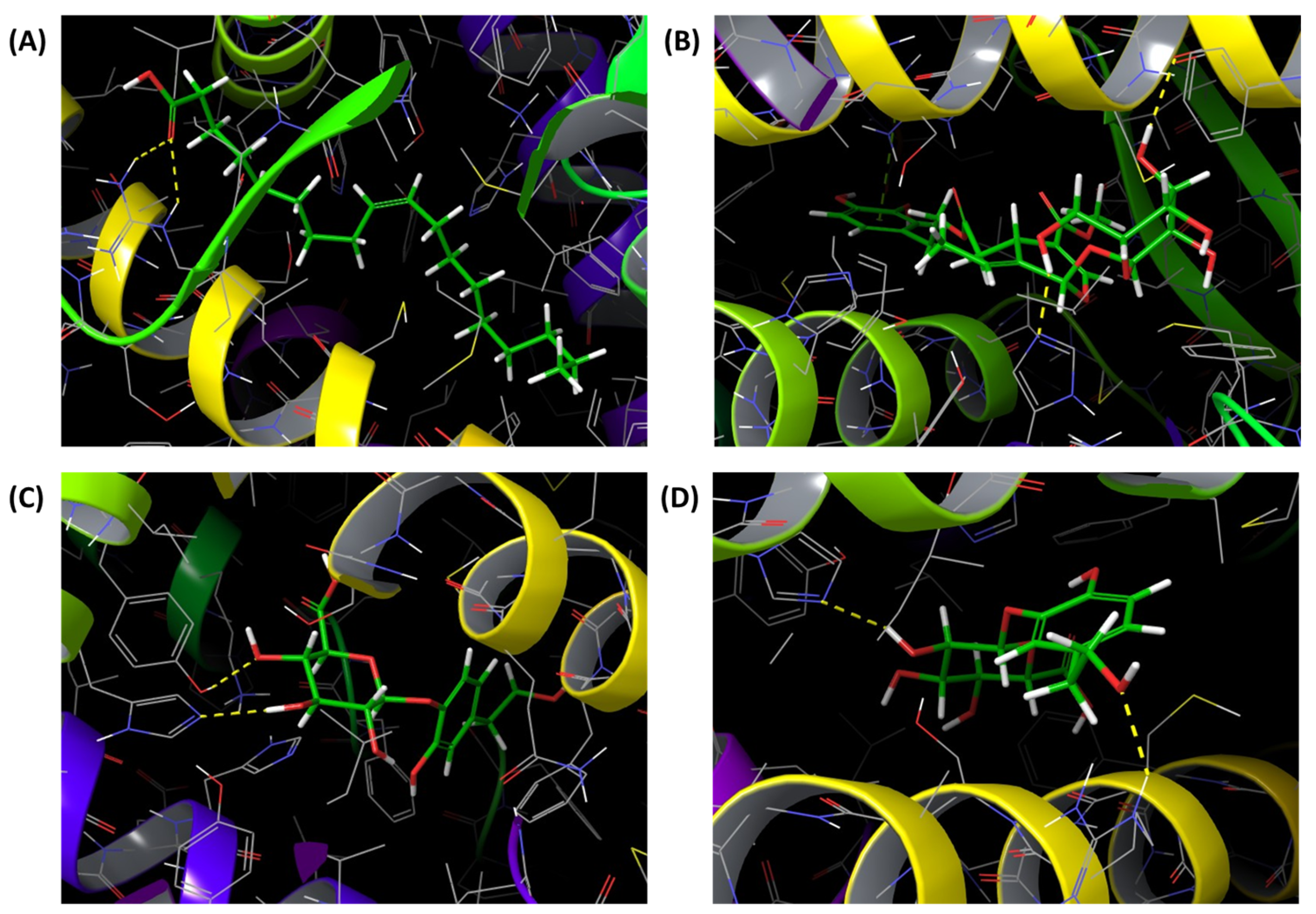
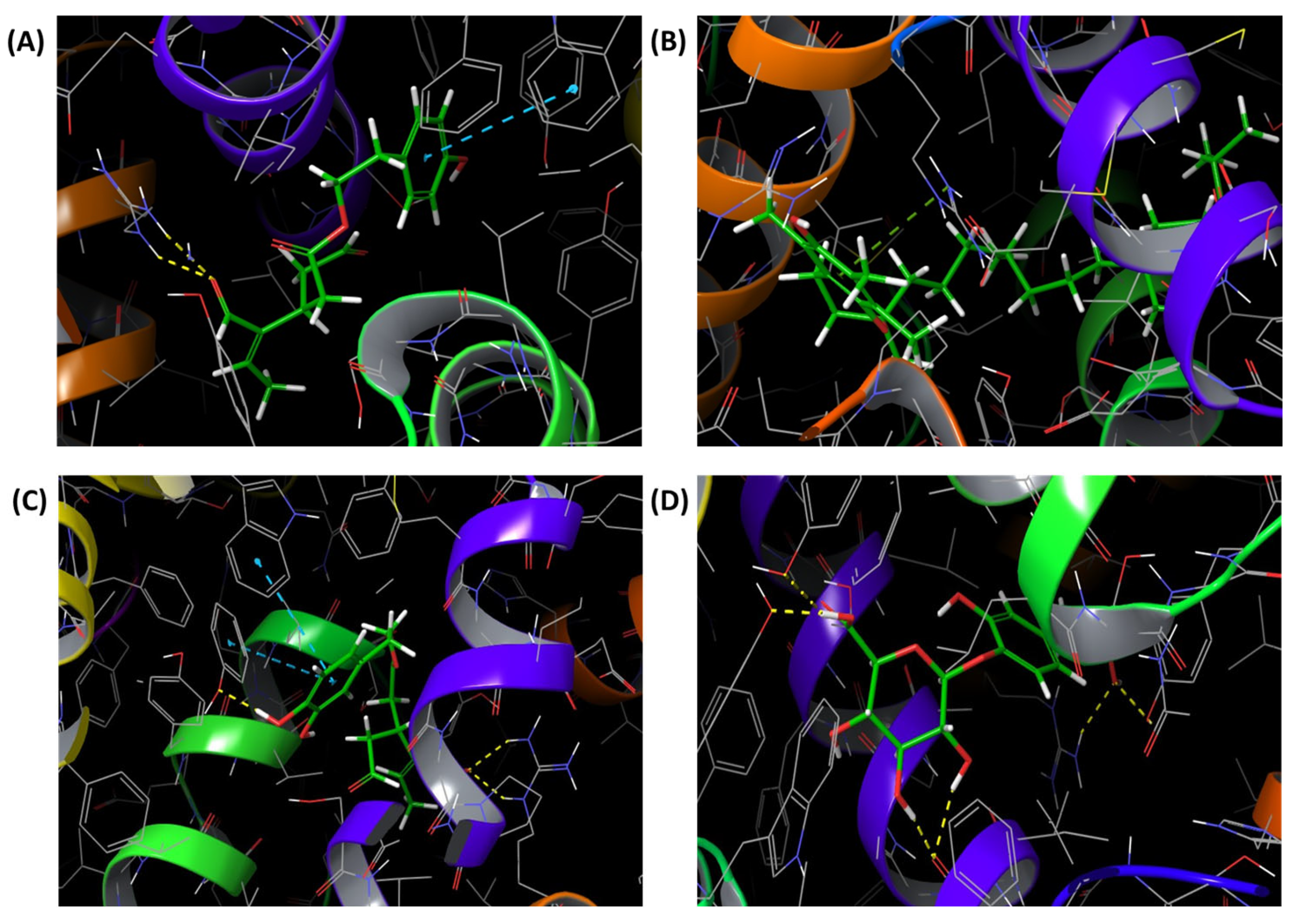
About HMG-CoA reductase, docking analysis indicated that oleuropein, oleacein and vitamin E formed an extensive hydrogen-bond network within the catalytic site, whereas squalene, despite high scores, did not exhibit relevant binding interactions (Figure 6). Within the complex mainly hydrogen-bond-driven network, oleuropein, vitamin E, oleacein, and even oleuropein aglycone demonstrated interactions with Arg590, resembling the interaction pattern of the reference ligand mevastatin. In addition, oleuropein formed an additional hydrogen bond with the Asp690 amino-acid residue similar to mevastatin.
For both COX isoforms, oleocanthal and oleacein exhibited the most consistent interaction patterns. In addition to π-π interactions, those compounds formed hydrogen-bond networks involving amino-acid residues surrounding the active sites of both COX-1 and COX-2. Again, complexes with hydroxytyrosol 4′-glucuronide were stabilized using hydroxyl groups in a dense network despite the lower ChemPLP values. Conversely, highly hydrophobic compounds such as squalene again displayed high scores but poor interaction profiles (Figure 7 and Figure 8).

4. Discussion

Considering the complex etiology of MASLD as well as limited pharmacological options, lifestyle modifications including diet remain the gold standard in the prevention and treatment of this disease [7]. Hence, constituents of diet with multi-target properties may be ideal candidates for MASLD mitigation and management [17,19]. The combination of QSAR-based algorithms, empirical rules, and the molecular docking approach was applied in this research in order to get insights into the drug-likeness properties, potential oral bioavailability as well as interaction mechanisms of multiple active components present in EVOO.
EVOO is rich in lipophilic compounds. The glyceride fraction with up to 85% of unsaturated acid, particularly oleic acid, is the major constituent of olive oil. The less than 2% of olive oil is based on phenolic compounds and vitamin E [20]. Hence, the EVOO compounds diverse in molecular properties were studied in the context of drug-likeness properties and bioavailability. The applied in silico algorithms evaluated absorption and permeability based on passive diffusion processes [21]. According to the predictions conducted by the ADMETlab 3.0 online software tool, poor oral bioavailability was attributed to lipophilic compounds such as squalene, β-sitosterol, vitamin E and oleic acid due to the high logP/logD7.4 values. Phenolic compounds also expressed low predicted intestinal absorption due to their high solubility in water. Apart from the molecular descriptors observed in drug-likeness empirical rules (i.e., nRot, nHA, nHD, TPSA, logP), Fsp3 is a relatively new parameter considered during the initial phases of drug discovery. The values of 0.42 and higher are usually targeted for compounds with an optimal aqueous solubility and pharmacokinetic profile [22]. Phenolic compounds such as tyrosol, hydroxytyrosol and their sulfate derivatives did not meet the set threshold. While most compounds followed Lipinski’s rule of five and Veber’s rule [15,16], oleuropein exhibited structural constraints (Table 2 and Table 3), suggesting limited passive intestinal permeability and low predicted oral bioavailability. Data from in vitro and in vivo studies confirmed relatively poor and highly variable oleuropein bioavailability [23,24,25]. However, the absorption of EVOO constituents is far more complex. After oral intake, oleuropein is partially hydrolyzed to oleuropein aglycone, hydroxytyrosol and other bioactive metabolites that may also contribute to its biological activity. In addition, hydroxytyrosol forms sulfate and glucuronide conjugates due to the intensive first past metabolism. The bioavailability of phenolic compounds is also gender related and their plasma concentrations are strongly influenced by the type of formulation. For example, liquid forms of oleuropein resulted in better plasma concentrations compared to capsules [26]. The bioavailability of a lipophilic compound is strongly dependent on emulsification with bile salts, the presence of other dietary lipids, and the transport proteins and cellular uptake in the intestinal membrane [23,24]. Taken together, the performed evaluation of molecular descriptors and drug-likeness properties imply that special formulation strategies are required to overcome poor solubility and/or membrane permeability of the studied compounds. Nanoemulsions, liposomes, niosomes, phytosomes as well as polymeric encapsulation systems could offer an alternative approach to enhance the stability and bioavailability of EVOO compounds [27,28]. Considering that poor pharmacokinetic behavior is the most common reason of the failure of promising drug candidates in clinical phases [29], the obtained results might serve as a baseline step for selection of best candidates.
Combined molecular docking and binding-mode analyses supported the hypothesis that EVOO-derived compounds may have acted as multi-target modulators of MASLD-related pathways (Table 4). Molecular targets involved in disease pathogenesis were carefully chosen based on their involvement in disease onset and progression as well as the activity of their co-crystalized ligands. For example, T0901317, lanifibranor, and GW1929 act as agonists for LXRα/β and PPARα/γ, respectively [29,30,31], while mevastatin, flurbiprofen and naproxen behave as inhibitors of HMG-CoA reductase, COX-1 and COX-2, respectively [32,33,34]. Despite the still-debated role of LXR in MASLD, for more than a decade, LXR α/β agonists have been considered for the treatment of dyslipidemia and atherosclerosis [35]. Binding-mode analysis demonstrated that phenolic compounds present in EVOO are able to interfere with key amino acids within the LXRα binding cavity (His421), specifically oleuropein, tyrosol, hydroxytyrosol, and its glucuronide and sulfate derivatives. Moreover, tyrosol, hydroxytyrosol, and hydroxytyrosol 3′-sulfate derivatives, like endogenous agonists oxycholesterols, established an additional hydrogen bond with Trp443 [36]. A novel in vivo study highlighted that the intestinal activation of LXRα resulted in decreased inflammation, steatosis, and liver fibrosis in MASLD [37]. Interestingly, the most abundant phytosterol in olive oil, β-sitosterol, reproduced a hydrogen bond with His435 as proven LXRβ agonist, T0901317. Despite lower ChemPLP values, hydroxytyrosol 4′-glucuronide and hydroxytyrosol 3′-sulfate reassembled the same binding mode of interaction while oleuropein, vitamin E and oleocanthal interacted via hydrogen bonds with Thr272, a residue reported to participate in ligand recognition within the LXRβ binding pocket [38]. Oleuropein aglycon and oleocanthal also had hydrophobic interactions involving Phe329 at the binding site, similar to other ligands for therapeutic utility [39].
PPARα/γ are primary targets in MASLD treatments due to their important role in lipid metabolism, insulin sensitivity, and inflammation [40]. Fibrates, like the co-crystallized ligand, lanifibranor, promote HDL formation and decrease triglyceride levels in blood via PPARα receptor activation [41]. The increased stability of phenolic compounds inside the binding pocket was attributed to the multiple hydrogen bonds. Previous in silico studies of PPARα agonists highlighted the role of hydrogen bonds with Ser280 and His440 amino-acid residues [42]. Oleacein and hydroxytyrosol 4′-glucuronide established hydrogen bonds with Ser280 analogous to fibrates, while oleuropein and hydroxytyrosol 4′-glucuronide interacted with the receptor, creating hydrogen bonds with His440 [42,43]. In the literature, the critical role of Phe273 in ligand-binding selectivity, stability and PPARα conformation is firmly established [44]. Hence, hydrogen bonds involving this residue additionally increased the stability of oleuropein, vitamin E, and oleocanthal within the bonding cavity. Targeting PPARγ represents a well-known approach in MASLD treatment, due to its ability to improve insulin sensitivity and reduce blood glucose levels [17]. Multiple hydrogen bonds contributed to the stability of lipophilic and hydrophilic EVOO compounds within the binding cavity. PPARγ analysis showed a hydrogen-bond binding mode involving hydrophilic residues His323, Tyr327, Lys367, His449, similar to the typical agonist rosiglitazone. In addition, bonding with Phe282 within hydrophobic region was also observed [45]. Despite the lower ChemPLP scores, 3′-hydroxytyrosol 3′-glucuronide, hydroxytyrosol 4′-glucuronide, oleuropein aglycon and oleocanthal established multiple hydrogen interactions.
Despite a range adverse effects, statins, as HMG-CoA reductase inhibitors, are commonly administered to MASLD patients. The inhibition of this enzyme is a primary therapeutic strategy for the reduction in cardiovascular risk as well as for the prevention of hepatic steatosis and fibrosis [46]. A dense network of interactions with key residues common for statins Arg590, Asp690, Lys691, Lys735 was observed in the case of oleuropein [47]. Vitamin E also formed hydrogen bonds with Arg590 and Lys691. Oleacein and oleuropein aglycone stability were increased via bonding with Arg590, Lys735 and Ala751. Importantly, oleacein was the only compound that established hydrogen bond with key residue Ser684, similar to statins [17,47].
Keeping in mind the pro-inflammatory role of prostaglandins in MASLD, COX-1 and COX-2 were examined as potential targets for EVOO compounds. Docking analysis suggested that these compounds might mitigate hepatic inflammation, lipid accumulation, and progression of the disease through the combination of hydrogen and π-π interactions with residues located within the enzyme’s binding site. The active sites of both isoforms are created of almost the same amino-acid residues, and hydrogen-bond interactions with Arg120 and Tyr355 have been reported as characteristic features of the binding modes of many COX inhibitors [48]. Based on the target-specific binding-mode analysis, oleocanthal, oleacein, vitamin E, oleic acid reassembled the hydrogen bond with Arg120 while hydroxytyrosol 4′-glucuronide, despite the lower ChemPLP values, established additional hydrogen bond with Tyr355.
Although lacking experimental validation and molecular dynamics simulations, this in silico study provides an initial insight into the drug-likeness properties, binding affinities, and interaction profiles of EVOO-derived compounds against key targets involved in MASLD pathogenesis. PPARα and PPARγ are currently among the most extensively investigated pharmacological targets, with compounds such as co-crystallized lanifibranor showing promising results in clinical studies. LXR receptors remain experimental targets but play a central role in the regulation of lipid and cholesterol metabolism. HMG-CoA reductase is the well-established target of statins, drugs widely used in patients with MASLD because of their cardiometabolic benefits. In addition, COX-mediated pathways contribute to hepatic and systemic inflammation, suggesting that the modulation of these enzymes may represent an additional mechanism through which EVOO-derived compounds exert beneficial effects in MASLD. Based on these findings oleuropein, oleocanthal, and oleacein should be considered in the management of MASLD due to the exhibited moderate to high potential for a multi-target approach (Table 5). However, further molecular dynamics simulations and experimental assays should provide additional physiologically relevant assessments of compound and target engagement.
One of the major limitations of the performed docking analysis is the application of scoring functions for the evaluation of binding affinities. ChemPLP has been widely used as the default scoring function in GOLD due to the highest success rates (for both pose prediction and virtual screening experiments) in comparison with diverse validation test sets [49]. However, the obtained high ChemPLP scores are not always associated with biologically relevant binding modes. The score is calculated based on an empirical approach that combines van der Waals interactions, hydrogen bonding and electrostatic interactions [50]. Hydrophobic compounds with high logP values such as squalene, vitamin E, β-sitosterol and oleic acid, frequently exhibited elevated scores despite lacking stabilizing interactions within the ligand-binding domain. Hence, the high hydrophobicity of these compounds should not be neglected in the discussion of the obtained ChemPLP scores. As stated in this research, the binding sites of the co-crystallized ligands were used to significantly increase the docking efficiency. Consequently, ChemPLP scores could only be used in comparison with the score obtained for the co-crystallized ligand for each target and could not be directly compared across targets with different pocket properties. In addition, the molecular docking results were discussed based on the binding profiles and established interactions within the ligand-binding pocket. A limitation of the present docking protocol is that all crystallographic water molecules were removed during target preparation to ensure methodological consistency across all targets. This approach neglected the structurally conserved water molecules that could mediate water-bridged hydrogen-bond interactions, particularly within the COX-2 active site and the ligand-binding domains of PPAR receptors. Although interactions with certain amino-acid residues have been reported for recognized agonists or inhibitors, similar behaviors may also occur in complexes with ligands exhibiting different functional profiles. Therefore, the observed binding modes should not be interpreted as evidence of agonistic or inhibitory activity without further in vitro and in vivo experiments. In addition, the relatively low predicted oral bioavailability of oleuropein might be overcome with advances in drug delivery technologies and pharmacokinetic evidences about its metabolism to oleuropein aglycone and hydroxytyrosol, which may also contribute to its biological activity. Hence, both the molecules and their metabolites were also tested using docking analysis. But the molecular species interacting with the target receptors in vivo may differ from the tested compounds due to gastrointestinal and metabolic conversion.
From a clinical perspective, the present findings support the growing body of evidence suggesting that EVOO should be considered not only as a dietary component but also as a source of bioactive molecules potentially capable of modulating multiple pathogenic pathways involved in MASLD. The multi-target interactions identified for oleuropein, oleocanthal, and oleacein are particularly relevant, as MASLD is characterized by a complex interplay between liver steatosis, insulin resistance, chronic inflammation, dyslipidemia, and cardiovascular risk [5,51]. The simultaneous modulation of PPARα/γ signaling, cholesterol metabolism, inflammatory pathways, and lipid homeostasis may provide a mechanistic explanation for the beneficial effects consistently observed in the Mediterranean diet intervention [6]. Therefore, therapeutic strategies targeting a single molecular pathway often provide only partial benefits. In this context, EVOO-derived compounds may represent attractive adjunctive agents capable of exerting complementary effects on both hepatic and extrahepatic manifestations of the disease. Although the present results are limited to in silico predictions and cannot be directly translated into clinical recommendations, they provide a biologically plausible rationale for further experimental and clinical investigations.
Future studies should focus on validating these molecular interactions in cellular and animal models, assessing the bioavailability of individual EVOO phenolics in optimized formulations, and determining whether specific compounds or combinations can enhance the therapeutic effects of lifestyle interventions. Ultimately, a better understanding of the pharmacological potential of EVOO-derived molecules may contribute to the development of novel nutraceutical or adjunctive therapeutic approaches for MASLD, particularly in patients with high cardiometabolic risk.

5. Conclusions

This integrated in silico approach, combining molecular docking, drug-likeness evaluation, and binding-mode analysis, identified several EVOO-derived bioactive compounds with potential relevance for MASLD management. Among the investigated molecules, oleuropein, oleacein, and oleocanthal emerged as the most promising multi-target candidates, showing consistent interaction profiles across molecular pathways involved in lipid metabolism, inflammation, and cardiometabolic regulation. Importantly, the combined assessment of docking scores and binding interactions highlighted the need for cautious interpretation of highly lipophilic compounds, whose elevated docking scores may not necessarily reflect biologically meaningful target engagement. Although oleuropein exhibited the most favorable overall interaction profile, its predicted pharmacokinetic limitations suggest that bioavailability and metabolic transformation should be carefully considered when translating these findings into biological settings. Overall, the present study provides a mechanistic framework supporting the beneficial effects of EVOO within the Mediterranean diet and identifies promising candidate molecules for future translational and experimental studies in MASLD.

Author Contributions

Conceptualization, L.A. and N.M. (Nataša Milić); methodology, M.L.G., N.M. (Nataša Milošević) and N.M. (Nataša Milić); software, N.M. (Nataša Milić); validation, L.A., M.M. and G.G.M.S.; formal analysis, N.M. (Nataša Milić); investigation, M.M., G.G.M.S., M.L.G. and N.M. (Nataša Milošević); resources, L.A.; data curation, G.G.M.S. and N.M. (Nataša Milić); writing—original draft preparation, L.A., M.M. and G.G.M.S.; writing—review and editing, N.M. (Nataša Milošević) and L.A.; visualization, N.M. (Nataša Milić); supervision, L.A. All authors have read and agreed to the published version of the manuscript.

Funding

This research received no external funding.

Institutional Review Board Statement

Not applicable.

Informed Consent Statement

Not applicable.

Data Availability Statement

The original contributions presented in this study are included in the article. Further inquiries can be directed to the corresponding author.

Conflicts of Interest

The authors declare no conflicts of interest.

Abbreviations

The following abbreviations are used in this manuscript:
BBBBlood–Brain Barrier
COX-1Cyclooxygenase-1
COX-2Cyclooxygenase-2
EVOOExtra Virgin Olive Oil
Fsp3Fraction of sp3 Carbon Atoms
GOLDGenetic Optimisation for Ligand Docking
HIAHuman Intestinal Absorption
HMG-CoA Reductase3-Hydroxy-3-Methylglutaryl-Coenzyme A Reductase
LXRαLiver X Receptor Alpha
LXRβLiver X Receptor Beta
MASLDMetabolic Dysfunction-Associated Steatotic Liver Disease
MWMolecular Weight
nHANumber of Hydrogen-Bond Acceptors
nHDNumber of Hydrogen-Bond Donors
nRingNumber of Rings
nRotNumber of Rotatable Bonds
PDBProtein Data Bank
PPARαPeroxisome Proliferator-Activated Receptor Alpha
PPARγPeroxisome Proliferator-Activated Receptor Gamma
RMSDRoot Mean Square Deviation
TPSATopological Polar Surface Area
VVolume

References

  1. Younossi, Z.M.; Kalligeros, M.; Henry, L. Epidemiology of metabolic dysfunction-associated steatotic liver disease. Clin. Mol. Hepatol. 2025, 31, S32–S50. [Google Scholar] [CrossRef] [PubMed]
  2. Lim, T.S.; Kwon, S.; Bae, S.A.; Chon, H.Y.; Jang, S.A.; Kim, J.K.; Kim, C.S.; Park, S.W.; Kim, K.M. Association Between Handgrip Strength and Cardiovascular Disease Risk in MASLD: A Prospective Study from UK Biobank. J. Cachexia Sarcopenia Muscle 2025, 16, e13757. [Google Scholar] [CrossRef] [PubMed]
  3. Caddeo, A.; Romeo, S. Precision medicine and nucleotide-based therapeutics to treat steatotic liver disease. Clin. Mol. Hepatol. 2025, 31, S76–S93. [Google Scholar] [CrossRef] [PubMed]
  4. Abenavoli, L.; Kobyliak, N.; Kukharchuk, A.; Chervona, O.; Shvets, Y.; Scarlata, G.G.M.; Morano, D.; Christian, E.; Molochek, N.; Lynchak, O.; et al. The Mediterranean diet: Historical benefits and contemporary challenges in Southern Italy. Nutr. Res. 2026, 145, 37–47. [Google Scholar] [CrossRef] [PubMed]
  5. Suresh, M.G.; Mohamed, S.; Geetha, H.S.; Prabhu, S.; Trivedi, N.; Mehta, P.D.; Damodaran, U.K.; Brar, A.; Sohal, A.; Hatwal, J.; et al. Cardiovascular Implications in Metabolic Dysfunction-Associated Steatotic Liver Disease (MASLD): A State-of-the-Art Review. Korean Circ. J. 2026, 56, 103–130. [Google Scholar] [CrossRef] [PubMed]
  6. Hsieh, M.L.; Su, T.H.; Lin, Y.C.; Chen, Y.Y.; Tung, C.F.; Huang, L.S.; Wu, C.H.; Peng, Y.C.; Hsieh, V.C. Mediterranean Diet Adherence Is Associated with Reduced Liver Fibrosis Risk in Metabolic Dysfunction-Associated Steatotic Liver Disease. J. Gastroenterol. Hepatol. 2026, 41, 1041–1051. [Google Scholar] [CrossRef] [PubMed]
  7. Abenavoli, L.; Procopio, A.C.; Paravati, M.R.; Costa, G.; Milić, N.; Alcaro, S.; Luzza, F. Mediterranean Diet: The Beneficial Effects of Lycopene in Non-Alcoholic Fatty Liver Disease. J. Clin. Med. 2022, 11, 3477. [Google Scholar] [CrossRef] [PubMed]
  8. Abenavoli, L.; Scarlata, G.G.M.; Gambardella, M.L.; Morano, D.; Milošević, N.; Milanović, M.; Milić, N. Annurca Apple Extract and Colorectal Cancer Prevention: Preliminary In Silico Evaluation of Chlorogenic Acid. Diseases 2026, 14, 33. [Google Scholar] [CrossRef] [PubMed]
  9. Yetisgin, A.M.; Bartolomeo, G.; Cicero, N.; Micale, N.; Costa, R.; Cristani, M. Exploring the Nutraceutical Potential of Extra Virgin Olive Oils from Sicilian Well-Known and Lesser-Known Cultivars. Chem. Biodivers. 2026, 23, e03286. [Google Scholar] [CrossRef] [PubMed]
  10. Bernardino, M.; Tiribelli, C.; Rosso, N. Exploring the Role of Extra Virgin Olive Oil (EVOO) in MASLD: Evidence from Human Consumption. Nutrients 2025, 17, 2932. [Google Scholar] [CrossRef] [PubMed]
  11. Carpi, S.; Scoditti, E.; Massaro, M.; Polini, B.; Manera, C.; Digiacomo, M.; Esposito Salsano, J.; Poli, G.; Tuccinardi, T.; Doccini, S.; et al. The Extra-Virgin Olive Oil Polyphenols Oleocanthal and Oleacein Counteract Inflammation-Related Gene and miRNA Expression in Adipocytes by Attenuating NF-κB Activation. Nutrients 2019, 11, 2855. [Google Scholar] [CrossRef] [PubMed]
  12. Gabbia, D.; Carpi, S.; Sarcognato, S.; Cannella, L.; Colognesi, M.; Scaffidi, M.; Polini, B.; Digiacomo, M.; Esposito Salsano, J.; Manera, C.; et al. The Extra Virgin Olive Oil Polyphenol Oleocanthal Exerts Antifibrotic Effects in the Liver. Front. Nutr. 2021, 8, 715183. [Google Scholar] [CrossRef] [PubMed]
  13. Gabbia, D. Beneficial Effects of Tyrosol and Oleocanthal from Extra Virgin Olive Oil on Liver Health: Insights into Their Mechanisms of Action. Biology 2024, 13, 760. [Google Scholar] [CrossRef] [PubMed]
  14. Schirone, L.; Overi, D.; Carpino, G.; Carnevale, R.; De Falco, E.; Nocella, C.; D’Amico, A.; Bartimoccia, S.; Cammisotto, V.; Castellani, V.; et al. Oleuropein, a Component of Extra Virgin Olive Oil, Improves Liver Steatosis and Lobular Inflammation by Lipopolysaccharides-TLR4 Axis Downregulation. Int. J. Mol. Sci. 2024, 25, 5580. [Google Scholar] [CrossRef] [PubMed]
  15. Lipinski, C.A.; Lombardo, F.; Dominy, B.W.; Feeney, P.J. Experimental and computational approaches to estimate solubility and permeability in drug discovery and development settings. Adv. Drug Deliv. Rev. 2001, 46, 3–26. [Google Scholar] [CrossRef] [PubMed]
  16. Veber, D.F.; Johnson, S.R.; Cheng, H.Y.; Smith, B.R.; Ward, K.W.; Kopple, K.D. Molecular properties that influence the oral bioavailability of drug candidates. J. Med. Chem. 2002, 45, 2615–2623. [Google Scholar] [CrossRef] [PubMed]
  17. Paravati, M.R.; Procopio, A.C.; Milanović, M.; Scarlata, G.G.M.; Milošević, N.; Ružić, M.; Milić, N.; Abenavoli, L. Onion Polyphenols as Multi-Target-Directed Ligands in MASLD: A Preliminary Molecular Docking Study. Nutrients 2024, 16, 1226. [Google Scholar] [CrossRef] [PubMed]
  18. Pinter, D.; Milošević, N.; Milanović, M.; Vidović, D.; Kvrgić, J.; Kojić, V.; Jakimov, D.; Drljača Lero, J.; Milić, N.; Božić, B.; et al. 1-Aryl-3-Ethyl-3-Methyl- and 1-Aryl-3-Methylsuccinimides as Drug Candidates for Cancer: Toxicity Prediction, Molecular Docking, and In Vitro Assessment. J. Biochem. Mol. Toxicol. 2025, 39, e70313. [Google Scholar] [CrossRef] [PubMed]
  19. Milošević, N.; Milanović, M.; Medić Stojanoska, M.; Tipmanee, V.; Smyrnioudis, I.; Dedoussis, G.V.; Milić, N. Triterpenoids from Chios Mastiha Resin Against MASLD-A Molecular Docking Survey. Curr. Issues Mol. Biol. 2025, 47, 51. [Google Scholar] [CrossRef] [PubMed]
  20. Jimenez-Lopez, C.; Carpena, M.; Lourenço-Lopes, C.; Gallardo-Gomez, M.; Lorenzo, J.M.; Barba, F.J.; Prieto, M.A.; Simal-Gandara, J. Bioactive Compounds and Quality of Extra Virgin Olive Oil. Foods 2020, 9, 1014. [Google Scholar] [CrossRef] [PubMed]
  21. Ancuceanu, R.; Lascu, B.E.; Drăgănescu, D.; Dinu, M. In Silico ADME Methods Used in the Evaluation of Natural Products. Pharmaceutics 2025, 17, 1002. [Google Scholar] [CrossRef] [PubMed]
  22. Wei, W.; Cherukupalli, S.; Jing, L.; Liu, X.; Zhan, P. Fsp3: A new parameter for drug-likeness. Drug Discov. Today 2020, 25, 1839–1845. [Google Scholar] [CrossRef] [PubMed]
  23. Nikou, T.; Sakavitsi, M.E.; Kalampokis, E.; Halabalaki, M. Metabolism and Bioavailability of Olive Bioactive Constituents Based on In Vitro, In Vivo and Human Studies. Nutrients 2022, 14, 3773. [Google Scholar] [CrossRef] [PubMed]
  24. Reboul, E. Vitamin E Bioavailability: Mechanisms of Intestinal Absorption in the Spotlight. Antioxidants 2017, 6, 95. [Google Scholar] [CrossRef] [PubMed]
  25. de la Torre, R. Bioavailability of olive oil phenolic compounds in humans. Inflammopharmacology 2008, 16, 245–247. [Google Scholar] [CrossRef] [PubMed]
  26. de Bock, M.; Thorstensen, E.B.; Derraik, J.G.; Henderson, H.V.; Hofman, P.L.; Cutfield, W.S. Human absorption and metabolism of oleuropein and hydroxytyrosol ingested as olive (Olea europaea L.) leaf extract. Mol. Nutr. Food Res. 2013, 57, 2079–2085. [Google Scholar] [CrossRef] [PubMed]
  27. Hendawy, O.M.; Al-Sanea, M.M.; Elbargisy, R.M.; Rahman, H.U.; Gomaa, H.A.M.; Mohamed, A.A.B.; Ibrahim, M.F.; Kassem, A.M.; Elmowafy, M. Development of Olive Oil Containing Phytosomal Nanocomplex for Improving Skin Delivery of Quercetin: Formulation Design Optimization, In Vitro and Ex Vivo Appraisals. Pharmaceutics 2023, 15, 1124. [Google Scholar] [CrossRef] [PubMed]
  28. Nasr, M.; Katary, S.H. From Olive Tree to Treatment: Nano-Delivery Systems for Enhancing Oleuropein’s Health Benefits. Pharmaceuticals 2025, 18, 573. [Google Scholar] [CrossRef] [PubMed]
  29. Lou, R.; Cao, H.; Dong, S.; Shi, C.; Xu, X.; Ma, R.; Wu, J.; Feng, J. Liver X receptor agonist T0901317 inhibits the migration and invasion of non-small-cell lung cancer cells in vivo and in vitro. Anticancer Drugs 2019, 30, 495–500. [Google Scholar] [CrossRef] [PubMed]
  30. Barb, D.; Kalavalapalli, S.; Godinez Leiva, E.; Bril, F.; Huot-Marchand, P.; Dzen, L.; Rosenberg, J.T.; Junien, J.L.; Broqua, P.; Rocha, A.O.; et al. Pan-PPAR agonist lanifibranor improves insulin resistance and hepatic steatosis in patients with T2D and MASLD. J. Hepatol. 2025, 82, 979–991. [Google Scholar] [CrossRef] [PubMed]
  31. Pang, X.; Wei, Y.; Zhang, Y.; Zhang, M.; Lu, Y.; Shen, P. Peroxisome proliferator-activated receptor-γ activation inhibits hepatocellular carcinoma cell invasion by upregulating plasminogen activator inhibitor-1. Cancer Sci. 2013, 104, 672–680. [Google Scholar] [CrossRef] [PubMed]
  32. Amin-Hanjani, S.; Stagliano, N.E.; Yamada, M.; Huang, P.L.; Liao, J.K.; Moskowitz, M.A. Mevastatin, an HMG-CoA reductase inhibitor, reduces stroke damage and upregulates endothelial nitric oxide synthase in mice. Stroke 2001, 32, 980–986. [Google Scholar] [CrossRef] [PubMed]
  33. Sidhu, R.S.; Lee, J.Y.; Yuan, C.; Smith, W.L. Comparison of cyclooxygenase-1 crystal structures: Cross-talk between monomers comprising cyclooxygenase-1 homodimers. Biochemistry 2010, 49, 7069–7079. [Google Scholar] [CrossRef] [PubMed]
  34. Duggan, K.C.; Walters, M.J.; Musee, J.; Harp, J.M.; Kiefer, J.R.; Oates, J.A.; Marnett, L.J. Molecular basis for cyclooxygenase inhibition by the non-steroidal anti-inflammatory drug naproxen. J. Biol. Chem. 2010, 285, 34950–34959. [Google Scholar] [CrossRef] [PubMed]
  35. Griffett, K.; Burris, T.P. Development of LXR inverse agonists to treat MAFLD, NASH, and other metabolic diseases. Front. Med. 2023, 10, 1102469. [Google Scholar] [CrossRef]
  36. Tice, C.M.; Noto, P.B.; Fan, K.Y.; Zhuang, L.; Lala, D.S.; Singh, S.B. The medicinal chemistry of liver X receptor (LXR) modulators. J. Med. Chem. 2014, 57, 7182–7205. [Google Scholar] [CrossRef] [PubMed]
  37. Lioci, G.; Gurrado, F.; Panera, N.; Bianchi, M.; De Stefanis, C.; D’Oria, V.; Cicolani, N.; Santini, S.J.; Schiadà, L.; Alisi, A.; et al. Intestinal Activation of LXRα Counteracts Metabolic-Associated Steatohepatitis Features in Mice. Nutrients 2025, 17, 1349. [Google Scholar] [CrossRef] [PubMed]
  38. Buñay, J.; Fouache, A.; Trousson, A.; de Joussineau, C.; Bouchareb, E.; Zhu, Z.; Kocer, A.; Morel, L.; Baron, S.; Lobaccaro, J.A. Screening for liver X receptor modulators: Where are we and for what use? Br. J. Pharmacol. 2021, 178, 3277–3293. [Google Scholar] [PubMed]
  39. Komati, R.; Spadoni, D.; Zheng, S.; Sridhar, J.; Riley, K.E.; Wang, G. Ligands of Therapeutic Utility for the Liver X Receptors. Molecules 2017, 22, 88. [Google Scholar] [CrossRef] [PubMed]
  40. Madariaga Traconis, A.P.; Uribe-Esquivel, M.; Barbero Becerra, V.J. Exploring the Role of Peroxisome Proliferator-Activated Receptors and Endothelial Dysfunction in Metabolic Dysfunction-Associated Steatotic Liver Disease. Cells 2024, 13, 2055. [Google Scholar] [CrossRef] [PubMed]
  41. Kim, N.H.; Kim, S.G. Fibrates Revisited: Potential Role in Cardiovascular Risk Reduction. Diabetes Metab. J. 2020, 44, 213–221. [Google Scholar] [CrossRef] [PubMed]
  42. Subramani, P.A.; Panati, K.; Narala, V.R. Molecular docking of Glyceroneogenesis pathway intermediates with Peroxisome Proliferator- Activated Receptor-Alpha (PPAR-α). Bioinformation 2013, 9, 629–632. [Google Scholar] [CrossRef] [PubMed]
  43. Dhoke, G.V.; Gangwal, R.P.; Sangamwar, A.T. A combined ligand and structure based approach to design potent PPAR-alpha agonists. J. Mol. Struct. 2012, 1028, 22–30. [Google Scholar] [CrossRef]
  44. Yue, L.; Ye, F.; Xu, X.; Shen, J.; Chen, K.; Shen, X.; Jiang, H. The conserved residue Phe273(282) of PPARalpha(gamma), beyond the ligand-binding site, functions in binding affinity through solvation effect. Biochimie 2005, 87, 539–550. [Google Scholar] [CrossRef] [PubMed]
  45. Gim, H.J.; Choi, Y.S.; Li, H.; Kim, Y.J.; Ryu, J.H.; Jeon, R. Identification of a Novel PPAR-γ Agonist through a Scaffold Tuning Approach. Int. J. Mol. Sci. 2018, 19, 3032. [Google Scholar] [CrossRef] [PubMed]
  46. Commins, I.; Clayton-Chubb, D.; Janko, N.; Majeed, A.; Kemp, W.; Roberts, S.K. Efficacy and Safety of Statins in MASLD and Other Chronic Liver Diseases. Med. Sci. 2026, 14, 84. [Google Scholar] [CrossRef]
  47. Antony, P.; Baby, B.; Vijayan, R. QSAR and scaffold-based optimization of HMGR inhibitors using cheminformatics and machine learning. Front. Bioinform. 2026, 6, 1764859. [Google Scholar] [CrossRef] [PubMed]
  48. Ahmadi, M.; Bekeschus, S.; Weltmann, K.D.; von Woedtke, T.; Wende, K. Non-steroidal anti-inflammatory drugs: Recent advances in the use of synthetic COX-2 inhibitors. RSC Med. Chem. 2022, 13, 471–496. [Google Scholar] [CrossRef] [PubMed]
  49. Tuccinardi, T.; Poli, G.; Romboli, V.; Giordano, A.; Martinelli, A. Extensive consensus docking evaluation for ligand pose prediction and virtual screening studies. J. Chem. Inf. Model. 2014, 54, 2980–2986. [Google Scholar] [CrossRef] [PubMed]
  50. Korb, O.; Stutzle, T.; Exner, T.E. Empirical Scoring Functions for Advanced Protein−Ligand Docking with PLANTS. J. Chem. Inf. Model. 2009, 49, 84–96. [Google Scholar] [CrossRef] [PubMed]
  51. U.S. Food and Drug Administration (FDA). FDA Completes Review of Qualified Health Claim Petition for Oleic Acid and the Risk of Coronary Heart Disease. Available online: https://www.fda.gov/food/hfp-constituent-updates/fda-completes-review-qualified-health-claim-petition-oleic-acid-and-risk-coronary-heart-disease (accessed on 8 June 2026).
Figure 1. Chemical structures of EVOO-derived compounds analyzed. SMILES notation is reported for each molecule.
Life 16 01146 g001
Figure 2. Three-dimensional representation of LXRα complexed with oleuropein (A), vitamin E (B), hydroxytyrosol 4′-glucuronide (C) and hydroxytyrosol 3′-sulfate (D). Yellow dash indicates hydrogen bonds.
Life 16 01146 g002
Figure 3. Three-dimensional representation of LXRβ complexed with β-sitosterol (A), oleuropein (B), vitamin E (C) and hydroxytyrosol 4′-glucuronide (D) Hydroxytyrosol 4′-glucuronide. Yellow dash indicates hydrogen bonds.
Life 16 01146 g003
Figure 4. Three-dimensional representation of PPARα complexed with oleuropein (A), vitamin E (B), oleacein (C) and hydroxytyrosol 4′-glucuronide (D) Hydroxytyrosol 4′-glucuronide. Yellow dash indicates hydrogen bonds.
Life 16 01146 g004
Figure 5. Three-dimensional representation of PPARγ complexed with oleic acid (A), oleuropein (B), hydroxytyrosol 4′-glucuronide (C), and 3′-hydroxytyrosol 3′-glucuronide (D). Yellow dash indicates hydrogen bonds.
Life 16 01146 g005
Figure 6. Three-dimensional representation of HMG-CoA reductase complexed with oleuropein (A), oleacein (B), vitamin E (C), and oleuropein aglycone (D). Yellow dash indicates hydrogen bonds while green dash is related to π–cation interaction.
Life 16 01146 g006
Figure 7. Three-dimensional representation of COX-1 complexed with oleocanthal (A), vitamin E (B), oleacein (C) and hydroxytyrosol 4′-glucuronide (D). Yellow dash indicates hydrogen bonds while blue dash is related to π-π interactions.
Life 16 01146 g007
Figure 8. Three-dimensional representation of COX-2 complexed with oleic acid (A), oleocanthal (B), oleacein (C) and hydroxytyrosol 4′-glucuronide (D). Yellow dash indicates hydrogen bonds while green dash is related to π–cation interactions.
Life 16 01146 g008
Table 1. Selected targets with co-crystallized ligands.
TargetPDB CodeCo-Crystalized Ligand
LXRα1UHLT0901317
LXRβ1UPVT0901317
PPARα8HUKlanifibranor
PPARγ6D8XGW1929
HMG-CoA reductase1HW8mevastatin
COX-13N8Zflurbiprofen
COX-23NT1naproxen
Abbreviations: PDB, Protein Data Bank; LXRα, Liver X Receptor alpha; LXRβ, Liver X Receptor beta; PPARα, Peroxisome Proliferator-Activated Receptor alpha; PPARγ, Peroxisome Proliferator-Activated Receptor gamma; HMG-CoA reductase, 3-Hydroxy-3-Methylglutaryl-Coenzyme A Reductase; COX-1, Cyclooxygenase-1; COX-2, Cyclooxygenase-2.
Table 2. Molecular properties of analyzed compounds obtained by ADMETlab 3.0.
Ligand/CompoundMWVnRingnRotnHAnHDTPSAFsp3
3′-Hydroxytyrosol 3′-Glucuronide316.12296.02586139.840.571
β-Sitosterol414.39482.068461120.230.931
Homovanillic Acid182.06180.279134266.760.222
Hydroxytyrosol 4′-glucuronide330.1302.1542596156.910.5
Hydroxytyrosol154.06156.829123360.690.25
Hydroxytyrosol 3′-sulfate234.02201.7091463104.060.25
Oleacein320.13328.31811062100.90.353
Oleanolic Acid456.36505.751513257.530.9
Oleic Acid282.26332.1920152137.30.833
Oleocanthal304.13319.5281105180.670.353
Oleuropein aglycone378.13371.9342883122.520.368
Oleuropein540.18511.104311136201.670.52
Squalene410.39511.6170150000.6
Tyrosol 4-sulfate218.02192.919145283.830.25
Tyrosol138.07148.039122240.460.25
Vitamin E430502.6982122129.460.793
Abbreviations: MW, molecular weight; V, volume; nRing, number of rings; nRot, number of rotatable bonds; nHA, number of hydrogen-bond acceptors; nHD, number of hydrogen-bond donors; TPSA, topological polar surface area; Fsp3, fraction of sp3 carbons.
Table 3. Lipophilicity, water solubility, intestinal absorption, and blood–brain barrier permeability parameters of the analyzed compounds.
Ligand/CompoundlogPLogD7.4LogSHIABBB
3′-Hydroxytyrosol 3′-Glucuronide−0.853−0.277−0.852+
β-Sitosterol8.0045.37−7.221
Homovanillic Acid0.8090.873−1.011
Hydroxytyrosol 4′-glucuronide−1.034−0.357−0.536
Hydroxytyrosol0.4530.512−0.566
Hydroxytyrosol 3′-sulfate−0.5730.482−1.329+
Oleacein1.9461.848−2.656
Oleanolic Acid4.113.544−5.061+
Oleic Acid7.0633.703−5.865
Oleocanthal2.0051.908−2.945
Oleuropein aglycone2.2422.305−3.266
Oleuropein0.7431.157−2.037
Squalene11.1695.744−10.531
Tyrosol 4-sulfate−0.1640.724−1.271+
Tyrosol0.5820.589−0.761
Vitamin E9.4775.539−8.105+
Abbreviations: logS, the logarithm of aqueous solubility value; logP, the logarithm of the n-octanol/water distribution coefficient; logD7.4, the distribution coefficient at physiological pH; HIA, human intestinal absorption; BBB, blood–brain barrier penetration.
Table 4. ChemPLP fitness score obtained from molecular docking of selected compounds towards selected targets in comparison to co-crystalized ligands.
Ligand/CompoundLXRαLXRβPPARαPPARγHMG-CoA ReductaseCOX-1COX-2
T090131768.54182.778/////
lanifibranor//91.781////
GW1929///113.039///
mevastatin////81.809//
flurbiprofen/////78.661/
naproxen//////74.702
3′-Hydroxytyrosol 3′-Glucuronide59.88250.80449.57961.16155.77850.97850.045
β-Sitosterol75.32376.92264.49170.78450.63736.07747.434
Homovanillic Acid38.91648.06944.14852.61140.97546.41546.675
Hydroxytyrosol 4′-glucuronide56.43055.13861.10068.44556.45360.81464.239
Hydroxytyrosol44.70055.13845.96448.74445.87545.39243.227
Hydroxytyrosol 3′-sulfate49.49242.25448.19353.61947.04849.08451.749
Oleacein64.05062.12965.23576.86760.58572.74674.332
Oleanolic Acid68.48244.99138.87419.97845.630//
Oleic Acid66.06672.51073.72177.63960.01768.09976.573
Oleocanthal62.29865.92960.38173.08056.42873.42676.293
Oleuropein aglycone61.63766.62969.48271.56160.59861.59061.464
Oleuropein84.21689.03780.18672.92769.82619.46037.828
Squalene86.93692.27998.227113.31772.88276.62989.432
Tyrosol 4-sulfate41.65945.68648.86052.39344.36150.29151.473
Tyrosol40.02441.40948.94549.63839.06245.74544.215
Vitamin E80.79486.32884.155103.52465.75072.06860.829
“/”—docking was not converged. Abbreviations: LXRα, Liver X Receptor alpha; LXRβ, Liver X Receptor beta; PPARα, Peroxisome Proliferator-Activated Receptor alpha; PPARγ, Peroxisome Proliferator-Activated Receptor gamma; HMG-CoA reductase, 3-Hydroxy-3-Methylglutaryl-Coenzyme A Reductase; COX-1, Cyclooxygenase-1; COX-2, Cyclooxygenase-2.
Table 5. Overall potential of analyzed compounds.
CompoundDrug-LikenessMulti-Target ActivityOverall Potential in MASLD
OleuropeinLowHighModerate
OleocanthalModerateModerate to highHigh
OleaceinModerateModerate to highHigh
Hydroxytyrosol derivativesHighModerateModerate
Vitamin ELowModerateModerate
SqualeneLowLowLow
The overall potential was evaluated using qualitative decision-making matrix that balances multi-target activity and drug-likeness properties; High—ChemPLP scores, relevant binding mode and adherence to drug-likeness rules; Moderate to high—certain binding modes require further optimization; Moderate—violation in up to two drug-likeness criteria or lower ChemPLP scores despite observed interactions; Low—violation of drug-likeness rules and absence of relevant interactions; Abbreviations: MASLD, Metabolic Dysfunction-Associated Steatotic Liver Disease.
Disclaimer/Publisher’s Note: The statements, opinions and data contained in all publications are solely those of the individual author(s) and contributor(s) and not of MDPI and/or the editor(s). MDPI and/or the editor(s) disclaim responsibility for any injury to people or property resulting from any ideas, methods, instructions or products referred to in the content.