Article

Biological Characteristics, Nutritional Composition, and Heavy Metal Safety of Mycelia from Gerhardtia borealis, a Rare Wild Edible Fungus


1
Agricultural College, Yanbian University, Yanbian 136200, China
2
Agricultural College, Jilin Agricultural Science and Technology University, Jilin City 132101, China
*
Authors to whom correspondence should be addressed.
Life 2026, 16(6), 967; https://doi.org/10.3390/life16060967
Submission received: 20 April 2026 / Revised: 16 May 2026 / Accepted: 19 May 2026 / Published: 8 June 2026
(This article belongs to the Special Issue New Developments in Mycology)

Abstract

The orthogonal design further optimized the culture medium to a combination of ammonium nitrate, sucrose, and magnesium sulfate, achieving a mycelial growth rate of 1.379 mm/d. The mycelia of Gerhardtia borealis contained 26.01% crude protein, 6.03% crude fat, and 1.24% crude polysaccharides. A total of 17 amino acids were detected, with a total content of 26.09 g/kg. The iron and zinc contents in the mycelia were 28.09 mg/kg and 22.17 mg/kg, respectively. The concentrations of arsenic, cadmium, mercury, and lead were all below the national food safety limits. This study provides fundamental data supporting the domestication and functional utilization of Gerhardtia borealis as an edible and medicinal resource.

1. Introduction

Gerhardtia borealis (Fr.) Contu & A.Ortega is a fungal species belonging to the phylum Basidiomycota, class Agaricomycetes, order Agaricales, and family Lyophyllaceae, within the genus Gerhardtia. The genus Gerhardtia was initially regarded as a subgenus of Lyophyllum but was later elevated to generic rank [1]. In 1994, Bon provided a comprehensive morphological description of G. borealis using traditional methods, marking its formal establishment as an independent taxonomic unit. The main characteristics of this genus include a variably colored pileus, a pileipellis that may be a cutis, trichoderm, or hymeniderm, and basidiospores that appear smooth or slightly undulate under light microscopy. Although several Gerhardtia species distributed in Europe and the Americas have been morphologically described, research on G. borealis in China remains scarce to date, with no detailed reports on its biological characteristics, nutritional composition, or artificial domestication potential.
Edible and medicinal fungi have garnered global attention not only for their culinary value but also for their significant nutritional and pharmacological properties. World-renowned commercial species such as Agaricus bisporus (white button mushroom), Pleurotus ostreatus (oyster mushroom), Lentinula edodes (shiitake), and Morchella spp. (morels) are cultivated on a large scale and have been extensively studied for their high-quality protein, dietary fiber, and bioactive compounds [2]. Similarly, ectomycorrhizal fungi like Tuber spp. (truffles) and Boletus edulis (porcini) command premium prices due to their unique flavors and aromas, with their nutritional and economic value being well documented [3]. The family Lyophyllaceae, to which G. borealis belongs, also comprises a group of edible and medicinal fungi with recognized nutritional properties. Species within this family, such as Lyophyllum cinerascens and Leucocybe connata, are characterized by high crude protein content (32–33%), low fat content (<2%), and the presence of bioactive compounds including polysaccharides, organic acids, and flavonoids [4,5]. For instance, the crude protein content in fruiting bodies of L. cinerascens reaches 33.46%, with essential amino acids accounting for 40.2% of total amino acids [4]. Lyophyllum decastes is also rich in beneficial minerals, containing 50.0 mg/kg zinc, 19.0 mg/kg copper, and 0.013 mg/kg selenium [6]. These nutritional attributes align well with modern dietary requirements for high-protein, low-fat, and functional foods.
Despite the well-documented nutritional and economic significance of many edible fungi worldwide, G. borealis remains a largely unexplored species. Unlike commercially established species such as A. bisporus, L. edodes, or Pleurotus spp., which benefit from decades of domestication and cultivation research, no studies have yet reported the biological characteristics, nutritional composition, or artificial domestication potential of G. borealis. This knowledge gap hinders the evaluation of its potential as a novel edible and medicinal resource. To address this, the present study focused on a wild strain of G. borealis collected from the Qinling Mountains in Gansu Province, China, which was identified through morphological characterization and ITS sequence analysis. The objectives were to (1) investigate the optimal culture conditions for mycelial growth using single-factor and orthogonal experimental designs, (2) determine the basic nutritional composition, amino acid profile, and trace element contents of the mycelia, and (3) evaluate the heavy metal safety of the mycelia in accordance with national food safety standards. The results are expected to provide a theoretical basis and data support for the artificial domestication, cultivation, and germplasm innovation of this species, thereby contributing to the diversification of edible and medicinal fungal resources.

2. Materials and Methods

2.1. Samples

The specimen of G. borealis was collected from the Qinling Mountains, Gansu Province, China. A voucher photograph was deposited under the number HMJU10471. The corresponding strain was isolated from the fruiting body.

2.2. Morphological and ITS-Based Identification

Macromorphological characteristics of G. borealis, including lamellae, pileus, stipe, size, color, and texture, were recorded. Identification was performed with reference to the Atlas of Large Scale Fungal Resources in China [7].
Genomic DNA was extracted using the Ezup Column Fungi Genomic DNA Purification Kit according to the manufacturer’s instructions. The internal transcribed spacer (ITS) region was amplified using primers ITS1F (5′-CTTGGTCATTTAGAGGAAGTAA-3′) and ITS4 (5′-TCCTCCGCTTATTGATATGC-3′) [8]. PCR amplification was performed in a 25 µL reaction mixture containing 10 µL 2 × M5 HiPer plus Taq HiFi PCR Mix,7 µL ddH2O, 7 µL genomic DNA, 0.5 µL forward primer, and 0.5 µL reverse primer. The amplification program consisted of an initial denaturation at 94 °C for 5 min, followed by 30 cycles of denaturation at 94 °C for 30 s, annealing at 53 °C for 30 s, and extension at 72 °C for 30 s, with a final extension at 72 °C for 5 min [9]. PCR products were analyzed by 1% agarose gel electrophoresis using a JY 600 electrophoresis system (Beijing Junyi Electrophoresis Co., Ltd., Beijing, China) and subsequently sequenced by BGI Co., Ltd. (Beijing, China).

2.3. Biological Characteristics Study

2.3.1. Effect of pH on Mycelial Growth

The pH of the modified PDA medium was adjusted to five levels (pH 5.0, 6.0, 7.0, 8.0, and 9.0) using 1 mol/L HCI and 1 mol/L NaOH. The medium composition was as follows: potato 200 g, glucose 20 g, potassium dihydrogen phosphate 2 g, magnesium sulfate 1 g, agar 20 g, and distilled water to 1000 mL [10]. After sterilization and cooling, the medium was poured and inoculated under aseptic conditions in a laminar flow hood. Mycelial plugs (0.5 cm in diameter) were excised from actively growing cultures using a sterile cork borer and placed at the center of each plate. The plates were incubated at 25 °C. Each treatment included five replicates. Mycelial morphology, including colony color and growth vigor, was recorded at regular intervals. After 7 days of inoculation, colony diameters were measured using the cross-measurement method, and the average daily growth rate was calculated as follows: Hyphal growth rate (mm/d) = [colony diameter (mm)−fungal block diameter (mm)]/number of growth days (d). In this unit, “d” stands for “day” [11]. Statistical analysis was performed using Duncan’s multiple range test in SPSS version 26.0 to determine significant differences among treatments.

2.3.2. Effect of Carbon Sources on Mycelial Growth

Glucose in the comprehensive PDA medium was individually replaced with sucrose, fructose, starch, or lactose as the sole carbon source. A carbon-free medium was used as the blank control. Each treatment was performed in five replicates. Experimental procedures and analytical methods were consistent with those described in Section 2.3.1.

2.3.3. Effect of Nitrogen Sources on Mycelial Growth

Five nitrogen sources, namely beef extract, peptone, ammonium nitrate, urea, and potassium nitrate, were incorporated into the comprehensive PDA medium. A nitrogen-free medium served as the blank control. Each treatment included five replicates. Experimental procedures and data analysis were conducted as described in Section 2.3.1.

2.3.4. Effect of Inorganic Salts on Mycelial Growth

Five inorganic salts, namely magnesium sulfate, ammonium sulfate, potassium chloride, sodium chloride, and ferric sulfate, were added to the comprehensive PDA medium. A medium without inorganic salts was used as the blank control. Each treatment was replicated five times. Experimental procedures and analytical methods followed those described in Section 2.3.1.

2.3.5. Effect of Temperature on Mycelial Growth

Using the comprehensive PDA medium as the basal medium, mycelial cultures were incubated at five temperatures (10 °C, 15 °C, 20 °C, 25 °C, and 30 °C) in a constant-temperature incubator. Each treatment was conducted with five replicates. Experimental procedures and analytical methods were consistent with those described in Section 2.3.1.

2.3.6. Orthogonal Experimental Design

Based on the results of the single-factor experiments, carbon source, nitrogen source, and inorganic salt were selected as the three experimental factors. An orthogonal design with three factors at three levels was constructed using the L9(33) orthogonal array (Table 1). Experimental procedures were performed as described in Section 2.3.1.

2.4. Determination of Basic Nutritional Components of Mycelia

2.4.1. Determination of Crude Polysaccharide, Crude Protein, and Crude Fat Contents

Crude Polysaccharide Content
The crude polysaccharide content of G. borealis mycelia was determined using the phenol–sulfuric acid spectrophotometric method as described in NY/T 1676–2023 [12], with slight modifications. Briefly, dried mycelial powder was extracted with hot water, and the extract was precipitated with ethanol. The precipitate was redissolved in distilled water and reacted with phenol and concentrated sulfuric acid. The absorbance was measured at 490 nm using a UV–Vis spectrophotometer(LJ–UV90A, Shandong Lanjing Electronic Technology Co., Ltd., Shandong, China), and the polysaccharide content was calculated against a glucose standard curve.
Crude Protein Content
The crude protein content was determined by the Kjeldahl method according to GB 5009.5–2016 [13], with slight modifications. Dried mycelial samples were digested with concentrated sulfuric acid in the presence of a catalyst, followed by distillation with sodium hydroxide. The liberated ammonia was absorbed in a boric acid solution and titrated with standard hydrochloric acid. The protein content was calculated by multiplying the nitrogen content by a conversion factor of 6.25.
Crude Fat Content
The crude fat content was determined using the Soxhlet extraction method (the first method in GB 5009.6–2016) [14], with slight modifications. Dried mycelial powder was extracted with anhydrous petroleum ether in a Soxhlet apparatus for 6–8 h. The solvent was then evaporated, and the residue was dried to constant weight at 105 °C. The crude fat content was calculated as the percentage of the extracted fat of the original sample weight.

2.4.2. Determination of Amino Acid Content

The amino acid composition of G. borealis mycelia was analyzed using an automatic amino acid analyzer according to GB 5009.124–2016 [15], with slight modifications. Dried mycelial samples were hydrolyzed with 6 mol/L hydrochloric acid under vacuum at 110 °C for 22 h. After hydrolysis, the acid was removed by evaporation, and the residue was dissolved in sodium citrate buffer (pH 2.2). The amino acids were separated by cation-exchange chromatography and detected by post-column ninhydrin derivatization, with identification and quantification performed by comparison with amino acid standard solutions. Tryptophan was not determined due to its destruction during acid hydrolysis.

2.4.3. Determination of Trace Elements and Heavy Metals

The contents of trace elements (Fe, Zn) and heavy metals (As, Cd, Hg, Pb, Tl) in G. borealis mycelia were determined by inductively coupled plasma mass spectrometry (ICP–MS) using the first method described in GB 5009.268–2016 [16], with slight modifications. Dried mycelial samples were digested with concentrated nitric acid in a microwave digestion system. After cooling, the digested solution was diluted to a fixed volume with ultrapure water and filtered through a 0.45 μm membrane. The concentrations of elements were determined using an ICP–MS instrument equipped with a collision/reaction cell to eliminate polyatomic interferences. Quantification was performed by external calibration with multi-element standard solutions, and the results were expressed as mg/kg dry weight.

3. Results

3.1. Morphological and ITS-Based Identification

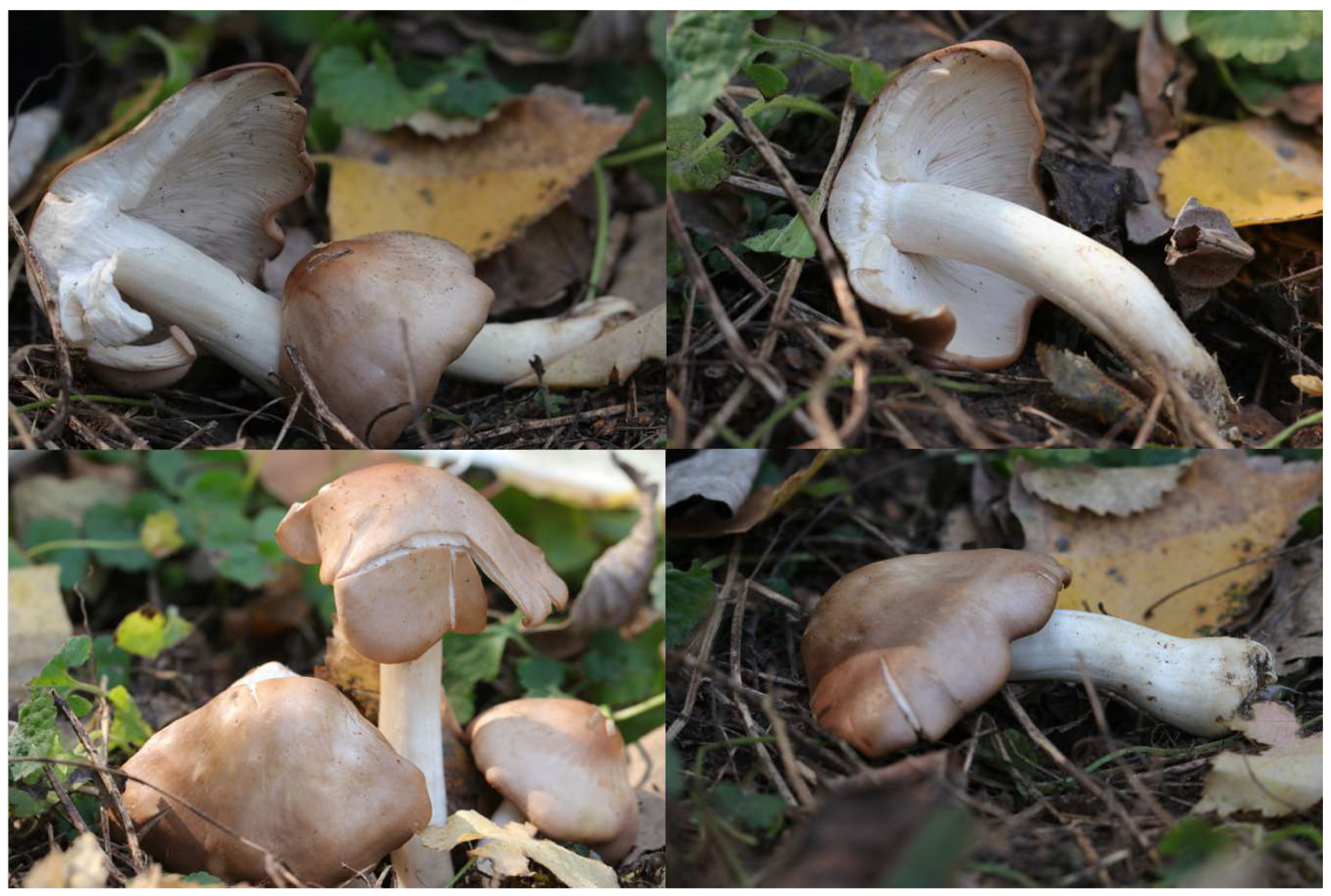
The pileus of strain HMJU10471 measured 25–68 mm in diameter, hemispherical, with an undulate margin. The surface was smooth, glabrous, dull, dry, and non-hygrophanous, ranging in color from yellowish-brown to light yellowish-brown, with a darker center. The lamellae were adnate, crowded, with smooth edges, and creamy white in color. The stipe measured 5.2–6.3 mm in length and 7–9 mm in width; it was cylindrical, equal in width, occasionally slightly curved, and creamy white to pale yellowish-brown. The basal surface was covered with silky fibrils and remained dry. The context was white (Figure 1).
A BLAST analysis of strain HMJU10471 against the NCBI database showed that it shared 100.00% sequence identity with G. borealis, with the GenBank accession number MW724270.1. Combined with the morphological observations, strain HMJU10471 was conclusively identified as G. borealis.

3.2. Biological Characteristics Analysis

3.2.1. Effect of pH on Mycelial Growth

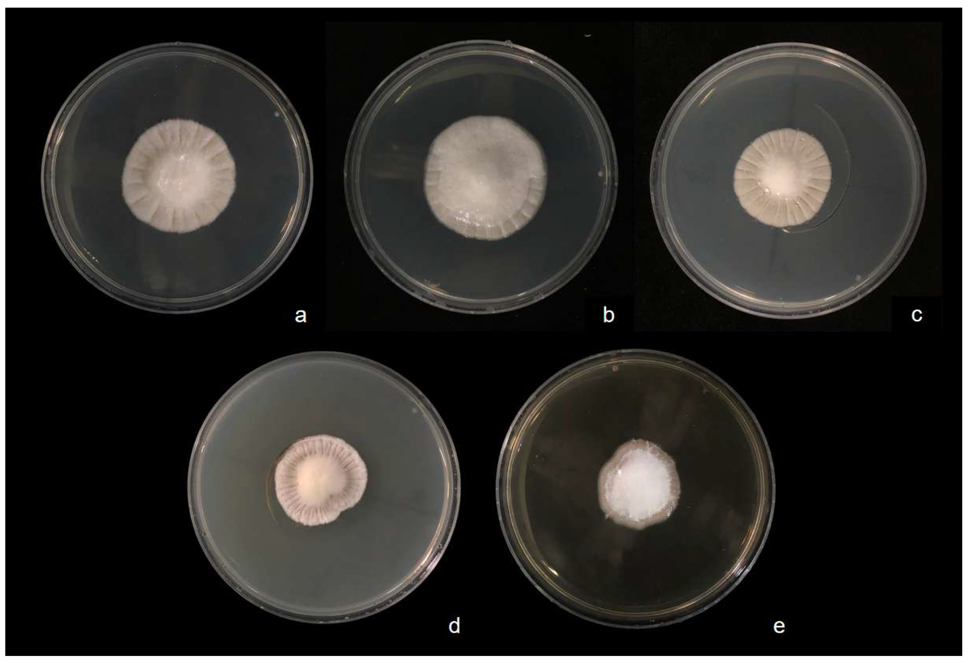
As shown in Figure 2 and Table 2, strain HMJU10471 was able to grow on the comprehensive PDA medium across a pH range of 5.0–9.0. However, significant differences in mycelial growth rate were observed among treatments. The growth rates ranked as follows: pH 6.0 > pH 5.0 > pH 7.0 > pH 8.0 > pH 9.0. The highest growth rate was recorded at pH 6.0 (0.939 ± 0.021 mm/d), which was significantly higher than those at pH 7.0, 8.0, and 9.0. Although the growth rate at pH 5.0 exceeded that at pH 7.0, the difference was not statistically significant. In terms of growth vigor, G. borealis exhibited robust and dense mycelial growth within the pH range of 5.0–6.0, with optimal vigor observed at pH 6.0. In conclusion, the optimal pH for mycelial growth of strain HMJU10471 was determined to be 6.0.

3.2.2. Effect of Carbon Sources on Mycelial Growth

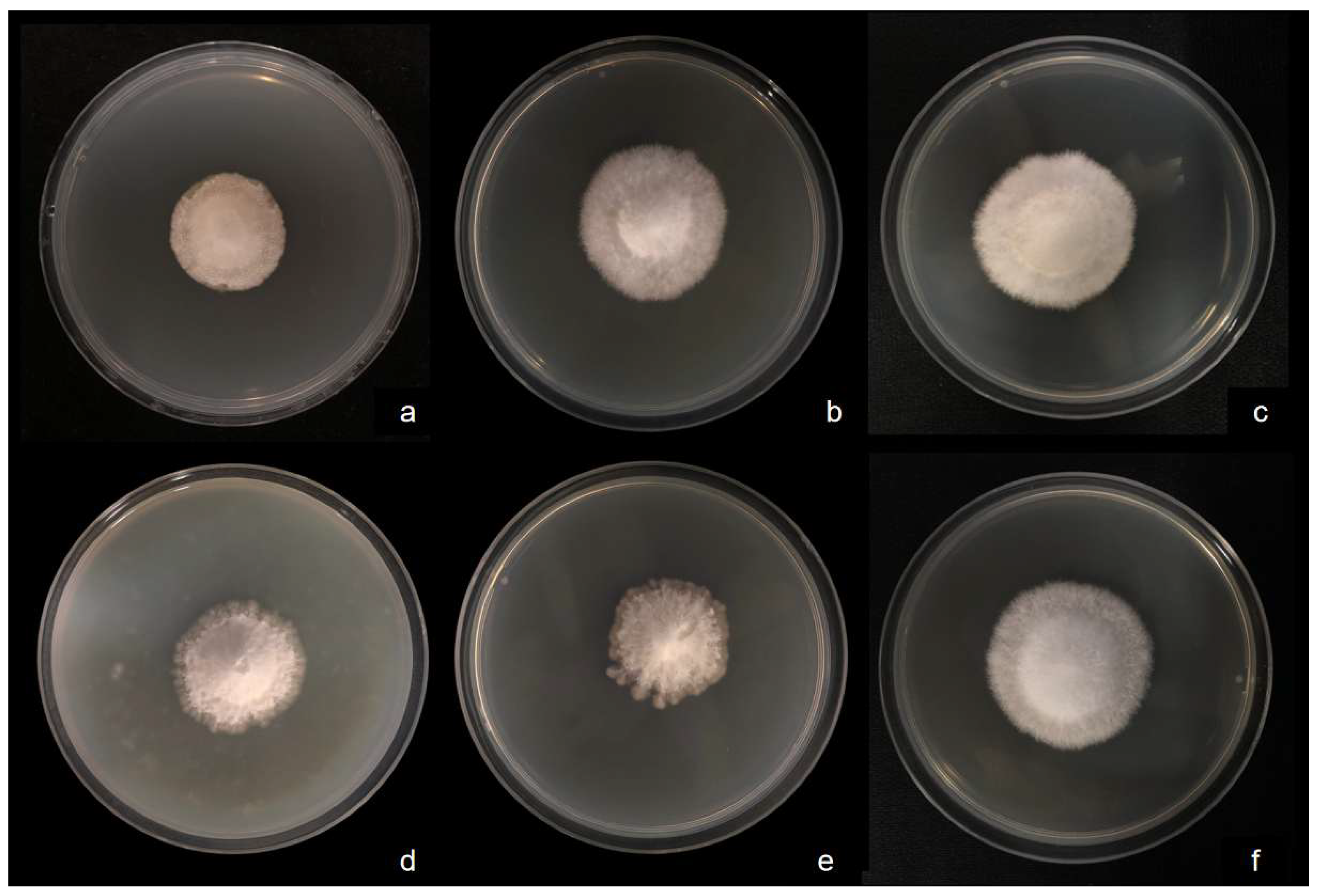
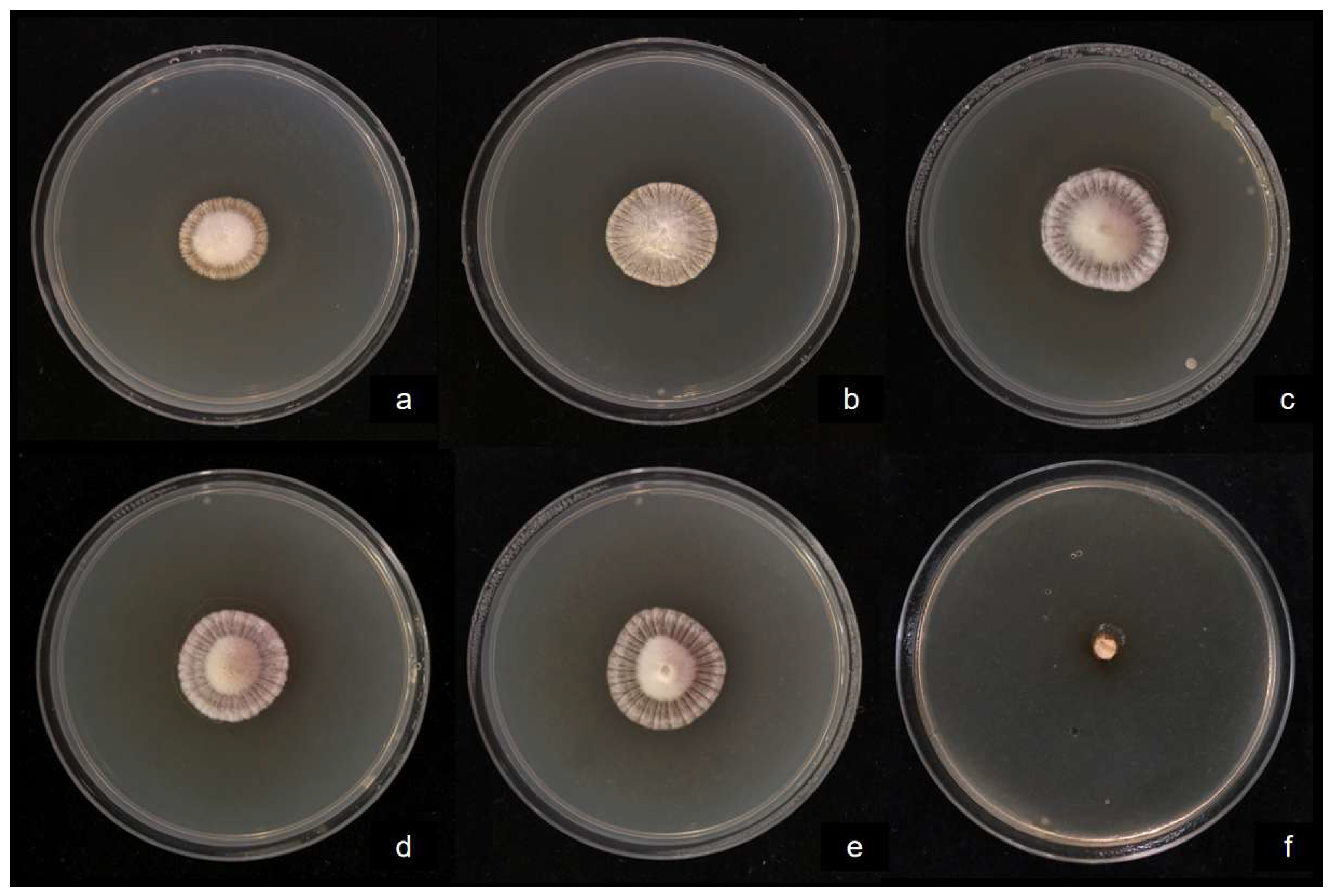
As shown in Figure 3, strain HMJU10471 was able to grow on media containing all tested carbon sources. The mycelial growth rates ranked as follows: lactose > sucrose > glucose > CK > fructose > starch. The highest growth rates were observed on media supplemented with lactose and sucrose, reaching 1.171 mm/d and 1.116 mm/d, respectively (Table 3), with dense and vigorous mycelial growth. Growth on glucose was slightly lower (1.084 mm/d), ranking third. In contrast, the slowest growth occurred on starch-containing medium (0.928 mm/d), where the mycelia were sparse and exhibited weak growth vigor. No significant differences were observed among glucose, sucrose, and lactose treatments. Based on a comprehensive evaluation of growth rate and mycelial vigor, lactose was identified as the optimal carbon source for mycelial growth of G. borealis.

3.2.3. Effect of Nitrogen Sources on Mycelial Growth

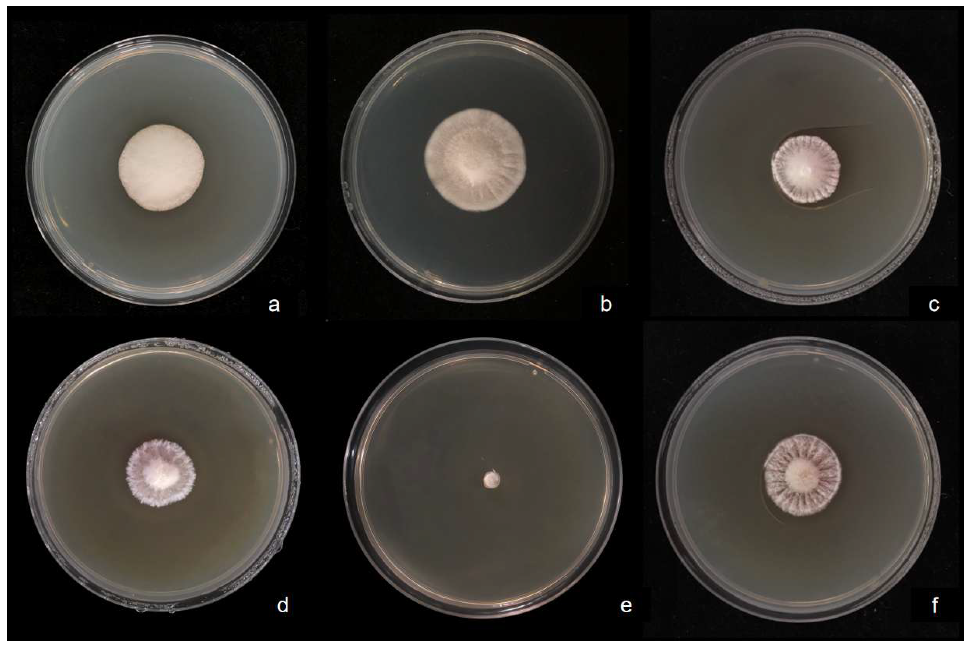
As shown in Figure 4 and Table 4, G. borealis was able to grow on media containing all tested nitrogen sources except urea. The mycelial growth rates follow: NH4NO3 > CK > KNO3 > peptone > beef extract > urea. The highest growth rate was observed on medium supplemented with NH4NO3 (1.070 mm/d), where the mycelia were dense and exhibited strong growth vigor. No mycelial growth was detected when urea was used as the sole nitrogen source. Based on a comprehensive assessment of growth rate and mycelial vigor, NH4NO3 was identified as the optimal nitrogen source for mycelial growth of G. borealis.

3.2.4. Effect of Inorganic Salts on Mycelial Growth

As shown in Figure 5 and Table 5, G. borealis was able to grow on media containing all tested inorganic salts except Fe2(SO4)3. The mycelial growth rates ranked as follows: NaCl > MgCl2 > KCl > (NH4)2SO4 > CK > Fe2(SO4)3. The highest growth rate was observed on medium supplemented with NaCl (0.926 mm/d), where the mycelia were dense and exhibited strong growth vigor. In contrast, no mycelial growth was detected when Fe2(SO4)3 was used. Based on a comprehensive evaluation of growth rate and mycelial vigor, NaCl was identified as the optimal inorganic salt for mycelial growth of G. borealis.

3.2.5. Effect of Temperature on Mycelial Growth

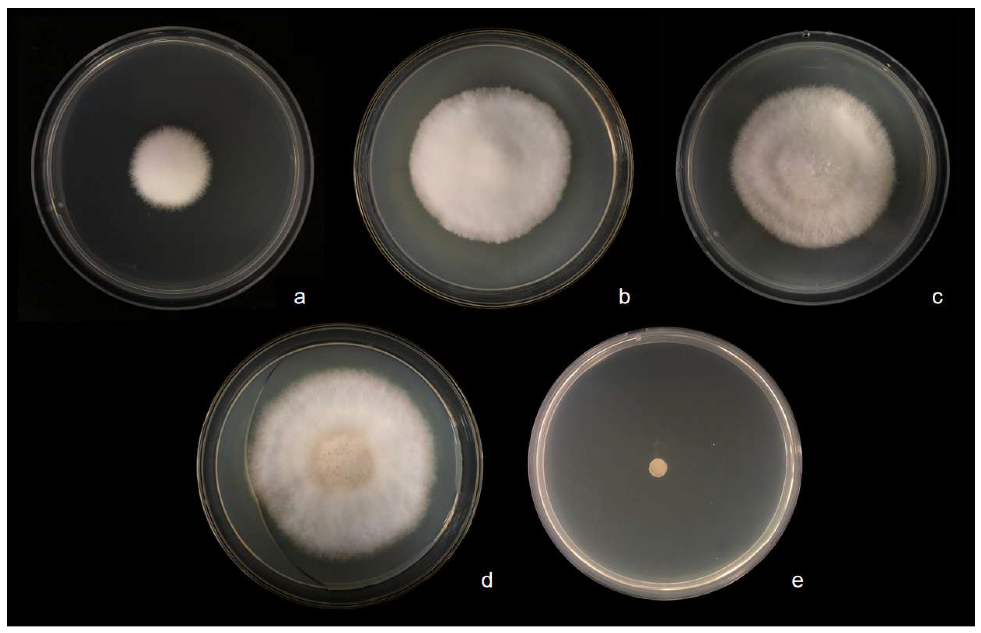
As shown in Figure 6 and Table 6, G. borealis exhibited mycelial growth within the temperature range of 10–25 °C, whereas no growth was observed at 30 °C. The growth rates ranked as follows: 25 °C > 20 °C > 15 °C > 10 °C > 30 °C. The highest growth rate was recorded at 25 °C (1.027 mm/d), with dense and vigorous mycelia, showing extremely significant differences compared with other temperature treatments. At 20 °C, the growth rate (0.836 mm/d) was slower than that at 25 °C; however, the mycelia remained dense and vigorous, with no significant difference compared to those at 15 °C. No mycelial growth was detected at 30 °C. Based on a comprehensive evaluation of growth rate and mycelial vigor, the optimal temperature for mycelial growth of G. borealis was determined to be 25 °C.

3.2.6. Results of the Orthogonal Experiment

Based on the results of single-factor tests, three factors that greatly influenced mycelial growth were selected: nitrogen source (A), carbon source (B), and inorganic salt (C). An L9(33) orthogonal design was adopted to carry out a three-factor, three-level experiment (Table 1), with mycelial growth rate as the response indicator. The results are shown in Table 7.
Range analysis indicated that the order of influence of the three factors on mycelial growth rate was carbon source (B) > nitrogen source (A) > inorganic salt (C), with range (R) values of 0.426, 0.392, and 0.148, respectively. The carbon source had the largest range value, suggesting that the type of carbon source is the most critical nutritional factor regulating mycelial growth in G. borealis. Analysis of the k values for the nitrogen source showed k1 (0.994) > k3 (0.945) > k2 (0.602); for the carbon source, k2 (1.093) > k3 (0.779) > k1 (0.668); and for the inorganic salt, k3 (0.897) > k1 (0.894) > k2 (0.749). Accordingly, the optimal theoretical combination was determined to be A1B2C3, namely ammonium nitrate as the nitrogen source, sucrose as the carbon source, and magnesium sulfate as the inorganic salt. Under this optimized combination (Combination 2), the mycelial growth rate reached 1.379 ± 0.010 mm/d, which was significantly higher than that of all other treatments except Combinations 1 and 6 (p < 0.01), with dense, white, and vigorously growing mycelia, confirming it as the optimal culture medium formulation.
Variance analysis of the orthogonal experiment (Table 8) further validated the conclusions of the range analysis. The carbon source exhibited the highest mean square (0.146) and F value (4.368), followed by the nitrogen source (mean square 0.137, F = 4.089), and the inorganic salt was the lowest (mean square 0.022, F = 0.646). The order of influence of the three factors on mycelial growth rate was consistent with that from the range analysis. Notably, although the p values for each factor did not reach the 0.05 significance level (p = 0.186–0.608), this may be attributable to the low degrees of freedom (df = 2) of the orthogonal design, leading to insufficient test power. Nevertheless, the F values clearly reflected the relative importance of each factor, with the effect sizes of carbon source and nitrogen source being much larger than that of inorganic salt, suggesting that precise regulation of carbon and nitrogen nutrition should be prioritized in subsequent domestication and cultivation efforts.
A comprehensive comparison of the single-factor and orthogonal experiment results revealed that, although lactose was the best carbon source in single-factor experiments, the combination with sucrose (A1B2C3) under the multi-factor interaction of the orthogonal design achieved superior growth performance. This phenomenon highlights the complex interactive effects among different nutritional components, indicating that the results of single-factor optimization cannot be simply extrapolated to a complex culture medium system. It further confirms the necessity of using orthogonal design or multi-factor response surface methodology for comprehensive optimization.
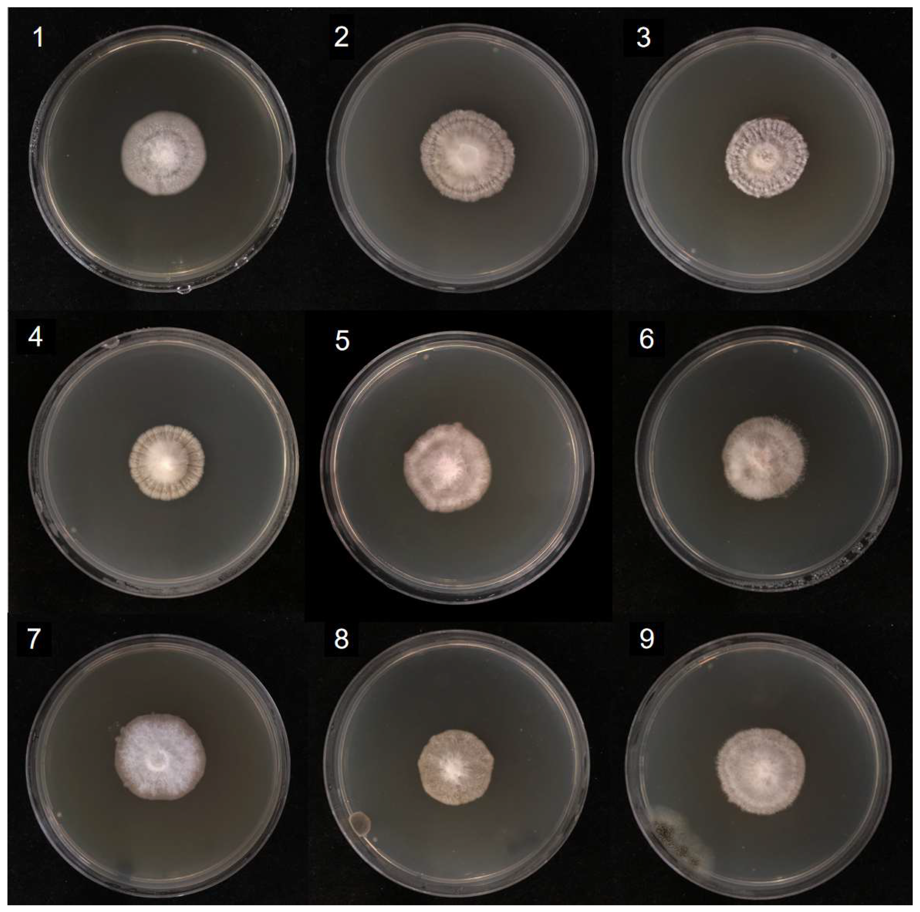
Based on the above analysis, the optimal mother culture medium formulation for mycelial growth of G. borealis was determined as follows: sucrose 20 g/L, ammonium nitrate 2 g/L, and magnesium sulfate 2 g/L. Under this formulation, the mycelial growth rate reached 1.379 mm/d, which was significantly better than that of the basal comprehensive PDA medium and other orthogonal combinations, providing key technical parameters for subsequent liquid spawn preparation and artificial domestication cultivation.The mycelial colony morphologies of the nine orthogonal test combinations are presented in Figure 7.

3.3. Analysis of Basic Nutritional Components of Mycelia

The contents of crude protein, crude fat, and crude polysaccharide in G. borealis mycelia are shown in Table 9. The results showed that the crude protein content reached 26.01%, which is higher than that reported for most edible mushrooms. The crude fat content was 6.03%, representing a moderate-to-relatively high level among edible fungi, whereas the crude polysaccharide content was comparatively low at 1.24%.
Essential amino acids accounted for 7.93 g/kg (30.39%) of the total, whereas non-essential amino acids comprised 18.16 g/kg (69.61%). Among the individual amino acids, arginine was the most abundant (5.50 g/kg, 21.08% of total amino acids), followed by alanine (4.33 g/kg, 16.60%). In contrast, aspartic acid and cystine were present at relatively low levels, at 0.18 g/kg (0.69%) and 0.08 g/kg (0.31%), respectively(Table 10).

3.4. Analysis of Trace Elements and Heavy Metals

The contents of trace elements and heavy metals in G. borealis mycelia are shown in Table 11. On a per-kilogram basis, the concentrations of essential elements were 28.09 mg/kg for iron and 22.17 mg/kg for zinc. The levels of potentially toxic elements were 0.06 mg/kg for arsenic, 0.13 mg/kg for cadmium, 0.01 mg/kg for mercury, and 0.19 mg/kg for lead. All measured values complied with the limits specified in GB 2762-2022 “Maximum Levels of Contaminants in Foods” [17] and the agricultural industry standard “Green Food—Edible Fungi” [18].

4. Discussion

Based on the biological characteristics study, the optimal temperature for G. borealis mycelial growth was 25 °C, with an optimal pH of 6.0. This result aligns with the characteristics of most known species within the Lyophyllaceae family. For instance, the optimal mycelial growth temperature for Lyophyllum decastes ranges from 20 to 25 °C, with an optimal pH of approximately 7.0 [19,20]. However, G. borealis exhibited a more pronounced preference for a weakly acidic environment (pH 6.0), mirroring the optimal pH of 5.0–6.0 observed in the confamilial species Flammulina yunnanensis [21], a preference potentially linked to its saprophytic habit and the slightly acidic soil of its native habitat. Regarding carbon source utilization, the optimal carbon source for this fungus was lactose, a finding that diverges from reports identifying maltose or glucose as the best carbon source for Flammulina rossica [22], suggesting potential specificity in the carbon metabolic pathways among different species. A particularly notable finding was the robust growth of this strain on the control medium devoid of any added nitrogen source. Without any nitrogen supplementation, the mycelial growth rate (1.036 mm/d) was not statistically different from that observed on the medium supplemented with ammonium nitrate, the optimal nitrogen source. This starkly contrasts with species such as Flammulina yunnanensis, which exhibit a strong preference for organic nitrogen sources like peptone [21]. This phenomenon may stem from the strain’s efficient utilization of endogenous nitrogen reserves within the potato-based medium or, more intriguingly, from a potential “nitrogen-saving” or enhanced nitrogen-scavenging capacity [23]. This trait holds significant biotechnological potential, warranting further investigation into its underlying enzymatic mechanisms (e.g., urease or protease activity) and the genomic basis of this response. Indeed, this finding complicates standard culture optimization approaches, indicating that the nitrogen requirements of this species are more nuanced than a simple source preference.
The results of the orthogonal experiment further challenge simplistic interpretations of nutritional optimization. We empirically determined the most effective medium combination to be ammonium nitrate, sucrose, and magnesium sulfate, which achieved a mycelial growth rate of 1.379 mm/d. Range analysis indicated that carbon and nitrogen sources were the most influential factors. Crucially, however, the analysis of variance revealed that the effects of the three tested factors—carbon source, nitrogen source, and inorganic salts—on the mycelial growth rate did not reach statistical significance at the p < 0.05 level. We interpret this as evidence of intricate interdependencies and interactive effects among the medium components within the tested concentration ranges. The shift in the most effective carbon source from lactose in the single-factor experiment to sucrose in the orthogonal combination is a clear empirical manifestation of these interactions, a phenomenon similarly documented in nutritional optimization studies of other edible fungi [24]. Given this, the proposed medium formulation should not be regarded as a definitive optimum but rather as an improved starting point for more sophisticated statistical optimization. For instance, response surface methodology (RSM) [25,26], which effectively resolves interactive effects among multiple factors by constructing continuous variable surface models, has been widely applied to culture medium optimization for edible fungi. Among phylogenetically close species within the Lyophyllaceae, Shan et al. [25] utilized Box–Behnken RSM to optimize the liquid spawn medium for Lyophyllum decastes, achieving a 7.34-fold increase in mycelial dry weight compared to the initial formulation, fully demonstrating the method’s strength in addressing complex nutrient factor interactions. Similarly, RSM has yielded favorable outcomes in optimizing multi-factor culture conditions for edible fungi within the genus Lepista [26]. Consequently, future research, building upon the empirically derived optimal formula (A1B2C3) from this experiment, should systematically optimize the concentrations of the carbon source, nitrogen source, and inorganic salts using RSM-centric continuous surface approaches to precisely pinpoint the global optimal medium formulation.
From a nutritional perspective, G. borealis mycelium presents a unique nutritional profile. Its high crude protein content (26.01%) aligns with the nutritional pattern reported for other Lyophyllaceae species, such as L. cinerescens and L. connata [4,5]. However, its crude fat content (6.03%) is notably higher than the characteristically low levels (typically below 2%) observed in many edible fungi, including the closely related L. connata, which possesses a crude fat content of only 1.22% [5]. This serves as a key distinguishing characteristic. The value of G. borealis mycelium may not reside in its simple classification as a low-fat food but rather in its moderate fat content, provided the fatty acid profile is favorable, rendering it a potential candidate ingredient for formulations requiring different energy densities or specific lipid compositions, rather than being merely a generic high-protein, low-fat supplement. A total of 17 amino acids were detected in the mycelium, with a total content reaching 26.09 g/kg, of which essential amino acids accounted for 30.39%. Notably, arginine exhibited the highest content at 5.50 g/kg. As a functional amino acid, arginine plays a significant role in enhancing immunity and promoting wound healing [27]. Moreover, the mycelium is rich in beneficial trace elements such as iron (28.09 mg/kg) and zinc (22.17 mg/kg), echoing reports of zinc and copper abundance in the fruiting bodies of Lyophyllum decastes. In terms of safety evaluation, the levels of hazardous heavy metals, including arsenic, cadmium, mercury, and lead, were all substantially below the limit requirements stipulated by both the National Food Safety Standard “Maximum Levels of Contaminants in Foods” [17] and the agricultural industry standard “Green Food—Edible Fungi” [18], indicating its excellent safety profile as a potential food ingredient.
In conclusion, the efficient mycelial growth achieved through optimized culture conditions not only advances the artificial domestication of this species but also provides a transferable technical pathway for the large-scale cultivation of other edible fungi. Overall, this approach unlocks the ecological value and application potential of the Lyophyllaceae family, thereby promoting its diversified applications in fields such as food safety, medicine and health, and environmental management.

5. Conclusions

Single-factor experiments on biological characteristics showed that the optimal carbon source for mycelial growth of Gerhardtia borealis was lactose, the optimal nitrogen source was ammonium nitrate, the optimal inorganic salt was sodium chloride, the optimal temperature was 25 °C, and the optimal pH was 6.0. Orthogonal experimental analysis indicated that the optimal culture medium formulation for G. borealis mycelia consisted of sucrose (20 g/L), ammonium nitrate (2 g/L), and magnesium sulfate (2 g/L) in 1 L of water. Under this formulation, the mycelial growth rate reached 1.379 mm/d, with dense and vigorous mycelial morphology. The crude protein, crude fat, and crude polysaccharide contents of G. borealis mycelia were 26.01%, 6.03%, and 1.24%, respectively. The total amino acid content was 26.09 g/kg, of which essential amino acids accounted for 7.93 g/kg (30.39%) and non-essential amino acids for 18.16 g/kg (69.61%). Among the detected amino acids, arginine was the most abundant (5.50 g/kg), accounting for 21.08% of the total.

Author Contributions

Literature review, Y.J.; Writing—original draft preparation, Y.J.; Writing—review and editing, Y.J., D.Z., Y.L. (Yuysen Liu), J.X. and Y.L. (Yunjiang Liang). All authors have read and agreed to the published version of the manuscript.

Funding

This research received no external funding.

Institutional Review Board Statement

Not applicable. The study involved only the cultivation and laboratory analysis of Gerhardtia borealis mycelia, a wild edible fungus, and did not involve any human subjects, human tissue, human data, or live vertebrate animals. Therefore, ethical approval from an Institutional Review Board was not required.

Informed Consent Statement

Not applicable. This study did not involve human participants, and therefore no informed consent was required.

Data Availability Statement

The original contributions presented in this study are included in the article. Further inquiries can be directed to the corresponding authors.

Acknowledgments

The authors would like to express their gratitude to all the students who participated in this study for their involvement and dedication. We also acknowledge Jilin Agricultural Science and Technology University for providing the experimental facilities and support.

Conflicts of Interest

The authors declare no conflicts of interest.

References

  1. Bon, M. Deux Lyophylloideae interessantes et le genre Gerhardtia st. et nom.nov. Doc. Mycol. 1994, 24, 65–68. [Google Scholar]
  2. Reis, F.S.; Barros, L.; Martins, A.; Ferreira, I.C.F.R. Chemical composition and nutritional value of the most widely appreciated cultivated mushrooms: An inter-species comparative study. Food Chem. Toxicol. 2012, 50, 191–197. [Google Scholar] [CrossRef]
  3. Wang, X.M.; Zhang, J.; Wu, L.H.; Zhao, Y.L.; Li, T.; Li, J.Q.; Wang, Y.Z.; Liu, H.G. A mini-review of chemical composition and nutritional value of edible wild-grown mushroom from China. Food Chem. 2014, 151, 279–285. [Google Scholar] [CrossRef] [PubMed]
  4. Li, X.; Zhang, S.Y.; Li, Y. Determination and evaluation of nutritional components in fruiting bodies of Lyophyllum cinerascens. North. Hortic. 2010, 6, 198–201. [Google Scholar]
  5. Zhang, S.Y.; Duan, X.L.; Li, X. Determination and evaluation of nutritional components in fruiting bodies of Lyophyllum connatum. Jilin Agric. 2010, 6, 73–75. [Google Scholar]
  6. Zhang, H.Y.; Zhang, F.Q.; Wang, X.M. Analysis and evaluation of nutritional components in fruiting bodies of Lyophyllum decastes. Mycosystema 2008, 27, 696–700. [Google Scholar]
  7. Li, Y.; Li, T.; Yang, Z.; Tuliguer; Dai, Y. China Large Scale Fungal Resources Atlas; Zhongyuan Farmers Publishing House: Zhengzhou, China, 2015. [Google Scholar]
  8. White, T.; Bruns, T.; Lee, S.; Taylor, J. Amplification and Direct Sequencing of Fungal Ribosomal RNA Genes for Phylogenetics. In PCR Protocols; Elsevier: Amsterdam, The Netherlands, 1990; pp. 315–322. [Google Scholar]
  9. Xu, J.; Yu, X.; Zhang, C.; Li, Y. Morphological characteristics and phylogenetic analyses revealed a new Calocybe (Lyophyllaceae, Basidiomycota) species from northeast China. Phytotaxa 2021, 490, 203–210. [Google Scholar] [CrossRef]
  10. Zheng, S.; Wang, M.; Gao, Y.; Guo, J.; Wang, C.; Zheng, J. Identification and Biological Characteristics of a Wild Tricholoma Strain. Food Res. Dev. 2023, 44, 171–177. [Google Scholar]
  11. Huang, H.; Li, X.; Hu, X.; Sun, H.; Zhang, J.; Wu, X.; Fu, J. Biological Characteristics, Domestication and Cultivation of Wild Tyromyces kmetii. Horticulturae 2024, 10, 1177. [Google Scholar] [CrossRef]
  12. NY/T 1676-2023; Determination of Crude Polysaccharides in Edible Mushroom—Spectrophotometric Method. Ministry of Agriculture and Rural Affairs of the People’s Republic of China. China Agriculture Press: Beijing, China, 2023; pp. 1–4.
  13. GB 5009.5-2016; National Food Safety Standard—Determination of Protein in Foods, State Food and Drug Administration, National Health and Family Planning Commission. China Standards Press: Beijing, China, 2016; pp. 1–7.
  14. GB 5009.6-2016; National Food Safety Standard—Determination of Fat in Foods, State Food and Drug Administration, National Health and Family Planning Commission. China Standards Press: Beijing, China, 2016; pp. 1–11.
  15. GB 5009.124-2016; National Food Safety Standard—Determination of Amino Acids in Foods, State Food and Drug Administration; National Health and Family Planning Commission. China Standards Press: Beijing, China, 2016; pp. 1–6.
  16. GB 5009.268-2016; National Food Safety Standard—Determination of Multi-Elements in Foods, State Food and Drug Administration, National Health and Family Planning Commission. China Standards Press: Beijing, China, 2016; pp. 1–14.
  17. GB 2762-2022; National Food Safety Standard—Maximum Levels of Contaminants in Foods; National Health Commission of the People’s Republic of China; State Administration for Market Regulation. Standards Press of China: Beijing, China, 2022. (In Chinese)
  18. NY/T 749-2023; Green Food—Edible Fungi; Ministry of Agriculture and Rural Affairs of the People’s Republic of China. China Agriculture Press: Beijing, China, 2023. (In Chinese)
  19. Li, X.; Li, S.J.; Dai, J.J.; Zhao, J.C.; Li, Z.W. Preliminary study on biological characteristics of wild Lyophyllum decastes strains from Huangsongdian. Edible Fungi 2016, 38, 14–16. [Google Scholar]
  20. Liu, N.; Zhang, M.; Zhang, P.; Zhang, Y.C.; Zhan, J.Y.; Song, Y. Identification and biological characteristics of wild Lyophyllum decastes strains. North. Hortic. 2023, 9, 119–125. [Google Scholar]
  21. Wang, X.; Zhu, J.T.; Chen, Y.; Yu, C.H.; Nie, Z.F.; He, J.Q. Biological characteristics and cultivation of wild Flammulina yunnanensis from Tibet. Mycosystema 2026. Epub ahead of printing. [Google Scholar]
  22. He, J.; Luo, Z.L.; Deng, R.M.; Su, K.M.; Li, S.H.; Su, H.Y. Isolation, biological characteristics and cultivation of wild Flammulina rossica and Flammulina filiformis. Mycosystema 2022, 41, 630–646. [Google Scholar]
  23. Ray, P.; Abraham, P.E.; Guo, Y.; Giannone, R.J.; Engle, N.L.; Yang, Z.K.; Jacobson, D.; Hettich, R.L.; Tschaplinski, T.J.; Craven, K.D. Scavenging organic nitrogen and remodelling lipid metabolism are key survival strategies adopted by the endophytic fungi, Serendipita vermifera and Serendipita bescii to alleviate nitrogen and phosphorous starvation in vitro. Environ. Microbiol. Rep. 2019, 11, 548–560. [Google Scholar] [CrossRef] [PubMed]
  24. Xi, Y.L.; Mao, A.L.; Wang, X.Q.; Wei, X.L.; Zhang, H.Y. Protein nutritional evaluation of fruit body, mycelia and fermentation broth of Lyophyllum decastes. Mycosystema 2010, 29, 603–607. [Google Scholar]
  25. Shan, C.C.; Ye, D.Z.; Guo, Y.; Chen, L.D.; Sun, S.J.; Jin, W.S.; Zhang, H.Y.; Jin, L.Y.; Hu, K.H. Response surface optimization of medium formulation for culture of Lyophyllum decastes. Fujian J. Agric. Sci. 2023, 38, 723–731. [Google Scholar]
  26. Zhao, B.H.; Liu, H.; Guo, L.J.; Fan, L. Optimization of culture conditions for three edible Lepista mushrooms. Microbiol. China 2019, 46, 2230–2241. [Google Scholar]
  27. Wu, G.; Bazer, W.F.; Davis, A.T.; Kim, S.W.; Li, P.; Rhoads, J.M.; Satterfield, M.C.; Smith, S.B.; Spencer, T.E.; Yin, Y. Arginine metabolism and nutrition in growth, health and disease. Amino Acids 2009, 37, 153–168. [Google Scholar] [CrossRef] [PubMed]
Figure 1. Gerhardtia borealis: Basidiomata.
Life 16 00967 g001
Figure 2. Effects of different pH on mycelial growth of G. borealis. Representative images of mycelial growth at various pH levels: (a) pH5.0; (b) pH6.0; (c) pH7.0; (d) pH8.0; (e) pH9.0.
Life 16 00967 g002
Figure 3. Effects of different carbon sources on mycelial growth of G. borealis. Representative images of mycelial growth in various media: (a) CK; (b) glucose; (c) sucrose; (d) fructose; (e) starch; (f) lactose.
Life 16 00967 g003
Figure 4. Effects of different nitrogen sources on mycelial growth of G. borealis. Representative images of mycelial growth in various media: (a) CK; (b) NH4NO3; (c) beef powder peptone; (d) yeast powder; (e) urea; (f) KNO3.
Life 16 00967 g004
Figure 5. Effects of different inorganic salts on mycelial growth of G. borealis. Representative images of mycelial growth in various media: (a) CK; (b) MgSO4; (c) (NH4)2SO4; (d) KCl; (e) NaCl; (f) Fe2(SO4)3.
Life 16 00967 g005
Figure 6. Effects of different temperatures on mycelial growth of G. borealis. Representative images of mycelial growth at different temperatures: (a) 10 °C; (b) 15 °C; (c) 20 °C; (d) 25 °C; (e) 30 °C.
Life 16 00967 g006
Figure 7. Mycelial growth of Gerhardtia borealis in the nine orthogonal test combinations (1–9) listed in Table 7. Each number corresponds to a specific combination of nitrogen source, carbon source, and inorganic salt: (1) no nitrogen, glucose, NaCl; (2) NH4NO3, sucrose, MgSO4; (3) no nitrogen, lactose, MgSO4; (4) NH4NO3, glucose, KCl; (5) KNO3, glucose, MgSO4; (6) no nitrogen, sucrose, KCl; (7) KNO3, sucrose, NaCl; (8) KNO3, lactose, KCl; (9) NH4NO3, lactose, NaCl. The mycelial growth rate and vigor for each combination are presented in Table 7.
Life 16 00967 g007
Table 1. Factors and levels of orthogonal tests.
LevelFactor
Nitrogen Source (A)Carbon Source (B)Inorganic Salt (C)
1NH4NO3LactoseNaCl
2KNO3SucroseKCl
3No nitrogen sourceGlucoseMgSO4
Table 2. The effect of different pH on mycelial growth.
pHMycelial Growth Rate (mm/d)Mycelium ColorMycelium VigorSignificance of
Difference
0.050.01
5.00.914 ± 0.093white+++abA
6.00.939 ± 0.021white+++aA
7.00.836 ± 0.059white++bA
8.00.514 ± 0.084white+cB
9.00.357 ± 0.078white+dC
Note: Different uppercase letters indicate significant differences (p < 0.01); different lowercase letters indicate significant differences (p < 0.05); “+++” indicates dense, robust mycelium; “++” indicates slightly denser mycelium and slightly better growth; “+” indicates sparse, poorly growing mycelium.
Table 3. The effect of different carbon sources on mycelial growth.
Carbon SourcesMycelial Growth Rate (mm/d)Mycelium ColorMycelium VigorSignificance of
Difference
0.050.01
CK1.063 ± 0.033white++bcABC
Glucose1.084 ± 0.077white+++abcAB
Sucrose1.116 ± 0.072white+++abAB
Fructose1.000 ± 0.079white++cdBC
Starch0.928 ± 0.018white++dC
Lactose1.171 ± 0.681white+++aA
Note: Different uppercase letters indicate significant differences (p < 0.01); different lowercase letters indicate significant differences (p < 0.05); “+++” indicates dense, robust mycelium; “++” indicates slightly denser mycelium and slightly better growth.
Table 4. The effect of different nitrogen sources on mycelial growth.
Nitrogen SourcesMycelial Growth Rate (mm/d)Mycelium ColorMycelium VigorSignificance of
Difference
0.050.01
CK1.036 ± 0.090white+++aA
NH4NO31.070 ± 0.036white+++aA
beef extract0.496 ± 0.011white+cC
peptone0.653 ± 0.030white+bB
ureadD
KNO30.980 ± 0.060white++aA
Note: Different uppercase letters indicate significant differences (p < 0.01); different lowercase letters indicate significant differences (p < 0.05); “+++” indicates dense, robust mycelium; “++” indicates slightly denser mycelium and slightly better growth; “+” indicates sparse, poorly growing mycelium; the symbol "–" indicates no growth.
Table 5. The effect of different inorganic salts on mycelial growth.
Inorganic SaltsMycelial Growth Rate (mm/d)Mycelium ColorMycelium VigorSignificance of
Difference
0.050.01
CK0.686 ± 0.050white++cC
MgSO40.873 ± 0.011white+++abA
(NH4)2SO40.753 ± 0.060white++cBC
KCl0.836 ± 0.030white+++bAB
NaCl0.926 ± 0.045white+++aA
Fe2(SO4)3dD
Note: Different uppercase letters indicate significant differences (p < 0.01); different lowercase letters indicate significant differences (p < 0.05); “+++” indicates dense, robust mycelium; “++” indicates slightly denser mycelium and slightly better growth; the symbol "–" indicates no growth.
Table 6. The effect of different temperatures on mycelial growth.
TemperatureMycelial Growth Rate (mm/d)Mycelium ColorMycelium VigorSignificance of
Difference
0.050.01
10 °C0.188 ± 0.034white+cC
15 °C0.769 ± 0.071white++bB
20 °C0.836 ± 0.031white+++bB
25 °C1.027 ± 0.201white+++aA
30 °CdD
Note: Different uppercase letters indicate significant differences (p < 0.01); different lowercase letters indicate significant differences (p < 0.05); “+++” indicates dense, robust mycelium; “++” indicates slightly denser mycelium and slightly better growth; “+” indicates sparse, poorly growing mycelium; the symbol "–" indicates no growth.
Table 7. Orthogonal test result of G. borealis mycelial growth.
CombinationFactorMycelial Growth Rate (mm/d)Mycelium ColorMycelium Vigor
Nitrogen Source (A)Carbon Source (B)Inorganic Salt (C)
13 (no nitrogen)3 (glucose)1 (NaCl)1.103 ± 0.061 bBwhite+++
21 (NH4NO3)2 (sucrose)3 (MgSO4)1.379 ± 0.010 aAwhite+++
33 (no nitrogen)1 (lactose)3 (MgSO4)0.699 ± 0.018 dCDwhite+
41 (NH4NO3)3 (glucose)2 (KCl)0.711 ± 0.032 dCDwhite+
52 (KNO3)3 (glucose)3 (MgSO4)0.613 ± 0.065 dDEwhite++
63 (no nitrogen)2 (sucrose)2 (KCl)1.122 ± 0.181 bBwhite+++
72 (KNO3)2 (sucrose)1 (NaCl)0.779 ± 0.132 cdCDwhite+++
82 (KNO3)1 (lactose)2 (KCl)0.413 ± 0.101 eEwhite+
91 (NH4NO3)1 (lactose)1 (NaCl)0.891 ± 0.121 cBCwhite+++
K12.9812.0032.683
K21.8053.2802.246
K32.8342.3372.691
k10.9940.6680.894
k20.6021.0930.749
k30.9450.7790.897
R0.3920.4260.148
Note: Different uppercase letters indicate significant differences (p < 0.01); different lowercase letters indicate significant differences (p < 0.05); “+++” indicates dense, robust mycelium; “++” indicates slightly denser mycelium and slightly better growth; “+” indicates sparse, poorly growing mycelium.
Table 8. Analysis of variance of orthogonal test results.
SourceType III Sum of SquaresdfMean SquaresFp
Corrected Model0.609 a60.1023.0340.269
Intercept6.45216.452192.7560.005
A0.27420.1374.0890.197
B0.29220.1464.3680.186
C0.04320.0220.6460.608
Error0.06720.033
Total7.1289
Corrected Total0.6768
Note: a R2 = 0.901 (Corrected Model SS/Corrected Total SS).
Table 9. Basic nutrition ingredients of G. borealis.
Basic Nutritional CompositionContent
Crude protein26.01 ± 0.04
Crude fat6.03 ± 0.25
Crude polysaccharides1.24 ± 0.18
Table 10. Composition and content of amino acids in mycelium of G. borealis (g/kg).
Amino Acid NameContent
Aspartic acid0.18 ± 0.23
Glutamic acid3.64 ± 0.03
Serine1.48 ± 0.04
Glycine0.85 ± 0.06
Histidine0.43 ± 0.11
Arginine5.50 ± 0.08
Threonine *0.93 ± 0.04
Alanine4.33 ± 0.02
Proline0.79 ± 0.05
Tyrosine0.88 ± 0.09
Valine *1.17 ± 0.15
Methionine *0.33 ±0.22
Cysteine0.08 ± 0.06
Isoleucine *0.90 ± 0.02
Leucine *1.97 ± 0.06
Phenylalanine *1.36 ± 0.03
Lysine *1.27 ± 0.05
Essential amino acid content7.93
Non–essential amino acid content18.16
Total26.09
Note: * Represents essential amino acids.
Table 11. Contents of heavy metals and trace elements in mycelia of G. borealis (mg/kg).
Element NamesContentGB 2762–2022
Limit for Contaminants in Foods (mg/kg)
NY/T 749–2023
Green Food-Edible Fungi (mg/kg)
Fe28.09 ± 0.04
Zn22.17 ± 0.17
Tlnot detected (<0.005)
As0.06 ± 0.010.51.0
Cd0.13 ± 0.03 0.21.0
Hg0.01 ± 0.000.10.2
Pb0.19 ± 0.020.52.0
Disclaimer/Publisher’s Note: The statements, opinions and data contained in all publications are solely those of the individual author(s) and contributor(s) and not of MDPI and/or the editor(s). MDPI and/or the editor(s) disclaim responsibility for any injury to people or property resulting from any ideas, methods, instructions or products referred to in the content.