Systematic Review

Surgical Intervention for Filariasis-Induced Lymphedema: A Systematic Review of Ablative and Physiological Approaches


by
Rani Septrina
1,*,
Irra Rubianti Widarda
1,
Putie Hapsari
2 and
Valeska Siulinda Candrawinata
3
1
Division of Plastic and Reconstructive Surgery, Department of Surgery, Faculty of Medicine, Universitas Padjadjaran/Dr. Hasan Sadikin General Hospital, Bandung 40161, Indonesia
2
Division of Vascular and Endovascular Surgery, Department of Surgery, Faculty of Medicine, Universitas Padjadjaran/Dr. Hasan Sadikin General Hospital, Bandung 40161, Indonesia
3
Department of Surgery, Universitas Padjadjaran/Dr. Hasan Sadikin General Hospital, Bandung 40161, Indonesia
*
Author to whom correspondence should be addressed.
Life 2026, 16(6), 942; https://doi.org/10.3390/life16060942
Submission received: 25 March 2026 / Revised: 19 May 2026 / Accepted: 26 May 2026 / Published: 2 June 2026

Abstract

Advanced filariasis-induced lymphedema causes irreversible fibrosis, severe disability, recurrent infections, and major psychosocial burden, often with poor response to conservative therapy. Surgical management in this type of lymphedema is rarely addressed. We conducted a PRISMA 2020-guided systematic review evaluating surgical interventions versus conservative management or no intervention in adults with filarial lymphedema, involving various anatomical locations. Of 580 records identified, 29 studies were included for qualitative synthesis. The evidence base predominantly consisted of case reports, with surgical indications frequently involving disabling elephantiasis or diagnostic excision of filarial masses. While ablative procedures (excisional debulking) remained the primary salvage strategy, specific studies documented physiological techniques, including nodovenous shunts and vascularized lymph node transfer (VLNT). Quantitative limb-volume reduction was sparsely reported; however, qualitative assessments demonstrated significant improvements in ambulation, cosmesis, and patient-reported quality of life. Postoperative complications, primarily superficial surgical site infections, were generally low and manageable. Ablative surgical procedures appear to be the most common salvage strategy for advanced filarial lymphedema, but the evidence base remains limited to low-level, heterogeneous observational data. However, with the emergence of physiological lymphatic surgery, a combination of ablative procedures and physiological procedures should be considered. Prospective controlled studies with standardized outcomes are urgently needed.

1. Introduction

Lymphatic filariasis remains a leading cause of permanent and long-term disability worldwide [1]. According to the World Health Organization, while millions are infected globally, approximately 36 million people are visibly disfigured and incapacitated by chronic complications such as lymphedema and elephantiasis [2]. The disease progressively damages the lymphatic system, and in its advanced stages, it presents a severe clinical challenge characterized by irreversible fibrosis in the subcutaneous tissue that results in massive limb enlargement and highly debilitating recurrent secondary infections (acute attacks) [3]. For adult patients, particularly those residing in endemic areas, these physical manifestations not only severely impair mobility but also inflict profound psychological and socioeconomic burdens, underscoring the critical need for effective, durable interventions [4].
The traditional cornerstone of care for early-stage filarial lymphedema relies heavily on conservative management, including strict hygiene protocols, compression therapy (bandaging), and pharmacologic regimens such as doxycycline or antifilarials [5]. However, adults presenting with advanced filariasis lymphedema frequently become unresponsive to these non-operative measures, as conservative care is generally insufficient to reverse established fibrotic tissue or adequately halt recurrent infections [6,7]. Consequently, surgical intervention has emerged as a vital salvage strategy. Contemporary surgical approaches range from physiological procedures aimed at restoring lymphatic flow to ablative techniques, such as liposuction and excisional debulking of fibrotic tissue [8]. The choice of physiological surgery, such as lymphovenous anastomosis (LVA) or vascularized lymph node transfer (VLNT), is rarely done in advanced stage of filariasis lymphedema due to less satisfactory results [5].
These surgical approaches have demonstrated growing clinical interest and highly promising outcomes in cases of primary lymphedema, such as 20% to 50% reductions in limb volume, along with improved cosmesis and quality of life in 70% to 90% of advanced cases at 1–2 years of follow-up, the overall comparative efficacy of these techniques remains fragmented [9,10]. The current literature varies widely in surgical approach, comparator groups, and follow-up metrics. Therefore, this systematic review aims to comprehensively evaluate the efficacy of surgical interventions compared to conservative management or no intervention in adults with filarial lymphedema.

2. Materials and Methods

  • Protocol and Guidelines
This systematic review was formulated and executed in strict accordance with the Preferred Reporting Items for Systematic Reviews and Meta-Analyses (PRISMA) guideline 2020 [11]. To ensure a rigorous and reproducible approach to synthesizing the available clinical evidence and to maintain methodological transparency, this study is registered in the PROSPERO system (ID: CRD420261343996) and can be accessed at: https://www.crd.york.ac.uk/PROSPERO/view/CRD420261343996 (registered at 25 March 2026).
  • Eligibility Criteria
Study inclusion was prospectively defined utilizing the Population, Intervention, Comparison, and Outcomes (PICO) framework. The target population was restricted to adult patients, predominantly situated in endemic regions, who presented with filarial lymphedema. It should be noted that individual studies may have used alternative classification systems, such as the International Society of Lymphology (ISL) staging system, and such discrepancies are indicated in the corresponding table entries. Eligible clinical profiles required the presence of irreversible fibrosis, elephantiasis, recurrent secondary infections, and significant mobility impairment that had proven refractory to standard conservative management strategies, such as Complete Decongestive Therapy (CDT) and antifilarial pharmacotherapy. The interventions under investigation encompassed a spectrum of surgical modalities, ablative surgical techniques, including excisional debulking of fibrotic tissue and liposuction, as well as physiological approaches such as lymphovenous anastomosis (LVA) and vascularized lymph node transfer (VLNT). Studies exploring the combined application of these techniques, such as vascularized lymph node transfer paired with excision, were also deemed eligible.
For comparative purposes, acceptable control groups included patients undergoing conservative management, encompassing strict hygiene protocols, routine bandaging, and doxycycline administration, or those receiving no intervention. The primary outcome measure was the quantifiable reduction in limb volume. Secondary clinical outcomes of interest included changes in the frequency of acute attacks, postoperative cosmesis, and patient-reported quality of life. Methodologically, the review incorporated a broad range of primary study designs, explicitly accepting randomized controlled trials, prospective cohort studies, case–control studies, case series, and case reports. Conversely, the literature was excluded if the studies were preclinical, based on non-human or in vitro models, or categorized as review articles. Furthermore, logistical constraints necessitated that inclusion be strictly limited to English-language publications that were either fully open access or retrievable through our university institutional accounts. Although the target population was adults, certain studies containing mixed-age cohorts, including a minority of pediatric cases, were retained when they contributed relevant surgical data that could not be disaggregated; this deviation is acknowledged as a limitation.
  • Information Sources and Search Strategy
A comprehensive systematic search was conducted across three primary academic databases: PubMed, EMBASE, and Scopus. The search parameters encompassed all available literature from the respective inceptions of these databases through to 31 January 2026. The search strategy was meticulously developed by synthesizing controlled vocabulary, including MeSH terms, with free-text keywords centralized around the core concepts of lymphedema, filariasis, and surgical interventions. To achieve literature saturation and mitigate the risk of omitting highly relevant data, a supplementary manual screening of the reference lists from all formally included studies and pertinent review articles was also performed.
  • Risk of Bias Assessment
To ensure the methodological rigor and validity of the synthesized evidence, a comprehensive risk of bias assessment was conducted for all included studies. The Joanna Briggs Institute (JBI) critical appraisal tools were selected for the evaluation of case reports and case series due to their validated applicability for these specific study designs, while the Newcastle-Ottawa Scale (NOS) was applied to the observational cross-sectional study as recommended by the Cochrane Collaboration for non-randomized studies. Two authors independently applied these standardized tools to evaluate each study’s methodological quality, resolving any discrepancies through mutual agreement. This rigorous, independent review approach guaranteed that potential biases related to patient selection, outcome ascertainment, and data reporting were systematically identified.
A meta-analysis was not feasible due to the extreme heterogeneity in study designs, outcome measures, anatomical sites, and follow-up durations across the included studies. Consequently, a qualitative narrative synthesis was determined to be the only appropriate analytical approach for this dataset.
  • Study Selection and Data Extraction
Following the application of the search algorithms across all databases, the aggregated citations were subjected to a deduplication process. The initial phase of study selection involved a rigorous screening of titles and abstracts to evaluate their alignment with the predefined eligibility criteria. Articles that satisfied these parameters, as well as those providing insufficient data in the abstract for a definitive exclusion, were subsequently advanced to a full-text review. Upon finalization of the included literature cohort, relevant data pertaining to study design, demographic characteristics, specific surgical interventions utilized, and the predefined clinical outcomes were systematically extracted to facilitate the final qualitative synthesis.

3. Results

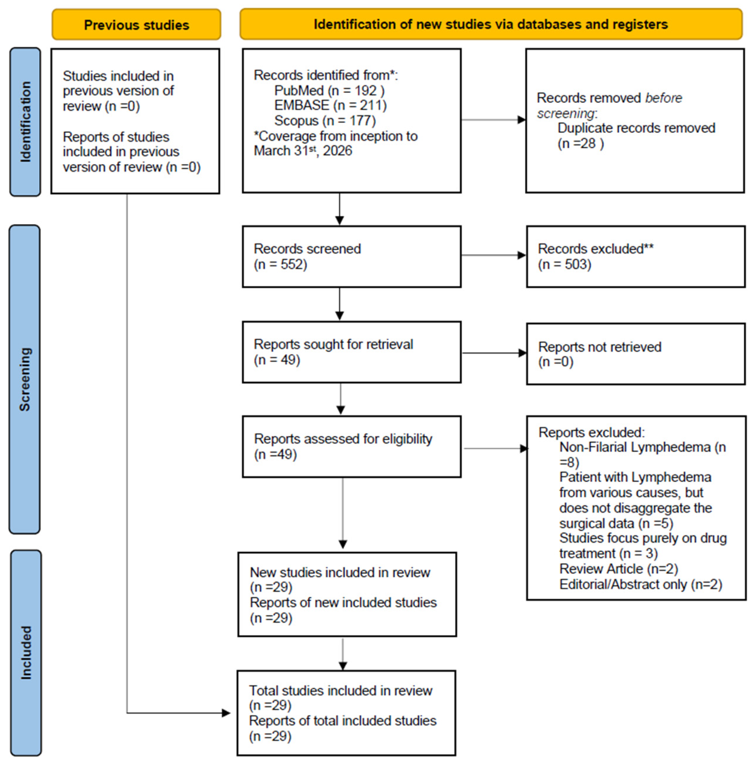
The study selection process is illustrated in Figure 1. A total of 580 records were identified from PubMed (n = 192), EMBASE (n = 211), and Scopus (n = 177); after removal of duplicate records (n = 28), 552 records were screened, of which 503 were excluded. Full-text reports were sought for 49 records, and all 49 were retrieved and assessed for eligibility (0 not retrieved). Of these, 20 reports were excluded for the following reasons: non-filarial lymphedema (n = 8), inclusion of patients with lymphedema from various causes without disaggregating surgical data (n = 5), studies focused purely on drug treatments (n = 3), review article (n = 2), and editorial/abstract-only publications (n = 2), leaving 29 studies included in the review (Figure 1) [12,13,14,15,16,17,18,19,20,21,22,23,24,25,26,27,28,29,30,31,32,33,34,35,36,37,38,39,40].

3.1. Study Characteristics and Patient Demographics

A total of twenty-nine studies met the eligibility criteria and were included in the final qualitative synthesis (Table 1). The extracted literature predominantly comprised single-patient case reports, alongside one case series, one retrospective database analysis, and one large-scale cross-sectional epidemiological study [12,13,14,15,16,17,18,19,20,21,22,23,24,25,26,27,28,29,30,31,32,33,34,35,36,37,38,39,40]. Geographically, the data reflect the endemic nature of lymphatic filariasis, with a significant majority of the studies originating from India. Additional cases were reported from diverse global locations, including the United States, Thailand, the United Kingdom, Spain, Canada, Denmark, Indonesia, Tanzania, and Pakistan. Excluding the large epidemiological study and the retrospective database analysis, the individual clinical cases represented a diverse age demographic ranging from eleven to seventy-three years [12,13,14,15,16,17,18,19,20,21,22,23,24,25,26,27,28,29,30,31,32,33,34,35,36,37,38,39,40].
Sex distribution was heavily dependent on the specific clinical manifestation, with male patients exclusively represented cases of genitourinary involvement, while female patients predominantly presented with breast-related filarial nodules or cervical masses. Where reported, the duration of clinical symptoms prior to surgical intervention varied drastically, ranging from six months to thirty years. While the initial inclusion criteria targeted advanced lymphedema, documentation of baseline lymphedema staging was notably sparse across the included studies, with only a fraction explicitly confirming grade three or grade four disease [12,13,14,15,16,17,18,19,20,21,22,23,24,25,26,27,28,29,30,31,32,33,34,35,36,37,38,39,40]. Prior conservative management was similarly underreported, though several cases noted the utilization of oral antibiotics, regular wound dressings, and targeted filariasis treatments before surgical escalation [12,13,14,15,16,17,18,19,20,21,22,23,24,25,26,27,28,29,30,31,32,33,34,35,36,37,38,39,40].

3.2. Anatomical Locations, Surgical Indications and Interventions

The anatomical distribution of filarial lymphedema was heterogeneous, although the lower extremities and genitourinary regions represented the most common sites of involvement. Lower extremity disease was reported in at least 9 studies and was frequently associated with advanced WHO Grade 3–4 lymphedema, chronic ulceration, impaired ambulation, and recurrent infections. Genitourinary involvement, including penoscrotal and scrotal lymphedema, was reported in 9 studies and often occurred after prolonged disease duration ranging from 3 to 32 years. Less frequent anatomical sites included the facial/periorbital region (2 studies), cervical lymph nodes (1 study), breast (3 studies), upper extremity involvement (3 studies), retroperitoneal or adrenal involvement (3 studies), and hepatic/systemic disease (1 study) [12,13,14,15,16,17,18,19,20,21,22,23,24,25,26,27,28,29,30,31,32,33,34,35,36,37,38,39,40].
The clinical indications prompting surgical intervention across the included cohort were highly heterogeneous (Table 2) and frequently diverged from classical extremity lymphedema management. A substantial proportion of surgeries were indicated for diagnostic uncertainty, such as the evaluation of suspected breast neoplasms, cervical lymphadenopathy, and unexplained facial or retroperitoneal cysts, which were subsequently identified as filarial in origin. Another major surgical indication was the presence of massive, disabling genitourinary elephantiasis, often complicated by secondary infections, chronic ulcerations, or the development of superimposed squamous cell carcinoma.
Among the 29 included studies, excisional and debulking procedures were the predominant surgical approaches, reported in 19 studies. These included subtotal scrotectomy, reduction scrotoplasty, modified Charles procedure, wide local excision, excisional biopsy, cyst excision, and reconstructive procedures involving skin grafts or flap reconstruction. Physiologic or microsurgical lymphatic procedures were less common, reported in 3 studies, and included nodovenous shunt surgery, nodo-venal shunt procedures, and vascularized lymph node transfer (VLNT). Minimally invasive or diagnostic procedures, including fine-needle aspiration cytology (FNAC), fine-needle aspiration (FNA), ultrasound-guided core needle biopsy, nephrostomy, and ureteric stenting, were reported in 5 studies. One study described conservative non-surgical management alone, while several epidemiologic or descriptive studies did not report a definitive operative intervention [12,13,14,15,16,17,18,19,20,21,22,23,24,25,26,27,28,29,30,31,32,33,34,35,36,37,38,39,40].

3.3. Clinical Outcomes and Postoperative Complications

An analysis of the defined primary and secondary outcomes revealed significant gaps in the current literature. Most notably, the primary outcome of percentage limb volume reduction was not quantified or reported in any of the included studies, largely due to the anatomical focus of the surgeries shifting toward genitourinary reconstruction and localized nodule excision rather than whole-limb debulking [12,13,14,15,16,17,18,19,20,21,22,23,24,25,26,27,28,29,30,31,32,33,34,35,36,37,38,39,40]. However, qualitative assessments of secondary outcomes demonstrated that surgical intervention generally yielded favorable symptomatic and psychosocial results. Patients undergoing massive excisional debulking for scrotal elephantiasis consistently reported significant relief from physical discomfort, restored ambulation, and highly acceptable cosmetic outcomes that fundamentally improved their quality of life [31,34]. In cases where surgery was performed for localized filarial cysts or nodules, complete symptomatic resolution and disease-free margins were frequently achieved within weeks of the procedure [13,14,15,16,17,19,20,21,22,23,24,25,26,27,28,29,30,31,32,33,34,35,36,37,38,39,40].
The postoperative follow-up durations varied widely, ranging from two weeks to ongoing monitoring over several months, with one database analysis noting significant loss to follow-up. Postoperative outcomes generally demonstrated significant functional and symptomatic improvement across intervention types. Excisional and reconstructive procedures for genital lymphedema consistently resulted in improved mobility, hygiene, cosmesis, psychosocial well-being, and quality of life, with several reports describing minimal recurrence and satisfactory wound healing. Lower extremity procedures, including modified Charles procedure, VLNT, and nodovenous shunt surgeries, demonstrated reductions in limb circumference and improvements in ambulation and daily functioning. Victor et al. reported limb circumference reductions exceeding 5 cm after debulking surgery, whereas Chilgar et al. demonstrated significant postoperative reductions in limb circumference at multiple levels following VLNT combined with excisional procedures [25,37]. Functional and psychological recovery were particularly emphasized among patients with advanced-stage disease. In contrast, minimally invasive or diagnostic procedures such as FNAC and aspiration mainly provided diagnostic clarification or temporary symptomatic relief rather than definitive disease control [12,13,14,15,16,17,18,19,20,21,22,23,24,25,26,27,28,29,30,31,32,33,34,35,36,37,38,39,40].
Complication profiles varied according to surgical technique and disease severity. Excisional and reconstructive procedures were primarily associated with wound-related complications, including superficial surgical site infection, serous discharge, edema, venous congestion, hypertrophic scarring, flap necrosis, cellulitis, and partial skin graft loss. The modified Charles procedure demonstrated the highest rate of postoperative hypertrophic scarring, reported in all patients, in addition to wound infection and ulceration. Microsurgical procedures such as VLNT and nodovenous shunts generally demonstrated favorable outcomes but remained associated with flap-related complications and seroma formation. Mortality and severe systemic deterioration were primarily observed in disseminated or progressive disease rather than as direct surgical complications. Overall, the comparative findings suggest that excisional debulking remains the most commonly utilized and effective intervention for advanced chronic filarial lymphedema, particularly in fibrotic disease, whereas physiologic procedures such as VLNT and nodovenous shunts may provide additional benefit in selected patients with preserved lymphatic function and less extensive tissue fibrosis [12,13,14,15,16,17,18,19,20,21,22,23,24,25,26,27,28,29,30,31,32,33,34,35,36,37,38,39,40].

3.4. Risk of Bias

The overall risk of bias across the included studies was generally low to moderate. All case reports assessed using the JBI checklist fulfilled all methodological domains, including clear reporting of patient demographics, clinical presentation, diagnostic methods, interventions, outcomes, adverse events, and key lessons learned, suggesting a consistently low risk of reporting bias among these studies. Similarly, the cohort studies evaluated using the Newcastle–Ottawa Scale (NOS) achieved high scores ranging from 6 to 7 stars, reflecting acceptable methodological quality with adequate selection, comparability, and outcome assessment domains overall. These findings support a reasonable level of confidence in the consistency of the reported clinical outcomes, although some methodological limitations remain present.
However, some concerns remain regarding the case series study assessed with the JBI checklist. Specifically, several domains were rated as “Unclear,” including inclusion criteria, completeness of participant inclusion, and reporting of participant demographics. These unclear items may indicate insufficient reporting transparency and introduce potential selection bias, as it is uncertain whether all eligible patients were included consecutively or whether important baseline characteristics were omitted.
Consequently, the representativeness and generalizability of the findings from this study may be limited. Although the overall body of evidence demonstrates acceptable methodological quality, the presence of unclear methodological domains reduces certainty in the pooled conclusions and should be considered when interpreting the findings. Therefore, conclusions drawn from this review should be interpreted with moderate caution, particularly regarding the external validity and reproducibility of outcomes across broader patient populations.

4. Discussion

The pathophysiology of lymphatic filariasis is fundamentally driven by the lodging of adult nematode worms within the lymphatic vessels, which triggers an intense local inflammatory response, endothelial dysfunction, and profound lymphatic dilation [41]. In the earlier phase, lymphedema caused thickening of the skin and structural changes in the subcutaneous tissue. As the disease progresses, the damage to the subcutaneous tissue also progresses; this might be due to greater fibrosis or fluid accumulation [42]. In the advanced stages of lymphedema targeted in this review, the lymphatic architecture undergoes irreversible fibrotic remodeling and sclerosis, severely compromising lymph flow and creating an immunologically compromised environment highly susceptible to acute dermatolymphangioadenitis (ADLA) attacks. Globally, it is estimated that over 15 million individuals suffer from filariasis-induced lymphedema, yet our systematic search revealed a striking discrepancy between modern lymphedema surgical paradigms and the realities of clinical management of filariasis [43]. While contemporary lymphedema treatment heavily emphasizes physiological reconstructions such as VLNT and lymphovenous anastomosis (LVA), our extracted data demonstrated an exclusive reliance on excisional debulking procedures. This divergence is likely rooted in the underlying pathology; by the time filarial patients reach the more advanced stage, the requisite lymphatic channels are functionally destroyed and physically obliterated by dense fibrosis, rendering delicate microvascular anastomoses anatomically unfeasible and necessitating ablative salvage techniques [44,45].
Furthermore, the clinical data highlighted a predominant focus on massive genitourinary involvement, specifically penoscrotal elephantiasis, rather than lower extremity lymphedema. The scrotal lymphatics serve as a common anatomic reservoir for adult filarial worms, leading to massive fluid accumulation, skin thickening, and, ultimately, superimposed fungal or bacterial infections [46]. Excisional debulking with complex genitourinary reconstruction, demonstrated high effectiveness for scrotal lymphedema [20,27,28,40,47]. By mechanically excising the fibrotic burden and removing the calcified remnants of dead worms that serve as a nidus for recurrent sepsis, patients experienced dramatic, life-altering improvements in mobility and psychosocial well-being [48]. Although historical perspectives often view excisional procedures as highly morbid, the studies synthesized in this review reported exceptionally low rates of severe surgical complications, with adverse events largely limited to manageable, superficial surgical site infections occurring in fewer than 10% of documented cases [49].
Despite these positive qualitative outcomes, the evidence base for the surgical management of filariasis remains critically underdeveloped compared with that for non-endemic, oncology-related lymphedema. The complete absence of comparative efficacy data in our findings underscores a significant failure of paradigm in tropical medicine research [50]. None of the twenty-nine included studies utilized conservative management as a defined control group, nor did any adequately quantify the primary outcome of percentage limb volume reduction using standardized objective metrics such as water displacement volumetry or bioimpedance spectroscopy. The literature is currently sustained by isolated case reports and small series that lack standardized longitudinal follow-up, highlighting an urgent clinical mandate to transition from anecdotal reporting to structured, comparative investigations that can rigorously define the optimal timing and modality of surgical intervention in advanced filarial disease.
  • Complete Decongestive Therapy
Complete Decongestive Therapy (CDT) remains a foundational component in the management of chronic lymphedema, even when definitive surgical treatment is being considered. CDT typically combines meticulous skin care, manual lymphatic drainage, compression bandaging or garments, and exercise, with the goal of reducing edema volume, softening fibrotic tissue, lowering bacterial and fungal colonization, and decreasing recurrent inflammatory attacks. In advanced filarial lymphedema, CDT alone may be insufficient to reverse established elephantiasis, but it can optimize the limb before surgery and help preserve postoperative gains afterward. Reports from the uploaded literature describe multimodal strategies in which CDT or related decongestive physical therapy was used preoperatively and postoperatively to improve tissue condition, facilitate wound healing, and reduce recurrence risk, supporting its role as an adjunct rather than a competing alternative to surgery [36,38,39].
However, the role of CDT in advanced filarial lymphedema appears largely supportive rather than curative [51]. Once patients progress to late-stage disease with dense fibrosis, marked skin thickening, and irreversible architectural destruction of the lymphatic system, conservative measures alone are often insufficient to restore meaningful drainage or reverse deformity [52]. This helps explain why many surgically treated patients in the present review had already reached a stage at which excisional procedures were used as salvage interventions. Accordingly, CDT should be viewed not as an alternative to surgery in all advanced cases, but as an essential component of perioperative optimization and long-term postoperative maintenance [53].
  • Physiologic and Reconstructive Surgical Procedures
Physiologic procedures aim to restore lymphatic drainage rather than simply remove diseased tissue. Lymphovenous anastomosis (LVA) diverts lymph from functioning lymphatic channels into nearby venules and is generally considered most effective when patent lymphatics remain. VLNT involves transfer of healthy lymph node–bearing tissue to the affected region to promote lymphangiogenesis and improve drainage, while nodovenous or lymphonodovenous shunts create an alternative pathway between lymphatic structures and the venous system. Although these approaches are well described conceptually, their application in filariasis-induced lymphedema remains limited and appears highly dependent on disease stage, residual lymphatic function, and local expertise. Available reports suggest that combining physiologic reconstruction with excisional debulking may offer better control of limb size, heaviness, and recurrent lymphangitis than debulking alone in selected patients, but robust comparative evidence is still lacking [35,36,37,38,39].
  • Comparison of Surgical Intervention to Conservative Management
Current evidence suggests that surgical intervention offers substantially greater functional and symptomatic improvement than conservative management alone in adults with filariasis-induced lymphedema, particularly in advanced-stage disease characterized by fibrosis, deformity, recurrent infection, and impaired mobility. Some cases of filariasis-induced lymphedema occurring in unusual anatomical locations as presented in this review, have also demonstrated that surgical intervention of mass excision, such as lumpectomy, cyst excision and mastectomy, can be a beneficial option in reducing lymphedema. Conservative measures—including complex decongestive therapy, compression, manual lymphatic drainage, antibiotics, hygiene optimization, and antiparasitic medications such as diethylcarbamazine or albendazole—remain important first-line approaches during early disease stages and for perioperative optimization. However, conservative treatment alone is often insufficient once irreversible lymphatic fibrosis and tissue hypertrophy develop. Patients undergoing excisional debulking procedures, modified Charles procedures, nodovenous shunts, or vascularized lymph node transfer (VLNT) consistently showed improved ambulation, reduction in limb circumference, enhanced hygiene, improved cosmesis, and marked psychosocial recovery compared with preoperative conservative management [25,37,39].
  • Advanced Diagnostic Imaging Modalities
Imaging is central to selecting appropriate candidates for physiologic lymphatic surgery and to understanding the severity of lymphatic dysfunction. Lymphoscintigraphy remains the most commonly reported functional imaging modality in the filariasis literature and can demonstrate delayed transport, dermal backflow, interrupted channels, and residual nodal function; these findings may help determine whether procedures such as nodovenous shunting or VLNT are feasible and can also be used for postoperative assessment. In contrast, indocyanine green (ICG) lymphography provides high-resolution real-time visualization of superficial lymphatic channels and is particularly useful for planning supermicrosurgical procedures such as LVA. However, ICG appears to be rarely reported in filariasis-specific surgical studies, likely reflecting limited availability in endemic settings and the fact that many patients present with advanced fibrosis, where superficial channels may already be severely damaged. As a result, lymphoscintigraphy currently remains more established in this field, while ICG represents a promising but underreported tool for future patient selection and operative planning [36,37].
  • Research Gaps and Future Directions
The current landscape of filarial lymphedema research reveals a critical paradigm failure, primarily due to the complete absence of comparative efficacy data. Currently, the literature is sustained by isolated case reports and small series that lack standardized longitudinal follow-up, making the evidence base highly susceptible to publication bias. None of the evaluated studies utilized defined conservative management as a control group, nor did they quantify postoperative volumetric reductions using standardized, objective tools such as bioimpedance spectroscopy or water displacement volumetry. None of the studies mention the evolution of subcutaneous changes post-surgical procedure. To establish definitive, evidence-based clinical guidelines, future research must urgently transition from anecdotal observational reporting to well-designed, prospective controlled trials that utilize standardized outcome metrics to clearly delineate the optimal timing, safety, and modality of surgical intervention [35,36,37,38,39].
  • Study Limitation
The findings of this systematic review must be interpreted within the context of several significant methodological limitations inherent to the primary literature. The overwhelming preponderance of single-patient case reports and small retrospective series introduces a high risk of publication bias, as successful or highly unusual clinical outcomes are disproportionately represented while surgical failures or modest improvements remain unpublished. Additionally, the complete lack of randomized controlled trials or prospective cohort studies with defined comparator groups entirely precludes the ability to perform a quantitative meta-analysis or establish definitive causality regarding surgical efficacy versus conservative medical management. Furthermore, the extracted literature demonstrated a profound heterogeneity in follow-up durations and a widespread failure to utilize validated, standardized assessment tools for quality of life or precise volumetric measurements, limiting the generalizability of the outcomes and preventing a standardized assessment of long-term surgical durability. The geographic distribution of included studies was heavily skewed toward India, with limited representation from other endemic regions such as sub-Saharan Africa and Southeast Asia, which may constrain external validity. Furthermore, the dataset was characterized by a pronounced imbalance between one large epidemiological study and numerous single-patient case reports, further limiting the robustness of pooled conclusions. The inclusion of studies with pediatric patients, despite the stated adult-focused inclusion criteria, represents an additional methodological inconsistency that should be considered when interpreting the findings.

5. Conclusions

In conclusion, surgical intervention remains an important therapeutic option for adult patients with advanced, medically refractory filariasis-induced lymphedema, particularly in cases associated with severe functional impairment. Current evidence suggests that excisional debulking procedures are commonly utilized in advanced-stage disease due to the extensive fibrosis and chronic lymphatic damage frequently observed in endemic filariasis. These procedures have been associated with symptomatic improvement, enhanced quality of life, and acceptable perioperative outcomes in several observational studies. However, the available evidence is limited predominantly to low-level, non-comparative studies, thereby precluding definitive conclusions regarding the relative effectiveness or superiority of specific surgical approaches. As physiological and microsurgical techniques continue to advance, a combination or staged approach of ablative and physiological surgery may prove useful to restore lymphatic flow, although their role in advanced filarial lymphedema remains insufficiently defined. Future well-designed prospective and controlled studies using standardized objective outcome measures are needed to clarify surgical indications, evaluate comparative effectiveness, optimize operative strategies, and guide evidence-based management for this debilitating disease.

Author Contributions

R.S.: Conceptualization, Investigation, Methodology, Data Curation, Data Analysis, Formal Analysis, Project Administration, Resources, Visualization, Supervision, Validation, Writing—original draft, Writing—Review and Editing. I.R.W.: Conceptualization, Investigation, Methodology, Supervision, Validation, Writing—Review and Editing. P.H.: Conceptualization, Investigation, Methodology, Supervision, Validation, Writing—Review and Editing. V.S.C.: Conceptualization, Investigation, Methodology, Data Curation, Data Analysis, Formal Analysis, Project Administration, Resources, Visualization, Supervision, Validation, Writing—original draft, Writing—Review and Editing. All authors have read and agreed to the published version of the manuscript.

Funding

This research received no external funding.

Institutional Review Board Statement

Not applicable.

Informed Consent Statement

Not applicable.

Data Availability Statement

The original data presented in the study are openly available in https://doi.org/10.5281/zenodo.20110806.

Acknowledgments

This publication charge is funded by Unpad through the Indonesian Endowment Fund for Education (LPDP) on behalf of the Indonesian Ministry of Higher Education, Science and Technology and managed under the EQUITY Program (Contract No. 4303/B3/DT.03.08/2025 and 3927/UN6. RKT/HK.07.00/2025).

Conflicts of Interest

The authors declare no conflicts of interest.

Abbreviations

The following abbreviations are used in this manuscript:
ADLAAcute dermatolymphangioadenitis
CDTComplete Decongestive Therapy
DECDiethylcarbamazine
FNAFine Needle Aspiration
FNACFine Needle Aspiration Cytology
ICGIndocyanine green
ISLInternational Society of Lymphology
JBIJoanna Briggs Institute
LVALymphovenous Anastomosis
MLDManual Lymph Drainage
NOSNewcastle-Ottawa Scale
PICOPopulation, Intervention, Comparison, and Outcomes
PRISMAPreferred Reporting Items for Systematic Reviews and Meta-Analyses
VLNTVascularized Lymph Node Transfer
WHOWorld Health Organization

References

  1. Goldin, J.; Juergens, A.L. Filariasis. In StatPearls; StatPearls Publishing: Treasure Island, FL, USA, 2025. Available online: http://www.ncbi.nlm.nih.gov/books/NBK556012/ (accessed on 1 February 2026).
  2. World Health Organization. Lymphatic Filariasis. 1 January 2025. Available online: https://www.who.int/news-room/fact-sheets/detail/lymphatic-filariasis (accessed on 1 February 2026).
  3. Nimmana, B.K.; Kimyaghalam, A.; Manna, B. Lymphedema. In StatPearls; StatPearls Publishing: Treasure Island, FL, USA, 2025. Available online: http://www.ncbi.nlm.nih.gov/books/NBK537239/ (accessed on 1 February 2026).
  4. Vasconez-Gonzalez, J.; Miño, C.; Noboa, M.D.L.; Tello-De-la-Torre, A.; Izquierdo-Condoy, J.S.; Ortiz-Prado, E. The psychosocial and emotional burden of lymphatic filariasis: A systematic review. PLoS Negl. Trop. Dis. 2025, 19, e0013073. [Google Scholar] [CrossRef] [Scilit] [PubMed]
  5. Mackenzie, C.D.; Kapa, D.R.; Krishnasastry, S.; Douglass, J.; Hoerauf, A.; Ottesen, E.A. Managing Lymphedema Induced by Lymphatic Filariasis: Implementing and Improving Care at the Individual and Programmatic Levels. Am. J. Trop. Med. Hyg. 2024, 111, 3–21. [Google Scholar] [CrossRef] [Scilit]
  6. Lee, B.-B.; Bergan, J.; Rockson, S.G. (Eds.) Lymphedema: A Concise Compendium of Theory and Practice; Springer: London, UK, 2011. [Google Scholar] [CrossRef] [Scilit]
  7. Molyneux, D. Lymphatic Filariasis (Elephantiasis) Elimination: A public health success and development opportunity. Filaria J. 2003, 2, 13. [Google Scholar] [CrossRef] [Scilit]
  8. Chen, J.; Feng, X.; Zhou, Y.; Wang, Y.; Xiao, S.; Deng, C. Outcomes after liposuction-based treatment of lymphedema: A systematic review and meta-analysis. Front. Oncol. 2025, 15, 1651472. [Google Scholar] [CrossRef] [Scilit]
  9. Senger, J.-L.B.; Kadle, R.L.; Skoracki, R.J. Current Concepts in the Management of Primary Lymphedema. Medicina 2023, 59, 894. [Google Scholar] [CrossRef] [Scilit]
  10. Fallahian, F.; Tadisina, K.K.; Xu, K.Y. Efficacy of Microsurgical Treatment of Primary Lymphedema: A Systematic Review. Ann. Plast. Surg. 2022, 88, 195–199. [Google Scholar] [CrossRef] [Scilit]
  11. Page, M.J.; McKenzie, J.E.; Bossuyt, P.M.; Boutron, I.; Hoffmann, T.C.; Mulrow, C.D.; Shamseer, L.; Tetzlaff, J.M.; Akl, E.A.; Brennan, S.E.; et al. The PRISMA 2020 statement: An updated guideline for reporting systematic reviews. BMJ 2021, 372, n71. [Google Scholar] [CrossRef] [Scilit] [PubMed]
  12. Azhar, S.M.; Devi, S.; Patro, B.K.; Panigrahi, S.; Jose, S.; Jacob, J.; Issac, A.; Stephen, S. Clinical Manifestations of Lymphatic Filariasis and Practice of Vector Control Measures after 15 Years of Mass Drug Administration: A Rapid Epidemiological Study from Odisha. Indian J. Community Med. Off. Publ. Indian Assoc. Prev. Soc. Med. 2025, 50, S243–S248. [Google Scholar] [CrossRef] [Scilit]
  13. Connor, M.J.; Habib, A.; Wen, D.; Kubba, F.; Raza, A. Filariasis an Unusual Cause of Retroperitoneal Fibrosis, Hydronephrosis, and Infertility in a Young Adult Male. J. Endourol. Case Rep. 2019, 5, 113–116. [Google Scholar] [CrossRef] [Scilit]
  14. Daggett, R.L.B.; Farishta, D.; Cuellar, H.; Nathan, C.-A.O. Substernal goitre presenting with upper and lower extremity oedema. BMJ Case Rep. 2021, 14, e245036. [Google Scholar] [CrossRef] [Scilit] [PubMed]
  15. Diwakar, D.K.; Wadhwani, N.; Prasad, N.; Gupta, A. Primary retroperitoneal filariasis: A common disease of tropics with uncommon presentation and review of literature. BMJ Case Rep. 2018, 2018, bcr2018226217. [Google Scholar] [CrossRef] [Scilit] [PubMed]
  16. Gahine, R.; Das, A.; Jain, V.K.; Agrawal, A. Rare association of microfilaria with poorly differentiated mucin-secreting metastatic carcinoma in liver aspirate cytology. BMJ Case Rep. 2019, 12, e231618. [Google Scholar] [CrossRef] [Scilit]
  17. Gonzálvez-Gasch, A.M.; Lopardo Villarino, G.; Bernal Morell, E.; Moltó Marhuenda, J.; Martínez Goñi, R.; Rolo Téllez, M.; Martín-Higuera, M.C.; Cañon Rodríguez, R.M.; Costell, J.E.; Valles Bergada, M. Rare case of extralymphatic dirofilariasis presenting as a facial subcutaneous nodule in non-endemic region of Spain. IDCases 2025, 42, e02369. [Google Scholar] [CrossRef] [Scilit] [PubMed]
  18. Jaiswal, S.; Chand, G.; Lal, H.; Vij, M.; Pandey, R. Microfilaria in association with adrenal lymphoma diagnosed on cytology: An extremely rare case report. Turk. Patoloji Derg. 2013, 29, 143–145. [Google Scholar] [CrossRef] [Scilit] [PubMed]
  19. Kumar, S.; Saha, A.; Kumar, S.; Singh, P.; Singh, K.K. Giant Scrotal Lymphoedema: A Case Series. Cureus 2023, 15, e48248. [Google Scholar] [CrossRef] [Scilit]
  20. Lema, D.D.; Ngowi, B.N.; Mbwambo, O.J.; Bright, F.; Mwakibibi, D.; Mremi, A. Surgical management of the giant scrotal lymphedema: A case report at a tertiary facility in northern Tanzania. Int. J. Surg. Case Rep. 2025, 133, 111622. [Google Scholar] [CrossRef] [Scilit]
  21. Mangla, A.; Singh, A.K.S.; Goel, S. Squamous cell carcinoma of skin in a case of post-filariasis chronic lower limb lymphedema- a case study and review of literature. Int. J. Surg. Case Rep. 2024, 116, 109355. [Google Scholar] [CrossRef] [Scilit]
  22. Mendu, D.R.; Sternlicht, H.; Ramanathan, L.V.; Pessin, M.S.; Fleisher, M.; Dalbagni, G.; Jaimes, E.A.; Kaltsas, A.; Glezerman, I.G. Two cases of spontaneous remission of non-parasitic chyluria. Clin. Biochem. 2017, 50, 886–888. [Google Scholar] [CrossRef] [Scilit]
  23. Nayak, M.; Patra, S.; Meher, S.; Sasmal, P.K. Amelanotic Melanoma Arising in Filarial Leg: A Report of a Rare Case. J. Clin. Diagn. Res. 2017, 11, ED07–ED09. [Google Scholar] [CrossRef] [Scilit] [PubMed]
  24. Nunthanid, P.; Roongruanchai, K.; Wongkamchai, S.; Sarasombath, P.T. Case Report: Periorbital Filariasis Caused by Brugia malayi. Am. J. Trop. Med. Hyg. 2020, 103, 2336–2338. [Google Scholar] [CrossRef] [Scilit]
  25. Victor, J.; Stephen, T.; Guin, D.; Victor, J. The nodovenous shunt and reduction surgery for post-filarial lymphedema—surgical technique and clinical outcomes. Indian J. Plast. Surg. 2021, 54, 75–81. [Google Scholar] [CrossRef] [Scilit]
  26. Panda, D.K.; Mohapatra, D.P.; Mohapatra, M.M. Filarial breast lump. BMJ Case Rep. 2017, 2017, bcr-2017-221536. [Google Scholar] [CrossRef] [Scilit]
  27. Sahu, G.; Verma, Y.; Jajoo, S.; Shrivastava, P. Filariasis with Squamous Cell Carcinoma: A Hidden Surprise. Int. J. Appl. Basic Med. Res. 2020, 10, 59–61. [Google Scholar] [CrossRef] [Scilit]
  28. Saqlain, M.; Ur-Rauf Khan Hiraj, M.; Ans, M.; Usama Ilyas Ahmed, M. Massive Scrotal Lymphedema in an Adolescent with Intellectual Disability: A Rare Case of Infection-Related Scrotal Enlargement Requiring Subtotal Scrotectomy. Cureus 2025, 17, e81821. [Google Scholar] [CrossRef] [Scilit] [PubMed]
  29. Simmonds, J.C.; Mansour, M.K.; Dagher, W.I. Cervical Lymphatic Filariasis in a Pediatric Patient: Case Report and Database Analysis of Lymphatic Filariasis in the United States. Am. J. Trop. Med. Hyg. 2018, 99, 104–111. [Google Scholar] [CrossRef] [Scilit]
  30. Tandon, N.; Bansal, C.; Sharma, R.; Irfan, S. Role of fine needle aspiration cytology in diagnosing filarial arm cysts. BMJ Case Rep. 2013, 2013, bcr2013009677. [Google Scholar] [CrossRef] [Scilit]
  31. Tekin, H.G.; Andersen, K.; Bakholdt, V.; Sørensen, J.A. Resection and reconstruction of idiopathic scrotal elephantiasis: Surgical approach and quality of life assessment. BMJ Case Rep. 2021, 14, e244018. [Google Scholar] [CrossRef] [Scilit] [PubMed]
  32. Thongpiya, J.; Sa-Nguanraksa, D.; Samarnthai, N.; Sarasombath, P.T. Filariasis of the breast caused by Brugia pahangi: A concomitant finding with invasive ductal carcinoma. Parasitol. Int. 2021, 80, 102203. [Google Scholar] [CrossRef] [Scilit]
  33. Tummidi, S.; Kothari, K.; Patil, R.; Singhal, S.S.; Keshan, P. Incidental detection of two adult gravid filarial worms in breast: A case report. BMC Res. Notes 2017, 10, 412. [Google Scholar] [CrossRef] [Scilit] [PubMed]
  34. Wijaya, I.; Pangestu, W. Giant Scrotal Hydrocele and Bilateral Leg Lymphedema as Clinical Manifestation of Chronic Lymphatic Filariasis. Acta Med. Indones. 2016, 48, 239–241. [Google Scholar]
  35. Islam, M.T.; Rahman, T.R.; Islam, M.S.; Mohiuddin, M.; Jony, N.H. Modified charles procedure using delayed skin graft for advanced lymphatic filariasis: A single centre study. MedPulse Int. J. Surg. 2022, 23, 1–6. [Google Scholar]
  36. Sivaprakasam, R.; Anuradha, R.; Bethunaickan, R.; Manokaran, G. Successful multi-modal treatment of grade IV lymphedema in lymphatic filariasis: A case study. Lymphology 2018, 51, 89–92. [Google Scholar]
  37. Chilgar, R.M.; Khade, S.; Chen, H.-C.; Ciudad, P.; Yeo, M.S.-W.; Kiranantawat, K.; Maruccia, M.; Li, K.; Zhang, Y.X.; Nicoli, F. Surgical treatment of advanced lymphatic filariasis of lower extremity combining vascularized lymph node transfer and excisional procedures. Lymphat. Res. Biol. 2019, 17, 637–646. [Google Scholar] [CrossRef] [Scilit]
  38. Kasim, K.N.; Wahab, S.; Djawad, K.; Wospodo, N.N. Lymphedema resulting from filariasis successfully treated by surgery. J. Public Health Afr. 2020, 11, 1059. [Google Scholar] [CrossRef] [Scilit] [PubMed]
  39. Istranov, A.L.; Makarov, I.G.; Makarova, N.V.; Tulina, I.; Ulasov, I.V.; Isakova, Y.I. Combination of conservative and surgical methods in the treatment of giant lymphedema of the scrotum: A case report. Front. Surg. 2023, 10, 1048159. [Google Scholar] [CrossRef] [Scilit]
  40. Coșeriu, A.; Prodan, L.; Fetti, A.; Filip, C. Surgical management of massive penoscrotal lymphedema. Case report and literature review. Ann. Ital. Di Chir. 2022, 11, S2239253X22035927. [Google Scholar]
  41. Chakraborty, S.; Gurusamy, M.; Zawieja, D.C.; Muthuchamy, M. Lymphatic filariasis: Perspectives on lymphatic remodeling and contractile dysfunction in filarial disease pathogenesis. Microcirculation 2013, 20, 349–364. [Google Scholar] [CrossRef] [Scilit] [PubMed]
  42. Pirri, C.; Ferraretto, C.; Pirri, N.; Bonaldo, L.; De Caro, R.; Masiero, S.; Stecco, C. Ultrasound Examination of Skin, Fasciae and Subcutaneous Tissue: Optimizing Rehabilitation for Secondary Upper Limb Lymphedema. Diagnostics 2024, 14, 2824. [Google Scholar] [CrossRef] [Scilit]
  43. Kazura, J.W. Narrowing the Gap in Managing the Lymphedema of Lymphatic Filariasis. Am. J. Trop. Med. Hyg. 2024, 111, 1–2. [Google Scholar] [CrossRef] [Scilit]
  44. Medeiros, Z.M.; Vieira, A.V.B.; Xavier, A.T.; Bezerra, G.S.N.; de Fátima C Lopes, M.; Bonfim, C.V.; Aguiar-Santos, A.M. Lymphatic Filariasis: A Systematic Review on Morbidity and Its Repercussions in Countries in the Americas. Int. J. Environ. Res. Public. Health 2021, 19, 316. [Google Scholar] [CrossRef] [Scilit]
  45. Melrose, W.D. Lymphatic filariasis: New insights into an old disease. Int. J. Parasitol. 2002, 32, 947–960. [Google Scholar] [CrossRef] [Scilit] [PubMed]
  46. Irdam, G.A.; Fadhly, S. Scrotal reconstruction on scrotal lymphedema. Urol. Case Rep. 2018, 20, 9–11. [Google Scholar] [CrossRef] [Scilit] [PubMed]
  47. Lucas, J.W.; Lester, K.M.; Chen, A.; Simhan, J. Scrotal reconstruction and testicular prosthetics. Transl. Androl. Urol. 2017, 6, 710–721. [Google Scholar] [CrossRef] [Scilit]
  48. Dreyer, G.; Norões, J.; Figueredo-Silva, J.; Piessens, W.F. Pathogenesis of lymphatic disease in bancroftian filariasis: A clinical perspective. Parasitol. Today 2000, 16, 544–548. [Google Scholar] [CrossRef] [Scilit] [PubMed]
  49. Rezaei, A.R.; Zienkiewicz, D.; Rezaei, A.R. Surgical site infections: A comprehensive review. J. Trauma. Inj. 2025, 38, 71–81. [Google Scholar] [CrossRef] [Scilit]
  50. Herold, G.; Justickis, V.; Maneikienė, V.; Maneikis, K.; Trinkauskas, P.; Palkova, K. A Physician-Driven Patient Safety Paradigm: The “Pitfall Bank” as a Translational Mechanism for Medical Error Prevention. Healthcare 2025, 13, 2248. [Google Scholar] [CrossRef] [Scilit]
  51. Ogino, R.; Yokooji, T.; Hayashida, M.; Suda, S.; Yamakawa, S.; Hayashida, K. Emerging Anti-Inflammatory Pharmacotherapy and Cell-Based Therapy for Lymphedema. Int. J. Mol. Sci. 2022, 23, 7614. [Google Scholar] [CrossRef] [Scilit]
  52. Brown, S.; Dayan, J.H.; Kataru, R.P.; Mehrara, B.J. The Vicious Circle of Stasis, Inflammation, and Fibrosis in Lymphedema. Plast. Reconstr. Surg. 2023, 151, 330e–341e. [Google Scholar] [CrossRef] [Scilit]
  53. Flores, A.M.; Fu, M.R.; Bock, K.J.; Campione, E.; Francis, K.; Kellerman, C.; Norris, T.L.; Tylka, S. Post-operative care for patients following surgical treatment of lymphedema. Med. Oncol. 2024, 41, 296. [Google Scholar] [CrossRef] [Scilit]
Figure 1. PRISMA flow diagram. * Automation tools were not used for screening. ** No additional records were identified from reference list screening.
Life 16 00942 g001
Table 1. Baseline Demographics and Study Characteristics.
ReferenceCountryStudy DesignTotal Sample SizeAgeSex DistributionMean Duration of Lymphedema Prior to SurgeryBaseline Lymphedema StagePrior Conservative ManagementAnatomical Location
Gonzálvez-Gasch et al., 2025 [17]SpainCase Report149 years 100% Male Not reportedNot reportedNot reportedFace (subcutaneous)
Nunthanid et al., 2020 [24]ThailandCase Report170 years 100% Female Not reportedNot reportedReceived dicloxacillin 250 mg four times a day for 2 weeks prior to surgeryFace/periorbital
Simmonds, 2018 [29]USACase Report and Retrospective Database Analysis1 case report + 865 database admissions11 years (case report); 64.5 years (database average)100% Female (case);Not reportedNot reportedNot reportedCervical (neck)
Panda, 2017 [26]IndiaCase Report128 years100% FemaleNot reportedNot reportedNot reportedBreast
Thongpiya, 2021 [32] ThailandCase Report164 years100% FemaleNot reportedNot reportedReceived anastrozole for two months before surgery to treat concomitant carcinomaBreast
Tummidi, 2017 [33]IndiaCase Report135 years100% FemaleNot reportedNot reportedNot reportedBreast
Tandon et al., 2013 [30] India Case Report 2 Mean: 36.5 years 50% Male, 50% Female Not reportedNot reportedNot reportedUpper extremity
Daggett et al., 2021 [14]USACase Report170 years 100% Male 5 yearsSevere bilateral lymphedema with chronic wounds (WHO Grade 4)Lasix prescribed as needed for edema, regular wound care, and empiric treatment with doxycycline and ivermectin was recommendedUpper and lower extremity
Gahine et al., 2019 [16]IndiaCase Report135 years 100% Male Not reportedNot reportedNot reportedHepatic/systemic
Jaiswal et al., 2013 [18]IndiaCase Report155 years 100% Male Not reportedNot reportedNot reportedAdrenal/retroperitoneal
Diwakar et al., 2018 [15]IndiaCase Report152 100% Male 6 months (abdominal pain duration)Not reportedOver-the-counter analgesics, antacids, and a proton pump inhibitor Retroperitoneal
Connor et al., 2019 [13]UKCase Report140 100% Male Not specifiedNot reportedTrial of steroids, bilateral Double-J stents, ivermectin, and doxycyclineRetroperitoneal
Kumar et al., 2023 [19] India Case Series 3 Mean: 37.3 years 100% Male 9.0 years WHO Grade 4 Antibiotics and regular dressings utilized to control infection prior to surgical debulking Genitourinary (scrotum/penis)
Mendu et al., 2017 [22] USA Case Reports 2 Mean: 73.0 years 100% Female Not reportedNot reportedNot reportedGenitourinary (chyluria)
Sahu et al., 2020 [27]IndiaCase Report170 100% Male 30 yearsNot reportedNot reportedGenitourinary (penis/scrotum)
Tekin et al., 2021 [31]DenmarkCase Report141 100% Male 3 yearsNot reportedExtensive diagnostic workup (conservative therapies were noted as ineffective)Genitourinary (scrotum)
Lema et al., 2025 [20]TanzaniaCase Report138 100% Male 28 yearsNot reportedSought assistance from traditional healersGenitourinary (penoscrotal)
Saqlain et al., 2025 [28]PakistanCase Report117 100% Male 5 yearsNot reportedNot reportedGenitourinary (scrotum)
Coseriu et al., 2022 [40]RomaniaCase Report150 Male 32 yearsNot reportedProphylactic Intravenous broad-spectrum antibioticsGenitourinary (penoscrotal)
Istranov et al., 2023 [39]RussiaCase Report148 Male 31 yearsNot reportedComplex Decongestive Therapy.Genitourinary (scrotum), lower extremity
Wijaya et al., 2016 [34]IndonesiaCase Report151 100% Male 17 years (scrotum), 5 years (left leg)Not reportedOral albendazole, diethylcarbamazine (DEC), captopril, sodium bicarbonate, folic acid, vitamin B12, and iron sucrose injection Genitourinary (scrotum), lower extremity (bilateral legs)
Nayak et al., 2017 [23]IndiaCase Report160 years 100% Male 30 yearsNot reportedManaged with dressing and oral antibiotics in another hospitalLower extremity
Mangla et al., 2024 [21]IndiaCase Report169100% Female4 yearsNot reportedMultiple filariasis treatments at a local hospitalLower extremity
Victor et al., 2021 [25]IndiaRetrospective study16Mean: 52.6 years 75% Male, 25% Female Not reportedWHO Grade 4Penicillin prophylaxis—Inj. Benzathine penicillin 1.2 million units intramuscular once every 3 weeks or tablet penicillin G 400 mg OD and daily foot care.Lower extremity
Kasim et al., 2020 [38]IndonesiaCase Report128 Male 4 years Albendazole therapy 1 × 400 mg, diethyl carbamazine 5 × 100 mg, cetirizine 2 × 10 mg, and vitamin B complex 2 × 1 tab.Lower extremity
Chilgar et al., 2019 [37]IndiaRetrospective study17Average 43 (±15, range 17–67). 52.9% Male, 47.1%
Female
Not reported29.4% ISL Grade 2, 70.6% ISL Grade 3Not reportedLower extremity
Sivaprakasam et al., 2018 [36]IndiaCase Report137 Female 27 yearsWHO Grade 4A course of oral Penicillin (800 mg/twice daily) for one week, Manual Lymph Drainage (MLD), Complete Decongestive Therapy (CDT), Respiratory physiotherapy and walking.Lower extremity
Islam et al., 2022 [35]BangladeshProspective Study1145.45% in range 25–45; 54.55% in range 46–6554.55% Male, 45.45% Female Not reportedAdvanced stage (Stage 3)Not reportedLower extremity
Azhar et al., 2025 [12]IndiaCross-sectional rapid epidemiological study1226Ranges: 67.6% were 15–59 years54.2% Female, 45.8% MaleNot reported19.3% had WHO Grade 3, 10.5% had WHO Grade 4Not reportedUpper extremity, lower extremity, genitourinary (hydrocele)
Note: Baseline lymphedema staging was reported using different classification systems across studies. The eligibility criteria for this review used the WHO filariasis staging system (stages 3–6). Where individual studies reported staging using the International Society of Lymphology (ISL) grading system or other frameworks, the original classification is preserved as reported by the study authors.
Table 2. Surgical Interventions, Comparators, and Clinical Outcomes.
ReferenceSurgical IndicationsSurgical ApproachComparator GroupFollow-Up DurationPercentage Limb Volume ReductionSecondary Outcomes (QoL, Cosmesis, Symptom Relief)Complications or Adverse Events
Gonzálvez-Gasch et al., 2025 [17] Painful subcutaneous facial swelling/cyst Excisional (Complete excision of the cyst containing the worm) Not reportedNot reportedNot reported All symptoms resolved within 48 h after surgical removal Not reported
Nunthanid et al., 2020 [24] Erythematous periorbital nodule/infected thin-walled cyst Excisional (Mass excision of the cyst) Not reported Not specified Not reported Surgical wound completely healed without secondary bacterial infection None reported
Simmonds, 2018 [29]Diagnosis of an enlarged, painless left cervical lymph nodeExcisional biopsy of the left neck massNot reportedPatient did not present for follow-up careNot reportedNot reportedTolerated without complications.
Panda, 2017 [26]Suspected breast neoplasm/organized breast abscessLumpectomyNot reported5 months post
operatively (for recurrence); 6 weeks post-medical therapy
Not reportedPatient was symptomatically relieved and the lesion disappeared 6 weeks following subsequent medical therapy (diethylcarbamazine, amoxicillin + clavulanic acid, and doxycycline).Occasional pain and recurrent nodularity 5 months post-surgery.
Thongpiya, 2021 [32]Invasive ductal carcinoma (left breast); Filarial nodule (right breast) initially planned for removalLeft modified radical mastectomy; Ultrasound-guided core needle biopsy (right breast)Not reported2 monthsNot reportedThe right breast lesion resolved spontaneously without filarial medication.Not reported
Tummidi, 2017 [33]Diagnostic evaluation of a breast lumpFine Needle Aspiration Cytology (FNAC)Not reportedNot reportedNot reportedNot specifiedNone reported.
Tandon et al., 2013 [30]Diagnostic Fine Needle Aspiration (FNA) only.None reported.None reported.Not reportedNot reportedSwelling shrank in size following aspiration in Case.None reported.
Daggett et al., 2021 [14] Substernal goiter causing external compression of the thoracic duct and resulting in severe lymphedema Left hemithyroidectomy via cervical incision and median sternotomy Not reported Approximately 6 months Not reported Patient regained mobility after being bed-bound for 5 years; quality of life greatly enhanced No intraoperative or postoperative complications
Gahine et al., 2019 [16]Not reportedNot reportedNot reported Followed for 1 month Not reportedNot reported Died within 1 month due to complications and hepatic failure (disease progression)
Jaiswal et al., 2013 [18]Not reportedNot reportedNot reportedNot reportedNot reportedNot reportedNot reported
Diwakar et al., 2018 [15]Diagnostic uncertainty; failure of non-surgical medical therapies to relieve abdominal pain.Excisional (Cyst excision).Not reported6 months.Not reportedDisease-free with clear ultrasound.Uneventful postoperative period.
Connor et al., 2019 [13]Atypical retroperitoneal fibrosis causing hydronephrosis and renal failure.Stent insertion, nephrostomy.Not reportedUntil death.Not reportedNo clinical improvement noted.Multiorgan failure leading to death after repeated infections.
Kumar et al., 2023 [19]Advanced disease stages where fibrosis has set in, causing severe physical limitations, disability, social stigma, and inability to maintain hygieneExcisional debulking (reduction scrotoplasty) combined with reconstructive surgery of the external genitalia.Not reported3 months.Not reportedAchieved acceptable/promising cosmetic results; alleviating psychosocial and physical distress; minimal to no recurrence.Serous discharge requiring daily dressings; minor wound infection on postoperative day 5 requiring antibiotics; required subsequent revision surgery/grafting for penile coverage.
Mendu et al., 2017 [22]Conservative management only: low fat, high fiber diet).None reported.None reported.Several weeks to months.None reported.Spontaneous remission and disappearance of chyluria.None reported.
Sahu et al., 2020 [27]Discovery of a cauliflower-like squamous cell carcinoma growth under the prepuce of a ram’s horn penis.Excisional debulking (Scrotectomy and total amputation of penis).Not reported2 weeks.Not reportedNegative for inguinal metastasis; patient put on regular follow-up.Uneventful postoperative recovery.
Tekin et al., 2021 [31]Emergency admission due to grossly enlarged, hemorrhagic scrotum.Excisional debulking (Subtotal scrotectomy and penoscrotal reconstruction).Not reported6 months.Not reportedComplete wound closure, no lymphedema recurrence, and substantial improvement in QoL scores (ICIQ, SF-36).Superficial surgical site infections (responded to antibiotics).
Lema et al., 2025 [20]Chronic massive penoscrotal lymphedema complicated by an ulcer and secondary infection.Excisional debulking (Urgent debridement, scrotoplasty, and penile skin grafting).Not reported6 months.Not reportedGood functional and cosmetic outcomes maintained; satisfactory clearance of warts after adjunct medical therapy.No complications observed postoperatively.
Saqlain et al., 2025 [28]Discomfort, difficulty ambulating, impaired hygiene, and recurrent skin infections/ulcerations.Excisional debulking (Subtotal scrotectomy with penile preservation).Not reportedNot reportedNot reportedSignificant relief from discomfort and improved mobility.Uneventful postoperative recovery.
Coseriu et al., 2022 [40]Massive penoscrotal edemaAblative surgery, bilateral orchidopexy, abdominal advancement flaps, split-thickness skin graft.Not reported5 months post
operatively
Not reportedSatisfactory esthetic and functional result.Venous congestion of the flaps
Istranov et al., 2023 [39]Repeated episodes of erysipelas and transient lymphorrhea, infertility.Excision and Reconstructive plastic surgery: removal of the affected
tissues of the urogenital region, phalloplasty, and scrotoplasty with
rotation skin flaps
Not reported3 months post
operatively
Right lower limb: 10.5 cm; Left lower limb: 6.0 cm; Scrotum: 62 cmSignificant emotional recovery, along with functional (activities and sexual) recovery.Edema
Wijaya et al., 2016 [34]Giant recurrent scrotal swelling and bilateral leg swelling.Excisional debulking (Scrotal reconstruction surgery).Not reportedNot reportedNot reportedNot reportedNot reported
Nayak et al., 2017 [23] Foul-smelling multiple ulcero-proliferative lesions/amelanotic melanoma on a filarial leg Excisional (Wide local excision of the tumor followed by skin grafting) Not reported9 months post
operatively until brain metastasis developed; 3 months post-brain surgery
Not reported Symptomatic improvement after the subsequent brain surgery Developed right-sided hemiparesis and brain metastasis after 9 months (disease progression, not a direct surgical complication)
Mangla et al., 2024 [21]Non-healing painful ulcer diagnosed as squamous cell carcinoma.Excisional debulking (Wide local excision with primary closure).Not reportedOngoing.Not reportedPatient was started on immunotherapy for nodal metastasis.No direct surgical complications reported; progression of swelling noted.
Victor et al., 2021 [25]WHO Grade 4Nodovenous shunt + debulking surgery = 14.
Nodovenous shunt + sculpting surgery = 2.
Not reported1 to 3 yearsNodovenous shunt surgery: Limb circumference reduction of 2.6 cm;
Debulking surgery: Limb circumference reduction of > 5 cm
Not reportedSkin flap necrosis in three patients, seroma at the site of the nodovenous shunt in one patient, and cellulitis in two patients.
Kasim et al., 2020 [38]Fever, pain, swollen legsRemoval of fibrotic tissue and edematous subcutaneous tissueNot reported2 years postoperativelyNot reportedSatisfactory leg size reductionNone reported
Chilgar et al., 2019 [37]Not reportedVascularized lymph node transfer (VLNT) along with excisional procedureNot reported3 to 16 months postoperativelySignificant reduction in leg circumference at Above knee, Below knee, Above ankle, Below ankle (p < 0.005)Satisfactory improvement of limb size and functional outcome, significant reduction in feeling of heaviness and episodes of acute lymphangitis after surgery, significant increase in level of self-confidence after surgery.Donor site seroma was present only in one case. In six patients, the skin graft on flap site was partially lost.
Sivaprakasam et al., 2018 [36]Not reportedSupra-fascial excision of alternate lumps in three stages at an interval of 6 weeks, followed by a nodo-venal shuntNot reported10 yearsNot reportedNot reportedNot reported
Islam et al., 2022 [35]Discomfort, infections, ulceration, nodularity, warty lesion, loss of movement, and the inability to wear conventional clothing and shoesModified Charles ProcedureNot reported5.6 monthsNot reportedContinuation of normal function and ambulation, and considerable improvements in quality of life, recovery of depressive illness.Wound infection in 18.18%, Ulceration in 18.18%, Hypertrophic scar in 100% patients.
Azhar et al., 2025 [12]Not reportedNot reportedNot reportedNot reportedNot reportedNot reportedAfter mass drug administration (not surgery), 2% reported side effects like headache, nausea, and weakness.
Disclaimer/Publisher’s Note: The statements, opinions and data contained in all publications are solely those of the individual author(s) and contributor(s) and not of MDPI and/or the editor(s). MDPI and/or the editor(s) disclaim responsibility for any injury to people or property resulting from any ideas, methods, instructions or products referred to in the content.