Article

Chromosomal Microarray Analysis in Critically Ill Neonates and Children: Diagnostic Yield and Clinical Utility

by
Joshua Meyer
1,†,
Emily Hershman
2,†,
Ananditha Sivakumaran
3,
Vinisha Venugopal
2,
Derek Neilson
4,5,
Theresa A. Grebe
4,5 and
Theru A. Sivakumaran
2,5,6,7,*,‡
1
School of Medicine, Creighton University, Phoenix, AZ 85012, USA
2
Division of Pathology and Laboratory Medicine, Phoenix Children’s Hospital, Phoenix, AZ 85016, USA
3
Sri Ramachandra Medical College & Research Institute, Chennai 600116, India
4
Division of Genetics and Metabolism, Phoenix Children’s Hospital, Phoenix, AZ 85016, USA
5
Department of Child Health, University of Arizona College of Medicine-Phoenix, Phoenix, AZ 85004, USA
6
Department of Pathology, University of Arizona College of Medicine-Phoenix, Phoenix, AZ 85004, USA
7
Department of Pathology, Creighton University School of Medicine, Phoenix, AZ 85012, USA
*
Author to whom correspondence should be addressed.
These authors contributed equally to this work.
Current address: Division of Human Genetics, Cincinnati Children’s Hospital Medical Center, Cincinnati, OH 45229, USA.
Life 2026, 16(6), 1034; https://doi.org/10.3390/life16061034
Submission received: 9 May 2026 / Revised: 13 June 2026 / Accepted: 15 June 2026 / Published: 22 June 2026
(This article belongs to the Special Issue Updates in Human Disease Genetic Research)

Abstract

Chromosomal microarray analysis (CMA) is widely used to detect chromosomal aneuploidies and copy number variants (CNVs) in pediatric patients with congenital anomalies or developmental concerns. However, its diagnostic utility in critically ill neonates and children admitted to intensive care units (ICUs) remains undercharacterized. We conducted a retrospective review of 679 patients admitted to the neonatal, pediatric, or cardiovascular intensive care units (NICU, PICU, CVICU) at Phoenix Children’s Hospital between 2019 and 2024 who underwent CMA. Demographic data, clinical indications, and CMA results were extracted from electronic medical records to assess diagnostic yield and variant patterns. CMA identified a clinically relevant finding in 102 of 679 patients, resulting in an overall diagnostic yield of 15.0% (95% CI: 12.3–17.7%). Clinically relevant findings included pathogenic (P) variants (n = 88), likely pathogenic (LP) variants (n = 12), and large regions of absence of heterozygosity (AOH) consistent with uniparental disomy (UPD) (n = 2). A variant of uncertain significance (VUS) was detected in 139 patients (20.5%). Among the pathogenic and likely pathogenic variants, CMA identified recurrent CNVs (n = 49), nonrecurrent CNVs (n = 17), aneuploidies (n = 22), and patients with two pathogenic or likely pathogenic CNVs (n = 10). Diagnostic yields of 48.4% (95% CI: 38.5–58.4%) and 8.4% (95% CI: 6.0–11.5%) were observed in patients with single or multiple congenital anomalies including a congenital heart defect (CA + CHD), and in patients with an isolated CHD, respectively. CMA demonstrates significant diagnostic value in critically ill neonates and children, particularly among those with multisystem congenital anomalies. These findings support the routine integration of CMA in genomic evaluation protocols for ICU populations to guide diagnosis, management, and counseling.

1. Introduction

Chromosomal microarray analysis (CMA) is a cornerstone of clinical genetic testing, particularly for detecting chromosomal aneuploidies and submicroscopic copy number variants (CNVs) in individuals with unexplained developmental delay (DD), intellectual disability (ID), congenital anomalies (CA), and autism spectrum disorder (ASD) [1,2,3]. Since 2010, CMA has been recognized as a first-tier cytogenetic test in both the United States and Europe, due to its superior diagnostic yield compared with conventional karyotyping [3,4,5].
In neonatal and pediatric intensive care units (NICU, PICU, and CVICU), where patients often present with complex, multisystem anomalies, a rapid genetic diagnosis can meaningfully inform and guide clinical care [6]. Recent implementation of rapid ES/GS programs in the NICU setting, including initiatives such as NICUSeq, BabySeq, and Project Baby Bear, has further expanded the role of genomic testing in critically ill infants by demonstrating improved diagnostic yield, reduced time to diagnosis, and potential impacts on clinical management and healthcare utilization [7,8,9,10]. These programs highlight the increasing integration of rapid sequencing technologies into NICU workflows, particularly for patients with suspected monogenic disorders or unexplained multisystem disease.
Although professional societies such as the American Academy of Pediatrics (AAP) and American College of Medical Genetics and Genomics (ACMG) recommend exome sequencing (ES) or genome sequencing (GS) as first-tier testing for critically ill patients, access to these modalities remains limited across many inpatient settings [11]. A survey of level IV NICUs found that, although ES/GS was universally available, access was restricted at 81% of centers, most commonly requiring specialist approval [12]. Additional barriers include insurance limitations, institutional variability in test availability, insufficient genetics workforce capacity, and concerns surrounding informed consent processes [12,13,14]. As a result, CMA continues to serve as a practical first-tier diagnostic modality in many inpatient settings, particularly where timely access to ES/GS remains constrained by these institutional and systemic barriers. In addition, the latest AAP guidelines continue to recommend CMA as a first-tier agnostic evaluation along with sequential or concurrent ES [11].
Despite continued utilization in intensive care populations, the diagnostic yield and clinical impact of CMA in critically ill neonates and children remains incompletely characterized in the literature.
This retrospective study examines the diagnostic yield and clinical significance of CMA in a cohort of 679 patients admitted to intensive care units at Phoenix Children’s Hospital (PCH) between 2019 and 2024. By examining CMA results alongside patient characteristics and testing indications, this study aims to (1) quantify the diagnostic yield of CMA, (2) describe the spectrum of pathogenic and likely pathogenic CNVs identified, and (3) assess the clinical relevance of these findings in the context of intensive care. These findings contribute to the growing evidence base supporting utilization of CMA in critically ill pediatric populations and help inform best practices for integrating genomic testing into ICU workflows.

2. Materials and Methods

2.1. Patient Samples

This retrospective study evaluated patients who underwent constitutional chromosomal microarray analysis (CMA) testing at the Phoenix Children’s Clinical Genomics Laboratory from 2019 to 2024 while admitted to the neonatal intensive care unit (NICU), pediatric intensive care unit (PICU), or cardiovascular intensive care unit (CVICU). In accordance with ACMG recommendations during the study period, CMA was typically ordered for critically ill patients with CA, DD, dysmorphic features, congenital heart defects, or other clinical findings raising suspicion for an underlying chromosomal abnormality or genomic syndrome. A total of 679 unrelated patients were included. When clinically indicated, confirmatory or follow-up testing was recommended by the laboratory and ordered at the discretion of the treating provider. However, not all findings underwent confirmatory evaluation due to factors including loss to follow-up, insurance limitations, clinical circumstances, or provider preference. This study was granted a waiver of consent by the Phoenix Children’s Institutional Review Board.

2.2. Microarray Analysis

Genomic DNA was isolated from peripheral blood, or buccal swab samples using the QIAsymphony DSP DNA Midi Kit on the QIAsymphony instrument (Qiagen Inc., Germantown, MD, USA). DNA samples were digested, amplified, labeled, and hybridized to the CytoScan HD microarray following the manufacturer’s protocol (Thermo Fisher Scientific, Inc., Waltham, MA, USA). Raw data were analyzed using Chromosomal Analysis Suite (ChAS) software versions 4.0 and 4.3 to identify copy-number variants (CNVs) and regions of homozygosity (ROH) ≥ 5 Mb. Genomic coordinates were based on the human genome reference sequence GRCh37/hg19. CNVs were classified as pathogenic (P), likely pathogenic (LP), variants of uncertain significance (VUS), likely benign, or benign following American College of Medical Genetics and Genomics recommendations [15].

2.3. Electronic Medical Record Review

Electronic medical records for all 679 patients were reviewed to obtain detailed phenotypic information and demographic characteristics, as well as results from ancillary laboratory studies.

2.4. Data and Statistical Analysis

Descriptive statistics summarized patient characteristics, including age at testing and clinical indications. Diagnostic yield was defined as the proportion of patients with clinically significant findings, including pathogenic/likely pathogenic (P/LP) variants and clinically significant AOH/UPD findings, among all tested patients within each clinical indication category. Clinical indication groups included congenital heart defects with or without other findings (CHD ± others), isolated CHD, congenital heart defects with central nervous system abnormalities or developmental delay (CHD + CNS/DD), CHD including single or multiple other congenital anomalies (CHD + CA), single or multiple congenital anomalies without CHD (CA—CHD), CNS/DD (isolated), and other. The “Other” category comprised lower-frequency indications, including respiratory, musculoskeletal, and other miscellaneous clinical presentations. Diagnostic yield was estimated along with the corresponding 95% confidence interval (CI) using the Wilson method.
Associations between diagnostic yield and demographic or clinical variables were evaluated using Fisher’s exact test. For comparisons involving more than two categories (e.g., race and grouped clinical indications), Fisher’s exact test with Monte Carlo simulation was performed to estimate global p-values. These analyses were designed to assess overall associations between clinical categories and diagnostic outcomes rather than differences between individual subgroup pairs; therefore, post hoc pairwise comparisons and multiple-comparison corrections were not performed. This approach was selected because several clinical indication groups had limited sample sizes, which could reduce the reliability and interpretability of multiple pairwise comparisons. All statistical analyses were performed in R 4.5.0, and two-sided p-values < 0.05 were considered statistically significant.

3. Results

3.1. Patient Cohort

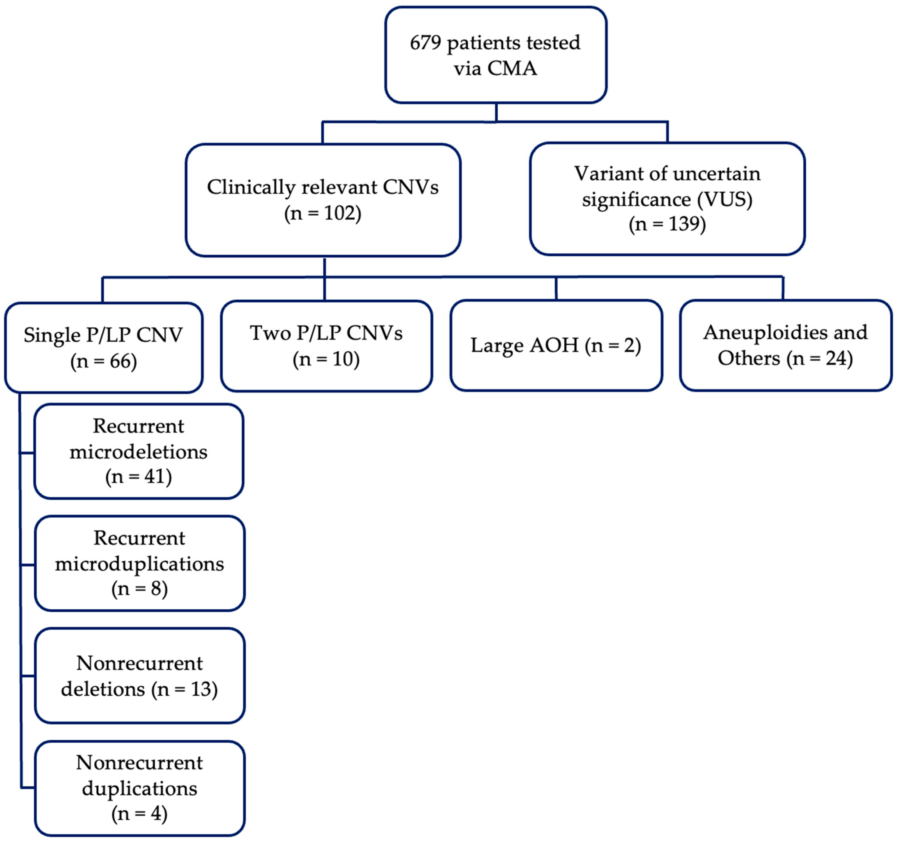
A total of 679 patients admitted to the NICU, PICU, or CVICU at Phoenix Children’s Hospital underwent chromosomal microarray analysis (CMA) between 2019 and 2024. Of those tested, 45.9% (n = 312) were female and 54.1% (n = 367) were male. The median age at testing was 7 days (IQR: 4–14 days; range: 0–15 years). More than 85% (578/679) were tested before one month of age; 11.8% (80/679) between 1 and 6 months, 1.2% (8/679) between 6 and 12 months, and 1.9% (13/679) were older than one year at testing. Of these 679 patients, 102 (15.0%) had a clinically relevant copy number variant (CNV), and 139 (20.5%) had a variant of uncertain significance (VUS). Among the 102 clinically relevant CNVs, 88 were classified as pathogenic (P), 12 as likely pathogenic (LP), and 2 demonstrated large regions of absence of heterozygosity (AOH) consistent with uniparental disomy (UPD). A summary of CMA findings for the cohort is shown in Figure 1.
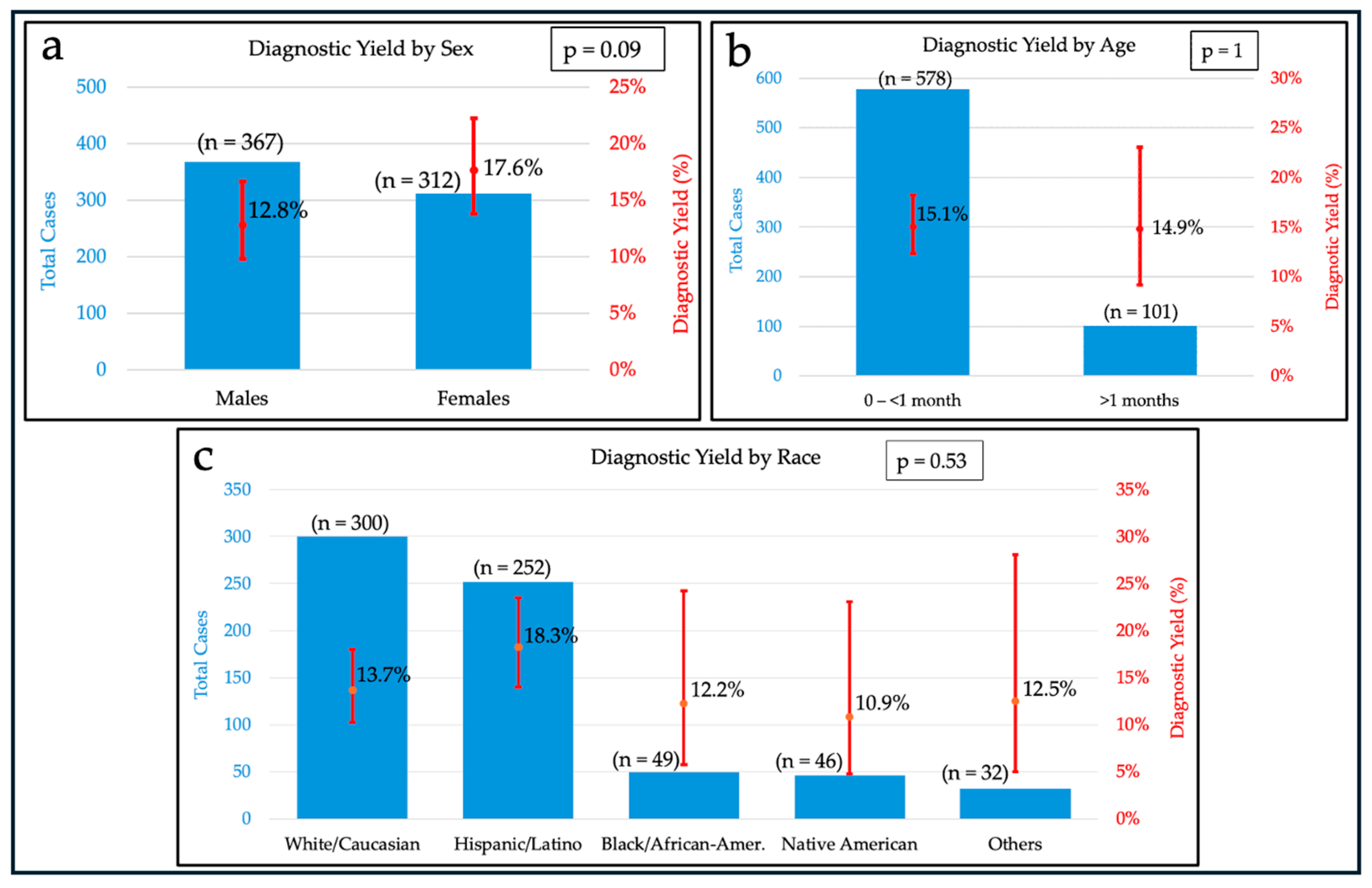
A breakdown of sex, age, and race distribution, along with the diagnostic rate by subgroup is provided in Figure 2. The diagnostic rate represents the proportion of patients within each subgroup who were found to have a clinically relevant chromosomal abnormality (i.e., pathogenic or likely pathogenic CNV) on CMA testing.
Diagnostic yield did not differ significantly by age, sex, or race. The yield was comparable between patients tested at <1 month (15.1%, 87/578; 95% CI 12.4–18.2%) and those tested at >1 month (14.9%, 15/101; 95% CI 9.2–23.1%), with no evidence of an association (Fisher exact p = 1.0). Similarly, although females demonstrated a higher diagnostic yield (17.6%, 55/312; 95% CI 13.8–22.2%) compared to males (12.8%, 47/367; 95% CI 9.8–16.6%), this difference did not reach statistical significance (p = 0.085). Across racial groups, diagnostic yield ranged from 10.9% to 18.3% (Black/African American: 12.2%, 6/49; Hispanic/Latino: 18.3%, 46/252; Native American: 10.9%, 5/46; Other: 12.5%, 4/32; White/Caucasian: 13.7%, 41/300), with no statistically significant differences observed (Fisher exact test with Monte Carlo simulation, p = 0.53). Racial categories with low sample sizes (Asian, Unknown, Hawaiian/Pacific Islander, and Other) were combined into a single group to ensure adequate counts for analysis. Collectively, these findings indicate that demographic variables were not significantly associated with diagnostic yield in this cohort.

3.2. Overview of Clinically Significant CNVs by Genetic Subtype

To facilitate a comprehensive characterization of patients with clinically significant CMA findings, individuals with pathogenic or likely pathogenic CNVs were systematically categorized according to genetic subtype, as presented in Figure 1. Four independent groups were observed: those with a single pathogenic or likely pathogenic CNV (n = 66; 64.7%), those with two identified pathogenic or likely pathogenic CNVs (n = 10; 9.8%), those with large regions of AOH consistent with possible UPD (n = 2; 2.0%), and those with aneuploidies along with sex chromosome-related disorders of sexual development (n = 24, 23.5%). Single pathogenic or likely pathogenic CNVs were further subdivided into the following categories: recurrent microdeletion syndromes, recurrent microduplication syndromes, nonrecurrent microdeletions, and nonrecurrent microduplications. Recurrent CNVs are defined as deletions or duplications that occur repeatedly in unrelated individuals at approximately the same chromosomal location, usually with similar breakpoints. Nonrecurrent CNVs are defined as deletions or duplications that occur at variable genomic regions and/or have unique breakpoints that differ between individuals. A complete breakdown of clinically relevant CNVs by genetic subtype and their relative proportions is provided in Table 1.

3.3. Recurrent CNVs Among Affected Patients

Of the 66 patients with a single CNV, 41 (62.1%) were diagnosed with recurrent microdeletion syndromes. Deletions involving 22q11.21 were the most common among patients with single CNV, identified in 40.9% (27/66), followed by 7q11.23 in 6.1% (4/66), 15q11.2 in 6.1% (4/66), 16p11.2 in 4.5% (3/66), 1q21.1 in 3.0% (2/66), and 17q12 in 1.5% (1/66). Notably, one patient (CMRES-2183) was identified with two independent pathogenic CNVs, including a 22q11.21 deletion, further supporting the recurrent occurrence of this microdeletion within the ICU cohort. In addition, eight patients (12.1%) were diagnosed with recurrent microduplication syndromes, most commonly involving 22q11.2 in 7.6% (5/66) of cases. Duplications at 16p11.2, 16p13.11, and 17q12 each were observed in 1.5% (1/66) of cases.

3.4. Nonrecurrent CNVs Among Affected Patients

Pathogenic and likely pathogenic nonrecurrent CNVs were identified across multiple chromosomal regions and varied substantially in size, gene content, and known clinical associations. Several CNVs overlapped, in whole or in part, genomic intervals that have been reported in association with established syndromic disorders or recognizable genotype–phenotype patterns. These included deletions involving 1p36.32, partially overlapping the distal 1p36 critical region associated with 1p36 deletion syndrome; 2q37.3, overlapping the region associated with 2q37 deletion syndrome; 9p24.3–p22.2, consistent with reported terminal 9p deletion phenotypes; 9q34.3, involving a deletion including NOTCH1; 13q12.3–q21.33, consistent with 13q deletion syndrome; and 22q13.2–q13.33, consistent with Phelan-McDermid syndrome. A deletion involving 14q32.2–q32.33 encompassed an imprinted region associated with Kagami–Ogata and Temple syndromes depending on parent-of-origin mechanisms. A smaller deletion at 12q15–q21.1, including CNOT2, overlapped deletions previously reported as 12q15 microdeletion syndrome in the literature. Additional nonrecurrent CNVs did not correspond to widely recognized recurrent genomic disorder intervals but have been reported in association with neurodevelopmental delay and congenital anomalies. These included interstitial deletions at 1q25.1–q32.1, 3p14.1–p12.2, 4q31.21–q31.22, and 18p11.32–p11.31, as well as duplications involving 2p22.1–p15, 8q12.1–q21.11, 8q21.2–q24.13, and 9p24.3–p21.3. A detailed summary of these CNVs, associated gene content, and phenotypes from the most recent chart review is provided in Table 2. Given the retrospective nature of this study and the variable expressivity associated with many CNVs, several reported clinical findings should be interpreted cautiously, as definitive genotype–phenotype relationships cannot be established in all cases, and some observed features may be incidental or multifactorial.

3.5. Two Pathogenic or Likely Pathogenic CNVs

Among the 102 patients with pathogenic or likely pathogenic CNVs or with AOH consistent with UPD, CMA identified two pathogenic or likely pathogenic CNVs in 10 patients (9.8%). Importantly, these CNVs were not uniformly interpreted as independent events. In at least half of these patients, the CNV patterns, typically consisting of a terminal deletion on one chromosome and a terminal duplication on another, were consistent with an underlying unbalanced translocation, raising the possibility of a balanced translocation in one of the parents. These cases included CMRES-1947, CMRES-1388, CMRES-0763, CMRES-1864, and CMRES-1150. Conventional chromosome analysis was recommended in these cases to further evaluate for potential underlying structural rearrangements.
In one patient (CMRES-0834), two CNVs involving chromosome 8 were identified, including an approximately 6.9 Mb terminal deletion at 8p23.3p23.1 and an approximately 31.2 Mb adjacent duplication at 8p23.1p11.1. This pattern of a terminal deletion with a more proximal duplication is characteristic of an inverted duplication with a terminal deletion of 8p (invdupdel [8p]), a recognized complex rearrangement mechanism associated with developmental delay, congenital anomalies, and other multisystem features. These findings were therefore interpreted as consistent with a possible invdupdel (8p) rearrangement rather than two independent pathogenic events.
In another case (CMRES-1392), CMA identified a duplication involving 17q that was suspected to represent a supernumerary marker chromosome, such as a ring chromosome 17, based on the size and distribution of duplicated material. An additional approximately 11.8 Mb duplication at 2q33.1q34, encompassing multiple genes including BMPR2, CPS1, SUMO1, IDH1, CASP8, and CASP10, was also detected. Partial duplications of 2q33 have been rarely reported but are associated with developmental delay and minor anomalies. Because these findings involved different chromosomes and lacked a shared structural mechanism, the 2q duplication was considered a possible independent pathogenic event in addition to the suspected marker chromosome.
Only cases with interstitial CNVs without evidence of a shared structural mechanism were considered likely to represent independent pathogenic events. These included CMRES-2183 and CMRES-3688; CMRES-1392 also met criteria for a possible independent pathogenic CNV in addition to the suspected marker chromosome.
For CMRES-2469, two adjacent CNVs involving 5p were identified, including an approximately 5.6 Mb deletion at 5p15.33p15.32 and a larger approximately 10.3 Mb mosaic deletion at 5p15.32p15.1, present in about 70% of cells. The mosaic loss was directly adjacent to the nonmosaic terminal deletion, raising the possibility that these findings represent a single complex or evolving structural event rather than two independent pathogenic CNVs. Both regions overlap the well-described 5p deletion region associated with Cri-du-chat syndrome. In addition, mosaic copy-neutral absence of heterozygosity of approximately 48 Mb involving the entire short arm of chromosome 11 (11p), encompassing the Beckwith–Wiedemann/Russell–Silver syndrome critical region, was identified, consistent with possible mosaic uniparental disomy; however, confirmatory UPD testing was not performed. Due to the lack of confirmatory testing, these results should be interpreted as provisional.
Overall, these findings indicate that the presence of two pathogenic or likely pathogenic CNVs in a single individual frequently reflects an underlying chromosomal rearrangement rather than multiple independent mutational events. This distinction is clinically relevant, as rearrangement-mediated CNVs have implications for parental carrier status and recurrence risk, whereas truly independent interstitial CNVs suggest separate pathogenic mechanisms. A detailed summary of these cases is provided in Table 3.

3.6. Absence of Heterozygosity Consistent with UPD

Of the 102 total patients with clinically significant CMA findings, two patients (2.0%) demonstrated large contiguous regions of absence of heterozygosity (AOH) involving a single chromosome, suggestive of possible uniparental disomy (UPD). CMA did not identify any pathogenic or likely pathogenic copy number variants in either case.
The first case (CMRES-0544) was a 5-day-old male with prenatal polyhydramnios and postnatal findings of ear malformations, musculoskeletal contractures, vertebral anomalies including scoliosis, and clinical features overlapping Jeune syndrome. CMA identified two large AOH intervals on the long arm of chromosome 14, spanning 14q11.2–q12 (~10.77 Mb) and 14q24.2–q32.33 (~34.72 Mb), raising concern for UPD(14). This patient has been comprehensively characterized clinically and genetically in a separate publication [16].
The second case (CMRES-3315) involved a 12-week-old premature male who presented with sepsis, respiratory distress, global hypotonia, lethargy, and limited behavioral responsiveness. CMA identified two large AOH segments on chromosome 15: a ~13.64 Mb interval spanning 15q11.2–q14, including the Prader–Willi/Angelman syndrome (PWS/AS) critical region (BP1–BP3), and a second ~26.28 Mb interval within 15q23–q26.2. Follow-up testing confirmed maternal UPD(15), establishing a diagnosis of Prader–Willi syndrome.

3.7. Aneuploidies

Of the 102 total patients with clinically significant CMA findings, aneuploidies were identified in 22 (21.6%), including Trisomy 21 in 16 (15.7%), Trisomy 18 in 3 (2.9%), Trisomy 13 in 1 (1.0%), and Monosomy X in 2 (2.0%). Follow-up chromosome analysis was not routinely available for patients with aneuploidies identified by CMA. Therefore, association between free trisomy, Robertsonian translocations-associated trisomy, and other chromosomal mechanism could not be consistently assessed. In addition to whole-chromosome aneuploidies, two cases demonstrated sex chromosome abnormalities: One patient exhibited 45,X/46,XY mosaicism, indicating the presence of both monosomic (45,X) and typical male (46,XY) cell lines, while another patient was a phenotypic female with a 46,XY karyotype. Both patients with sex chromosome abnormalities possessed congenital heart defects in addition to other congenital anomalies which prompted ICU admission and subsequent CMA.

3.8. Diagnostic Yield by Clinical Indication

To enable clinically meaningful interpretation of genotype–phenotype relationships and to evaluate diagnostic yield across presenting phenotypes, patients with clinically relevant CNVs were categorized according to the primary clinical indication at the time of referral into the following groups: congenital heart defects with or without other findings (CHD ± others), isolated CHD, congenital heart defects with central nervous system abnormalities or developmental delay (CHD + CNS/DD), CHD including single or multiple other congenital anomalies (CHD + CA), single or multiple congenital anomalies without CHD (CA—CHD), CNS/DD (isolated), and other. The CNS/DD group contained patients possessing structural brain malformations, abnormal electroencephalograms (EEG), or both. Conditions categorized as “other” included conditions that could not be reasonably assigned to the predefined diagnostic categories, including but not limited to cardiac abnormalities not associated with structural malformations (e.g., tachycardia) and perinatal conditions (e.g., intrauterine growth restriction). The categories of isolated CHD, CHD + CNS/DD, and CHD + CA were all subgroups of the CHD ± others category.
The most common clinical indications for which CMA was ordered were CHD ± others (73.8%, n = 501) and CA—CHD (17.2%, n = 117). Overall, the distribution of clinically significant CNVs across indication categories generally mirrored the relative frequency with which CMA was ordered in the ICU cohort; however, several notable exceptions were observed. For example, although CHD + CA accounted for only 13.7% (n = 93) of all CMA orders, this group represented 44.1% (n = 45) of the 102 clinically significant CNVs identified, yielding a detection rate of 48.4%. A similar trend was observed for the CHD + CNS/DD category, which accounted for only 1.9% (n = 13) of cases in which CMA was ordered, yet represented 6.8% (n = 7) of the clinically significant CNVs identified, yielding a diagnostic rate of 53.8% (95% CI: 29.1–76.8%). However, a limitation of this study was the small sample size within this group and thus additional studies with larger sample sizes are needed to confirm this trend observed in the CHD + CNS/DD subgroup. In contrast to the CHD + CA and CHD + CNS/DD categories, isolated CHD represented the most common indication for CMA, comprising 58.2% (n = 395) of cases while only 32.4% (n = 33) of clinically relevant CNVs were identified in this group, resulting in a diagnostic yield of 8.4% (95% CI: 5.6–11.1%). A complete breakdown of diagnostic yields across these clinical categories is provided in Table 4.
Diagnostic yield varied considerably across clinical indication categories. The highest detection rate was observed in patients with CHD + CNS/DD (53.8%); however, this estimate is exploratory and requires further confirmation in larger cohorts. Higher detection rates were also observed in the CHD + CA (48.4%) group. In contrast, substantially lower yields were observed in isolated CHD (8.4%), CA—CHD (10.3%), and isolated CNS abnormalities or developmental delay (10.7%). Indications with low sample sizes (Respiratory, Other, and Musculoskeletal) were grouped together as “Other Disorders” to meet statistical assumptions. A global Fisher exact test with Monte Carlo simulation demonstrated a significant association between clinical indication and diagnostic outcome (p = 1 × 10⁻⁶), indicating that diagnostic yield differed across clinical indication categories overall. Because this analysis assessed global association across all groups simultaneously, it does not identify which specific indication categories differed from one another. Post hoc pairwise analyses were not performed, as the primary objective was to evaluate overall variation in diagnostic yield across clinical presentations.

4. Discussion

In this study, we evaluated the diagnostic utility of CMA among 679 critically ill neonates and children admitted to intensive care units at Phoenix Children’s Hospital between 2019 and 2024. Clinically significant copy number variants were identified in 102 (15.0%) patients, and variants of uncertain significance were observed in 139 (20.5%). Notably, the diagnostic yield of CMA is often higher in inpatient cohorts with syndromic or multisystem presentations. For example, Sanri et al. reported a diagnostic yield of 20.2% among patients with syndromic features, compared to 11.8% in those with isolated developmental delay or intellectual disability, underscoring the enhanced value of CMA in acutely ill or diagnostically complex populations [17]. These findings are consistent with prior reports of 10–20% yield in pediatric populations and reinforce CMA’s role as a first-tier diagnostic tool in the inpatient setting, especially for patients with complex phenotypes [6,7,18,19].
When considering clinical indications, the highest diagnostic yield in our study was observed in patients with multiple congenital anomalies including a congenital heart defect (CHD + CA, 48.4%). This finding is consistent with previous studies demonstrating a substantially higher diagnostic yield among patients with CHDs and other congenital anomalies compared with those with isolated CHDs [20]. Although a high diagnostic yield was also observed among patients with concurrent congenital heart defects and central nervous system abnormalities or developmental delay (CHD + CNS/DD, 53.8%), the limited number of affected individuals with pathogenic or likely pathogenic CMA findings (7 of 13) warrants cautious interpretation, and validation in larger, independent cohorts is necessary to confirm this association. These findings highlight the enhanced utility of CMA in patients with multisystem involvement and structural anomalies, where the likelihood of an underlying genomic etiology is increased. In contrast, lower yields were observed in patients with isolated findings, such as isolated congenital heart defects (CHD, 8.4%) and isolated central nervous system abnormalities or developmental delay (CNS/DD, 10.7%), which may reflect non-genomic etiologies, polygenic contributions, environmental influences, monogenic causes, or variants undetectable by CMA. Due to these reasons, ES/GS may have increased utility in isolated CHD cases when CMA is nondiagnostic. These diagnostic yields are consistent with those previously reported in cohorts with isolated CHD [21,22,23,24] and isolated DD [25].
Recurrent CNV syndromes accounted for the majority of positive findings in this cohort (n = 49, 48.0%), with deletions involving 22q11.2 being the most frequently identified (n = 27, 26.5%). This finding is consistent with prior literature characterizing 22q11.2 deletions as a leading cause of conotruncal congenital heart defects, immune dysfunction, and neurodevelopmental manifestations [26,27]. Other recurrent CNVs included 7q11.23 deletions (n = 4, 3.9%) and 16p11.2 deletions or duplications (n = 4, 3.9%), both of which are well-recognized contributors to syndromic and neurodevelopmental phenotypes [28,29,30].
Population-based and cohort studies suggest that 22q11.2 deletions account for approximately 1–5% of all congenital heart defects, with higher frequencies often in the 10–20% range and up to 20% or more for specific defects such as tetralogy of Fallot or interrupted aortic arch type B in cohorts enriched for conotruncal defects [31,32,33,34,35,36]. By contrast, studies of hospitalized infants with severe or surgically managed CHD report abnormal CMA yields of approximately 15–35%, with 22q11.2 deletions consistently among the most common recurrent pathogenic CNVs [22,37]. Collectively, these comparisons support the interpretation that critically ill ICU patients with multisystem involvement represent an enriched subgroup in whom recurrent genomic syndromes, particularly 22q11.2 deletions, are detected more frequently than in unselected outpatient CHD cohorts.
Beyond recurrent syndromes, 17 patients carried nonrecurrent CNVs overlapping genomic regions previously associated with syndromic disease. These findings underscore the challenges of establishing genotype–phenotype correlations in neonates, where variable expressivity and incomplete penetrance may obscure early recognition of classic syndromes. Furthermore, because 85% of patients in this cohort underwent testing within the first month of life, many had not yet developed distinguishing clinical features. These observations emphasize the importance of longitudinal follow-up and age-appropriate reevaluation to refine interpretation over time. This is particularly relevant for variants of uncertain significance, as ongoing phenotypic evolution, accumulation of published case data, and advances in CNV interpretation frameworks may result in future variant reclassification and refinement of genotype–phenotype comparisons. Importantly, earlier molecular diagnosis may also facilitate syndrome-specific surveillance, anticipatory management, and diagnosis-guided health monitoring before the full phenotypic spectrum becomes clinically apparent. In some cases, early genomic diagnosis may additionally help expand the recognized phenotypic spectrum associated with these syndromes.
Rare and complex genomic findings were also detected, including 22 cases of aneuploidy, 2 cases of suspected uniparental disomy (UPD14 and UPD15), and additional cases involving sex chromosome mosaicism or karyotype–phenotype discordance. The observed aneuploidy diagnostic rate of 3.2% (22/679) in our cohort is consistent with rates reported in prior CMA studies [19]. These results highlight the value of single nucleotide polymorphism (SNP) based microarray platforms in detecting alterations that may be missed by traditional karyotyping or array comparative genomic hybridization (array-CGH), such as regions of absence of heterozygosity, imprinting disorders, and chromosomal mosaicism.
Importantly, 10 patients were identified with two pathogenic or likely pathogenic CNVs. These cases often presented with complex, multisystem phenotypes, and in several instances, clinical features appeared to reflect combined effects from both CNVs. While rare, multilocus genomic variation is increasingly recognized in clinical genomic testing and underscores the need for comprehensive genomic evaluation in cases with atypical or discordant presentations [9].
Beyond establishing a genetic diagnosis, CMA findings may provide clinically actionable information relevant to ICU management. Identification of pathogenic CNVs may inform prognosis, guide syndrome-specific surveillance for associated systemic complications, support anticipatory management strategies, and facilitate subspecialty referral. In critically ill infants with complex congenital anomalies, early genomic diagnosis may also assist with surgical planning, clarify recurrence risk for families, guide genetic counseling, and support informed medical decision-making regarding escalation of care and long-term management. Additionally, recognition of specific syndromic diagnosis may help avoid unnecessary diagnostic testing and streamline inpatient evaluation.
This study has several strengths, including a large cohort size, a focus on critically ill pediatric patients, and systematic clinical-genomic correlation. However, limitations include the retrospective design, variability in phenotypic documentation, inability to follow up certain cases with confirmatory testing (e.g., CMRES-1392, CMRES-2469), and the absence of longitudinal follow-up to assess long-term outcomes. The cohort included patients from multiple intensive care settings, including the NICU, PICU, and CVICU, encompassing a broad spectrum of clinical phenotypes, illness severities, and pretest probabilities for underlying genetic disease. This heterogeneity may have influenced diagnostic yield estimates and may limit the generalizability of findings to specific ICU subpopulations or more clinically homogenous cohorts. CMA also lacks the resolution to detect single nucleotide variants or small insertions and deletions, which may explain undiagnosed cases within the cohort.
In accordance with the recent AAP recommendations, genome or exome sequencing is increasingly recommended as a first-tier diagnostic test for children with global developmental delay or intellectual disability given its superior yield and cost-effectiveness. Accordingly, there has been an increase in the implementation of rapid ES/GS programs in the NICU setting. However, despite the growing role of rapid sequencing, CMA continues to provide important diagnostic value due to its robust detection of pathogenic CNVs, aneuploidy, regions of AOH, and structural chromosomal abnormalities that may not be consistently identified in all sequencing pipelines. In many institutions, CMA remains an important complementary genomic tool that may be performed sequentially or concurrently with exome sequencing, to optimize detection of copy number and structural chromosomal abnormalities [11].
These findings support the continued integration of CMA into early diagnostic workflows in neonatal and pediatric ICU patients, particularly those with multiple congenital anomalies or complex clinical presentations. Given its high diagnostic yield and rapid turnaround time, CMA remains a valuable component of genomic evaluation workflows for critically ill neonatal and pediatric patients. Future studies incorporating longitudinal follow-up and complementary genomic technologies will be essential for enhancing diagnostic accuracy and advancing precision care in this high-risk population.

5. Conclusions

CMA identified clinically relevant chromosomal abnormalities in 15.0% of cases in our cohort, including recurrent and non-recurrent CNVs, common aneuploidies, and complex genomic alterations such as UPD and sex chromosome mosaicism.
Our findings reinforce the clinical utility and diagnostic value of chromosomal microarray analysis in the ICU population, particularly among neonates and infants with congenital anomalies, dysmorphic features, or unexplained critical illness.
These results underscore the continued clinical utility of CMA within contemporary genomic evaluation workflows in the ICU setting and highlight the value of early genetic evaluation to inform clinical management, prognosis, and family counseling. Notably, many patients in this cohort were tested within the first days or weeks of life, when hallmark features of syndromic conditions may not yet be evident. Accordingly, longitudinal clinical follow-up is critical for delineating the evolving phenotype and refining the interpretation of large or atypical CNVs identified during the neonatal period. Future efforts should prioritize integrating CMA with next-generation sequencing approaches to further increase diagnostic yield and uncover additional genomic contributors to complex phenotypes in this vulnerable population.

Author Contributions

Conceptualization, J.M., E.H. and T.A.S.; CNV interpretation, T.A.S. and E.H.; formal analysis, J.M., E.H., A.S., V.V. and T.A.S.; writing—original draft preparation, J.M., E.H. and T.A.S.; writing—review and editing, J.M., E.H., A.S., V.V., D.N., T.A.G. and T.A.S. All authors have read and agreed to the published version of the manuscript.

Funding

This research received no external funding.

Institutional Review Board Statement

The study was conducted in accordance with the Declaration of Helsinki and approved by the Institutional Review Board of Phoenix Children’s Hospital (IRB-22-230; 7 September 2022).

Informed Consent Statement

Patient consent was waived because this research is conducted on material collected for diagnosis or medical treatment.

Data Availability Statement

All shareable data from patients are presented in the manuscript. De-identified case-level data are not available for sharing under the terms of the IRB approval.

Acknowledgments

The authors thank all the staff of the Clinical Genomics Laboratory at Phoenix Children’s Hospital for their technical support with array processing and initial analysis. The authors also acknowledge Lucia Mirea, and Perla Aguilera, Department of Clinical Research, Phoenix Children’s, for their contributions to statistical support.

Conflicts of Interest

T.A.S., E.H. and V.V. are employees of Clinical Genomics Laboratory at the Phoenix Children’s Hospital, which offers commercial CMA testing. All other authors declare no conflicts of interest.

References

  1. Arican, P.; Olgac Dundar, N.; Ozyilmaz, B.; Cavusoglu, D.; Gencpinar, P.; Erdogan, K.; Saka Guvenc, M. Chromosomal Mi-croarray Analysis in Children with Unexplained Developmental Delay/Intellectual Disability. J. Pediatr. Genet. 2019, 8, 1–9. [Google Scholar] [CrossRef]
  2. Rosenfeld, J.; Patel, A. Chromosomal Microarrays: Understanding Genetics of Neurodevelopmental Disorders and Congenital Anomalies. J. Pediatr. Genet. 2016, 6, 42–50. [Google Scholar] [CrossRef] [PubMed]
  3. Miller, D.T.; Adam, M.P.; Aradhya, S.; Biesecker, L.G.; Brothman, A.R.; Carter, N.P.; Church, D.M.; Crolla, J.A.; Eichler, E.E.; Epstein, C.J.; et al. Consensus Statement: Chromosomal Microarray Is a First-Tier Clinical Diagnostic Test for Individuals with Developmental Disabilities or Congenital Anomalies. Am. J. Hum. Genet. 2010, 86, 749–764. [Google Scholar] [CrossRef] [PubMed]
  4. Krepischi, A.C.V.; Villela, D.; Da Costa, S.S.; Mazzonetto, P.C.; Schauren, J.; Migliavacca, M.P.; Milanezi, F.; Santos, J.G.; Guida, G.; Guarischi-Sousa, R.; et al. Chromosomal Microarray Analyses from 5778 Patients with Neurodevelopmental Dis-orders and Congenital Anomalies in Brazil. Sci. Rep. 2022, 12, 15184. [Google Scholar] [CrossRef] [PubMed]
  5. Žilina, O.; Teek, R.; Tammur, P.; Kuuse, K.; Yakoreva, M.; Vaidla, E.; Mölter-Väär, T.; Reimand, T.; Kurg, A.; Õunap, K. Chromosomal Microarray Analysis as a First-tier Clinical Diagnostic Test: Estonian Experience. Mol. Genet. Genom. Med. 2014, 2, 166–175. [Google Scholar] [CrossRef] [PubMed]
  6. Carroll, J.; Wigby, K.; Murray, S. Genetic Testing Strategies in the Newborn. J. Perinatol. 2020, 40, 1007–1016. [Google Scholar] [CrossRef] [PubMed]
  7. NICUSeq Study Group. Effect of Whole-Genome Sequencing on the Clinical Management of Acutely Ill Infants with Suspected Genetic Disease: A Randomized Clinical Trial. JAMA Pediatr. 2021, 175, 1218–1226. [Google Scholar] [CrossRef] [PubMed]
  8. Marom, D.; Mory, A.; Reytan-Miron, S.; Amir, Y.; Kurolap, A.; Cohen, J.G.; Morhi, Y.; Smolkin, T.; Cohen, L.; Zangen, S.; et al. National Rapid Genome Sequencing in Neonatal Intensive Care. JAMA Netw. Open 2024, 7, e240146. [Google Scholar] [CrossRef] [PubMed]
  9. Smith, H.S.; Zettler, B.; Genetti, C.A.; Hickingbotham, M.R.; Coleman, T.F.; Lebo, M.; Nagy, A.; Zouk, H.; Mahanta, L.; Christensen, K.D.; et al. The BabySeq Project: A Clinical Trial of Genome Sequencing in a Diverse Cohort of Infants. Am. J. Hum. Genet. 2024, 111, 2094–2106. [Google Scholar] [CrossRef] [PubMed]
  10. Kingsmore, S.F.; Cakici, J.A.; Clark, M.M.; Gaughran, M.; Feddock, M.; Batalov, S.; Bainbridge, M.N.; Carroll, J.; Caylor, S.A.; Clarke, C.; et al. A Randomized, Controlled Trial of the Analytic and Diagnostic Performance of Singleton and Trio, Rapid Genome and Exome Sequencing in Ill Infants. Am. J. Hum. Genet. 2019, 105, 719–733. [Google Scholar] [CrossRef] [PubMed]
  11. Rodan, L.H.; Stoler, J.; Chen, E.; Geleske, T. Genetic Evaluation of the Child with Intellectual Disability or Global Developmental Delay: Clinical Report. Pediatrics 2025, 156, e2025072219. [Google Scholar] [CrossRef] [PubMed]
  12. Wojcik, M.H.; Callahan, K.P.; Antoniou, A.; Del Rosario, M.C.; Brunelli, L.; ElHassan, N.O.; Gogcu, S.; Murthy, K.; Rumpel, J.A.; Wambach, J.A.; et al. Provision and Availability of Genomic Medicine Services in Level IV Neonatal Intensive Care Units. Genet. Med. 2023, 25, 100926. [Google Scholar] [CrossRef] [PubMed]
  13. Goldin, M.R.; Ruderfer, D.M.; Bick, A.; Roden, D.M.; Schuler, B.A.; Robinson, J.R. Benefits and Barriers to Broad Implementa-tion of Genomic Sequencing in the NICU. Am. J. Hum. Genet. 2025, 112, 1270–1285. [Google Scholar] [CrossRef] [PubMed]
  14. Lee, W.; Yan, J.; Fooks, K.; Barwick, M.; Dobrow, M.; Friedman, J.M.; Marshall, C.R.; Hayeems, R.Z. Barriers and Facilitators to Implementing Clinical Genome-Wide Sequencing: A Scoping Review of the Global Landscape. Genet. Med. 2026, 28, 101665. [Google Scholar] [CrossRef] [PubMed]
  15. Riggs, E.R.; Andersen, E.F.; Cherry, A.M.; Kantarci, S.; Kearney, H.; Patel, A.; Raca, G.; Ritter, D.I.; South, S.T.; Thorland, E.C.; et al. Technical Standards for the Interpretation and Reporting of Constitutional Copy-Number Variants: A Joint Consensus Recommendation of the American College of Medical Genetics and Genomics (ACMG) and the Clinical Genome Resource (ClinGen). Genet. Med. 2020, 22, 245–257. [Google Scholar] [CrossRef] [PubMed]
  16. Sivakumaran, T.A.; Azab, B.; Hershman, E.; Sivakumaran, A.; Friedman, N.R.; Raz, D.; Neilson, D.; Grebe, T.A. Clinical Utility of Runs of Homozygosity in the Identification of Genetic Causes in Individuals with Rare Diseases. Genet. Med. Open 2025, 3, 103454. [Google Scholar] [CrossRef] [PubMed]
  17. Sanri, A.; Mutlu, M.B.; Sezer, O. Diagnostic Utility of Chromosomal Microarray Analysis in a Turkish Pediatric Cohort: Insights from 1,022 Patients with Neurodevelopmental Disorders and Congenital Anomalies. Cytogenet. Genome Res. 2025, 166, 144–177. [Google Scholar] [CrossRef] [PubMed]
  18. Mathew, M.T.; Antoniou, A.; Ramesh, N.; Hu, M.; Gaither, J.; Mouhlas, D.; Hashimoto, S.; Humphrey, M.; Matthews, T.; Hunter, J.M.; et al. A Decade’s Experience in Pediatric Chromosomal Microarray Reveals Distinct Characteristics Across Or-dering Specialties. J. Mol. Diagn. 2022, 24, 1031–1040. [Google Scholar] [CrossRef] [PubMed]
  19. Lu, X.-Y.; Phung, M.T.; Shaw, C.A.; Pham, K.; Neil, S.E.; Patel, A.; Sahoo, T.; Bacino, C.A.; Stankiewicz, P.; Kang, S.-H.L.; et al. Genomic Imbalances in Neonates with Birth Defects: High Detection Rates by Using Chromosomal Microarray Analysis. Pediatrics 2008, 122, 1310–1318. [Google Scholar] [CrossRef] [PubMed]
  20. Guo, R.; Duan, C.; Zarrei, M.; Reuter, M.S.; Dong, R.; Zhang, G.; Yang, X.; Zhang, H.; Wang, Y.; Scherer, S.W.; et al. Genetic Findings of Children with Congenital Heart Diseases Using Chromosomal Microarray and Trio-Based Whole Exome Se-quencing. Sci. Rep. 2025, 15, 27312. [Google Scholar] [CrossRef] [PubMed]
  21. Geng, J.; Picker, J.; Zheng, Z.; Zhang, X.; Wang, J.; Hisama, F.; Brown, D.W.; Mullen, M.P.; Harris, D.; Stoler, J.; et al. Chro-mosome Microarray Testing for Patients with Congenital Heart Defects Reveals Novel Disease Causing Loci and High Diag-nostic Yield. BMC Genom. 2014, 15, 1127. [Google Scholar] [CrossRef] [PubMed]
  22. Helm, B.M.; Landis, B.J.; Ware, S.M. Genetic Evaluation of Inpatient Neonatal and Infantile Congenital Heart Defects: New Findings and Review of the Literature. Genes 2021, 12, 1244. [Google Scholar] [CrossRef] [PubMed]
  23. Hureaux, M.; Guterman, S.; Hervé, B.; Till, M.; Jaillard, S.; Redon, S.; Valduga, M.; Coutton, C.; Missirian, C.; Prieur, F.; et al. Chromosomal Microarray Analysis in Fetuses with an Isolated Congenital Heart Defect: A Retrospective, Nationwide, Multi-center Study in France. Prenat. Diagn. 2019, 39, 464–470. [Google Scholar] [CrossRef] [PubMed]
  24. Salzer-Sheelo, L.; Polak, U.; Barg, A.; Kahana, S.; Yacobson, S.; Agmon-Fishman, I.; Klein, C.; Matar, R.; Rurman-Shahar, N.; Sagi-Dain, L.; et al. Prenatal and Postnatal Chromosomal Microarray Analysis in 885 Cases of Various Congenital Heart De-fects. Arch. Gynecol. Obstet. 2022, 306, 1007–1013. [Google Scholar] [CrossRef] [PubMed]
  25. Oğuz, S.; Arslan, U.E.; Kiper, P.Ö.Ş.; Alikaşifoğlu, M.; Boduroğlu, K.; Utine, G.E. Diagnostic Yield of Microarrays in Individuals with Non-syndromic Developmental Delay and Intellectual Disability. J. Intellect. Disabil. Res. 2021, 65, 1033–1048. [Google Scholar] [CrossRef] [PubMed]
  26. Agarwal, M.; Kumar, V.; Dwivedi, A. Diagnosis of 22q11.2 Deletion Syndrome in Children with Congenital Heart Diseases and Facial Dysmorphisms. Med. J. Armed Forces India 2023, 79, S196–S201. [Google Scholar] [CrossRef] [PubMed]
  27. Putotto, C.; Pugnaloni, F.; Unolt, M.; Maiolo, S.; Trezzi, M.; Digilio, M.C.; Cirillo, A.; Limongelli, G.; Marino, B.; Calcagni, G.; et al. 22q11.2 Deletion Syndrome: Impact of Genetics in the Treatment of Conotruncal Heart Defects. Children 2022, 9, 772. [Google Scholar] [CrossRef] [PubMed]
  28. Lv, X.; Yang, X.; Li, L.; Yue, F.; Zhang, H.; Wang, R. Prenatal Diagnosis of 7q11.23 Microdeletion: Two Cases Report and Lit-erature Review. Medicine 2023, 102, e34852. [Google Scholar] [CrossRef] [PubMed]
  29. Mitrakos, A.K.; Kosma, K.; Makrythanasis, P.; Tzetis, M. The Phenotypic Spectrum of 16p11.2 Recurrent Chromosomal Re-arrangements. Genes 2024, 15, 1053. [Google Scholar] [CrossRef] [PubMed]
  30. Giuseppe, M.; Brunetti-Pierri, N.; Fusco, C. Copy Number Variants at Williams-Beuren Syndrome 7q11.23 Region. Hum. Genet. 2010, 128, 3–26. [Google Scholar] [CrossRef] [PubMed]
  31. Peyvandi, S.; Lupo, P.J.; Garbarini, J.; Woyciechowski, S.; Edman, S.; Emanuel, B.S.; Mitchell, L.E.; Goldmuntz, E. 22q11.2 Deletions in Patients with Conotruncal Defects: Data from 1,610 Consecutive Cases. Pediatr. Cardiol. 2013, 34, 1687–1694. [Google Scholar] [CrossRef] [PubMed]
  32. Trainer, A.H.; Morrison, N.; Dunlop, A.; Wilson, N.; Tolmie, J. Chromosome 22q11 Microdeletions in Tetralogy of Fallot. Arch. Dis. Child. 1996, 74, 62–63. [Google Scholar] [CrossRef] [PubMed][Green Version]
  33. Lee, M.-Y.; Won, H.-S.; Baek, J.W.; Cho, J.-H.; Shim, J.-Y.; Lee, P.-R.; Kim, A. Variety of Prenatally Diagnosed Congenital Heart Disease in 22q11.2 Deletion Syndrome. Obstet. Gynecol. Sci. 2014, 57, 11. [Google Scholar] [CrossRef] [PubMed][Green Version]
  34. Wonkam, A.; Toko, R.; Chelo, D.; Tekendo-Ngongang, C.; Kingue, S.; Dahoun, S. The 22q11.2 Deletion Syndrome in Congenital Heart Defects: Prevalence of Microdeletion Syndrome in Cameroon. Glob. Heart 2017, 12, 115. [Google Scholar] [CrossRef] [PubMed]
  35. Agergaard, P.; Hebert, A.; Sørensen, K.M.; Østergaard, J.R.; Olesen, C. Can Clinical Assessment Detect 22q11.2 Deletions in Patients with Cardiac Malformations? A Review. Eur. J. Med. Genet. 2011, 54, 3–8. [Google Scholar] [CrossRef] [PubMed]
  36. Gioli-Pereira, L.; Pereira, A.C.; Bergara, D.; Mesquita, S.; Lopes, A.A.; Krieger, J.E. Frequency of 22q11.2 Microdeletion in Sporadic Non-Syndromic Tetralogy of Fallot Cases. Int. J. Cardiol. 2008, 126, 374–378. [Google Scholar] [CrossRef] [PubMed]
  37. Buckley, J.R.; Kavarana, M.N.; Chowdhury, S.M.; Scheurer, M.A. Current Practice and Utility of Chromosome Microarray Analysis in Infants Undergoing Cardiac Surgery: CMA in Infants Undergoing Cardiac Surgery. Congenit. Heart Dis. 2015, 10, E131–E138. [Google Scholar] [CrossRef] [PubMed]
Figure 1. Flowchart of CMA findings in critically ill patients.
Life 16 01034 g001
Figure 2. Diagnostic rate by sex, age, and race among patients who underwent CMA. (a) Distribution of total cases and diagnostic rates by sex. (b) Distribution of total cases and diagnostic rates by age group. (c) Distribution of total cases and diagnostic rates by race. Total case numbers are shown as blue bars, and diagnostic rates are represented by red points.
Life 16 01034 g002
Table 1. Clinically significant CNVs and relative proportions by genetic subtype.
Clinically Significant CNVsRelative Proportions
(N = 102), n (%)
Single Pathogenic or Likely Pathogenic CNV66 (64.7%)
   Recurrent Microdeletion Syndromes41 (40.2%)
  1q21.12 (2.0%)
  7q11.234 (3.9%)
  15q11.24 (3.9%)
  16p11.23 (2.9%)
  17q121 (1.0%)
  22q11.2127 (26.5%)
   Recurrent Microduplication Syndromes8 (7.8%)
  16p11.21 (1.0%)
  16p13.111 (1.0%)
  17q121 (1.0%)
  22q11.25 (4.9%)
   Nonrecurrent Deletions13 (12.7%)
   Nonrecurrent Duplications4 (3.9%)
Two Pathogenic/Likely Pathogenic CNVs10 (9.8%)
Large AOH Consistent with UPD 2 (2.0%)
  UPD141 (1.0%)
  UPD151 (1.0%)
Aneuploidies and Others24 (23.5%)
   Trisomy 2116 (15.7%)
   Trisomy 183 (2.9%)
   Trisomy 131 (1.0%)
   Monosomy X2 (2.0%)
   46,X/46,XY Mosaicism1 (1.0%)
   Phenotypic Female with 46,XY Karyotype1 (1.0%)
Table 2. CMA results and clinical phenotype of patients with rare or non-recurrent clinically significant CNVs.
Case NumberAge at Testing; GenderClinical Phenotype from Chart ReviewCNV TypeSize (Kb)RegionCNV ClassAssociated with Known Syndrome
CMRES–29106 wk; FApnea, bronchiolitis, LVNCLoss2430arr[GRCh37] 1p36.32 (2,628,158–5,060,971) × 1LP1p36 microdeletion syndrome
CMRES–29262 wk; MASD, ear malformations, hypothermia, IUGR, VSDLoss25,200arr[GRCh37] 1q25.1–q32.1 (174,360,902–199,565,997) × 1PNo
CMRES–01966 d; MIUGR, oligohydramnios, GDM, elevated creatinine, hypotonia, hypospadias, inguinal hernia, low set ears, PRS, renal hypoplasia, respiratory failureGain23,000arr[GRCh37] 2p22.1–p15 (40,505,796–63,510,902) × 3LPNo
CMRES–02554 d; Moligohydramnios, DD, abnormal EEG, bilateral CHL, HRV, lung hypoplasia, pulmonary atresiaLoss2660arr[GRCh37] 2q37.3 (240,123,897–242,783,384) × 1P2q37 deletion syndrome
CMRES–17096 d; Fear malformations, eye anomalies, CA, VSD, high aortic arch, aortic dilatation, thickened pulmonary valve, aortopulmonary collateral, enlarged 4th ventricle, respiratory failure, hypotonia, short statureLoss14,300arr[GRCh37] 3p14.1–p12.2 (68,898,172–832,44,508) × 1P3p deletion syndrome
CMRES–16652 wk; FDD, epilepsy, cerebral infarction due to left MCA embolism, hemiplegia, hemiparesis, HLHS, VSD, PDA, PH, bilateral CHL, feeding difficulty, GERDLoss5000arr[GRCh37] 4q31.21–q31.22 (143,381,651–148,378,867) × 1LPNo
CMRES–02252 d; MASD, GI/GU abnormalities, imperforate anusGain16,500arr[GRCh37] 8q12.1–q21.11 (60,765,051–77,234,448) × 3P8q duplication syndrome
CMRES–25053 wk; MDD, feeding difficulties, ASD, PFO, dysmorphic facial features, brachycephalyGain39,631arr[GRCh37] 8q21.2–q24.13 (85,850,967–125,482,371) × 3PNo
CMRES–00401 yr; Mepilepsy, GDD, OSA, feeding difficulties, tracheomalacia, FTT, abnormal head movements, dysmorphic facial features, ear malformations, bilateral CHL, NTD, congenital sacral dimpleGain25,300arr[GRCh37] 9p24.3–p21.3 (203,861–25,537,584) × 3P9p duplication syndrome
CMRES–35832 wk; Fmacrosomia, limb anomaly, COA, omphaloceleLoss16,600arr[GRCh37] 9p24.3–p22.2 (203,862–16,767,774) × 1P9p deletion syndrome
CMRES–13144 d; FDD, feeding difficulties, COA, BAV, HLV, AKI, LA, elevated LFTs, possible seizure activity, abnormal brain MRILoss179arr[GRCh37] 9q34.3 (139,217,461–139,396,216) × 1LP9q34.3 deletion
CMRES–13764 d; MHLHSLoss85arr[GRCh37] 9q34.3 (139,341,866–139,427,066) × 1PNOTCH1–related condition
CMRES–13446 d; MDD, feeding difficulties, ear malformations, COA, PDA, VSD, respiratory failureLoss1110arr[GRCh37] 12q–15q21.1 (70,403,305–71,510,598) × 1LP12q15 microdeletion syndrome
CMRES–10199 wk; MIUGR, FTT, dysmorphic facial features, bilateral retinoblastomaLoss38,000arr[GRCh37] 13q12.3–q21.33 (30,706,913–68,730,584) × 1P13q deletion syndrome
CMRES–06782 wk; MGDM, IUGR, respiratory distress, focal seizures, COA, microcephaly, dysmorphic facial features, abnormal EEG, hypotonia, GERD, colobomaLoss5560arr[GRCh37] 14q32.2–q32.33 (100,765,047–106,329,074) × 1P14q32 imprinting disorder (Temple vs. Kagami–Ogata), origin not established
CMRES–10013 mo; MTOF, PAS, hepatic hemangiomaLoss6050arr[GRCh37] 18p11.32–p11.31 (136,226–6,185,609) × 1P18p deletion syndrome
CMRES–16984 d; FTAPVR, structural brain anomalyLoss8430arr[GRCh37] 22q13.2–q13.33 (42,755,628–51,183,872) × 1PPhelan–McDermid syndrome
Abbreviations: d, day; wk, week; mo, month; yr, year; F, female; M, male; LP, likely pathogenic; P, pathogenic; AKI, acute kidney injury; ASD, atrial septal defect; BAV, bicuspid aortic valve; CA, choanal atresia; CHL, conductive hearing loss; COA, coarctation of aorta; DD, developmental delay; EEG, electroencephalogram; FTT, failure to thrive; GDD, global developmental delay; GDM, gestational diabetes mellitus; GI/GU, gastrointestinal/genitourinary; HLHS, hypoplastic left heart syndrome; HLV, hypoplastic left ventricle; HRV, hypoplastic right ventricle; IUGR, intrauterine growth restriction; LA, lactic acidosis; LFT, liver function test; LVNC, left ventricular non-compaction; MCA, middle cerebral artery; NTD, neural tube defect; OSA, obstructive sleep apnea; PAS, pulmonary artery stenosis; PDA, patent ductus arteriosus; PH, pulmonary hypertension; PFO, patent foramen ovale; PRS, Pierre Robin sequence; TAPVR, total anomalous pulmonary venous return; TOF, tetralogy of Fallot; VSD, ventricular septal defect.
Table 3. CMA results and clinical phenotype of patients with two pathogenic or likely pathogenic CNVs.
Case NumberAge; GenderClinical PhenotypeCNV TypeSize (kb)RegionCNV Class
CMRES-19476 d; Fcoronary artery fistula, respiratory failure, metabolic acidosis, suspected seizure, hydrocephalus, dysmorphic features, ventriculomegaly, lissencephaly, polymicrogyria, PH, CPLoss4420arr[GRCh37] 1p36.33–p36.32 (849,466–5,264,535) × 1P
Gain18,600arr[GRCh37] 14q31.3–q32.33 (88,703,186–107,285,437) × 3P
CMRES-13927 d; Mpolyhydramnios, hypertonia, ear malformations, facial cleft, CHL, right hemifacial microsomiaGain11,800arr[GRCh37] 2q33.1–q34 (202,035,063–213,827,186) × 3P
Gain8120arr[GRCh37] 17p11.2–q11.2 (19,257,545–27,378,279) × 3P
CMRES-13882 wk; MIUGR, respiratory distress, ASD, congenital malformations of ribs, craniosynostosis, hemivertebrae, dysmorphic features, congenital ptosis, mandibular hypoplasia, short palpebral fissures, hypoplastic anterior fontanelle, sacral dimple, low set ears, feeding difficulties, FTT, scoliosisLoss3580arr[GRCh37] 2q37.3 (239,203,172–242,783,384) × 1P
Gain30,300arr[GRCh37] 6p25.3–p21.33 (156,974–30,487,974) × 3P
CMRES-24693 wk; FIUGR, FTT, laryngomalacia/stridor, respiratory distressLoss5610arr[GRCh37] 5p15.33–p15.31 (113,577–5,723,670) × 1P
Loss10,300arr[GRCh37] 5p15.33–p15.31 (5,771,241–15,984,451) × 1~2P
CMRES-07636 d; FHLHS, hypoplastic aortic arch, anterior anus, coloboma, cranial deformity, choanal atresia, thrombocytopenia, PHGain7170arr[GRCh37] 5p15.33–p15.31 (113,576–7,283,299) × 3P
Loss15,000arr[GRCh37] 11q23.3–q25 (119,975,949–134,938,470) × 1P
CMRES-18644 d; FTOF, agenesis of corpus callosum, absent septum pellucidum, optic nerve hypoplasia, feeding difficultiesLoss1830arr[GRCh37] 6q27 (169,085,221–170,919,482) × 1P
Gain17,800arr[GRCh37] 21q21.3–q22.3 (30,261,221–48,097,372) × 3P
CMRES-08345 wk; Mdysmorphic facial features, hypotonia, deep creases separating 1st toes bilaterally, inguinal hernia, respiratory distress, FTT, hypotonia, tracheomalacia, hearing loss, DD, multiple congenital malformations, diffuculty feeding, constipationLoss6890arr[GRCh37] 8p23.3–p23.1 (158,048–7,044,046) × 1P
Gain31,300arr[GRCh37] 8p23.1–p11.1 (12,528,482–43,786,723) × 3P
CMRES-218313 d; Ftruncus arteriosus, bilateral PA hypoplasia, renal dysplasia, growth retardation, microcephaly, DDLoss10,400arr[GRCh37] 9q21.11–q21.13 (68,734,572–79,156,769) × 1P
Loss2550arr[GRCh37] 22q11.21 (18,916,843–21,465,659) × 1P
CMRES-11505 d; FHLV, VSD, interrupted aortic arch, hypoplasia of corpus callosum, focal epilepsy, DDGain24,000arr[GRCh37] 12q24.11–q24.33 (109,732,471–133,777,902) × 3P
Loss4360arr[GRCh37] 11q24.3–q25 (130,574,610–134,938,470) × 1LP
CMRES-36884 d; MTOF, CP, dysmorphic facial features, ear malformationsGain25,900arr[GRCh37] 14q11.2–q21.2 (20,511,673–46,421,909) × 3P
Gain29,700arr[GRCh37] 3p26.3–p24.1 (61,892–29,803,397) × 3P
Abbreviations: d, day; wk, week; mo, month; yr, year; F, female; M, male; LP, likely pathogenic; P, pathogenic; ASD, atrial septal defect; CHL, conductive hearing loss; CP, cleft palate; DD, developmental delay; FTT, failure to thrive; HLHS, hypoplastic left heart syndrome; HLV, hypoplastic left ventricle; IUGR, intrauterine growth restriction; PA, pulmonary artery; PH, pulmonary hypertension; TOF, tetralogy of Fallot; VSD, ventricular septal defect.
Table 4. Diagnostic yield of chromosomal microarray (CMA) by primary clinical indication at time of referral.
Clinical Indications at Time of ReferralICU Cases and Relative Proportions (N = 679), n (%)Clinically Significant CNVs and Relative Proportions (N = 102), n (%)Detection Rate for Clinical Indication, % (95% CI)
CHD + others501 (73.8)85 (83.3)17% (13.9%–20.5%)
   CHD (isolated)395 (58.2)33 (32.4)8.4% (6.0%–11.5%)
   CHD + CNS/DD13 (1.9)7 (6.8)53.8% (29.1%–76.8%)
   CHD + CA93 (13.7)45 (44.1)48.4% (38.5%–58.4%)
CA—CHD117 (17.2)12 (11.8)10.3% (6.0%–17.1%)
CNS/DD (isolated)28 (4.1)3 (2.9)10.7% (3.7%–27.2%)
Other33 (4.9)2 (2.0)6.1% (1.7%–19.6%)
Disclaimer/Publisher’s Note: The statements, opinions and data contained in all publications are solely those of the individual author(s) and contributor(s) and not of MDPI and/or the editor(s). MDPI and/or the editor(s) disclaim responsibility for any injury to people or property resulting from any ideas, methods, instructions or products referred to in the content.